HAL Id: hal-00297916
https://hal.archives-ouvertes.fr/hal-00297916
Submitted on 27 Aug 2007HAL is a multi-disciplinary open access
archive for the deposit and dissemination of sci-entific research documents, whether they are pub-lished or not. The documents may come from teaching and research institutions in France or abroad, or from public or private research centers.
L’archive ouverte pluridisciplinaire HAL, est destinée au dépôt et à la diffusion de documents scientifiques de niveau recherche, publiés ou non, émanant des établissements d’enseignement et de recherche français ou étrangers, des laboratoires publics ou privés.
Three-dimensional Magnetic Resonance Imaging of
fossils across taxa
D. Mietchen, M. Aberhan, B. Manz, O. Hampe, B. Mohr, C. Neumann, F.
Volke
To cite this version:
D. Mietchen, M. Aberhan, B. Manz, O. Hampe, B. Mohr, et al.. Three-dimensional Magnetic Reso-nance Imaging of fossils across taxa. Biogeosciences Discussions, European Geosciences Union, 2007, 4 (4), pp.2959-3004. �hal-00297916�
BGD
4, 2959–3004, 2007 Fossil MRI D. Mietchen et al. Title Page Abstract Introduction Conclusions References Tables Figures ◭ ◮ ◭ ◮ Back Close Full Screen / EscPrinter-friendly Version Interactive Discussion
EGU
Biogeosciences Discuss., 4, 2959–3004, 2007 www.biogeosciences-discuss.net/4/2959/2007/ © Author(s) 2007. This work is licensed
under a Creative Commons License.
Biogeosciences Discussions
Biogeosciences Discussions is the access reviewed discussion forum of Biogeosciences
Three-dimensional Magnetic Resonance
Imaging of fossils across taxa
D. Mietchen1,2,3, M. Aberhan4, B. Manz1, O. Hampe4, B. Mohr4, C. Neumann4, and F. Volke1
1
Fraunhofer Institute for Biomedical Engineering (IBMT), 66386 St. Ingbert, Germany 2
University of the Saarland, Faculty of Physics and Mechatronics, 66123 Saarbr ¨ucken, Germany
3
Friedrich-Schiller University Jena, Department of Psychiatry, 07740 Jena, Germany 4
Humboldt-Universit ¨at zu Berlin, Museum f ¨ur Naturkunde, 10099 Berlin, Germany Received: 22 August 2007 – Accepted: 27 August 2007 – Published: 27 August 2007 Correspondence to: D. Mietchen (daniel.mietchen@uni-jena.de)
BGD
4, 2959–3004, 2007 Fossil MRI D. Mietchen et al. Title Page Abstract Introduction Conclusions References Tables Figures ◭ ◮ ◭ ◮ Back Close Full Screen / EscPrinter-friendly Version Interactive Discussion
EGU
Abstract
The visibility of life forms in the fossil record is largely determined by the extent to which they were mineralised at the time of their death. In addition to mineral structures, many fossils nonetheless contain detectable amounts of residual water or organic molecules, the analysis of which has become an integral part of current palaeontological research.
5
The methods available for this sort of investigations, though, typically require dissolu-tion or ionisadissolu-tion of the fossil sample or parts thereof, which is an issue with rare taxa and outstanding materials like pathological or type specimens. In such cases, non-destructive techniques could provide an interesting methodological alternative. While Computed Tomography has long been used to study palaeontological specimens, a
10
number of complementary approaches have recently gained ground. These include Magnetic Resonance Imaging (MRI) which had previously been employed to obtain three-dimensional images of pathological belemnites non-invasively on the basis of intrinsic contrast. The present study was undertaken to investigate whether 1H MRI can likewise provide anatomical information about non-pathological belemnites and
15
specimens of other fossil taxa. To this end, three-dimensional MR image series were acquired from intact non-pathological invertebrate, vertebrate and plant fossils. At rou-tine voxel resolutions in the range of several dozens to some hundreds of micrometers, these images reveal a host of anatomical details and thus highlight the potential of MR techniques to effectively complement existing methodological approaches for
palaeon-20
tological investigations in a wide range of taxa. As for the origin of the MR signal, relaxation and diffusion measurements as well as 1H and 13C MR spectra acquired from a belemnite suggest intracrystalline water or hydroxyl groups, rather than organic residues.
BGD
4, 2959–3004, 2007 Fossil MRI D. Mietchen et al. Title Page Abstract Introduction Conclusions References Tables Figures ◭ ◮ ◭ ◮ Back Close Full Screen / EscPrinter-friendly Version Interactive Discussion
EGU
1 Introduction
When an organism dies, it is usually quickly decomposed but special conditions – namely the presence of biomineralised structures – sometimes allow for part of its mor-phological or biochemical characteristics to be preserved (for reviews, see
Behrens-meyer et al.,2000;Briggs,2003;Durand, 2003;Weiner and Dove,2003;Middelburg
5
and Meysman, 2007). Though these conditions only apply to a tiny minority of in-dividual life forms at a given time, vast amounts of biogenic deposits have been ac-cumulated over geological periods, most famously perhaps in the form of fossil fuels (e.g. Treibs,1934;Brocks et al.,1999;Vandenbroucke, 2003) and sedimentary rock (Albrecht and Ourisson,1971). Fossils, too, were increasingly often found to contain
10
organic matter (Kidwell and Holland,2002;Behrensmeyer et al.,2000;Briggs,2003), be it in cephalopod shells (Abelson,1954;Westbroek et al., 1979), belemnite rostra (Bandel and Spaeth, 1988; Florek et al., 2004), bones (Abelson, 1954; Schweitzer
et al.,2005,2007;Asara et al.,2007), or wood (Boyce et al.,2001;Siurek et al.,2004). Such observations led to the suggestion that part of the organic material detected
15
in fossils might actually represent the most stable portion of the molecules originally constituting the individual at the time of its death (e.g.Abelson, 1954; Florkin,1965;
Westbroek et al.,1979;Eglinton and Logan,1991;Engel et al.,1994;Schweitzer et al.,
2007), which opened the door for palaeobiochemical investigations (Blumer, 1965;
Albrecht and Ourisson, 1971; Niklas and Gensel, 1976; Weiner et al., 1976;
West-20
broek et al.,1979;Lowenstein,1981;Ourisson and Nakatani,1994;Waggoner,2002;
Schweitzer,2003;P ¨a ¨abo et al.,2004;Asara et al.,2007), provided that the specimens were excavated and stored in a suitable manner (cf.Pruvost et al.,2007).
Unfortunately, chemical analyses generally consume the specimens or parts thereof, thus reducing the morphological information they contain (Albrecht and Ourisson,
25
1971;Sælen,1989;Schweitzer et al.,2005;Dunca et al.,2007;Pruvost et al.,2007;
Asara et al.,2007). While a number of techniques exist that can non-destructively im-age the surface of a specimen (see, e.g., Sælen, 1989; Scott and Collinson, 2003),
BGD
4, 2959–3004, 2007 Fossil MRI D. Mietchen et al. Title Page Abstract Introduction Conclusions References Tables Figures ◭ ◮ ◭ ◮ Back Close Full Screen / EscPrinter-friendly Version Interactive Discussion
EGU
serial grinding techniques combined with digital photography have long been the only method allowing to reconstruct the three-dimensional structure of fossils at a high spa-tial resolution (Luo and Eastman, 1995; Luo and Marsh, 1996; Sutton et al., 2001;
Siveter et al., 2004;Sutton et al., 2005), yet they trade this achievement for a com-plete loss of the specimen. Due to these technical limitations, obtaining chemical and
5
morphological information from within fossils has generally been mutually exclusive but progress in non-invasive imaging techniques of geomaterials in general has impor-tant spill-over effects for palaeontological investigations (Rothwell and Vinegar,1985;
Carlson,2006).
One non-destructive approach to construct 3-D representations of porous or
other-10
wise heterogeneous materials at microscopic resolution is x-ray Computed Tomogra-phy (CT) which enjoys considerable popularity for fossil analysis (for an overview, see
Zollikofer and Ponce de L ´eon,2005) and can still be enhanced by monochromatisation, leading to synchrotron radiation CT (SR-CT; cf.Tafforeau et al.,2006;Mazurier et al.,
2006;Donoghue et al.,2006).
15
Two other tomographic techniques have recently been demonstrated to allow for non-invasive in saxo imaging of three-dimensional fossil morphologies: Neutron To-mography (NT; cf.Domanus,1992) has successfully been used to acquire images of sauropod vertebrae (Schwarz et al.,2005), while Magnetic Resonance Imaging (MRI; for detailed description, seeCallaghan,1991) can either be used to generate negative
20
images of mouldic or cavernous fossil samples by immersing them in or filling them with materials that provide MR signal (e.g. water or oil, possibly containing contrast agents;Sebes et al.,1991;de Swiet et al.,1998;Steiger et al.,1997;Steiger,2001;
Clark et al.,2004), or the fossil morphology can be imaged directly, solely on the basis of intrinsic contrast, as shown for pathological belemnite rostra (Mietchen et al.,2005).
25
Like CT, both NT and MRI can reach microscopic resolutions, and for all three tomo-graphic modalities, spectroscopic sister techniques exist that allow a detailed chemical characterisation of a given specimen (Boyce et al.,2002;Gabel et al.,2002;Abragam,
BGD
4, 2959–3004, 2007 Fossil MRI D. Mietchen et al. Title Page Abstract Introduction Conclusions References Tables Figures ◭ ◮ ◭ ◮ Back Close Full Screen / EscPrinter-friendly Version Interactive Discussion
EGU
In this study, we concentrated on in saxo MRI whose signal intensity had been found to co-vary with pathological alterations of biomineralisation in the belemnite specimens mentioned above. Specifically, we addressed the issue whether the method would equally be applicable to non-pathological rostra and to fossils other than belemnites – as previous studies of fluids in porous rocks (de Swiet et al.,1998) and of extant
biomin-5
eralised samples (Majumdar et al.,1998;Borah et al.,2001;Tsai et al.,2004;M ¨uller
et al., 2006) would suggest – and used MRI to investigate the internal morphology of non-pathological fossils of invertebrate, vertebrate and plant origins from different geological settings.
The fossil material figured here is housed at the Museum f ¨ur Naturkunde der
10
Humboldt-Universit ¨at zu Berlin, Germany (acronym MB.) and in the Museum f ¨ur Natur und Umwelt in L ¨ubeck, Germany (acronym MNU).
2 Diagenesis of biomineralised structures
When considering the use of new methodologies like MRI for the structural investiga-tion of fossils, it is necessary to reflect on the material characteristics of the skeleton,
15
the extent to which the original morphologic structures have been diagenetically altered and how this could affect the acquisition and interpretation of the data. The following sections will hence review biomineralisation and diagenetic processes in selected tax-onomic groups, with a focus on preservation of organic matter in the mineral matrix (the preservation of water will be discussed in Sect.5.4). Several reviews are available
20
that treat taphonomy, particularly chemical and microbial degradation of the organic and mineral fractions, in more detail (Behrensmeyer,1978;Behrensmeyer et al.,2000;
BGD
4, 2959–3004, 2007 Fossil MRI D. Mietchen et al. Title Page Abstract Introduction Conclusions References Tables Figures ◭ ◮ ◭ ◮ Back Close Full Screen / EscPrinter-friendly Version Interactive Discussion
EGU
2.1 Invertebrates 2.1.1 Overview
Shells of macroinvertebrates composed of calcium carbonate abound in the Phanero-zoic fossil record. Calcite is the more stable crystal form, though shells composed thereof may have experienced diagenetic recrystallisation. Aragonitic shells have
com-5
monly been dissolved during diagenesis or replaced by other minerals, mainly calcite. The organic matrix has been attributed several functions in biomineralisation, including nucleation of the mineral, determination of the mineral phase, control of the orientation and growth of carbonate crystals, and enhancement of mechanical properties of the crystals (e.g.Crenshaw,1990). Biochemically, it consists of proteins, poly-saccharides,
10
and water. A major portion of the soluble fraction of organic matrix macromolecules are aspartic acid-rich glycoproteins, whereas important constituents of the insoluble frac-tion are glycine, alanine and chitin (for details, seeLowenstam and Weiner,1989).
The properties of organic matrix components are subject to often severe postmortem alterations – proteins, for instance, decompose to individual amino acids which can
un-15
dergo isomerisation and further decomposition down to simple hydrocarbons (Collins
and Gernaey-Child,2001). Several studies have documented the presence of amino acids in fossil shells as old as about 360 Ma (summarised inWeiner,1979). However, the amino acid composition of a Late Cretaceous (80 Ma before present) ammonoid shell bears no resemblance to those found in a close extant relative, the cephalopod
20
Nautilus (Weiner,1979). 2.1.2 Belemnites
Another group of cephalopods, now extinct, are the belemnites. They had an en-doskeleton whose most distal part – the rostrum or guard that helped to maintain a horizontal swimming posture (Naef,1922) – is frequently preserved. It consists of the
25
BGD
4, 2959–3004, 2007 Fossil MRI D. Mietchen et al. Title Page Abstract Introduction Conclusions References Tables Figures ◭ ◮ ◭ ◮ Back Close Full Screen / EscPrinter-friendly Version Interactive Discussion
EGU
solidum (e.g.Sælen,1989, cf. Fig.1). It is further characterised by (1) the apical line, which represents the axis of the rostrum and marks the trajectory of the apex (the most posterior part of the rostrum) during successive growth stages, (2) composite radial structures formed by crystals radiating out from the apical line to the margin and (3) commarginal (concentric) growth lines (also known as growth rings) which stem from
5
periodical accretions of radial structures that resulted in spatial variations of the or-ganic content. This variation is often subtle, so that growth rings may be difficult to define (Sælen,1989).
Apart from the primordial rostrum and very early growth stages, the original mineral-ogy of belemnite rostra was low-Mg calcite (Veizer,1974). As such they are relatively
10
stable even under freshwater influence (meteoric diagenesis). The microstructure con-sists of regular, fine prisms with parallel crystal axes, arranged in well-ordered prismatic layers (Bandel and Spaeth,1988) which can still be discerned in even strongly recrys-tallised specimens. Diagenetic exchange between rostra and enclosing rock appears to be limited (up to about 10% by weight, according toVeizer,1974) and may be caused
15
by solution-precipitation phenomena or filling of the pore space. The latter was either primary or diagenetically generated by the decay of organic matter. Growth lines are frequently preserved.
With respect to organic matrix macromolecules in belemnites, Westbroek et al.
(1979) analysed rostra of two late Cretaceous taxa, Gonioteuthis and Belemnitella.
20
In the soluble macromolecular fraction of well-preserved Gonioteuthis rostra, these authors identified components with peptidic and saccharidic properties as well as an amino acid composition very similar to that of Nautilus, dominated by glycine and ala-nine. Even original antigenic properties of certain fractions were still preserved, sug-gesting that the biochemical materials derived from Gonioteuthis were original
belem-25
nite compounds which only experienced minor alterations during diagenesis. The observed enrichment in polyphenols may be due to reactions between peptides and carbohydrates during diagenesis. In contrast to the exceptionally well-preserved ros-tra of Gonioteuthis, those of Belemnitella were strongly recrystallised. Although the
BGD
4, 2959–3004, 2007 Fossil MRI D. Mietchen et al. Title Page Abstract Introduction Conclusions References Tables Figures ◭ ◮ ◭ ◮ Back Close Full Screen / EscPrinter-friendly Version Interactive Discussion
EGU
amino acid composition is similarly dominated by glycine (12.6 mol%) and alanine (11.5 mol%), less stable (threonine, serine, arginine – 5.5, 6.2 and 4.9 mol%, respec-tively) and even very labile amino acids (methionine, 2.2 mol%) were also present.
Westbroek et al. concluded thus that the primary organic composition of the Belem-nitella rostra was contaminated during or after recrystallisation by percolating ground
5
water.
2.1.3 Crinoids
As in all echinoderms, the crinoid stalk ossicles belong to the mesodermal endoskele-ton and are composed of magnesium calcite which is arranged in the typical form of a stereom (a meshwork of anastomosing trabeculae and pillars). In the living crinoid,
10
the interspace in the stereom is filled with soma which contains living cells, mainly sclerocytes and phagocytes (Heinzeller and Welsch,1994). Columnals bearing cirri are nodals, those without cirri are internodals. Connections of the ossicles are non-muscular and exclusively by elastic ligaments of mutable collagenous tissue. Short, intercolumnar ligaments connect each pair of adjacent columnals. Longer, continuous
15
ligaments connect each set of internodals and one associated nodal (Ausich et al.,
1999).
Columnals of Isselicrinus buchii are cylindrical, rarely subpentagonal, with a smooth surface. The corresponding articulations of adjacent ossicles bear a characteristic pat-tern: Interlocking grooves and ridges of densified stereom occur in a petaloid pattern,
20
giving the stem a certain flexibility. A crenulation is also visible at the margin of most ossicles, and the stem possesses a small central canal which represents a tubular cavity with extensions of the coelom and nervous system (Ausich et al.,1999).
Skeletal particles of echinoderms are usually preserved as large single crystals of calcite. During diagenesis, the metastable high Mg-calcite of the echinoderm skeleton
25
is replaced by low Mg-calcite. This transformation occurs by means of a solid-state process known as incongruent dissolution. Unidirectional growth of calcite crystals gives rise to syntaxial cements, the formation of which starts with the infilling of the
BGD
4, 2959–3004, 2007 Fossil MRI D. Mietchen et al. Title Page Abstract Introduction Conclusions References Tables Figures ◭ ◮ ◭ ◮ Back Close Full Screen / EscPrinter-friendly Version Interactive Discussion
EGU
pore space. Occasionally, the microstructure may be preserved when the porespace is infilled with clay preventing precipitation of spar. Even more rarely, the echinoderm crystals can be diagenetically transformed into microcristalline calcite (also known as micrite;Neugebauer,1978). Under favourable conditions (rapid burial, low pore water circulation, an- or dysoxic conditions), organic remains such as color pigments can be
5
preserved within the stereom (Blumer,1965;Wolkenstein et al.,2006). Especially in the Cretaceous chalk sea, dissolution or corrosion of echinodermal magnesium calcite occured either on the sea floor or during diagenesis (Ernst,1963;Neugebauer,1978). 2.2 Vertebrates
2.2.1 Overview
10
Diagenetic processes acting upon vertebrate remains are far from being completely understood but for most practical purposes, they can be reduced to the diagenesis of bone and its collagenous protein matrix. Although this is still a complex, multistage process, notoriously dependent on external biochemical, hydrological and taphonomi-cal factors of the embedding environment and internal parameters such as bone size,
15
histological structure and collagen content (Martill,1991; Hedges and Millard,1995;
Schweitzer et al.,2007), it has been relatively well-studied in archaeological sciences, especially in relation to collagen decomposition (Nielsen-Marsh et al.,2000). Similar to most other proteins, collagen is composed mostly of carbon, hydrogen, nitrogen and a minor content of sulphur, and its initial decay during early diagenesis is of great
20
importance for the final preservation and chemical composition of the bone (Hedges
et al.,1995;Nielsen-Marsh and Hedges,2000;Pfretzschner,1998,2000;Schweitzer
et al.,2005,2007). Generally, microbial degradation of collagen accelerates destruc-tion (Collins et al.,1995) and therefore hinders fossil preservation of bone. However, microbial activity depends on a number of chemical parameters – including
tempera-25
ture, pH and the availability of oxygen and water – whose combination might lead to reduced microbial degradation (Bocherens et al., 1997; Nielsen-Marsh and Hedges,
BGD
4, 2959–3004, 2007 Fossil MRI D. Mietchen et al. Title Page Abstract Introduction Conclusions References Tables Figures ◭ ◮ ◭ ◮ Back Close Full Screen / EscPrinter-friendly Version Interactive Discussion
EGU 2000), which is an advantage for later fossilisation. The proteolysis of collagen,
es-pecially under water, is determined by local pH as well as the redox potential of bone during early diagenesis (Pfretzschner,1998), and this influences the ion and isotope exchange rates between the phosphorous mineral phase of the skeletal remains and the ambient fluid and sediment. Reaction conditions, moreover, differ between
fresh-5
water and marine environments. Finally, even if two samples have been taken from the same fossil specimen, the circumstances of their excavation, handling, storage and analysis can severely influence the residual biochemical information contained therein, as has been shown for DNA contents (Pruvost et al.,2007) but can be assumed to be the case for other macromolecules and water as well.
10
2.2.2 Bone preservation and taphonomy
Bone material consists of the biomineral hydroxy apatite – Ca5(PO4)3OH – which can be modified during fossilisation by various chemical interactions with the environment and the sediment covering the carcass (Rottl ¨ander,1979). Depending on the environ-mental distribution of reactants (Pfretzschner,1998), the mobile ions within the apatite
15
crystal – H+, Ca2+, HPO2−4 and OH− – can be substituted, asPiepenbrink(1989) has shown for subfossil human bone. Ca2+, for example, is often replaced by UO2+2 (Millard
and Hedges,1995;Pfretzschner,1997) or Sr2+, or 2Ca2+ by Na+and Y3+. Likewise, the phosphate group HPO2−4 can exchange with SO2−4 or CO2−3 . SiO2, V4+or As3+can also move into the crystal lattice, while F− and Cl− can substitute for OH− (Newesely,
20
1989;Pfretzschner,2000;Trueman,1999). The histological structure of bone is impor-tant for the interaction with the ambient fluid. The pore structure, which often follows a trajectory pre-defined by in vivo biomechanical load, determines the internal surface area and therefore the capacity for surface reactions (Hedges and Millard,1995).
BGD
4, 2959–3004, 2007 Fossil MRI D. Mietchen et al. Title Page Abstract Introduction Conclusions References Tables Figures ◭ ◮ ◭ ◮ Back Close Full Screen / EscPrinter-friendly Version Interactive Discussion
EGU
2.2.3 Fossil ear bones
With the exception of some phylogenetically early species (cf.Luo et al., 2007), the mammalian ear bones typically comprise the tympanic bulla, the middle ear with the three ossicles malleus, incus and stapes, and the periotic bone with the cochlear por-tion containing the spiral cavity which forms the organ of sound receppor-tion (Berta and
5
Sumich,1999). The cochlea is coiled and divided lengthwise into three parallel tubular channels (Liem et al.,2001, Fig. 12-22).
In cetaceans, the periotic is the most compact skull bone and of a comparatively stable and chemically resistent nature. As an adaptation to underwater hearing, it is displaced from the skull by pneumatic spaces (e.g.Purves and Pilleri,1983) and only
10
loosely connected to it via ligaments to the mastoid process (Pilleri et al.,1987). The detachment allows separate reception of sound and isolated vibrations of the ear bones (Miller,1923;Fleischer,1978;Pilleri et al.,1987). The anatomy of the cetacean organ of hearing is well described (Pilleri et al.,1987) but was usually studied by producing serial sections by grinding the petrosals (e.g.Luo and Eastman,1995;Luo and Marsh,
15
1996), resulting in the loss of the unique fossil specimens. To our knowledge, non-invasive imaging of fossil ear bones has not been reported so far, though petrosals obtained from contemporary animals (e.g.Ketten and Wartzok,1990;Nummela et al.,
1999) have successfully been imaged by CT, as have entire fossil cetacean skulls (at resolutions not suitable for petrosal investigations, cf.Marino et al.,2003).
20
Incidentally, the first MR imaging of fossils that we know of (Sebes et al., 1991) has been performed on the vertebrae of a Miocene dolphin, though the specimen was immersed in water which provided the contrast. The specimen was not specified in that report but determined as Xiphiacetus bossi (Kellogg) USNM 10480 (D. Bohaska and B. Rothschild, personal communication). At the time of writing, it was not known
25
whether the water immersion had had any impact on the state of preservation of these vertebrae, as compared to non-immersed vertebrae of the same specimen.
BGD
4, 2959–3004, 2007 Fossil MRI D. Mietchen et al. Title Page Abstract Introduction Conclusions References Tables Figures ◭ ◮ ◭ ◮ Back Close Full Screen / EscPrinter-friendly Version Interactive Discussion
EGU
2.3 Plants
Petrified plant remains, mainly silicified wood, are a frequent feature in the fossil record. In ion exchange reactions similar to those desribed above for bone, silica minerals are deposited in cracks, openings between cells, and spaces left by cell fluids. These processes take place while the wood is still relatively intact. Consequently, petrified
5
plant parts exhibit preserved morphological patterns down to the cellular scale, and they often contain organic and carbon compounds. Silicification depends on the ther-mal conditions (Sigleo, 1978) and can occur over a wide pH range and sometimes very rapidly (in vitro as fast as in 24 h, according toDrum,1968). The most probable mechanism for wood silicification is hydrogen bonding between silicic acid, Si(OH)4,
10
and the hydroxyl groups in cellulose. When studying Araucarioxylon arizonicum fossils from the Triassic Chinle Formation of Petrified Forest National Park in Arizona for lignin derivates, Sigleo (1978) was able to identify various organic compounds in silicified wood by sequential high vacuum pyrolysis and gas chromatography mass spectrom-etry. These organic components included phenols, methylphenols, alkyl substituted
15
benzenes and benzofurans. Further support for organic remains in plant fossils comes from elemental mapping of silicified wood from Neoproterozoic up to Miocene sites (Boyce et al.,2001), and Siurek et al.(2004) – who investigated silicified wood from Chile and the Barton Peninsula (King George Island, Antarctica) – found carbon in each volume element they tested, in most cases convincingly attributable to the
rem-20
nant primary organic matter.
As for noninvasive imaging, x-ray tomography was applied successfully to pyritized fossil fruits from the Lower Eocene London Clay flora, namely to the visualisation of in-ternal structures, including small seeds, within Myrtaceaen fruits (DeVore et al.,2006), and further applications are about to emerge, e.g. for the study of internal structures
25
(down to the cellular level) within charcoalified three-dimensionally preserved Creta-ceous flowers (Friis et al.,2006). MR imaging has, however, found multiple applications to extant plant specimens (Chudek and Hunter,1997), including conifer cones (e.g.Mill
BGD
4, 2959–3004, 2007 Fossil MRI D. Mietchen et al. Title Page Abstract Introduction Conclusions References Tables Figures ◭ ◮ ◭ ◮ Back Close Full Screen / EscPrinter-friendly Version Interactive Discussion EGU et al.,2001). 3 Specimen description 3.1 Invertebrates 3.1.1 Belemnites
The analysed belemnite rostra, identified as Belemnopsis sp. byDietrich(1933), were
5
collected by the German Tendaguru Expedition (1909–1912, cf.Janensch,1914). The fossil locality of Tendaguru, famous for its diverse dinosaur assemblages, is located ap-proximately 60 km northwest of the seaport of Lindi in southeastern Tanzania. The Late Jurassic to Early Cretaceous (ca. 155–130 Ma before present) Tendaguru Beds reach a thickness of 110 m and consist of three fine-grained dinosaur-bearing sequences
10
which are intercalated with sandstone-dominated sequences containing a predomi-nantly marine fauna (Aberhan et al.,2002). The rostra were embedded in a medium-to coarse-grained sandsmedium-tone of Late Jurassic (Tithonian) age at the transition between the so-called Trigonia smeei Bed and the base of the Upper Saurian Bed at Tendaguru site IX, about 1.4 km northeast of Tendaguru Hill. Most specimens are fragmented and
15
the outer surface appears pitted due to intense weathering. Recent sedimentological and palaeoecological analyses of the Tendaguru Beds (Aberhan et al.,2002) suggest that deposition of the Trigonia smeei Bed took place in lagoon-like, shallow marine environments above fair weather wave base and with evidence of tides and storms. Sediments of the Upper Saurian Bed represent extended siliciclastic tidal flat
environ-20
ments including brackish coastal lakes and ponds. The Late Jurassic palaeoclimate of the Tendaguru area was subtropical to tropical, characterised by seasonal rainfall alternating with a pronounced dry season.
The analysed rostrum of Belemnella (Pachybelemnella) sumensis Jeletzky is from the boreal Upper Cretaceous (Lower Maastrichtian, ca. 69 Ma before present) white
BGD
4, 2959–3004, 2007 Fossil MRI D. Mietchen et al. Title Page Abstract Introduction Conclusions References Tables Figures ◭ ◮ ◭ ◮ Back Close Full Screen / EscPrinter-friendly Version Interactive Discussion
EGU
chalk of the Stubbenkammer, Isle of R ¨ugen (Northeastern Germany). The sedimentary matrix is a weakly lithified coccolith limestone, representing fully marine, distal open shelf conditions well below storm wave base.
3.1.2 Crinoids
The crinoid specimen (MNHB/MB.E 5730) used for this study belongs to the species
Is-5
selicrinus buchii (Roemer, 1840) was found together with the above-described Belem-nella (PachybelemBelem-nella) sumensis sample in the white chalk of Stubbenkammer, Isle of R ¨ugen, Germany. Isselicrinus buchii is the most abundant crinoid taxon of the chalk soft bottom ecosystem during the Maastrichtian. As a stalked crinoid, Isselicrinus was highly adapted for a life on a muddy substrate. It possesssed a relay strategy, using
10
pre-existing upright stem columns as anchorage (Fujiwara et al.,2004). The long stem raised the cup highly above the sea floor into the water current. As a passive suspen-sion feeder, this crinoid was dependent on currents transporting plankton to the catch apparatus formed by the crinoid’s arms (Oji, 1985, 1990). We used only columnals (stem elements) for MR imaging.
15
3.2 Vertebrates
The bone used for the current study was a periotic originating from a partial skeleton of a kentriodontid dolphin (Cetacea: Odontoceti) belonging to the genus Atocetusde
Muizon 1988 of the subfamily Pithanodelphininae Barnes 1985. This Atocetus fossil was discovered in a commercial gravel pit near Groß Pampau in Schleswig-Holstein,
20
northern Germany, and is stored since then in the Museum f ¨ur Natur und Umwelt in L ¨ubeck (MNU-071-18). The site is famous for several whale remains (e.g.H ¨opfner,
1991;Hampe,1999). The horizon containing the whale fossils belonged to a dark mica clay (“Oberer Glimmerton” in regional stratigraphy; cf.Hinsch,1990) that was deposited between 10.6 Ma and 11.8 Ma (Spiegler and G ¨urs, 1996) after Bolboforma biozones
25
BGD
4, 2959–3004, 2007 Fossil MRI D. Mietchen et al. Title Page Abstract Introduction Conclusions References Tables Figures ◭ ◮ ◭ ◮ Back Close Full Screen / EscPrinter-friendly Version Interactive Discussion
EGU
silicate muscovite and occasionally in pyrite, glauconite, and carbonate (Gripp,1964). 3.3 Plants
For this study, two silicified fossils have been chosen that exhibited some well-preserved anatomical details under the light microscope. They were collected by M. Wehrfeld in North Patagonia, Argentina, in 1937 and later donated to W. Gothan (cf.
5
Gothan,1950). The material comes from the “classic” locality Cerro Cuadrado, where silicified conifer remains are preserved in a volcanic ash. The age of these fossils is considered to be mid- to late Jurassic (ca. 150–160 Ma;Calder, 1953;Men ´endez,
1960). Cones in the same state of preservation have been described earlier (
Spegazz-ini,1924;Gothan, 1925;Darrow,1936; Calder,1953;Stockey,1977,1978). A
com-10
prehensive book on Cerro Cuadrado conifer fossils has been published byDernbach
et al. (1992). The cones are completely silicified by alpha-quartz, as demonstrated by x-ray diffraction (Stockey,1975). This quartz, known as chalcedon, spots a wide variety of colours. In case of the conifer cones, abundantly interspersed hematite renders the fossils reddish brown. However, it seems that different seed tissues of these cones tend
15
to show differently coloured quartz (Darrow,1936). Consequently, the parts of these cones can be easily distinguished by light microscope when cut. Two fossils were used for this study. The first one (MB.Pb. 2006/511) is a silicified twig of about 9 cm length and approximately 1.3 cm diameter from an araucarian conifer and appears poorly pre-served. The piece is broken off at the base and at the top. The Brachyphyllum-type
20
foliage is dense and seems to be helically arranged. The leaves show rhomboidal leaf cushions and acute apices. In most cases, only the leaf base is preserved, and no ob-vious wood or leaf structures are visible under the light microscope. The second piece (MB.Pb. 2006/512), a cone of a conifer of uncertain affinities, Pararaucaria patagonica (Wieland, 1935), is approximately 3.3 cm in length and 1.5 cm in maximal diameter
25
and exhibits a good three-dimensional preservation under the microscope (cf. Fig.5a). This cone belongs to a group of three specimens in the collection that had already partly been sectioned horizontally and vertically (MB.Pb. 2004/948), so as to reveal
BGD
4, 2959–3004, 2007 Fossil MRI D. Mietchen et al. Title Page Abstract Introduction Conclusions References Tables Figures ◭ ◮ ◭ ◮ Back Close Full Screen / EscPrinter-friendly Version Interactive Discussion
EGU
the internal structures under the light microscope. Consequently, light microscopic observations could serve as comparisons to the MR images to be acquired. Cones of Pararaucaria patagonica have previously been described in detail (Stockey,1977). Nu-merous helically arranged cone-scale complexes are arranged around a slender cone axis. Each cone scale complex is composed of a large woody ovuliferous scale
sub-5
tended by a smaller, flattened woody bract. Externally, cone-scales resemble those of some members of the Taxodiaceae, e.g., Sciadopitys and Sequoiadendron but the bract and ovuliferous scale of Pararaucaria are not fused and instead free for almost their entire length. Seed tissue is partly preserved, too: one flattened, winged, roughly heart-shaped seed is embedded in the upper surface of each ovuliferous scale.
10
4 Magnetic Resonance Imaging and Spectroscopy
As for MR imaging, the same methodology as inMietchen et al.(2005) was employed. The MRI experiments were thus performed on a Bruker Avance NMR spectrometer (Bruker, Rheinstetten, Germany) operating at a1H resonance frequency of 400 MHz with standard Micro2.5 microimaging equipment and a maximum gradient strength of
15
0.95 T/m.
Typical images were recorded using a standard 3D spin-echo imaging sequence (for details, see Ernst et al., 1997) with a Field of view (FOV) of 15×15×30 mm3, a matrix size (MTX) of 128×128×256 voxels, an echo time TE=1.3 ms, a repetition
timeTR=1 s, giving an isotropic voxel resolution of 80 µm (note that resolution in the
20
different directions can be handled independently). With number of averages (NA)=2,
this results in a total experimental time (Texp) of 18 h. The maximum gradient used
for MR imaging was 0.95 T/m. Deviations from this parameter set are given in the figure captions. All measurements were performed at a temperature of (22±1)◦C if not mentioned otherwise. The images were visualised and processed with the help of
25
ImageJ Institutes of Health and available online viahttp://rsb.info.nih.gov/ij/.
BGD
4, 2959–3004, 2007 Fossil MRI D. Mietchen et al. Title Page Abstract Introduction Conclusions References Tables Figures ◭ ◮ ◭ ◮ Back Close Full Screen / EscPrinter-friendly Version Interactive Discussion
EGU
quickly spun around an axis inclined to the static magnetic field, such that anisotropic interactions between MR-sensitive nuclei average out (reviewed inAndrew,1981;
Gar-roway et al.,1981;Veeman,1997). For MAS NMR spectroscopy, the powdered sample was placed inside a standard ZrO2MAS rotor of 4 mm outer diameter. The data were
acquired at a spinning speed of 10 kHz and a repetition time of 3 s. For the1H spectra,
5
32 transients were averaged, and 22 200 for the13C spectra.
5 Results and discussion
5.1 Invertebrates 5.1.1 Belemnites
In cross-sectional MR images of the Belemnopsis samples (cf. Fig. 2C,E),
concen-10
tric circles reflecting radially oscillating signal intensity can easily be identified – with higher signal intensity indicating a higher number of mobile1H nuclei, and lower sig-nal indicating lower1H contents, lower mobility or a combination of both. As for the molecular affiliation of these nuclei (see also Sect.5.4), water would perhaps provide the most parsimonious explanation but organic remains of the original material would
15
also be compatible with recently published findings (Florek et al.,2004): organic and inorganic signals in electron microprobe measurements showed very similar oscillatory patterns in belemnite rostra, yet with opposite sign, along a radial line extending from the central channel. The signal oscillations correspond to growth rings reflecting a lay-ered microstructure similar to the one described for nacre (Jackson et al.,1988;Feng
20
et al.,1999,2000), where inorganic layers (aragonite in bivalves, calcite in belemnites) basically alternate with organic layers, with each of both phases forming a continuum through small bridges (cf. Fig. 10ff inJackson et al.,1988). The latter, however, can not be identified in our MRI data. In the longitudinal sections, the apical line or central channel is marked by low signal intensities over its entire preserved length, from the
BGD
4, 2959–3004, 2007 Fossil MRI D. Mietchen et al. Title Page Abstract Introduction Conclusions References Tables Figures ◭ ◮ ◭ ◮ Back Close Full Screen / EscPrinter-friendly Version Interactive Discussion
EGU
apex to the alveolus at the distal tip of the phragmocone. Interestingly, the apical line is marked by high signal intensity in the otherwise rather featureless Belemnella sam-ple (cf. Fig.2d, f), thereby effectively reversing the image contrast with respect to the Belemnopsis guard.
5.1.2 Crinoids
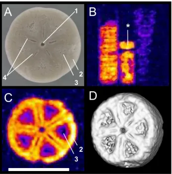
5
Figure 3 depicts MR images obtained from the Isselicrinus columnals from R ¨ugen (MNHB/MB.E 5730). Visible are the small central canal and the grooves and riches serving as attachment surfaces for connective ligaments as well as crenellae along the margin. The spatial resolution achieved with this sample was 108 µm in the image plane depicted here (subsequently, it was artificially increased by a factor of two via
10
zerofilling of the data set before Fourier transformation) and 50 µm perpendicular to it – the highest resolution achieved in the MR images described in this study.
5.2 Vertebrates
The MR images of the whale periotics (cf. Fig. 4) reveal a strong signal in the pos-terior cochlear part and the pospos-terior process of the bone. At the site of Groß
Pam-15
pau, the fossil bones are associated with diagenetically produced minerals like glau-conite and pyrite. The latter is a product of a reaction between iron (Fe released by haemoglobin degradation) and sulphur (H2S released by protein degradation, cf.
Pfretzschner,2000). FeS2(pyrite) develops under alkaline conditions, e.g. in the pres-ence of water-dissolved NH3 produced during collagen decay. It is less clear,
how-20
ever, what the destiny was of the hydrogen and carbon of the decomposed protein. It appears possible, though, that part of these molecules still reside in the bone and contribute to the high signal intensity.
BGD
4, 2959–3004, 2007 Fossil MRI D. Mietchen et al. Title Page Abstract Introduction Conclusions References Tables Figures ◭ ◮ ◭ ◮ Back Close Full Screen / EscPrinter-friendly Version Interactive Discussion
EGU
5.3 Plants
In the MR image depicted in Fig.5b, several anatomical details of the fir cone (MB.Pb. 2006/512) are visible: The cone axis is situated in the centre, surrounded by scales. The embedded seeds on each ovuliferous scale show a high signal intensity. This even allows to discern seed-internal patterns, though it remains unclear whether these
5
reflect part of the former biological structures (like seed integuments or embryos) or diagenetic alterations thereof.
The MR images (not shown) obtained from the Jurassic araucarian twig (MB.Pb. 2006/511) exhibited a much lower overall signal intensity than the Pararaucaria cone. One possible explanation for this could be long-term waterlogging, by means of which
10
the original organic matter would already have been decayed to a high degree before the onset of silicification – it is known that the physical appearance of fossil conifer remains is a function of the residence time either in a terrestrial or aquatic setting (Gastaldo, 1991). Experiments on extant leaf litter have demonstrated degradation in floating plant material: The breakdown starts already with the leaching of
water-15
soluble substances and is accelerated by higher water temperatures. This phase is followed by invasion of micro-organisms, mainly fungi and bacteria (Ferguson,1985). The fossil conifer twigs used for this study have most likely undergone both types of degradation while floating in the water column, perhaps for as long as several months. If the different fossilisation history of the cone, on the other hand, led to a complete
20
silicification shortly after abscission from the tree, it would appear plausible that some organic compounds might still be in place and possibly contributing to the MR image contrast.
5.4 Origin of the MR signal
The molecules that give rise to these signals can be characterised in multiple ways,
25
e.g. by using a Pulsed Gradient Spin Echo MR experiment (Stejskal and Tanner,1965) which measures their self-diffusion coefficient, an estimate of their translational
mobil-BGD
4, 2959–3004, 2007 Fossil MRI D. Mietchen et al. Title Page Abstract Introduction Conclusions References Tables Figures ◭ ◮ ◭ ◮ Back Close Full Screen / EscPrinter-friendly Version Interactive Discussion
EGU
ity. The highest self-diffusion coefficients we found in fossil samples (in some belem-nites) reached 10−11m2
s−1, which is comparable to that of water bound to cell
mem-branes (Volke et al.,1994), and more than two orders of magnitude smaller than the 2×10⁻⁹m2s−1typically found in free water (Mills,1973).
In most of the fossils we tested, no translational mobility was detectable. With the
5
given equipment and experimental parameters, it can thus be estimated that the self-diffusion coefficients of the1H-containing molecules within our fossil samples is gener-ally below 10−13m2s−1. Interestingly, diffusion determines decay in a recent physical
model of kerogenesis (Rothman and Forney,2007).
The transverse relaxation times (T2) in the fossil samples ranged in the order of
10
1 ms, which is three orders below free water but still several orders above those in solid crystals (Mansfield,1965), indicating that the MR-visible molecules retain a certain ro-tational mobility despite the absence of translational motion. This would be compatible with the idea of organic contributions to the signal, as the oscillating biomineralisation patterns described byFlorek et al.(2004, see slso Sect.5.1.1) would suggest.
15
To further test whether free water contributes to the MR signal, we froze one of the non-pathological Tendaguru belemnites with a self-diffusion coefficient near 10−11m2s−1 (Belemnopsis sp.; MB.C. 3700.11) down to –20◦C, which did not bring about any significant signal change indicative of a phase transition, nor did the signal change when the sample was heated up to 70◦C. We thus heated it in an oven for 8 h at
20
200◦C, which did not result in any observable weight loss on a milligram scale, and the MR spectra and images obtained thereafter showed no difference to those obtained before the temperature experiments.
In order to address the issue of the chemical nature of the MR signal in more de-tail, a piece of this belemnite was powdered and subjected to1H and 13C NMR MAS
25
spectroscopy. The 13C spectrum (cf. Fig. 6) shows a peak (at about 180 ppm) re-flecting C=O or C=S double bonds, while no other signal can be clearly identified, and namely no CHxgroups which would hint at organic material. These observations
BGD
4, 2959–3004, 2007 Fossil MRI D. Mietchen et al. Title Page Abstract Introduction Conclusions References Tables Figures ◭ ◮ ◭ ◮ Back Close Full Screen / EscPrinter-friendly Version Interactive Discussion
EGU
where CHxpeaks were readily observable (Hemsley et al.,1995;Lambert et al.,2000) but they fit nicely with the results of organochemical analysis of the belemnite’s pow-der by successive extractions with dichlormethyl/methanol, dichlormethyl/ethanol and ultrasonication – which did not reveal any traces of lipids (P. Albrecht and A. Charri ´e, personal communication) – and with the finding that total organic contents in (even
5
recent) echinoid calcite was less than 0.2 wt% (Gaffey,1995).
The1H spectrum (cf. inset in Fig.6), on the other hand, is dominated by the water and hydroxyl peak at 4.8 ppm but also shows a small peak between 0 and 1 ppm, which is indicative of cyclopropyl, metal-bound methyl groups or mineral hydroxyl groups (
Kali-nowski et al., 1984; Gaffey, 1995). So, at least in this belemnite, water or hydroxyl
10
groups (the latter perhaps bound to the mineral matrix) seem to have been the major signal contributor to our1H MR images, and the question about the origin of the signal translates into a question about the origin of these water or hydroxyl groups. We are not aware of any studies focusing on this issue specifically but given that temperature-stable water was found to exist in dental enamel (Myers,1965, using NMR) as well as
15
in recent and fossil shells (Hudson,1967, using gas chromatography), it was proposed that hydroxyl groups sometimes take the place of oxygen atoms in the crystal grid (
Mar-tin and Donnay,1972), which has since been affirmed by infrared and NMR techniques (Aines and Rossman,1984;Gaffey,1988,1995;Rossman,2006). Analyses of spots with high signal intensity (e.g. “3” in Fig.4) could provide further insights into the signal
20
origins in other fossils.
The strong but narrow peak in the1H spectrum (cf. Fig.6) can thus perhaps best be explained in terms of rigidly bound H2O molecules or OH groups being close enough to H2O molecules in a more liquid state, so as to allow for proton exchange. This fits well
with MR spectroscopic observations in echinoid calcite where liquid-like protons were
25
found to amount to about 60% of the total H2O content (Gaffey,1995). The latter var-ied around 2–3wt%, both between individuals and species but even more so between ossicles of the same specimen, which would suggest that total H2O might be indicative
BGD
4, 2959–3004, 2007 Fossil MRI D. Mietchen et al. Title Page Abstract Introduction Conclusions References Tables Figures ◭ ◮ ◭ ◮ Back Close Full Screen / EscPrinter-friendly Version Interactive Discussion
EGU
MR images and spectra acquired from neo- to palaeolithic mammalian bone and teeth samples suggests that neither 1H nor 13C MR signals predict PCR-amplifiable DNA content (D. Mietchen, M. Hofreiter, E.-M. Geigl, unpublished data).
5.5 Perspectives for fossil MRI
Perhaps the most obvious limitation for1H MRI of fossils is their typically low contents
5
of soft-bound1H. A possible strategy to deal with low intrinsic signal intensities is to fill cavities in the specimen of interest with solutions containing sufficient amounts of MR-visible1H (Sebes et al.,1991;Doughty and Tomutsa,1996;Steiger et al.,1997;
de Swiet et al.,1998;Steiger,2001;Clark et al., 2004) but such treatment might im-pede future chemical analyses, especially of biological macromolecules (Pruvost et al.,
10
2007). In this respect, it would be interesting to find out whether samples that expe-rienced such liquid treatment show any peculiarities in their state of preservation if compared to non-treated samples from the same specimen, though the replacement of the liquids by inert gases (cf. Seeley et al., 2004) could eventually alleviate these concerns.
15
Besides 1H, both MR spectroscopy and MR imaging are in principle possible with all isotopes that possess a net nuclear magnetic moment, i.e. those exhibiting odd numbers of protons or neutrons (e.g.2H,11B,13C,14N,15N,17O,19F,23Na,25Mg,29Si,
31
P and 39K). Since many of these elements play important roles during diagenesis (cf. introduction), such non-1H constituents might be of special interest for a variety of
20
palaeontological and related studies. The major limiting factor here is sensitivity, whose upper limit in a given static magnetic field depends on the gyromagnetic ratio of the target isotope and on its abundance within the sample, while the practically achievable value is further dependent upon acquisition parameters (for details, see Abragam,
1961;Callaghan,1991). 13C MR spectroscopy (as in Fig.6) has already found multiple
25
applications in archaeological research (Lambert et al.,2000), and 31P MRI has been applied to fresh bone and teeth (Li,1991;Wu et al.,1999).
BGD
4, 2959–3004, 2007 Fossil MRI D. Mietchen et al. Title Page Abstract Introduction Conclusions References Tables Figures ◭ ◮ ◭ ◮ Back Close Full Screen / EscPrinter-friendly Version Interactive Discussion
EGU
signal correlates with spin (and thus mass) density, the MR signal obtainable from solid samples (like most fossils) will normally be above that from gaseous and – due to chemical binding strength – below that from liquid samples of similar chemical compo-sition. From this perspective, it is perhaps surprising that we know of only one report of an MRI examination of a mummy and that imaging was not even attempted after
5
examining the induction decay signal (Notman et al.,1986). It seems reasonable for us to assume that the technological advances over the two decades since then justify a reconsideration of MRI for the study of mummified or otherwise preserved ancient tissue samples, e.g. soft tissue recovered from within dinosaur bones (cf.Schweitzer
et al.,2005,2007;Asara et al.,2007), which we expect to yield a stronger MR signal
10
per voxel than the completely mineralised samples shown here.
Mildly frozen specimens like mammoths uncovered from permafrost soil often show remarkable states of soft tissue preservation (Ezra and Cook,1959;Zimmerman and
Tedford,1976;Cooper,2006), which sometimes even allows for genome-level genetic analyses (Poinar et al.,2006). Such samples, despite being frozen, still contain
con-15
siderable amounts of unfrozen water (Koop,2004) and thus represent another window of opportunity for MR techniques, as demonstrated in sea ice (Eicken et al.,2000) or permafrost samples (Kleinberg and Griffin,2005).
Another important aspect to be considered is sample size. First, at a given resolution, imaging a larger sample means acquiring more data points and thus longer scanning.
20
Second, larger samples generally require larger coils (which will reduce the achievable resolution under otherwise identical conditions), and the ultimate coil size within a given imaging setup is given by the inner diameter of the gradient coils (in our case 38 mm, in human MRI scanners typically around 80 mm). Third, although an increase in field strength generally provides for an increase in signal, noise and signal-to-noise ratio,
25
a number of artifacts also get more pronounced then (e.g. susceptibility distortions near interfaces with different magnetic susceptibilities). Fourth, there are a number of further parameters relevant to MR measurements (e.g. field homogeneity, sample and coil temperature, gradient strength, filling factor, pulse sequence), and so the choice of
BGD
4, 2959–3004, 2007 Fossil MRI D. Mietchen et al. Title Page Abstract Introduction Conclusions References Tables Figures ◭ ◮ ◭ ◮ Back Close Full Screen / EscPrinter-friendly Version Interactive Discussion
EGU
the experimental conditions is vital (for details, seeAbragam,1961;Callaghan,1991;
Ernst et al.,1997).
In terms of materials, the only major restriction is that specimens should not exhibit a permanent magnetisation (due to ferro- or paramagnetic constituents) beyond about 10−7(i.e. 0.1 ppm) of the static magnetic field, as this would distort the latter and thus
5
interfere with the way spatial or spectral information are encoded. Fulfilment of this criterion will probably show (like the apical lines in Fig. 2c, d) some taxonomically relevant bias due to biomineralisation or diagenetic environment but this bias is unlikely to extend to higher taxonomic units, and so we suggest that the fossil invertebrate, vertebrate and plant taxa described here should not be seen as a limit but rather taken
10
as starting points for a more detailed screening of fossil lineages by MR techniques. Comparative evolutionary studies – especially between closely related species, of which only one exhibits a particular trait of interest, while the other does not – have proven useful in and now form the core of most if not all biological disciplines. Palaeon-tological investigations, specifically, could profit from comparative studies including
ex-15
tant species, thereby complementing the existing knowledge with anatomical and dy-namic details concerning processes like post mortem tissue decay (Weigelt,1927) and in vivo cellular metabolic activities like biomineralisation (e.g.Freytet et al.,1996;Levi
et al.,1998;Kolo and Claeys,2005) as well as embryological development (Xiao,2002) or locomotion (Gatesy et al.,1999). For all these applications, suitable MR techniques
20
are now in place (e.g.Ciobanu et al.,2003;Manz et al.,2003;M ¨uller et al.,2006;Lee
et al., 2006, 2007; Honda and Hata, 2007). This versatility of MR techniques, along with their non-invasiveness, renders them a very promising tool for such comparative investigations (e.g.Hopkins and Rilling,2000;Spoor et al.,2000;Glidewell et al.,2002). Ongoing developments in MR technology (Glover and Mansfield,2002) and the ever
25
wider availability of high-field imaging facilities suggest that MRI will continue to help bypass and circumvent current methodological limitations and to generate more appli-cations in the geosciences (Carlson,2006) and neighbouring fields. As an example, consider portable NMR devices (Eidmann et al., 1996; Prado, 2003; Bl ¨umich et al.,
BGD
4, 2959–3004, 2007 Fossil MRI D. Mietchen et al. Title Page Abstract Introduction Conclusions References Tables Figures ◭ ◮ ◭ ◮ Back Close Full Screen / EscPrinter-friendly Version Interactive Discussion
EGU 2002;Manz et al.,2006;Marble et al.,2006;Marko et al.,2007;McDonald et al.,2007)
which opened up the possibility to examine samples on the spot, be it in the field or in archives. While these devices are currently far from capable of producing images com-parable to those presented here, the characteristic relaxation parameters or spectral fingerprints they already can measure could become a valuable non-invasive
categori-5
sation tool when screening rock samples for embedded fossils.
6 Conclusions
The data presented here demonstrate that MR imaging allows micromorphological de-tails within intact fossils to be studied non-invasively at resolutions down to 50 µm, i.e. comparable to those of CT images and well below the 100 µm that “would be
satisfac-10
tory [. . .] for a majority of researchers and for most applications” (Lyons et al.,2000), at least after the Cambrian explosion. The intrinsic MR signal used to acquire the images appears to originate from mobile yet not diffusible water molecules, while the range of specimens suitable for MRI has been extended beyond liquid-filled mouldic or cav-ernous fossils (Sebes et al.,1991;Steiger,2001;Clark et al.,2004) and pathological
15
belemnites (Mietchen et al., 2005), so that it now comprises invertebrate, vertebrate and plant specimens obtained from a variety of sites. Moreover, after MRI scanning, all other palaeontological investigations still remain possible – which is not necessarily true in the opposite case. It should be noted that the digital availability of MRI data (as with CT and other modalities; cf. Zollikofer and Ponce de L ´eon, 2005) renders them
20
ideal for applications like computational morphology (Bookstein,1996) or rapid proto-typing (Zollikofer and Ponce de L ´eon, 1995) which have a high potential not only in research but also in science education at school or in museums. Taken together, the microscopic resolution currently achieved with MR techniques, their non-invasiveness, the possibility to obtain taxonomically relevant 3-D spatial as well as chemical
informa-25
tion, their potentially broad applicability and the multitude of ongoing efforts to further improve them all suggest they could open up complementary avenues for non-invasive
BGD
4, 2959–3004, 2007 Fossil MRI D. Mietchen et al. Title Page Abstract Introduction Conclusions References Tables Figures ◭ ◮ ◭ ◮ Back Close Full Screen / EscPrinter-friendly Version Interactive Discussion
EGU
approaches to a wide range of palaeobiological issues.
Acknowledgements. We wish to thank W. Eckloff and S. F ¨uting (L ¨ubeck) for providing the
ken-trodontid periotics, H. Keupp (Berlin) for helpful discussions and E.-M. Geigl (Paris) as well as Y. Fern ´andez-Jalvo (Madrid) for commenting on earlier drafts of the manuscript.
References
5
Abelson, P. H.: Organic constituents of fossils, Carnegie Inst. Wash. Year Book, 53, 97–101, 1954. 2961
Aberhan, M., Bussert, R., Heinrich, W.-D., Schrank, E., Schultka, S., Sames, B., Kriwet, J., and Kapilima, S.: Palaeoecology and depositional environments of the Tendaguru Beds (Late Jurassic to Early Cretaceous, Tanzania), Mitt. Mus. f. Naturkunde Berl. Geowiss. Reihe, 5,
10
19–44, 2002. 2971
Abragam, A.: The Principles of Nuclear Magnetism, Clarendon, Oxford, 1961. 2962, 2980,
2982
Aines, R. D. and Rossman, G. R.: Water in minerals? A peak in the infrared, J. Geophys. Res., 89B, 4059–4071, 1984. 2979
15
Albrecht, P. and Ourisson, G.: Biogenic substances in sediments and fossils, Angew. Chem. Int. Ed. Engl., 10, 209–225, 1971.2961
Andrew, E. R.: Magic Angle Spinning in Solid State n. m. r. Spectroscopy, Phil. Trans. R. Soc. Lond. A, 299, 505–520, 1981. 2975
Asara, J. M., Schweitzer, M. H., Freimark, L. M., Phillips, M., and Cantley, L. C.: Protein
Se-20
quences from Mastodon and Tyrannosaurus rex Revealed by Mass Spectrometry, Science, 316, 280–285, doi:10.1126/science.1137614, 2007.2961,2981
Ausich, W. I., Brett, C. E., Hess, H., and Simms, M. J.: Crinoid form and function, in: Fossil Crinoids, edited by Hess, H., Ausich, W. I., Brett, C. E., and Simms, M. J., 3–30, Cambridge University Press, Cambridge, 1999. 2966
25
Bandel, K. and Spaeth, C.: Structural differences in the ontogeny of some belemnite rostra, in: Cephalopods present and past, edited by Wiedmann, J. and Kullmann, J., 247–271, Schweizerbart, Stuttgart, 1988. 2961,2965
Barnes, L. G.: The late Miocene dolphin Pithanodelphis Abel, 1905 (Cetacea: Kentriodontidae) from California, Contr. Sci. Nat. Hist. Mus. Los Angeles Co., 367, 1–27, 1985. 2972
BGD
4, 2959–3004, 2007 Fossil MRI D. Mietchen et al. Title Page Abstract Introduction Conclusions References Tables Figures ◭ ◮ ◭ ◮ Back Close Full Screen / EscPrinter-friendly Version Interactive Discussion
EGU
Behrensmeyer, A. K.: Taphonomic and Ecologic Information from Bone Weathering, Paleobiol., 4, 150–162, 1978. 2963
Behrensmeyer, A. K., Kidwell, S. M., and Gastaldo, R. A.: Taphonomy and paleobiology, Pale-obiol., 26, 103–147, 2000.2961,2963
Berta, A. and Sumich, J. L.: Marine mammals: Evolutionary biology, Academic Press, San
5
Diego, 494p., 1999. 2969
Blumer, M.: Organic pigments: their long-term fate, Science, 149, 722–726, 1965.2961,2967
Bl ¨umich, B., Anferov, V., Anferova, S., Klein, M., Fechete, R., Adams, M., and Casanova, F.: Simple NMR-mouse with a bar magnet, Conc. Magn. Reson. B (Magn. Reson. Eng.), 15, 255–261, 2002. 2982
10
Bocherens, H., Tresset, A., Wiedemann, F., Giligny, F., Lafage, F., Lanchon, Y., and Mariotti, A.: Diagenetic evolution of mammal bones in two French Neolithic sites, Bull. Soc. G ´eol. France, 168, 555–564, 1997. 2967
Bookstein, F.: Biometrics, biomathematics and the morphometric synthesis, Bull. Math. Biol., 58, 313–365, 1996.2983
15
Borah, B., Gross, G. J., Dufresne, T. E., Smith, T. S., Cockman, M. D., Chmielewski, P. A., Lundy, M. W., Hartke, J. R., and Sod, E. W.: Three-dimensional microimaging (MRmicroI and microCT), finite element modeling, and rapid prototyping provide unique insights into bone architecture in osteoporosis, Anat. Rec., 265, 101–110, doi:10.1002/ar.1060, 2001.
2963
20
Boyce, C. K., Hazen, R. M., and Knoll, A. H.: Nondestructive, in situ, cellular-scale mapping of elemental abundances including organic carbon in permineralized fossils, Proc. Natl. Acad. Sci. USA, 98, 5970–5974, doi: 10.1073/pnas.101130598, 2001. 2961,2970
Boyce, C. K., Cody, G. D., Feser, M., Jacobsen, C., Knoll, A. H., and Wirick, S.: Organic chemical differentiation within fossil plant cell walls detected with X-ray spectromicroscopy,
25
Geology, 30, 1039–1042, 2002. 2962
Briggs, D. E. G.: The role of decay and mineralization in the preservation of soft-bodied fossils, Annu. Rev. Earth Planet. Sci., 31, 275–301, doi:10.1146/annurev.earth.31.100901.144746, 2003. 2961,2963
Brocks, J. J., Logan, G. A., Buick, R., and Summons, R. E.: Archean Molecular Fossils and the
30
Early Rise of Eukaryotes, Science, 285, 1033–1036, 1999. 2961
Calder, M. G.: A coniferous petrified forest in Patagonia, Bull. Brit. Mus. (Nat. Hist.) Geology, 2, 99–138, 1953.2973
BGD
4, 2959–3004, 2007 Fossil MRI D. Mietchen et al. Title Page Abstract Introduction Conclusions References Tables Figures ◭ ◮ ◭ ◮ Back Close Full Screen / EscPrinter-friendly Version Interactive Discussion
EGU
Callaghan, P. T.: Principles of Nuclear Magnetic Resonance Microscopy, Oxford University Press, Clarendon, 1991. 2962,2980,2982
Carlson, W. D.: Three-dimensional imaging of earth and planetary materials, Earth Planet. Sci. Lett., 249, 133–147, 2006. 2962,2982
Chudek, J. A. and Hunter, G.: Magnetic resonance imaging of plants, Progr. Nucl. Magn.
Re-5
son. Spectr., 31, 43–62, 1997. 2970
Ciobanu, L., Webb, A., and Pennington, C.: Magnetic resonance imaging of biological cells, Prog. Nucl. Magn. Reson. Spectrosc., 42, 69–93, 2003. 2982
Clark, N. D. L., Adams, C., Lawton, T., Cruickshank, A. R., and Woods, K.: The Elgin marvel: using magnetic resonance imaging to look at a mouldic fossil from the Permian of Elgin,
10
Scotland, UK, Magn. Reson. Imaging, 22, 269–273, doi: 10.1016/j.mri.2003.09.006, 2004.
2962,2980,2983
Collins, M. J. and Gernaey-Child, A. M.: Fossilized Materials: Proteins, in: Palaeobiology II, edited by Briggs, D. E. G. and Crowther, P. R., pp. 247–271, Blackwell Science Inc., Cam-bridge, Massachussetts, 2001. 2964
15
Collins, M. J., Riley, M. S., Child, A. M., and Turner-Walker, G.: A basic mathematical simulation of the chemical degradation of ancient collagen, J. Archaeol. Sci., 22, 175–183, 1995. 2967
Collins, M. J., Nielsen-Marsh, C. M., Hiller, J., Smith, C. I., Roberts, J. P., Prigodich, R. V., Wess, T. J., Csap `o, J., Millard, A. R., and Turner-Walker, G.: The Survival of Organic Matter in Bone: A Review, Archaeometry, 44, 383–394, 2002.2963
20
Cooper, A.: The Year of the Mammoth., PLoS Biol, 4, e78, 2006.2981
Crenshaw, M. A.: Biomineralization mechanisms, in: Skeletal Biomineralization: Patterns, Pro-cesses and Evolutionary Trends, edited by Carter, J. G., pp. 1–9, Van Nostrand Reinhold, New York, 1990. 2964
Darrow, B. S.: A fossil araucarian embryo from the Cerro Cuadrado of Patagonia, Bot. Gaz.,
25
98, 328–337, 1936.2973
Dauphin, Y.: Fossil organic matrices of the Callovian aragonitic ammonites from Lukow (Poland): location and composition, Int. J. Earth Sci., 93, 1071–1080, 2002. 2963
de Muizon, C.: Les vert ´ebr ´es fossiles de la formation Pisco (Perou) III. Troisi `eme partie: Les Odontoc `etes (Cetacea, Mammalia) du Mioc `ene, Inst. Fr. ´Etud. And. M `em., 78, 1–240, 1988.
30
2972
de Swiet, T. M., Tomaselli, M., H ¨urlimann, M. D., and Pines, A.: In Situ NMR
BGD
4, 2959–3004, 2007 Fossil MRI D. Mietchen et al. Title Page Abstract Introduction Conclusions References Tables Figures ◭ ◮ ◭ ◮ Back Close Full Screen / EscPrinter-friendly Version Interactive Discussion
EGU
doi:10.1006/jmre.1998.1459, 1998.2962,2963,2980
Dernbach, U., Jung, W., Selmeier, A., G ¨otz, K., and Fine, H.: Araucaria. The petrified Arau-carias from the Cerro Cuadrado, Argentina, D’Oro Verlag, Lorsch, 1992. 2973
DeVore, M. L., Kenrick, P., Pigg, K. B., and Ketcham, R. A.: Utility of high resolution x-ray computed tomography (HRXCT) for paleobotanical studies: an example using London Clay
5
fruits and seeds, Am. J. Botany, 93, 1848, 2006. 2970
Dietrich, W.: Zur Stratigraphie und Palaeontologie der Tendaguruschichten, Palaeontographica, II (Suppl. VII, part 2), 1–86, 1933.2971
Domanus, J. C.: Practical Neutron Radiography, Kluwer, Dordrecht, Netherlands, 1992.2962
Donoghue, P. C., Bengtson, S., Dong, X. P., Gostling, N. J., Huldtgren, T., Cunningham, J. A.,
10
Yin, C., Yue, Z., Peng, F., and Stampanoni, M.: Synchrotron X-ray tomographic microscopy of fossil embryos., Nature, 442, 680–3, 2006. 2962
Doughty, D. A. and Tomutsa, L.: Multinuclear NMR microscopy of two-phase fluid systems in porous rock., Magn. Reson. Imaging, 14, 869–73, 1996. 2980
Drum, R. W.: Silification of Betula woody tissue in vitro, Science, 161, 175–176, 1968.2970
15
Dunca, E., Doguzhaeva, L., Sch ¨one, B. R., and Van de Schootbrugge, B.: Growth patterns in rostra of the middle jurassic belemnite Megateuthis giganteus: controlled by the moon?, Geolines, in press, 2007. 2961
Durand, B.: A History of Organic Geochemistry, Oil Gas Sci. Technol. Rev. IFP, 58, 203–231, 2003. 2961
20
Eglinton, G. and Logan, G. A.: Molecular preservation, Philos. Trans. R. Soc. Lond. B Biol. Sci., 333, 315–327, 1991. 2961
Eicken, H., Bock, C., Wittig, R., Miller, H., and Poertner, H.-O.: Magnetic resonance imaging of sea-ice pore fluids: methods and thermal evolution of pore microstructure, Cold Regions Sci. Technol., 31, 207–225, 2000.2981
25
Eidmann, G., Savelsberg, R., Bl ¨umler, P., and Bl ¨umich, B.: The NMR MOUSE, a Mobile Uni-versal Surface Explorer, J. Magn. Reson. A, 122, 104–109, 1996. 2982
Engel, M. H., Goodfriend, G. A., Qian, Y., and Macko, S. A.: Indigeneity of organic matter in fossils: a test using stable isotope analysis of amino acid enantiomers in Quaternary mollusk shells, Proc. Natl. Acad. Sci. U S A, 91, 10 475–10 478, 1994. 2961
30
Ernst, G.: Stratigraphische und gesteinschemische Untersuchungen im Santon und Campan von L ¨agerdorf (SW-Hostein), Mitt. Geol. Staatsinst. Hamburg, 32, 71–127, 1963. 2967