HAL Id: hal-02919104
https://hal.archives-ouvertes.fr/hal-02919104
Submitted on 7 Nov 2020HAL is a multi-disciplinary open access archive for the deposit and dissemination of sci-entific research documents, whether they are pub-lished or not. The documents may come from teaching and research institutions in France or abroad, or from public or private research centers.
L’archive ouverte pluridisciplinaire HAL, est destinée au dépôt et à la diffusion de documents scientifiques de niveau recherche, publiés ou non, émanant des établissements d’enseignement et de recherche français ou étrangers, des laboratoires publics ou privés.
Iron-biomineralizing organelle in magnetotactic bacteria:
function, synthesis and preservation in ancient rock
samples
Matthieu Amor, François Mathon, Caroline Monteil, Vincent Busigny,
Christopher Lefèvre
To cite this version:
Matthieu Amor, François Mathon, Caroline Monteil, Vincent Busigny, Christopher Lefèvre. Iron-biomineralizing organelle in magnetotactic bacteria: function, synthesis and preservation in ancient rock samples. Environmental Microbiology, Society for Applied Microbiology and Wiley-Blackwell, 2020, �10.1111/1462-2920.15098�. �hal-02919104�
Iron-biomineralizing organelle in magnetotactic bacteria: function, synthesis
1
and preservation in ancient rock samples
2 3
Matthieu Amor1, François P. Mathon1,2, Caroline L. Monteil1 , Vincent Busigny2,3, Christopher
4
T. Lefevre1
5 6
1Aix-Marseille University, CNRS, CEA, UMR7265 Institute of Biosciences and Biotechnologies
7
of Aix-Marseille, CEA Cadarache, F-13108 Saint-Paul-lez-Durance, France 8
2Université de Paris, Institut de Physique du Globe de Paris, CNRS, F-75005, Paris, France.
9
3Institut Universitaire de France, 75005 Paris, France
10 11
Abstract
12
Magnetotactic bacteria (MTB) are ubiquitous aquatic microorganisms that incorporate iron 13
from their environment to synthesize intracellular nanoparticles of magnetite (Fe3O4) or
14
greigite (Fe3S4) in a genetically controlled manner. Magnetite and greigite magnetic phases
15
that allow MTB to swim towards redox transitions zones where they thrive. MTB may 16
represent some of the oldest microorganisms capable of synthesizing minerals on Earth, and 17
have been proposed to significantly impact the iron biogeochemical cycle by immobilizing 18
soluble iron into crystals that subsequently fossilize in sedimentary rocks. In the present article, 19
we provide an overview of the mechanisms leading to iron mineralization in MTB. We then 20
describe their distribution in the environment, and discuss the possible function of the 21
magnetite and greigite nanoparticles. Finally, we update the methods used for the detection 22
of MTB crystals in sedimentary rocks and present their occurrences in the geological record. 23
Introduction
24
Biomineralization corresponds to the capacity for living organisms to produce minerals such 25
as carbonates, sulfides, phosphates and oxides. It can be an indirect process during which the 26
metabolic activity of organisms transforms the physico-chemical conditions of their 27
environment and induces the formation of mineral phases. Alternatively, biomineralization 28
can be genetically controlled and serve biological functions. Biomineralization is widespread 29
among living organisms, and can significantly impact biogeochemical cycles of elements (e.g. 30
C, Ca, P, S, Fe) both qualitatively and quantitatively (e.g. Ridgwell and Zeebe, 2005). 31
Magnetotactic Bacteria (MTB) constitute an elegant example of controlled biomineralization 32
(Fig. 1). They were first observed by Salvatore Bellini, a medical Doctor at the University of 33
Pavia in Italia in the late 1950s and early 1960s (Bellini, 2009a,b). They were then described 34
by Richard Blakemore from samples collected near Woods Hole, Massachusetts (Blakemore, 35
1975). They both noticed a group of bacteria whose swimming direction could be tuned using 36
a magnet. Such magnetic behavior was explained by R. Blakemore, who observed intracellular 37
crystals corresponding to either magnetite [Fe(II)Fe(III)2O4] or greigite [Fe(II)Fe(III)2S4] in these
38
bacteria (Blakemore, 1975; Mann et al., 1990). 39
Since their discovery, MTB have been found in a large variety of aquatic ecosystems including 40
lakes, rivers, oceans, estuaries and lagoons (Lin et al., 2014a). They incorporate a high amount 41
of dissolved iron from their environment for magnetite or greigite synthesis, which 42
corresponds to a mass of 10- to 100-fold higher than the one estimated in the bacterium 43
Escherichia coli (Amor et al., 2020a). When magnetotactic cells die, their crystals may be 44
deposited in sediments and preserved during diagenesis (i.e. the physico-chemical 45
transformation of sediments into sedimentary rock associated with natural burial), which 46
effectively removes iron from the soluble pool (Chen et al., 2014; Larrasoaña et al., 2014). This 47
raised the hypothesis that MTB could significantly contribute to the iron biogeochemical cycle 48
by sequestering a large amount of dissolved iron into insoluble crystals (Lin et al., 2014a; Amor 49
et al., 2020a). MTB could have also impacted the ancient Earth surface, since they were 50
proposed to have emerged during the Archean (Kopp and Kirschvink, 2008; Lin et al., 2017a). 51
In MTB, magnetite and greigite crystals are produced in organelles called magnetosomes. 52
They correspond to prokaryotic organelles made of a bi-layered lipid membrane containing 53
proteins and surrounding a single magnetite or greigite nanoparticle. Magnetosomes are 54
aligned as chains, and provide the cell with a magnetic dipole. It is assumed that chains of iron 55
nanoparticles are used as a compass, allowing the bacteria to passively align along the 56
geomagnetic field’s lines to efficiently reach the Oxic/Anoxic Transition Zone (OATZ) in water 57
columns or sediments where they thrive (Frankel et al., 1981). However, observation of MTB 58
at the geomagnetic equator where the Earth’s magnetic field has no vertical component 59
(Frankel et al., 1981) suggests that magnetite or greigite could have an additional function for 60
the bacteria such as energy storage (Byrne et al., 2015). 61
Here, we first describe the biodiversity, distribution and ecophysiology of MTB, and discuss 62
the proposed function for magnetosomes in the environment. Then, we provide an overview 63
of iron biomineralization in MTB. Mechanisms of iron incorporation and magnetite formation 64
discovered in model magnetotactic strains cultivated in the laboratory are presented. Finally, 65
we summarize occurrences of fossil MTB found in the geological record, as well as the 66
methods used for identification of biological magnetite and greigite. Based on paleontological 67
data, we speculate on the emergence of MTB and potential additional function(s) for 68
magnetosomes over geological times. 69
70
1. Ecophysiology of magnetotactic bacteria
Biodiversity of magnetotactic bacteria 72
MTB diversity is reflected by a great variation of their shape, physiology and taxonomy. All 73
known cultured and uncultured MTB are motile by means of flagella and have a cell wall 74
characteristic of Gram-negative bacteria. The arrangement of flagella differs among MTB and 75
can be either polar, bipolar or arranged in bundles. Another trait showing considerable 76
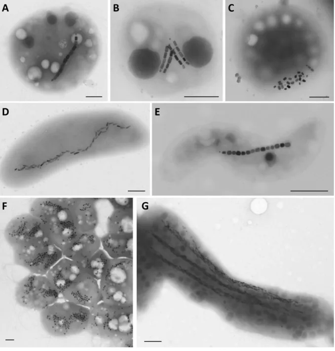
diversity is the arrangement of magnetosomes within the cell (Fig. 1). In the majority of MTB, 77
magnetosomes are aligned in one or more straight chains parallel to the cell’s long axis, which 78
maximizes the magnetic moment of the bacteria and reflects their evolutionary optimization 79
for magnetic navigation. However, disordered magnetosomes located at one side of the cell 80
occur in some magnetotactic cocci (Moench, 1988; Cox et al., 2002; Freitas et al., 2003) (Fig. 81
1C). Magnetosome mineral particles display diverse, well-defined and strain-specific 82
morphologies (Bazylinski et al., 1994). Four magnetite or greigite morphologies are found in 83
MTB: cuboctahedral, elongated prismatic, tooth-shaped and bullet-shaped (Balkwill et al., 84
1980; Mann et al., 1984a, 1984b; Mann et al., 1987a, 1987b; Heywood et al., 1990; Devouard 85
et al., 1998; Pósfai et al., 1998a, 1998b; Lefèvre et al., 2011a) (Fig. 1). In addition to 86
magnetosomes, other inclusions containing elemental sulfur, polyphosphate or poly-β-87
hydroxybutyrate (PHB) are common in MTB (Bazylinski et al., 2004; Schultheiss et al., 2005; 88
Rivas-Lamelo et al., 2017). A study of MTB from the Seine River (France) indicated that cells 89
of uncultured MTB contain barium-rich and calcium oxide inclusions (Isambert et al., 2007). 90
Some MTB from the Nitrospirae and Omnitrophica phyla also contain numerous sulfur 91
globules (Jogler et al., 2010; Lefèvre et al., 2011b; Kolinko et al., 2012; Qian et al., 2019). 92
Another feature illustrating MTB diversity is their type of magnetotactic behavior. A study 93
carried out on 12 cultured MTB from different taxa with various morphologies, physiologies, 94
and flagellar apparatus showed six different magnetotactic behaviors described as a 95
combination of three distinct mechanisms, including dipolar, axial and unipolar mechanisms 96
(Lefèvre et al., 2014). 97
A straightforward experiment to reveal the diversity of MTB consists in performing a 98
magnetic enrichment of an environmental sample containing sediment and water. In most 99
habitats the light microscope observation will allow the observation of various cell 100
morphotypes and behaviors. The main morphotypes include coccoid-to-ovoid cells (Fig. 1A-C), 101
rods (Fig. 1D), vibrios and spirilla (Fig. 1E) of various dimensions. Two unique morphotypes 102
include the multicellular magnetotactic prokaryotes (MMPs) (Keim et al., 2007) (Fig. 1F), and 103
the very large rod Candidatus Magnetobacterium bavaricum (Jogler et al., 2010) (Fig. 1G). 104
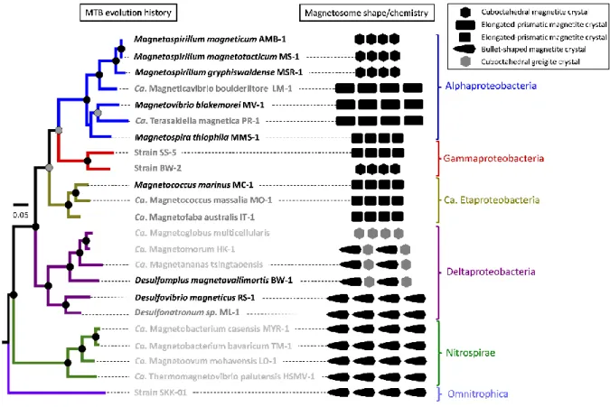
MTB do not represent a well-defined taxonomic group of prokaryotes: they are distributed 105
over several bacterial taxa containing non-magnetotactic species as well (Lefèvre and 106
Bazylinski, 2013) (Fig. 2). Most of our knowledge on MTB diversity is based on isolation, 107
cultivation, genomic, metagenomic and single-cell genomic studies (Jogler et al., 2011; Kolinko 108
et al., 2012; Lefèvre et al., 2012; Lin et al., 2018). The majority of MTB described so far belongs 109
to four subdivisions of Proteobacteria (Alpha, Beta, Gamma and Deltaproteobacteria classes), 110
and some are affiliated with the Nitrospirae and Omnitrophica phyla (Lefèvre and Bazylinski, 111
2013; Lin et al., 2017b) Recent metagenomic data have suggested that even some lineages 112
within the deep-branching Latescibacteria and Planctomycetes phyla may also produce 113
magnetosomes (Lin et al., 2018). With their deep-branched phylogenetic position, the latter 114
groups are particularly important to study since they would represent the earliest examples 115
of biomineralization on Earth (Parks et al., 2018). Magnetosome biogenesis is thus a 116
polyphyletic, possibly ancient trait associated with a large genetic, metabolic, and 117
morphological diversity. Interestingly, there is an obvious and apparently strong correlation 118
between the composition and morphology of magnetosome mineral crystals produced by 119
MTB and their phylogenetic affiliation (Lefèvre and Wu, 2013; Lefèvre et al., 2013; Pósfai et 120
al., 2013) (Fig. 2). 121
The physiology of known MTB, either determined experimentally with cultured strains or 122
inferred from genomic studies on uncultured strains, is also quite diverse. Cultured MTB 123
belong to three classes of the Proteobacteria phylum (Alpha, Gamma and 124
Deltaproteobacteria) (Fig. 2). They are obligate microaerophiles, anaerobes or both (Bazylinski 125
and Williams, 2007). Those that tolerate relatively high concentrations of oxygen do not 126
synthesize magnetite under these conditions. They are mesophilic with regard to growth 127
temperature, and none grow at temperatures much higher than 30°C. Known MTB are 128
chemoorganoheterotrophic and/or chemolithoautotrophic. In the model strains of the 129
Magnetospirillum genus, heterotrophic growth occurs while cells are using organic acids as a 130
source of carbon and electrons (Schleifer et al., 1991). MSR-1 is also capable of autotrophic 131
and mixotrophic growth using reduced sulfur compounds as a source of electrons (Geelhoed 132
et al., 2010). Species of the Magnetococcus (Lefèvre et al., 2009; Bazylinski, Williams, Lefèvre, 133
Berg, et al., 2013; Morillo et al., 2014) and Magnetospira genera (Zhu et al., 2010; Williams et 134
al., 2012) as well as strains SS-5 and BW-2 of the Gammaproteobacteria class (Lefèvre et al., 135
2012) are also growing autotrophically with reduced sulfur compounds. Species of the 136
Magnetovibrio (Bazylinski et al., 2013) and Terasakiella (Monteil et al., 2018) genera are 137
heterotrophic with Magnetovibrio blakemorei strain MV-1 that shows the greatest metabolic 138
versatility regarding the compounds that can be used as potential electron donors and carbon 139
sources for growth during microaerobic and anaerobic growth (Bazylinski et al., 2013). Indeed, 140
it has a respiratory metabolism, using oxygen, nitrate, and nitrous oxide (N2O) as terminal
141
electron acceptors (Bazylinski et al., 1988). It grows chemoorganoheterotrophically and 142
chemolithoautotrophically but also chemoorganoautotrophically with formate as the electron 143
donor. The pathway of autotrophy is the Calvin-Benson-Bassham cycle since ribulose-1,5-144
bisphosphate carboxylase/oxygenase (RubisCO) genes were found in the genome of most 145
autotrophic MTB of the Alpha and Gammaproteobacteria classes. Only Magnetococcus 146
species such as strain MC-1 appear to use the rTCA cycle for autotrophic carbon assimilation 147
(Bazylinski et al., 2013). Cultured strains of the Deltaproteobacteria, including several species 148
of the Desulfovibrionales (Sakaguchi et al., 2002; Lefèvre et al., 2011c) and Desulfobacterales 149
(Abreu et al., 2007; Descamps et al., 2017) orders grow anaerobically while reducing sulfate. 150
Cells of some strains of the Desulfovibrio genus also present the potential to grow as a 151
microaerophilic band of cells at the oxic–anoxic interface in media lacking sulfate (Lefèvre et 152
al., 2016). Desulfovibrio magneticus strain RS-1 is the only cultured magnetotactic bacterium 153
known to be capable of fermentation: pyruvate is fermented to acetate and hydrogen 154
(Sakaguchi et al., 2002). 155
Genomic, environmental and ultrastructural evidences seem to indicate that the deep-156
branched MTB including the Nitrospirae candidate genera Magnetobacterium (Jogler et al., 157
2011; Lin et al., 2014b; Kolinko et al., 2016) and Magnetoovum (Lefèvre et al., 2011; Kolinko 158
et al., 2016) as well as some magnetotactic cocci of the Magnetococcaceae family isolated 159
from the water column of the Black Sea (Schulz-Vogt et al., 2019) are growing anaerobically 160
using nitrate as respiratory electron acceptor and oxidizing reduced sulfur compounds. 161
Although some MTB have been shown to be sensitive to light (Frankel et al., 1997; Chen et al., 162
2011; Shapiro et al., 2011) with genomic evidences showing the presence of phototrophic 163
genes in some Magnetospirillum strains (Wang et al., 2019), no photosynthetic MTB have 164
been found. So far, no MTB were shown to be able to use reduced or oxidized forms of iron 165
for their energetic metabolism. 166
Environments hosting magnetotactic bacteria 168
MTB have been described worldwide and found on all continents. They are ubiquitous in 169
sediments of freshwater, brackish, marine, and hypersaline habitats (Lin et al., 2017b). The 170
occurrence of MTB appears to depend on the presence of an Oxic-Anoxic Transition Zone 171
(OATZ) consisting of opposing gradients of oxygen from the surface and reduced compounds 172
from the sediments (Frankel et al., 1997). The largest numbers of MTB are typically found at 173
or slightly below the OATZ (Jogler et al., 2010; Lefèvre et al., 2011b). The different species of 174
MTB show specific distributions within the OATZ. Magnetite-producing MTB are generally 175
found at or very close to the OATZ, while greigite producers are present in reducing biotopes, 176
below the OATZ, in the sulfidic anoxic zone (Moskowitz et al., 2008; Lefèvre et al., 2011c). 177
Although most environmental studies on MTB were conducted on sediments of aquatic 178
environments, some freshwater and saline lakes with water column stratification, seasonally 179
or permanently, were also found to be suitable to host diverse populations of MTB (Simmons 180
et al., 2004; Rivas-Lamelo et al., 2017; Schulz-Vogt et al., 2019). 181
Biogeochemical studies indicate that some environmental parameters such as salinity, 182
temperature, nitrate, or sulfur compounds could explain MTB abundance or community 183
differences (Martins et al., 2009, 2012; Lin et al., 2012, 2013). Even if the largest proportion 184
of MTB is detected within the suboxic zone, a strict correlation between the distribution of 185
MTB and specific geochemical parameters has never been demonstrated (Flies et al., 2005). 186
For example, the presence of MTB is not correlated with the concentration in dissolved iron. 187
Ferrous or ferric salts in the micromolar concentrations range are sufficient for growth and 188
magnetite production (Schüler and Baeuerlein, 1996, 1998; Amor et al, 2020a). Such 189
concentrations were shown to be typical of the free soluble iron found in environmental 190
sediments where MTB are most abundant (Flies et al., 2005). Thus, iron does not represent 191
an element limiting growth and magnetosome formation of natural populations of MTB. 192
For years, MTB were thought to be restricted to habitats with pH values near neutral and 193
ambient temperatures. However, moderate thermophilic, psychrophilic, alkaliphilic and 194
acidophilic MTB were described including: an uncultured, moderately thermophilic bacterium 195
found in hot springs located in northern Nevada with a likely upper growth limit of about 63 °C 196
(Lefèvre et al., 2010); a cocci isolated from the low-temperature (<1°C) Antarctic maritime 197
region (Abreu et al., 2016); several obligatory alkaliphilic strains isolated in pure culture from 198
different aquatic habitats in California, including the hypersaline and hyperalkaline Mono Lake, 199
with an optimal growth pH of >9.0 (Lefèvre et al., 2011c); and various morphotypes of MTB 200
isolated from an acidic freshwater lagoon in Brazil (pH ∼ 4.4) (Abreu et al., 2018). Deep-sea 201
sediments were also investigated for the presence of MTB. A study reported the presence of 202
alive MTB of different morphologies at water depths up to 3,000 m in the African continental 203
margin (Petermann and Bleil, 1993). Metagenomic analyses coupled to magnetic 204
measurements and electron microscope observations evidenced the occurrence of MTB in 205
deep-sea sediments from the eastern Pacific manganese nodule province at water depths of 206
4,970 - 5,620 m (Dong et al., 2016). Given that MTB are markers of chemical and redox 207
gradients in aquatic settings, all chemically stratified environments could potentially support 208
populations of MTB. 209
Finally, magnetosome-producing cells were also found in association with other living 210
organisms. The presence of magnetotactic symbionts was observed within the marine bivalve 211
Thyasira cf. gouldi (Dufour et al., 2014). This association is likely more related to predation 212
since the integrity of magnetosome chains and possibly flagellum is lost in the host. Predation 213
of MTB was also revealed in some protists such as bacterivorous ciliates that grazed MTB 214
(Bazylinski et al., 2000; Monteil et al., 2018). Interestingly, the accumulation of magnetite in 215
these microeukaryotes makes them sensitive to magnetic fields. In marine anoxic sediments, 216
another report evidenced a mutualistic symbiosis between excavate protists (Symbiontida, 217
Euglenozoa) and ectosymbiotic Deltaproteobacteria producing magnetosomes (Monteil et al., 218
2019). 219
220
Biological function of magnetosomes 221
The Earth’s core generates a permanent magnetic dipole with field vectors having a non-zero 222
vertical component at every location on the planet but at the magnetic equator (Guyodo and 223
Valet, 1999). The magnetosome chain in MTB imparts a permanent magnetic moment to the 224
cell, and acts as a compass needle that aligns along the geomagnetic field lines (Frankel, 1984). 225
MTB can thus use the geomagnetic field to swim upward or downward along chemical 226
gradients in a process called magnetotaxis. It represents a selective advantage for 227
microorganisms living in spatially fluctuating niches such as those of chemically stratified 228
environments, and limits environmental prospection for energy sources and electron 229
acceptors to bidirectional transects instead of a volume (Frankel et al., 1997). This trait 230
compensates unstable disturbances of vertical chemical gradients and help MTB to move 231
toward their optimal local niche. The geomagnetic field selects for a dominant cell polarity in 232
each hemisphere by favoring cells whose polarity caused them to swim downward along the 233
inclined field lines toward microaerobic/anaerobic sediments and away from high oxygen 234
concentrations in surface waters. This is supported by the observation of north-seeking MTB 235
that predominate in the Northern Hemisphere, and south-seeking cells predominating in the 236
Southern Hemisphere (Blakemore et al., 1980). At the magnetic equator, south-seeking and 237
north-seeking MTB were found in equal concentrations (Frankel et al., 1981). However, 238
magnetotaxis is not considered as a true taxis, but rather corresponds to a magnetically 239
assisted chemo-aerotaxis, directing the cells towards attractants or away from repellents 240
(Frankel et al., 1997; Klumpp et al., 2019). It is assumed that magnetic field passively 241
orientates the cells, which in turn actively swim using their flagellar apparatus (Frankel and 242
Bazylinski, 2006). In both hemispheres, magnetotaxis thus reduces the three-dimensional 243
random swim associated with chemo-aerotaxis to a one-dimensional search, supposedly 244
yielding an energy advantage over non-magnetotactic organisms (Klumpp et al., 2019). 245
However, there are some exceptions that reported species of south-seeking MTB from the 246
Northern Hemisphere and north-seeking MTB from the Southern Hemisphere (Simmons et al., 247
2006; Leão et al., 2016). The magnetotactic protists whose magnetotaxis originates from the 248
interaction with ectosymbiotic magnetosome-producing bacteria were also described with an 249
atypical orientation of the cells polarity regarding the magnetic moment (Monteil et al., 2019). 250
These specific behaviors, that could illustrate sensors having distinct response to chemical 251
gradients, seem to be restricted to anaerobes swimming upward to the bottom limit of the 252
OATZ. A variation in sensory response could also be at the origin of the different magneto-253
aerotactic behaviors observed in cultured MTB that formed aerotactic band under oxygen-254
gradient conditions (Lefèvre et al., 2014). Indeed, it was suggested that the three magneto-255
aerotactic mechanisms (i.e. polar, dipolar, and axial) are related to distinct oxygen sensing 256
mechanisms that regulate the direction of cells’ motility in an oxygen gradient. 257
Thus, the most currently accepted hypothesis regarding the function of magnetosomes 258
corresponds to the increase of efficiency in finding preferred biotope, but alternative 259
biological functions were also proposed. Indeed, similar to other complex functions, 260
magnetosome biomineralization requires the coordination of a complex set of interacting 261
proteins with a large range of functions and domains (Uebe and Schüler, 2016; McCausland 262
and Komeili, 2020). This raises questions about the evolutionary steps and ancestral functions, 263
as well as on the environmental conditions and selective pressures that led to the emergence 264
of such a complexity. Several observations support that magnetosomes of modern lineages 265
could be used for other purposes than cell orientation. For example, some MTB produce more 266
magnetosomes than necessary for magnetotaxis (Spring et al., 1993) (Figs. 1F and 1G), while 267
others thrive at the geomagnetic equator, where the Earth’s magnetic field has no vertical 268
component (Frankel et al., 1981). There, MTB cannot use the geomagnetic field to swim 269
upward or downward to find their optimal environment following redox gradients. When 270
grown in a metal chamber that cancels external magnetic field or without redox gradient, 271
some MTB keep producing magnetosomes (unpublished observations). Alternative 272
hypotheses for magnetosome function has thus been proposed. 273
It was originally speculated that magnetosomes could play a role in iron storage (Blakemore, 274
1982). Even though no evidence supports the role of magnetosomes as an iron-storage vesicle, 275
they could be used to prevent accumulation of free iron in the cytoplasm that would be 276
harmful to the cell (Imlay, 2003). 277
Alternatively, it has been proposed that magnetosomes could be used as energy-storing 278
systems (Kopp et al., 2004; Byrne et al., 2015). This could be achieved by oxidizing magnetite 279
into maghemite under oxidative conditions, thus releasing electrons that could be used as a 280
source of energy. Maghemite would then be reduced back to magnetite under reductive 281
conditions. 282
In addition, it was shown in MSR-1 and AMB-1 that proteins contained in the magnetosome 283
membrane exhibit a peroxidase-like activity (Guo et al., 2012; Li et al., 2015). Magnetosomes 284
were thus proposed to decrease and eliminate reactive oxygen species in the cell. However, 285
magnetite produces reactive oxygen species through Fenton reactions (He et al., 2015). 286
Moreover, such hypothesis cannot explain the presence of crystal phases as only the proteins 287
located in the magnetosome membrane would be needed to eliminate reactive oxygen 288
species. Therefore, the peroxidase activity of magnetosomes may instead be used to prevent 289
accumulation of free radicals generated by magnetite crystals in the bacteria. 290
291
2. Iron biomineralization in magnetotactic bacteria
292
Magnetosomes are produced from the invagination of the inner cell membrane (Komeili et al., 293
2006). In magnetite-forming MTB, a set of ~30 genes located in a distinct portion of the 294
genome, the Magnetosome Gene Cluster (MGC), is involved in the step-wise formation of 295
magnetosomes (Murat et al., 2010). Magnetosome genes are used for (i) the formation of 296
magnetosome vesicles from invagination of the inner cell membrane, (ii) the alignment of 297
magnetosome vesicles as a chain, (iii) iron trafficking to magnetosomes for magnetite 298
precipitation, and (iv) crystal growth and maturation with controlled size and shape. Two 299
recent review articles discussed in details the genetic factors leading to magnetosome 300
formation (Uebe and Schüler, 2016; McCausland and Komeili, 2020). In the present 301
contribution, we focus on the chemical reactions leading to iron incorporation and 302
transformation in MTB during magnetosome formation. Iron biomineralization was mostly 303
studied in magnetite-forming strains, and was extensively characterized in the three 304
Alphaproteobacteria Magnetospirillum magneticum AMB-1, Magnetospirillum 305
magnetotacticum MS-1, and Magnetospirillum gryphiswaldense MSR-1, as well as in the 306
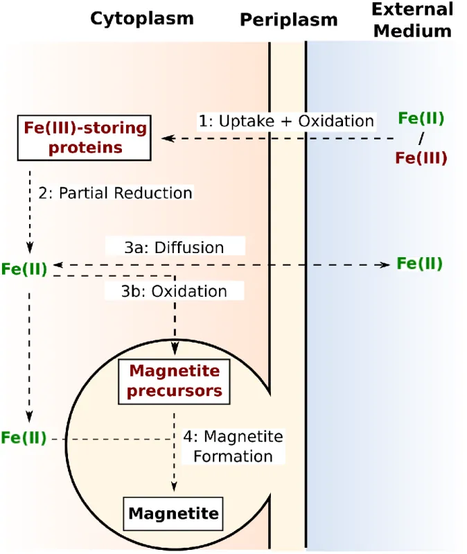
sulfate-reducing Deltaproteobacteria Desulfovibrio magneticus RS-1. A model compiling the 307
proposed pathways for iron biomineralization in MTB is illustrated in Figure 3. 308
309
Iron uptake 310
MTB can take either dissolved Fe(II) or Fe(III) up from their environment. Incorporation of 311
reduced iron species occurs as a diffusion process (Schüler and Baeuerlein, 1996, 1998; Amor 312
et al., 2018). Fe(III) assimilation requires energy-dependent mechanisms (Schüler and 313
Baeuerlein, 1996). In AMB-1 and MS-1, they involve biosynthesis of hydroxamate and catechol 314
siderophores, which are subsequently released to the external medium for Fe(III) 315
solubilization and incorporation (Paoletti and Blakemore, 1986; Calugay et al., 2003, 2006). 316
Under iron-rich conditions, genes encoding for high-affinity Fe(II) transport are up-regulated, 317
while Fe(III) acquisition systems are down-regulated under the same conditions (Suzuki et al., 318
2006). Contrastingly, siderophore-independent Fe(III) incorporation was observed for MSR-1 319
(Schüler and Baeuerlein, 1996). Finally, Fe(II) has been shown to be preferentially 320
incorporated by MTB when both oxidized and reduced species are available for the bacteria 321
(Amor et al., 2018). This illustrates that iron assimilation favors energy-independent 322
mechanisms under sufficient iron conditions (Suzuki et al., 2006). 323
324
Iron-storing mechanisms 325
Once incorporated, iron is first stored into the cell as Fe(III) species (Amor et al., 2018). In the 326
case of Fe(II) incorporation, iron is thus fully oxidized upon internalization (Amor et al., 2018). 327
The subcellular location of these species remains debated. They could be contained in the 328
cytoplasm and/or periplasm. Several proteins are known to store iron, including ferritins, 329
bacterioferritins and DNA-binding proteins (Dps) (Arosio et al., 2017; Uebe et al., 2019). 330
Ferritin-like structures have been identified in AMB-1 (Baumgartner et al., 2013), MSR-1 331
(Faivre et al., 2007; Fdez-Gubieda et al., 2013; Uebe et al, 2019) and MS-1 (Frankel et al., 1983). 332
From Mössbauer characterization of iron in MS-1 and MSR-1, it was proposed that oxidized 333
iron present as ferritin-like structures is then partially reduced into ferrous iron for trafficking 334
to magnetosomes and precipitation as magnetite (Frankel et al., 1983; Faivre et al., 2007). 335
These results were confirmed by measurement of iron isotopes in AMB-1 (Amor et al., 2016, 336
2018). RS-1 can also accumulate phosphate- and sulfur-associated Fe(II) species upon iron 337
assimilation (Baumgartner et al., 2016). 338
339
Iron delivery to magnetosomes 340
In contrast to ferric iron, ferrous iron is soluble at circumneutral pH, which can help iron 341
trafficking in the cell (Millero et al., 1995). Fe(II) could be addressed to magnetosomes via the 342
periplasm when the magnetosome membrane is still invaginating from the cell’s inner 343
membrane (Faivre et al., 2007; Jones et al., 2015). Alternatively, ferrous iron could be 344
transported to mature magnetosome vesicles across the magnetosome membrane. The latter 345
hypothesis was recently supported by two high-resolution electron microscopy and 346
fluorescence studies, which evidenced a layer of unmineralized ferrous ions surrounding 347
magnetite in the magnetosome membrane in both cultured and environmental bacteria 348
(Werckmann et al., 2017; Amor et al., 2020b). In that regard, genes encoding Fe(II) 349
transporters (feoAB operon) have been found in the vicinity of all MCGs of MTB with their 350
genome sequenced (Suzuki et al., 2006; Rong et al., 2008, 2012; Uebe and Schüler, 2016). 351
Deletion of these genes causes a strong impairment of magnetite biomineralization. 352
353
Magnetite formation and possible precursors 354
Fe(II) / Fe(III) ratio at the magnetosome location needs to be tightly controlled for magnetite 355
or greigite formation. This ratio is 0.5 in both mineral phases, although slightly oxidized 356
magnetite in MTB [i.e. Fe(II) / Fe(III) ratios lower than 0.5] has been consistently reported 357
(Weiss et al., 2004; Kopp et al., 2006; Moskowitz et al., 2008; Li et al., 2009). Several groups 358
proposed that magnetite precursors are first precipitated. In AMB-1, MS-1, and MSR-1, these 359
precursors include oxidized phases such as ferrihydrite [Fe(III)2O3.5H2O] (Frankel et al., 1983;
360
Faivre et al., 2007; Baumgartner et al., 2013; Fdez-Gubieda et al., 2013). One study also 361
suggested that hematite [-Fe(III)2O3] could represent a magnetite precursor (Le Nagard et
362
al., 2019). Fe(II) transported to magnetosomes would thus have to be oxidized for 363
precipitation of these precursors (Amor et al., 2018). The candidate proteins for oxidation 364
reactions include MamP, which contains a c-type cytochrome domain (Siponen et al., 2012, 365
2013; Jones et al., 2015). It has been detected in the magnetosome membrane (Raschdorf et 366
al., 2018). Fe(II) oxidation could thus happen during iron diffusion across the magnetosome 367
membrane or in the magnetosome lumen. Magnetite would then form upon further Fe(II) 368
addition in magnetosomes (Tronc et al., 1992; Ahn et al., 2012). This proposed 369
biomineralization pathway was recently challenged by Uebe and co-workers, who showed 370
that ferritins and ferritin-like structures (i.e. ferrihydrite) are not necessary for magnetite 371
precipitation in MSR-1 (Uebe et al., 2019). They produced a mutant strain lacking iron-storing 372
proteins, and characterized iron species in these bacteria as well as in wild-type cells. They 373
found that the presence of ferrihydrite in MSR-1 is tied to the presence of iron-storing proteins. 374
However, magnetite biomineralization was not altered in the mutant strain. They rather 375
proposed a distinct function for these proteins, which protected the bacteria from oxidative 376
stress. From these results, the authors concluded that iron-storing proteins are not involved 377
in biomineralization and that ferrihydrite does not represent a magnetite precursor. However, 378
the possibility of alternative biomineralization pathways triggered by the absence of iron-379
storing proteins cannot be completely ruled out. Moreover, the mutant strains lacking iron-380
storing proteins were cultivated under a limited number of growth conditions. It is thus 381
difficult to give a definitive conclusion about the role of ferritin-like structures. Finally, green 382
rust [a mixed Fe(II)/Fe(III) hydroxide] was proposed as a potential magnetite precursor in RS-383
1, a strain genetically distant from species of the Magnetospirillum genus (Baumgartner et al., 384
2016). Although the genetic content appears to be quite conserved among MTB, it is possible 385
that they developed different strategies for magnetite biomineralization depending on their 386
physiology and environment. 387
388
Iron distribution in magnetotactic bacteria 389
Physical and chemical characterizations of model magnetotactic strains demonstrated that 390
different iron species can be present in MTB. Time-course experiments conducted with AMB-391
1 and MSR-1 observed that virtually all iron was contained in magnetite when 392
biomineralization was complete (Baumgartner et al., 2013; Fdez-Gubieda et al., 2013). In 393
contradiction with these findings, direct measurements of iron in wild-type AMB-1 and MSR-394
1, as well as an AMB-1 mutant strain unable to form magnetosomes, evidenced a large pool 395
of iron distinct from magnetite (Amor et al., 2016, 2018, 2020a; Berny et al., 2020). Such 396
additional pool of iron can represent half of the total intracellular iron in wild-type AMB-1 and 397
MSR-1 (Amor et al., 2020b; Berny et al., 2020). The discrepancy between these findings and 398
the time-course experiments was proposed to raise from experimental conditions. Indeed, 399
time-course experiments require to first grow bacteria under iron-starving conditions (i.e. no 400
iron provided to the growth medium), which trigger overexpression of iron transporters 401
(Suzuki et al., 2006; Wang et al., 2017). This could optimize the transfer of iron to 402
magnetosomes for magnetite precipitation, and prevent accumulation of iron in a reservoir 403
distinct from magnetite. Interestingly, mutant AMB-1 strains unable to form magnetosomes 404
also accumulated iron (Amor et al., 2020a). However, the total mass of iron incorporated by 405
these mutant bacteria was significantly lower than the one contained in wild-type bacteria. 406
407
Fe(II) dynamics in magnetotactic bacteria 408
Finally, iron isotope modeling during magnetite biomineralization in AMB-1 allowed the 409
identification of Fe(II) diffusion from the cell’s internal medium to the external growth 410
medium (Amor et al., 2016, 2018). A release of intracellular Fe(II) to the external medium 411
could have resulted from cell lysis. However, monitoring of cell concentration during bacterial 412
cultures ruled out such possibility. Bacterial Fe(II) outward diffusion thus rather reflects 413
passive Fe(II) exchange with the external medium. 414
415 416
3. Tracking the footprint of MTB in the fossil record
417
Criteria for detection of MTB fossils 418
The specific mineralization pathway provides MTB magnetite with unique features that are 419
used for the detection of MTB fossils (hereafter referred to as magnetofossils) in ancient rock 420
samples. For several decades, magnetofossil identification relied on morphological, 421
crystallographic and magnetic criteria, which are now completed by chemical and isotopic 422
fingerprints, as well as additional magnetic methodologies. Thomas-Keprta and co-workers 423
(2000) defined six robust criteria for magnetofossil identification (see also review and 424
discussion in Jimenez-Lopez et al., 2010; Li et al., 2013). Here we summarize these criteria and 425
update them using the most recent discoveries (Fig. 4) 426
Unlike abiotic magnetite, MTB crystals show blunt edges associated with typical shapes (see 427
below) (Kopp and Kirschvink, 2008; Witt et al., 2005). Their size ranges from less than 35 nm 428
up to 120 nm (Konhauser et al., 1998). Because crystal growth in MTB is tightly controlled by 429
proteins, the particle sizes and shape factors (i.e. width/length ratios) in a given population 430
show a narrow skewed gaussian distribution (Devouard et al., 1998) compared to their abiotic 431
counterparts as well as magnetite produced through induced biomineralization (Loveley et al., 432
1987; Sparks et al., 1990; Arató et al., 2005). However, this criterion is not sufficient to 433
distinguish magnetofossil from abiotic magnetite in natural sedimentary rocks because 434
abundance peaks of biotic and abiotic magnetite can overlap and create more complex 435
distributions (Arató et al., 2005). 436
The size and shape of MTB nanoparticles fall into the stable single magnetic domain, which 437
maximizes the magnetic moment for each particle (Witt et al., 2005; Muxworthy and Williams, 438
2008). The alignment of Stable Single-Domain (SSD) magnetite particles in the magnetosome 439
chain imparts a strong magnetic moment to the bacteria. The unique chain arrangement of 440
SSD nanoparticles provides MTB with distinct magnetic features characterized by a large 441
remanent magnetization as well as a limited interaction field between particles (Li et al., 2012). 442
First Order Reversal Curves (FORC), a magnetic method based on the measurement of 443
hysteresis curves, as well as Ferromagnetic Resonance (FMR) spectroscopy have successfully 444
identified intact chains of fossilized magnetosomes (Roberts et al., 2013; Heslop et al., 2014; 445
Roberts et al., 2019). However, such magnetite alignment may be lost during sediment burial 446
and fossilization processes owing to the degradation of proteins assembling magnetosome 447
chains (Li et al., 2012). In that case, the typical FORC signatures of magnetosome chains are 448
lost. The Verwey temperature (i.e. the temperature below which magnetite undergoes a 449
phase transition associated with a modification of surface charge configuration and a sharp 450
change of susceptibility and remanent magnetization) has also been used to test the 451
magnetotactic origin of fossil magnetite samples (Chang et al., 2016). Finally, Isothermal 452
Remanent Magnetization (IRM) and Anhysteretic Isothermal Remanent (ARM) analyses 453
allowed to cluster the origins of magnetite and to estimate the proportion of biogenic 454
magnetite in a bulk sample using a non-negative matrix factorization (Egli, 2004; Heslop and 455
Dillon, 2007; Roberts et al., 2013). This method has been used efficiently on magnetite 456
samples from Eocene sedimentary rocks that survived diagenetic modifications (Day et al., 457
1977; Roberts et al., 2013; Roberts et al., 2019). 458
Transmission Electron Microscopy (TEM) analyses also showed that magnetite produced by 459
MTB usually grows along the [111] crystalline axis without lattice defects (Mann et al., 1987a; 460
Vali et al., 1987; Devouard et al., 1998). However, evidences of elongation anomalies along 461
the [110] or [100] axes were reported (Isambert et al., 2007; Benzerara and Menguy, 2009; Li 462
et al., 2015). So far, the crystal growth along the [111] axis still seems to be congruent for 463
prismatic magnetite. 464
Laboratory experiments demonstrated a low incorporation of trace elements contained in the 465
bacterial growth medium into AMB-1 magnetite (Amor et al., 2015). For most tested elements, 466
such incorporation was at least 100 times lower compared to abiotic magnetite samples 467
synthesized from co-precipitation of Fe(II) and Fe(III) ions in aqueous solutions (Amor et al., 468
2015). Chemical purity can thus be used as a chemical fingerprint of MTB magnetite. However, 469
a few elements such as molybdenum and tin showed preferential incorporation into AMB-1 470
magnetite. They could reflect metabolic reactions in which these two elements are involved, 471
and thus represent biological signatures of MTB activity recorded in magnetite. We note that 472
several publications reported improved incorporation of elements from the iron series such 473
as cobalt and manganese into MTB magnetite for biotechnology purposes (e.g. Staniland et 474
al., 2008; Keim et al., 2009). Trace elements incorporation was not compared with abiotic 475
conditions under such high concentrations. Therefore, they have limited paleontological 476
applications. 477
Finally, isotopic tools were used to search for magnetofossils in ancient rock samples. Oxygen 478
isotope composition of magnetite produced by the strains MS-1 and MV-1 has been 479
investigated, and showed a temperature dependency (Mandernack et al., 1999). However, no 480
difference was found between MTB magnetite and magnetite formed extracellularly by 481
Dissimilatory-Iron Reducing Bacteria (DIRB), a group of iron-metabolizing microorganisms that 482
perform magnetite induced biomineralization. In the same study, no specific iron isotope 483
signatures were evidenced in the magnetite produced by the magnetotactic strains MS-1 and 484
MV-1 (Mandernack et al., 1999). Contrastingly, magnetite produced by AMB-1 demonstrated 485
a significant depletion in heavy iron isotopes relative to the growth medium and the organic 486
fraction of the cell (Amor et al., 2016). Additionally, the odd iron isotope (57Fe) showed a
487
specific incorporation into AMB-1 magnetite. Such isotopic anomaly with preferential 488
incorporation of 57Fe compared to the three other isotopes (54Fe, 56Fe and 58Fe) has never
489
been observed before and may thus constitute a unique biosignature of magnetite produced 490
by MTB. However, the origin of this isotopic signature remains unclear and needs to be 491
investigated further on other MTB strains and natural samples. 492
So far, it can be highlighted that neither a single nor couples of criteria are sufficient to attest 493
for magnetite biogenicity in the past sedimentary record. A whole body of evidences based 494
on mineralogical, magnetic, chemical and isotopic tools is required for a firm identification of 495
magnetofossils (Kopp and Kirschvink, 2008). Such difficulty raises from the limited number of 496
magnetotactic strains tested for their morphologic, magnetic, chemical, and isotopic 497
specificities. Finally, additional work should be carried out to determine whether these criteria 498
can survive sediment burial and fossilization processes (see below). 499
500
Magnetofossil occurrences in the rock record 501
Magnetofossils were identified in sedimentary rocks from various ages, most of which were 502
observed in quaternary sediments from lakes, carbonate platforms, pelagic or hemi-pelagic 503
environments (e.g. Mc Neill et al., 1988; Yamazaki and Ioka, 1998). The oldest observation 504
during the current geologic eon Phanerozoic (0-540 Ma) was reported in Late Cretaceous 505
chalks from Southern England that were deposited about 85 Ma ago (Montgomery et al., 506
1998). This study reported detailed paleomagnetic data based on SSD magnetite-bearing 507
samples. Their observation using TEM demonstrated the presence of well-preserved chains of 508
magnetosomes, constituted mainly of prismatic magnetite although cuboctahedral and 509
bullet-shaped particles were also present. 510
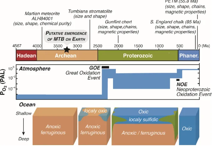
In contrast, scarce traces of magnetofossils were reported in rock records of the Precambrian 511
representing the earliest and longest period of Earth’s history, from 3.8 Ga to 540 Ma (Fig. 5). 512
In the 1980s, Chang and co-workers examined a number of Precambrian stromatolithic 513
limestones and cherts using rock magnetic methods and high-resolution electron microscopy 514
on magnetic extracts (Chang et al., 1989). They described several potential magnetofossils, 515
among which the most robust and ancient traces including magnetosome chains were 516
recorded in the 2.0 Ga-old Gunflint Formation, Western Australia. Several years later, Akai and 517
coworkers (1997) provided evidence for the oldest putative magnetofossils ever reported in 518
terrestrial samples in stromatolites from the Tumbiana Formation, Western Australia, formed 519
in a marine basin 2.7 Ga ago. Although very promising, these results rely only on the size (in 520
the SSD) and shape of magnetite crystals determined by TEM analyses (Akai et al., 1997); they 521
thus require to be validated by further investigations. 522
The presence of magnetofossils in ancient extraterrestrial material was also suggested, as 523
illustrated by the controversial studies of the Martian meteorite ALH84001 (McKay et al., 524
1996; Thomas-Keprta et al., 2000, 2009; Brearley, 2003; Treiman, 2003; Jimenez-Lopez et al., 525
2012). This sample might represent the oldest evidence for magnetofossils in the solar system. 526
The meteorite ALH84001 is an orthopyroxenite that crystallized 4.1 Ga ago (Lapen et al., 2010). 527
It contains carbonate globules formed in a late stage about 3.9 Ga ago, likely from low-528
temperature hydrothermal fluids. Nano-sized magnetite crystals with cuboctahedral, 529
teardrop, prismatic and irregular shapes were identified in the carbonate globules (McKay et 530
al., 1996; Thomas-Keprta et al., 2009). Large statistical TEM analyses indicate that 30% of the 531
nanomagnetites were elongated prisms showing a striking similarity with those formed on 532
Earth by the magnetotactic strain MV-1 (Thomas-Keprta et al., 2000). In addition, 533
extraterrestrial SSD nanomagnetites exhibited restricted anisotropy, crystallographic 534
perfection (i.e. free of internal defects), elongation along the [111] axis and chemical purity, 535
but no chain arrangement (Thomas-Keprta et al., 2000, 2009). It should be noted that chemical 536
purity was assessed from energy dispersive X-ray spectroscopy, a method that is not suitable 537
to strictly evaluate the biogenicity of magnetite based on its trace element content (Amor et 538
al., 2015). The nanomagnetite biogenicity in the ALH84001 meteorite is still a matter of debate 539
because alternative hypotheses have been proposed to explain their formation. These crystals 540
could also have been formed from shock-induced thermal decomposition and metamorphism 541
of Fe carbonates (Brearley, 2003; Treiman, 2003), even though chemically-pure magnetite can 542
hardly be produced from impure carbonates (Jimenez-Lopez et al., 2012). These last ten years, 543
new magnetofossil criteria were identified such as trace element and iron isotope 544
compositions, which could help to further decipher the origin of nanomagnetites in the 545
ALH84001 meteorite. 546
547
Alteration vs preservation of magnetofossils in sediments 548
As stated in the introduction, magnetite can be degraded during diagenesis. Three main 549
diagenetic processes can alter magnetite and prevent the identification of primary 550
magnetosomes: (i) oxidation, (ii) chain breakup, and/or (iii) reductive dissolution (see review 551
of Roberts, 2015). The first process happens when magnetite experiences oxidizing conditions, 552
due to diffusion of molecular oxygen in sediment or late exposition of sedimentary rock to the 553
atmosphere. This leads to partial or total transformation of magnetite to maghemite [-554
Fe(III)2O3] (Chang et al., 2013). In the second and third processes, one must consider that all
555
sediments contain a fraction of organic matter used as electron donor for aerobic or anaerobic 556
respiration processes. Various oxidants are utilized as electron acceptors following a well-557
established sequence depending on the free energy of each respiration pathway: O2, NO3-,
558
manganese oxides, iron oxides, and SO42- (Froelich et al., 1979). The magnetosome
559
membranes of MTB are mostly made of lipids, which can be degraded by microbial oxidative 560
process. The chain of magnetosomes is hold by a filament mainly constituted by the actin-like 561
MamK protein (Komeili, 2012; Uebe and Schuler, 2016), and can thus also be degraded by 562
diagenetic microbial processes. The last stage of anaerobic respiration during sediment burial 563
is based on microbial SO42- reduction that releases H2S in porewater. Many studies of marine
564
sediments demonstrated that H2S reduces and dissolves magnetite (e.g. Canfield and Berner,
565
1987; Mohamed et al., 2011), with magnetite half-life ranging from 5 to 1000 years (Canfield 566
and Berner, 1987; Emiroglu et al., 2004). If SO42- and organic matter are abundant in sediments,
567
one might expect magnetite to be mostly consumed and not preserved in rocks (Roberts, 568
2015). What might thus be a factor of magnetofossil preservation in sediments? SO42--poor
569
environments such as modern lake and river systems provide potentially favorable conditions 570
for preservation. Indeed, only a small amount of H2S can be produced in SO42--poor sediments
571
and reductive dissolution of magnetite may be limited. Significant amount of magnetofossils 572
could also be present in Archean sedimentary rocks (3.8 to 2.5 Ga old) since SO4
2-573
concentrations dissolved in the ocean at that time were extremely low (< 2.5 M; Crowe et 574
al., 2014), and organic matter supply to the sediments was likely weak due to low biological 575
productivity (Canfield, 2005). It can be noted that the biggest natural reservoir of magnetite 576
in the Earth’s crust is from the Archean banded iron formation, termed BIF (Konhauser et al., 577
2017). This indirectly confirms the high capacity of magnetite preservation in Archean 578
sediments even though BIFs have never been supposed to be formed from MTB remaining. 579
Magnetofossil preservation in sediments can also be inferred from data available for 580
Phanerozoic sedimentary rocks, in which a striking relationship was observed between 581
magnetofossil features and glacial-interglacial cycles (Paasche et al., 2004, 2011). Analysis of 582
sediments covering the last million years showed that glacial stages are associated with an 583
enrichment of organic matter but a decrease in magnetofossil abundance (Lean and McCave, 584
1998; Hesse, 1994; Yamazaki and Kawahata, 1998). These variations are interpreted as an 585
increase of biological productivity leading to higher organic matter flux and stronger reductive 586
dissolution of magnetite during sediment diagenesis. In contrast, interglacial periods 587
correspond to lower biological productivity, thus lowering organic carbon flux. The O2
588
diffusion from bottom water to sediment extended deeper in the sediment and increased the 589
size of the OATZ, where MTB thrive, up to tens of meters. This resulted in (i) a decrease of 590
organic matter content (inducing a lower H2S production and reductive dissolution) and (ii) an
591
increase of MTB in the larger OATZ (Roberts, 2015). Both effects globally increase the capacity 592
of magnetofossil preservation in sediments. Another scenario has also been suggested to 593
explain abundant magnetofossils in Phanerozoic sediments. Several authors proposed that 594
the amount of iron-rich dust could control the production and preservation of MTB (Roberts 595
et al., 2011; Yamazaki and Ikehara, 2012; Savian et al., 2014). Indeed, iron-dust particles 596
increase the primary productivity in the water column but can also be delivered to the 597
sediments and be used for anaerobic microbial oxidation of organic matter. This latter process 598
would increase the availability of dissolved Fe(II) in sediment porewater (used by MTB for 599
magnetite production), and decrease organic matter content, which in turn would decrease 600
the relative production of H2S (and dissolution of magnetite) by sulfate-reducing bacteria.
601
Finally, magnetofossil spikes were also evidenced during a global warming event ~55 Ma ago 602
known as the Paleocene-Eocene Thermal Maximum (PETM) (Kopp et al., 2007, 2009). 603
Unusually large magnetite crystals sharing morphological and crystallographic features typical 604
of MTB were observed during the PETM (Schumann et al., 2008). As suggested by the 605
glacial/interglacial cycles, warmer climate conditions could thus favor MTB magnetite 606
formation and/or preservation. 607
608
Tracking the age of the last common MTB ancestor
609
Resolving the evolution of controlled iron biomineralization in MTB is a major challenge in 610
evolutionary biology because this trait is polyphyletically distributed in taxa that are separated 611
by billion years of evolution from their most recent common ancestor. This intriguing 612
phylogenetic distribution of MTB raised three main questions tackled by the scientific 613
community over the last three decades: (i) has magnetosome formation a single genetic 614
origin?, (ii) was it an ancestral character shared by the ancestors of all phyla, genera and 615
species including MTB lineages?, and (iii) when this function emerged during the evolution of 616
life on Earth? To figure this out, several research groups performed whole genome sequencing 617
and inferred molecular phylogenies of MTB species and magnetosome genes (Abreu et al., 618
2011; Lefèvre et al., 2013; Lin et al., 2017a, 2018; Monteil et al., 2018). Over the years, 619
datasets got progressively stronger and stronger by including a higher taxonomic sampling 620
and a higher degree of phylogenetic signal thanks to whole genome datasets. Together with 621
molecular genetics, they provided evidence of a single genetic origin followed by a duplication 622
event that led to the emergence of the modern greigite and magnetite Magnetosome Gene 623
Clusters (MGCs) at some point in the MTB diversification. Two scenarios are currently 624
proposed: (i) a duplication of an unknown form of the MGCs that occurred well before the 625
emergence of the deep-branched PVC group (i.e. the superphylum gathering the 626
Planctomycetes, Verrucomicrobia and Chlamydiae phyla) that contain MTB, and (ii) a much 627
more recent duplication of the magnetite MGC in the Deltaproteobacteria that was 628
neofunctionalized to biomineralize greigite (Lin et al., 2018). Whenever this duplication 629
occurred, the distribution of magnetotaxis in the different bacterial phyla was mainly 630
explained by the vertical inheritance of the MGCs followed by numerous independent gene 631
losses in the non-magnetotactic lineages over phyla diversification (Lefèvre et al., 2013; Lin et 632
al., 2017a). Accordingly, controlled iron biomineralization emerged just before the emergence 633
of Nitrospirae and Proteobacteria phyla, which was estimated to have occurred during the 634
Archean over a timescale between 3.21 to 3.38 Ga ago with 95% confidence intervals ranging 635
over approximately 500 million years (Lin et al., 2017a). As a consequence, the ancestor of a 636
tremendous amount of modern bacterial lineages, but not all, could have performed 637
controlled iron biomineralization. 638
However, the above-mentioned phylogenies and the use of molecular clocks for prokaryotic 639
organisms were discussed by other research groups. Tackling MTB evolution is equivalent to 640
inferring the timing of the explosive radiation that led to the emergence of all major 641
Eubacteria phyla billions of years ago (Battistuzzi et al., 2004; Betts et al., 2018). At such 642
evolution timescale, methods in molecular phylogeny faces numerous limitations that 643
introduces uncertainties in reconstruction of tree topologies and estimation of event timing 644
[e.g. long branch attraction artifacts (Bergsten, 2005), impracticable and miscalibrated 645
molecular clocks (Graur et al., 2004), effect of gene flow blurred over timescales and site 646
saturation (Duchêne et al., 2016)]. According to some authors (Wang and Chen, 2017), the 647
current proposed scenario may not be parsimonious as long as it involves too many loss events 648
and rely on an uncertain tree topology. Additional investigations will help to refine these 649
inferences, and in the case of controlled iron biomineralization, magnetofossils could 650
represent a major asset to calibrate evolutionary history of life. 651
652
Predicting MTB evolution in relation to the Precambrian ocean chemistry 653
Geochemical traces of biogenic reduced carbon in turbiditic and pelagic sedimentary rocks 654
from the Isua supracrustal belt in west Greenland suggested that early life emerged long 655
before 3.7 Ga (Rosing, 1999). Molecular clock analyses later confirmed that the last universal 656
common ancestor of cellular life emerged ~4 Ga ago followed by an evolutionary burst of all 657
Eubacteria phyla that likely occurred all over the Archean eon (Battistuzzi et al., 2004; Betts 658
et al., 2018). Do current knowledge of MTB evolution and magnetofossils allows to estimate 659
the time of emergence of controlled iron biomineralization among prokaryotes and its role in 660
Earth’s evolution? 661
Although the presence of magnetofossils in the geological record before the end of the 662
Paleoproterozoic needs to be further investigated, controlled iron biomineralization could 663
have evolved before -2.7 Ga. From the angle of molecular phylogeny, this event could have 664
occurred a lot earlier. Indeed, the age of the last common MTB ancestor was estimated 665
between the Paleo- and Mesoarchean era, which corresponds to the timescale of the radiative 666
evolution at the origin of all the major Eubacteria phyla. On such evolutionary timescale, 667
deepest branching relationships of the tree of life are extremely difficult to reconstruct and 668
interpret because of the paucity of fossils and the tremendous uncertainty of phylogenetic 669
reconstructions. However, given the currently known MTB phylogenetic distribution, it seems 670
very unlikely that controlled iron biomineralization emerged before the radiation of the 671
Terrabacteria, Actinobacteria, Cyanobacteria, Firmicutes, and all candidate phyla radiations 672
revealed in the dark microbial matter (Hug et al., 2016). Although no geological data have ever 673
supported these inferences yet, paleontology may represent a major ally by reconstructing 674
the global chemical environment over time scales and help to determine if both biology and 675
environmental conditions were suitable for magnetosome biogenesis. 676
In the Archean eon (-4 to -2.5 Ga), early life undoubtedly thrived in the oceans that were 677
dominantly anoxic and ferruginous [rich in dissolved Fe(II)], with limited zones of surface 678
waters being oxygenated by photosynthetic activity (Lyons et al., 2014). During this period, 679
other strategies than photosynthesis emerged to exploit energy sources in anoxic conditions 680
including methanogenesis and methanotrophy (Rosing, 1999). Anoxic and ferruginous waters 681
of the Archean ocean, with local redox gradients such as those in which modern MTB are 682
observed, could have represented ideal conditions for the development and evolution of MTB. 683
Unlike modern oceans, redox boundary of the Archean ocean was likely located in the water 684
column (not in the sediment), and possibly extended in the photic zone (exposed to sunlight) 685
since O2 was produced in shallow water by photosynthesis. As mentioned above, MTB can
686
respire oxygen but can also get energy anaerobically by respiring nitrate, nitrous oxide or 687
sulfate. However, it is still unclear whether they can oxidize hydrogen, sulfur or iron, or even 688
reduce Fe(III), metabolism that would be more suitable in Archean environments. This gap is 689
mainly due to the lack of representative deep-branching MTB for which full genomes and 690
cultures are available, especially those from ferruginous environments (Rivas-Lamelo et al., 691
2017). 692
After 2.4 Ga, a progressive surface ocean oxygenation occurred in the early Paleoproterozoic 693
and diffused into the atmosphere, which is a stage known as the great oxidation event (GOE) 694
(Holland, 2002). As a consequence of atmospheric oxygenation, continental sulfides were 695
oxidized into highly soluble sulfates that were massively transferred to the ocean by river 696
systems (Canfield, 1998). From a paleontological perspective, it is widely accepted that 697
environmental conditions strongly influenced the evolution of life on Earth, and life in turn 698
impacted environmental conditions (Anbar and Knoll, 2002). It is likely that dissimilatory 699
sulfate-reducing prokaryotes have progressively reduced sulfates into sulfides in deep anoxic 700
ocean waters that precipitated as Fe sulfides. Such geochemical processes changed ocean 701
chemistry by decreasing the dissolved Fe concentrations. While dominated by iron in the early 702
Precambrian, deep ocean became S-dominated for a period that persisted at least from -1.8 703
Ga to -700 Ma (Canfield, 1998). Complete oxygenation of the oceans similarly to current state 704
occurred only between -700 and -600 Ma. The evolution of ocean chemistry towards more 705
sulfidic conditions after -2.4 to -1.8 Ga (i.e. Paleoproterozoic) (Canfield, 1998) could thus have 706
conducted to the development of other magnetotactic Deltaproteobacteria forming greigite 707
rather than magnetite. Although more speculative, this assertion could be tested by looking 708
in detail the magnetofossil record and crossing these data with phylogenetic reconstructions. 709
Overall, available data indicate that redox gradients generated by oxygen diffusion and 710
sustaining modern MTB could have existed since the Archean. This is critical to discuss the 711
evolution of MTB, since the magnetosome chains is thought to be used for navigation toward 712
optimal redox conditions in the water column or sediments. In the case of distinct function(s), 713
one cannot rule out the possibility that MTB magnetosomes evolved for purposes other than 714
magnetotaxis. Thus, first magnetosomes and MTB could have appeared even if no redox 715
gradients were established. Future studies will have to elucidate these points. 716