SIGNALING AND CELL PHYSIOLOGY
Evidence against a direct role of klotho in insulin resistance
Olivier Lorenzi&Christelle Veyrat-Durebex&Claes B. Wollheim&Pascal Villemin&
Françoise Rohner-Jeanrenaud&Anne Zanchi&
Ulrich M. Vischer
Received: 29 June 2009 / Revised: 30 August 2009 / Accepted: 2 September 2009 / Published online: 13 September 2009
# Springer-Verlag 2009
Abstract The klotho gene may be involved in the aging process. Klotho is a coactivator of FGF23, a regulator of phosphate and vitamin D metabolism. It has also been reported to be downregulated in insulin resistance syn-dromes and paradoxically to directly inhibit IGF-1 and insulin signaling. Our aim was to study klotho’s regulation and effects on insulin and IGF-1 signaling to unravel this paradox. We studied klotho tissue distribution and expres-sion by quantitative real-time polymerase chain reaction and Western blotting in obese Zucker rats and high-fat fed Wistar rats, two models of insulin resistance. Klotho was expressed in kidneys but at much lower levels (<1.5%) in
liver, muscle, brain, and adipose tissue. There were no significant differences between insulin resistant and control animals. We next produced human recombinant soluble klotho protein (KLEC) and studied its effects on insulin and IGF-1 signaling in cultured cells. In HEK293 cells, FGF23 signaling (judged by FRS2-α and ERK1/2 phosphoryla-tion) was activated by conditioned media from KLEC-producing cells (CM-KLEC); however, IGF-1 signaling was unaffected. CM-KLEC did not inhibit IGF-1 and insulin signaling in L6 and Hep G2 cells, as judged by Akt and ERK1/2 phosphorylation. We conclude that decreased klotho expression is not a general feature of rodent models of insulin resistance. Further, the soluble klotho protein does not inhibit IGF-1 and/or insulin signaling in HEK293, L6, and HepG2 cells, arguing against a direct role of klotho in insulin signaling. However, the hypothesis that klotho indirectly regulates insulin sensitiv-ity via FGF23 activation remains to be investigated. Keywords Klotho . Aging . Insulin resistance . FGF23
Introduction
The klotho gene has been proposed as a regulator of the aging process. Homozygous (−/−) klotho-deficient mice (“klotho mice”) have a reduced life span and display many features resembling human aging [1]. Conversely, an extended life span was demonstrated in transgenic mice engineered to express klotho in a ubiquitous manner [2]. Several cellular mechanisms underlying klotho’s antiaging effects have been proposed. The klotho gene encodes a 130-kD (1,014 amino acids) type 1 membrane protein, expressed mainly in mouse kidney and possibly at lower levels in other organs including the brain. The soluble
O. Lorenzi
:
C. B. Wollheim:
P. Villemin:
U. M. VischerDepartment of Cell Physiology and Metabolism, Faculty of Medicine, University of Geneva, Geneva, Switzerland
U. M. Vischer
Department of Rehabilitation and Geriatrics, Faculty of Medicine, University of Geneva,
Geneva, Switzerland
C. Veyrat-Durebex
:
F. Rohner-JeanrenaudDepartment of Internal Medicine, Division of Endocrinology, Diabetology and Nutrition, Faculty of Medicine,
University of Geneva, Geneva, Switzerland A. Zanchi
Division of Nephrology and Department of Medicine, University of Lausanne,
Lausanne, Switzerland
U. M. Vischer (*)
Department of Cell Physiology and Metabolism, Centre Médical Universitaire,
1, rue Michel Servet, 1211 Geneva 4, Switzerland e-mail: [email protected]
extracellular domain is also found in the circulation, presumably after shedding from renal cells, and may act as an endocrine factor [3, 4]. Klotho is a coactivator of FGF23, a hormone produced by bone-forming cells [5] that enhances renal phosphate excretion and inhibits vitamin D 1α-hydroxylation. Klotho converts the FGF receptor 1 (FGFR1, more specifically the FGFR1beta(III)c isoform) from a FGF2 receptor to a FGF23 receptor [6, 7]. FGF23 gain-of-function mutations underlie congenital hypophos-phatemic rickets, a rare autosomal dominant disease [8,9]. FGF23 knockout mice bear striking similarities with klotho mice [7, 10]. Shared features include growth retardation and soft tissue calcifications, which are in large part accounted for by high 1α-hydroxylase activity and elevated vitamin D levels [11]. Studies in klotho mice overexpress-ing FGF23 or klotho/FGF23 double knockout mice indicate an absolute requirement for klotho in FGF23 activity in vivo [12, 13]. These data indicate that inactivation of FGF23 signaling in renal tubular cells and the consequent abnormalities in calcium, phosphate, and vitamin D metabolism account for most, if not all, features of the aging phenotype observed in klotho mice. However, FGF23 overexpression is expected to induce a disease state [14]; it is, therefore, unlikely that klotho mediates increased longevity via FGF23 coactivation.
Several studies have indicated that klotho induces insulin resistance. Klotho mice display adipose tissue atrophy, increased insulin sensitivity, pancreatic islet atrophy, and hypoglycemia [1, 15]. Conversely, klotho transgenic mice with increased longevity are characterized by insulin resistance, judged from the glucose/insulin ratio and hyper-insulinemic euglycemic clamps. These abnormalities are observed in males only. However, mice from both genders are resistant to the hypoglycemic effect of insulin and IGF-1. The administration of murine soluble recombinant klotho (952 residue extracellular domain) induces an increase in blood glucose levels and a decreased hypoglycemic response to insulin in both genders. In cultured L6 myocytes, recombinant klotho inhibits insulin- and IGF-1-induced glucose transport and tyrosine phosphorylation of the insulin receptor (IR) and IRS-1. Klotho-induced insulin resistance is not explained by reduced insulin or IGF-1 binding to their receptors [2]. These in vitro experiments suggest that klotho may have direct effects on insulin signaling, independent of FGF23. Other studies have suggested that the soluble klotho protein confers resistance to insulin-induced oxidative stress via activation of FoxO transcription factors [16]. Klotho effects on insulin signaling or oxidative stress resistance may occur either via klotho expression in tissues involved in carbohydrate metabolism or as a hormone, after release of the klotho extracellular domain from renal tubular cells into the circulation [3].
The finding that klotho induces both insulin resistance and increased longevity is hard to reconcile with the notion that insulin resistance plays a central role in the metabolic syndrome, a cluster of cardiovascular risk factors. In the Otsuka–Long–Evans–Tokushima fatty rat (OLETF), an animal model of the metabolic syndrome (combining obesity, diabetes, hypertension, and dyslipidemia), a de-creased renal expression of klotho is observed although these animals display insulin resistance. Decreased klotho expression is corrected by treatment with the insulin sensitizer troglitazone [17]. In this model, gene therapy with klotho led to the correction of hypertension and of endothelium-dependent (ED) relaxation, possibly via im-provement in insulin resistance [18]. Klotho mice display impaired ED relaxation which is corrected by parabiosis with wild-type mice, compatible with the notion that circulating klotho regulates ED relaxation [19]. These preliminary studies suggest that klotho may (at least in part) correct the abnormalities which constitute the meta-bolic syndrome.
In a first step to clarify these contradictory findings, we undertook a study to determine the tissue distribution of klotho in a wide range of tissues, using quantitative real-time polymerase chain reaction (RT-PCR). We also inves-tigated whether klotho expression is regulated in two models of insulin resistance, the Zucker rat and high-fat feeding in Wistar rats, and whether it is influenced by the insulin sensitizer pioglitazone. We also studied the effects of the klotho soluble protein on insulin signaling in vitro.
Methods Reagents
Myc antibody was from Invitrogen (Basel, Switzerland), and all others antibodies were from Cell Signaling (Danvers, MA, USA): Phospho-FRS2-α Tyr-196 (CS#3864), Phospho p42/44 (CS#9106), P42/44 (CS#9102), Akt (CS#9272), and Phospho-Akt Ser-473 (CS#9271). FGF23 was from R&D systems (Minneapolis, MN, USA; 2604-FG). Pioglitazone was from Takeda pharmaceuticals.
Animal studies
Male obese Zucker (fa/fa) rats and lean controls (FA/?) rats were purchased from Charles River laboratories (L’Arbresle, France). They were housed under controlled temperature (22°C) and lighting (light on: 7 a.m.–7 p.m.). A first series of animals (five obese and five lean) was kept on a standard diet (RMI, SDS, Essex, UK) and sacrificed at age 10 weeks. A second group of ten obese 8-week-old rats was
treated with vehicle (n = 4) or pioglitazone (n = 6) for 4 weeks. They had ad libitum access to water and to a standard chow diet (consisting by energy: 52.8% carbohy-drates, 9.2% fat, 38% protein (diet 3200; Kliba)). Pioglita-zone was incorporated in the diet (0.24 g/kg). Insulin resistance was assessed by the homeostasis model of insulin resistance (HOMA-IR) [20]. The full metabolic character-ization of these animals has previously been reported [21]. Eight-week-old male Wistar rats were purchased from Charles River (L’Arbresle, France). They were housed under controlled temperature (22°C) and lighting (light on: 7 a.m.– 7 p.m.). After 1 week of acclimatization, rats were individually housed and randomly distributed into two groups and fed for 6 weeks with either a standard diet (by energy 7.42% fat, 75% carbohydrates, 17.5% protein, metabolized energy, 2.61 kcal/g) or a high-fat diet (by energy 41% fat, 40% carbohydrates, 19% protein, metabo-lized energy, 3.1 kcal/g; Ssniff RM/H, Ssniff Spezialdiaten, Soest, Germany). Body weight and food intake were recorded daily. Seven days prior to sacrifice, a glucose tolerance test was performed after 4 h fasting. A glucose load of 1.5 g/kg was administered i.p., and blood samples were collected by tail nicking at 0, 15, 30, 60, 120, 180, and 240 min. Glycemia was recorded using Glucotrend Active (Roche, Basel, Switzerland), and the glucose area under curve (AUC) was calculated at 240 min. Unpaired Student’s t test was used for group comparisons.
The animals were sacrificed using light isoflurane anesthesia (Halocarbon Laboratories, River Edge, NJ, USA) and rapid decapitation between 9 a.m. and 1 p.m. Tissues were rapidly removed, freeze-clamped, and stored at −80°C. All procedures were approved by the ethics committees of the Universities of Geneva or Lausanne and were in accordance with the Swiss guidelines for animal experimentation.
cDNA cloning of the human klotho extracellular domain Total human kidney RNA was used as the template for the synthesis of first-strand complementary DNA (cDNA) using ImProm-II Reverse Transcriptase (Promega, Dübendorf, Switzerland) and oligo(dT) primers. PCR was performed using GoTaq/Pfu DNA polymerase (Promega) according to the manufacturer’s instructions with primers for amplifica-tion of two overlapping cDNA fragments named A (N-term: 2,095 bp) and B (C-term: 1,062 bp) covering the whole coding sequence. These fragments were inserted into pGem-T Easy vector. The B fragment was used as a template to generate mutated fragments with the stop codon (full-length) or the transmembrane domain (extracellular domain) removed. Complete klotho full-length and extra-cellular domain constructs carrying polyhistidine and myc tags at the C-terminus were generated by three-piece
ligations into the pcDNA3.1 myc/his expression vector. Full sequencing of the constructs (Microsynth, Balgach, Switzerland) confirmed complete homology with the published sequence (Genbank accession number NM_004795.2). The klotho extracellular domain (KLEC) construct consists of residues 1–982, followed by a short four residues linker sequence and the polyhistidine and myc tags.
Cell culture and transfection
HEK293 cells were grown in DMEM (Gibco, Invitrogen Life Science, Carlsbad, CA, USA) supplemented with 10% fetal calf serum (FCS) and 1% P/S (Penicillin Streptomy-cin, Gibco). After transfection using TransIT-293 (Mirus), cells stably expressing the klotho extracellular domain (HEK-KLEC) were obtained by selection for 14 days in medium supplemented with 0.5 mg/mL G418. Klotho protein secreted into the conditioned medium of this cell line was purified to near-homogeneity using the nickel– agarose resin. The biological effects of klotho, either contained in conditioned media or in purified form, were tested on HEK293, rat L6, and human HepG2 cells. L6 and HepG2 cells were grown in MEM-α and DMEM media, respectively, supplemented with 10% FCS.
Western blot
Protein extracts were prepared from rat organs by mechan-ical homogenization in radioimmunoprecipitation assay buffer (NaCl 300 mM; Tris-HCl 100 mM; NP40 2%; SDS 0.2%; Deoxycholic acid 1%) with protease cocktail inhibitor (set V, animal free Merck), 2 mM NaVO4 and 10 mM NaF. Cultured cells were directly solubilized in the same buffer after three washes with phosphate-buffered saline at 4°C. Total proteins were separated by standard sodium dodecyl sulfate polyacrylamide gel electrophoresis techniques and transferred to nitrocellulose membranes. Proteins of interest were detected with specific primary antibodies, horseradish peroxidase-conjugated secondary antibodies, and an enhanced chemiluminescence detection system (Amersham, Otelfingen, Switzerland). Proteins were quantified using the ChemiDoc XRS from Bio-Rad (Bio-Rad, Reinach, Switzerland) and Quantity One software (Bio-Rad).
Real-time PCR
Total RNA was extracted from rat organs with Trizol reagent (Invitrogen), and reverse transcription was made by murine leukemia virus-R reverse transcriptase according to the manufacturer’s protocol (Promega). Quantitative real-time PCR (qPCR) was assessed in ABI Thermal Cycler
apparatus with PRISM® 7500 Sequence Detection System was programmed with the ABI standard cycling program of 50°C for 2 min, denaturation at 95°C for 2 min, 50 cycles of primer annealing at 95°C for 15 s, and primer extension at 60°C for 30 s. cDNA quantification was performed using FastStart universal SYBR Green Master (Roche) according to the manufacturer’s instructions. Rat klotho primers were designed using ABI software (P1: 5′-TGGACCCACCTT GAGTTTTCA-3′; P2: 5′-TTGCCTCAGGTTGGGAGATT-3′). Klotho messenger RNA (mRNA) values were normal-ized for cyclophilin mRNA used as a stable control.
Results
Klotho expression and tissue distribution in relation to insulin resistance in rats
We first studied the klotho tissue distribution in rat organs and tested whether it is modified by obesity/insulin resistance in Zucker rats. Ten-week-old obese Zucker rats (body weight 346.3± 8.0 g, mean±standard deviation (SD)) and control rats (284.5 ±9.2 g) were sacrificed, and various organs were collected for RNA and protein extraction (n= 5 in each group). Klotho mRNA levels were analyzed by quantitative RT-PCR (Fig. 1a). Klotho mRNA was readily detected in kidneys. However, only low levels (<1.5% relative to kidneys) were found in liver, white and red muscle, epididymal white adipose tissue, and brain (frontal lobe). Klotho mRNA levels were somewhat higher in the hypothalamus (2.17± 1.07%, mean ±standard error of the mean (SEM)) than in the brain (0.4 ± 0.15%), but the difference failed to achieve statistical significance (p=0.14 using unpaired Student’s t test). We found no differences in klotho mRNA levels between obese Zucker and control rats, neither in kidneys nor in other organs. Klotho protein levels tested by Western blot were similar in kidneys from obese Zucker and control rats (Fig.1b).
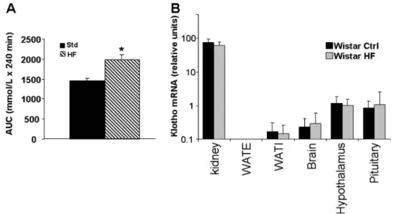
We also tested klotho expression and tissue distribution after high-fat feeding in Wistar rats, another model of insulin resistance. Four-month-old Wistar rats were fed a high-fat or control diet for 5 weeks prior to sacrifice (n=6 for each group). High-fat feeding resulted in higher body weight (379.3±6.1 g compared to 342.4±6.1 g in control rats, p< 0.002), higher fasting glucose level (6.0±0.2 mmol/l, versus 5.4 mmol/l in control rats, p<0.03), and a more pronounced rise in blood glucose during a glucose tolerance test (Fig. 2a), indicative of insulin resistance. Again, klotho mRNA was readily detected in the kidney, but only at low levels in inguinal and epidydimal white adipose tissue and brain (Fig.2b). Klotho mRNA was significantly higher (p< 0.001) in the hypothalamus (1.17±0.2%) and in the pituitary (0.83±0.17%) than in the whole brain (frontal lobe; 0.23±
0.06%). However, these values were again much lower than in the kidney. There was no significant change in klotho mRNA levels after high-fat feeding, neither in the kidney nor in other organs tested.
These findings indicate that renal klotho expression is unchanged in two rat models of insulin resistance, arguing against an obligatory role for regulated klotho expression in the pathogenesis of insulin resistance. Further, the expres-sion of klotho was restricted to the kidneys both in control and insulin resistant animals.
Klotho kidney induction after 4 weeks treatment with pioglitazone
While klotho expression was not regulated in our insulin resistance models, it may be modulated by drugs that modify insulin resistance. Some studies have suggested a role of thiazolidinediones (glitazones) in the regulation of renal klotho expression [17,22]. We next investigated the effects of glitazone treatment on renal klotho expression in obese Zucker rats. Zucker fatty rats were treated for 4 weeks with pioglitazone (see Methods). This treatment resulted in significant weight gain (503±7 g compared to 450±12 g in
Fig. 1 Klotho tissue distribution in Zucker rats. a Klotho mRNA levels were determined in rat organs from lean or obese (fa/fa) Zucker rats by quantitative RT-PCR. Results are shown on a log scale in arbitrary units, with a pool of renal total RNA defined as 100%. No signal was detected in the absence of reverse transcriptase. Results are shown as mean±SD, n=5. WATE epididymal white adipose tissue. b Klotho protein was detected in protein extracts from rat kidneys by Western blot. Mouse kidney homogenate (C57Bl6) was added as a positive control. No differences in renal klotho expression were observed either at the mRNA or the protein levels
control rats, mean±SEM, p<0.005). Insulin resistance was markedly decreased, as shown by reduced insulinemia (2.4± 0.1μU/ml compared to 24.6±4.2 μU/ml in obese untreated rats, p<0.001) and HOMA-IR index (0.46±0.07 compared to 7.30±1.81 in obese untreated rats, p< 0.008). In spite of these metabolic differences, there were no significant differ-ences in renal klotho expression in treated and untreated animals, whether judged by mRNA levels (Fig. 3a) or protein levels (Fig. 3b). These results do not confirm that
glitazone treatment increases renal klotho expression in insulin-resistant rats.
Effects of klotho on FGF23 and IGF-1 signaling in HEK293 cells
Circulating klotho may determine insulin sensitivity by direct regulation of insulin and IGF-1 signaling [2]. Both full-length and soluble klotho proteins are coactivators of
Fig. 2 Klotho tissue distribution in high-fat fed Wistar rats. a Wistar rats were fed a high-fat or a control diet. High-fat feeding resulted in glucose intolerance as judged by a higher glucose areas under the
curve (AUC; mean±SEM; n=5; *p=0.01, Student’s t test). b mRNA
expression was tested by quantitative RT-PCR in kidney, epididymal white adipose tissue (WATE ), inguinal white adipose tissue (WATI),
brain (frontal lobe), hypothalamus, and pituitary. Results are shown on a log scale in arbitrary units (mean ±SD), with a pool of rat total kidney RNA defined as 100%. Klotho expression was higher in hypothalamus and pituitary than in the brain. There were no differ-ences in klotho expression between high-fat fed and control animals
Fig. 3 Effect of pioglitazone treatment on renal klotho expression in obese Zucker rats. Obese Zucker rats were treated for 4 weeks with 20 mg/ml/kg of pioglitazone. a Renal klotho expression was measured by quantitative RT-PCR. Results are shown in arbitrary units, with a pool of renal total RNA defined as 100%. b Renal klotho expression was measured by Western blot in kidney protein extracts. The blot was
also revealed for glyceraldehyde-3-phosphate dehydrogenase (GAPDH) to verify equal protein load. The bands were quantified by densitometric scanning; results are shown in arbitrary units, the mean value of the control group being defined as 100%. Pioglitazone did not significantly modify renal klotho expression, neither at the mRNA nor the protein level
FGF23 in responsive cells [6, 7]. The klotho effects on insulin signaling have been reported in the absence of FGF23, pointing to a mechanism of action distinct from FGF23 coactivation [2]. We next studied the effects of the human klotho protein on both FGF23 and IGF-1 signaling. The human klotho cDNA was cloned and inserted into a eukaryotic expression vector to generate the extracellular domain carrying myc and polyhistidine tags at the C-terminus (KLEC, MW 116 kD). We next generated a HEK293 cell line stably transfected with this construct (HEK-KLEC). The KLEC protein was purified on nickel– agarose columns. However, this purified protein failed to inhibit insulin signaling in rat L6 cells, as judged by IRS-1 and Akt phosphorylation (not shown). It also failed to coactivate FGF23 signaling in HEK293 cells. We suspected that the purification procedure inactivated the protein and therefore, returned to testing the bioactivity of conditioned media from HEK-KLEC cells.
We first tested whether conditioned medium from HEK-KLEC cells can coactivate FGF23 signaling, as judged by FRS2-α (FGF receptor substrate 2-α) and ERK1/2 phos-phorylation by Western blot (Fig. 4). Confluent HEK-KLEC and control, wild-type HEK293 (HEK-WT) cells were grown in serum-free medium. After 24 h, the conditioned media were concentrated by ultrafiltration. The estimated final klotho concentration (calculated from the myc immunoreactivty compared to the purified protein) was 10–30 nmol/l. In HEK-WT cells, FGF2 (used as a positive control for FGFR1 activation) induced phosphor-ylation of both FRS2-α and ERK1/2. As expected, conditioned medium from HEK-KLEC had no effect when added alone. However, the addition of FGF23 resulted in activation of both FRS2-α and ERK1/2 when added
together with conditioned medium from HEK-KLEC but not from HEK-WT cells (Fig.4). These results confirm that the klotho recombinant protein contained in HEK-KLEC-conditioned media is an effective FGF23 coactivator in HEK cells.
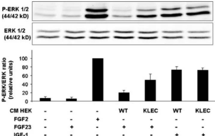
HEK cells respond to both FGF2 and IGF-1 in terms of ERK phosphorylation. We next explored whether the klotho protein inhibits IGF-1 signaling in HEK cells (Fig. 5). Again, the conditioned medium from HEK-KLEC but not from HEK-WT cells coactivated FGF23-induced ERK1/2 phosphorylation. However, in the same experiment, IGF-1-induced ERK1/2 phosphorylation was not inhibited by HEK-KLEC-conditioned medium, com-pared to HEK-WT-conditioned medium. Similar results were obtained whether IGF-1 was tested at 10 or 100 nmol/l. Thus, in the same cells, klotho coactivates FGF23 but fails to inhibit IGF-1 signaling.
Effects of klotho protein on insulin signaling
We next reinvestigated whether the soluble klotho protein modulates insulin signaling in insulin-responsive cell lines. Confluent L6 cells were preincubated with fresh control serum-free conditioned medium or conditioned medium from HEK-KLEC or HEK-WT, prior to stimulation with insulin or IGF-1 (Fig.6). Both insulin (10 and 100 nmol/l)
Fig. 5 Effect of klotho FGF23 and IGF-1 signaling in HEK cells. Conditioned media (CM) from wild-type HEK cells (HEK-WT) or from HEK cells expressing soluble klotho (HEK-KLEC) or control medium were concentrated by ultrafiltration and incubated with fresh HEK cells for 2 h. FGF2 (100 ng/ml), FGF23 (100 ng/ml), and IGF-1 (100 nmol/l) were then added for 15 min as indicated. Cell lysates were prepared for Western blots using antibodies against phospho-ERK1/2 and total phospho-ERK1/2. A representative blot is shown. The bar graphs show the quantification of band intensity (phosphorylated/total protein ratio) of three independent experiments (mean±SEM) in relative units, the value with FGF-2 (100 ng/ml) being defined as 100. FGF2 but not FGF23 added alone induced ERK1/2 phosphorylation. IGF-1-induced ERK1/2 phosphorylation was identical in CM from HEK-KLEC and HEK-WT cells. However, FGF23-induced ERK1/2 phosphorylation was clearly higher when added to CM from
HEK-KLEC than from HEK-WT cells (p<0.05 by paired Student’s t test)
Fig. 4 Soluble klotho protein coactivates FGF23 signaling in HEK cells. Conditioned media (CM) from wild-type HEK cells (HEK-WT) or from HEK cells expressing soluble klotho (HEK-KLEC) or control medium were concentrated by ultrafiltration and incubated with fresh HEK cells for 2 h. FGF2 (100 ng/ml) and FGF23 (100 ng/ml) were then added for 15 min as indicated. Cell lysates were then prepared for
Western blots using antibodies against Phospho-FRS-2α,
phospho-ERK1/2, and total ERK1/2. FGF2 stimulated both FRS-2α and
ERK1/2 phosphorylation as expected. FGF23 stimulated FRS-2α
and ERK1/2 phosphorylation only when added to HEK-KLEC but not to HEK-WT-conditioned medium
and IGF-1 (10 nmol/l) markedly increased Akt and ERK1/2 phosphorylation. In the presence of conditioned media, basal Akt and ERK1/2 phosphorylation was somewhat increased, presumably because of activation by nonspecific HEK cell-derived factors. However, insulin- and IGF-1-induced Akt and ERK1/2 phosphorylation was not significantly altered by HEK-KLEC-conditioned medium, compared to HEK-WT-conditioned medium (paired Student’s t test, n=3 indepen-dent experiments). We observed a similar lack of inhibition of insulin and IGF-1 signaling by HEK-KLEC-conditioned medium in human HepG2 cells (not shown).
Discussion
Studies in Caenorhabditis elegans, Drosophila, and mice have identified the insulin and/or IGF-1 receptors and
several downstream signaling molecules as aging genes [23]. For instance, the heterozygous IGF-1 receptor knockout mouse has an extended life span [24]. Similarly, inactivation of the insulin receptor gene in adipose tissue is also associated with an extended life span [25]. These studies indicate that insulin and/or IGF-1 resistance is associated with increased longevity. However, this notion remains to be reconciled with the well-established role of insulin resistance in the metabolic syndrome which under-lies a risk for diabetes and cardiovascular disease. The metabolic syndrome is a complex metabolic disorder, in which insulin resistance can be seen as the consequence of “fuel overload” leading to ectopic fat accumulation. It is, therefore, conceivable that insulin resistance (in a biochem-ical sense) antagonizes the pathogenesis of insulin resis-tance understood as a clinical syndrome [26]. Studies on the cellular mechanisms of action of klotho may contribute to an understanding of this paradox.
Our data indicate that klotho is not regulated in two rat models of insulin resistance, the obese Zucker rat and high-fat feeding in Wistar rats. Decreased renal klotho expres-sion was shown by Northern blot analysis in the OLETF rat, a model of the metabolic syndrome [17]. Our data, based on quantitative RT-PCR and Western blots, indicate that reduced renal klotho expression is not a general feature of insulin resistance in rodents. Previous studies have suggested that treatment with glitazones increases renal klotho expression. However, we failed to observe increased renal klotho expression in obese Zucker rats treated with pioglitazone, although this treatment markedly reduced insulin resistance as expected. Transgenic mice overex-pressing Klotho in a ubiquitous manner are characterized by insulin resistance. Insulin resistance is possibly con-ferred by klotho expression in insulin-sensitive tissues. However, we found only low levels of extrarenal klotho expression in liver, muscle, adipose tissue, and hypothala-mus/pituitary, both in insulin resistant and control animals. It is, therefore, unlikely that regulated extrarenal klotho expression plays an important role in insulin resistance syndromes. We, thus, found no evidence of regulated klotho expression in insulin resistance or its treatment. However, these data do not rule out that constitutively expressed klotho plays a role in insulin resistance.
Soluble klotho protein has been identified in serum and cerebrospinal fluid and may act as an endocrine factor. Our experiments with purified klotho protein failed to identify any klotho bioactivity. However, conditioned media from cells expressing the klotho protein were also unable to inhibit IGF-1 signaling in HEK cells, although they successfully coactivated FGF23, indicating that the klotho protein in conditioned media is bioactive. Further, klotho-containing media failed to inhibit IGF-1 and insulin signaling in rat L6 and human HepG2 cells. Our results
Fig. 6 Effect of klotho on IGF-1 and insulin signaling in L6 cells. Conditioned media (CM) from wild-type HEK cells (HEK-WT) or from HEK cells expressing soluble klotho (HEK-KLEC) or control medium (Ctrl) were concentrated by ultrafiltration and incubated with L6 cells for 2 h. Insulin (Ins) and IGF-1 were then added for 10 min. Cell lysates were prepared for Western blots using antibodies against phosphor-Akt, total Akt, phospho-ERK1/2, and total ERK1/2. Representative Western blots are shown. The corresponding bar graphs show the quantification of band intensity (phosphorylated/ total protein ratio) of three independent experiments (mean±SEM) in relative units, the value with insulin 100 nmol/l being defined as 100. Both insulin and IGF-1 induced Akt and ERK1/2 phosphorylation, an effect that was not significantly altered by HEK-KLEC-CM compared to HEK-WT-CM
stand in contrast with the data published by Kurosu et al. [2], who showed potent inhibition of insulin and IGF-1 signaling by purified murine recombinant klotho protein in L6 cells. These discrepancies may be due to inactivation of klotho bioactivity during our purification procedure (using a technique different from the one used by Kurosu et al.) or to species restrictions. However, since klotho-containing conditioned media coactivate FGF23 as previously de-scribed, the absence of inhibition of IGF-1 signaling (shown within the same experiment) cannot be attributed to the lack of klotho bioactivity. Further, klotho-containing media failed to inhibit insulin and IGF-1 signaling in both rat L6 and human HepG2 insulin-sensitive cells, arguing against a problem of species restriction. We cannot formally rule out the possibility that higher klotho protein concen-trations or multiple repeat experiments might have uncov-ered minor effects of soluble klotho on insulin or IGF-1 signaling. However, again, the klotho protein concentration in the HEK-KLEC-conditioned medium we used was sufficient for effective FGF23 coactivation. Our finding that native, bioactive klotho protein fails to inhibit insulin signaling raises the possibility that the direct effect of purified klotho on insulin signaling observed by Kurosu et al. results from a purification artifact. Our data certainly indicate that klotho’s molecular determinants for klotho-induced FGF23 coactivation and potential inhibition of insulin and IGF-1 signaling are distinct.
Taken together, our data indicate that regulated klotho expression (whether at the renal or extrarenal level) is not a general feature of insulin resistance syndromes. They further indicate that soluble klotho protein is not a direct regulator of insulin signaling in at least two insulin-sensitive cell lines. However, the hypothesis that klotho exerts indirect effects on insulin sensitivity remains to be investigated. As a FGF23 coactivator, klotho is involved in calcium, phosphate, and vitamin D metabolism. Klotho-deficient and FGF23 (−/−) mice are both characterized by hypoglycemia and increased insulin sensitivity [1,10,15], suggesting that the FGF23 axis is involved in the regulation of insulin sensitivity. FGF23 inhibits vitamin D and PTH synthesis and promotes renal phosphate excretion. Vitamin D deficiency is associated with impaired insulin secretion and corrected by vitamin D repletion [27]. Further, 25(OH) vitamin D levels are inversely correlated with the preva-lence of the metabolic syndrome [28]. However, klotho mice display high insulin sensitivity and reduced insulin secretion even though vitamin D levels are markedly elevated. Phosphate depletion is thought to induce insulin resistance. Insulin resistance is associated with low serum phosphate in a population study on nondiabetic individuals [29]. The prevalence of type 2 diabetes or impaired glucose tolerance is elevated in primary hyperparathyroidism, possi-bly via hypophosphatemia [30,31]. It is, therefore, possible
that klotho/FGF23-induced renal phosphate excretion or related changes play a role in the pathogenesis of insulin resistance, a hypothesis that remains to be investigated.
A role for klotho in the aging process was originally suspected by the reduced life span and phenotypic features (including skin atrophy, osteoporosis, and arteriosclerosis) in klotho mice [1]. These abnormalities can now largely be attributed to the loss of FGF23 function and consequent changes in calcium, phosphate, and vitamin D metabolism [11]. However, the extended life span of klotho transgenic mice remains to be explained. FGF23 gain-of-function mutations result in congenital hypophosphatemic rickets [8, 9], i.e., a disease state. However, is it conceivable that the weaker stimulation of FGF23 bioactivity afforded by klotho overexpression results in extended life span, without causing hypophosphatemia or bone disease. Such an effect may be associated with (or even mediated by) insulin resistance indirectly induced by hypophosphatemia or other FGF23 biological effects, as discussed above. Further, Liu et al. have suggested that Wnt signaling is involved stem cell depletion. Klotho may directly inhibit Wnt signaling, thereby preserving stem cell function [32]. The relative roles of FGF23 signaling, insulin resistance, and Wnt signaling in the extended life span seen with klotho overexpression remain to be established.
Acknowledgments This work was supported by a grant from the
ALFEDIAM (ALFEDIAM/Antadir Prize) and the EFSD/Sanofi-aventis European Programme on macrovascular complications and blood glucose abnormalities and by the Swiss National Science Foundation (grants number 310030-108062 to UMV and number 310000-120147 to FRJ).
Conflict of interest The authors declare that they have no conflict of
interest.
References
1. Kuro-o M, Matsumura Y, Aizawa H, Kawaguchi H, Suga T, Utsugi T, Ohyama Y, Kurabayashi M, Kaname T, Kume E, Iwasaki H, Iida A, Shiraki-Iida T, Nishikawa S, Nagai R, Nabeshima YI (1997) Mutation of the mouse klotho gene leads
to a syndrome resembling ageing. Nature 390:45–51
2. Kurosu H, Yamamoto M, Clark JD, Pastor JV, Nandi A, Gurnani P, McGuinness OP, Chikuda H, Yamaguchi M, Kawaguchi H, Shimomura I, Takayama Y, Herz J, Kahn CR, Rosenblatt KP, Kuro-o M (2005) Suppression of aging in mice by the hormone
Klotho. Science 309:1829–1833
3. Chen C-D, Podvin S, Gillespie E, Leeman SE, Abraham CR (2007) Insulin stimulates the cleavage and release of the extracellular domain of Klotho by ADAM10 and ADAM17. Proc
Natl Acad Sci 104:19796–19801
4. Imura A, Iwano A, Tohyama O, Tsuji Y, Nozaki K, Hashimoto N, Fujimori T, Nabeshima Y (2004) Secreted klotho protein in sera and CSF: implication for post-translational cleavage in release of klotho protein from cell membrane. FEBS Lett 565:
5. Riminucci M, Collins MT, Fedarko NS, Cherman N, Corsi A, White KE, Waguespack S, Gupta A, Hannon T, Econs MJ, Bianco P, Gehron Robey P (2003) FGF-23 in fibrous dysplasia of bone and its relationship to renal phosphate wasting. J Clin Invest 112:683–692
6. Kurosu H, Ogawa Y, Miyoshi M, Yamamoto M, Nandi A, Rosenblatt KP, Baum MG, Schiavi S, Hu MC, Moe OW, Kuro-o M (2006) Regulation of fibroblast growth factor-23 signaling by klotho. J Biol Chem 281:6120–6123
7. Urakawa I, Yamazaki Y, Shimada T, Iijima K, Hasegawa H, Okawa K, Fujita T, Fukumoto S, Yamashita T (2006) Klotho converts canonical FGF receptor into a specific receptor for
FGF23. Nature 444:770–774
8. Bai XY, Miao D, Goltzman D, Karaplis AC (2003) The autosomal dominant hypophosphatemic rickets R176Q mutation in fibroblast growth factor 23 resists proteolytic cleavage and enhances in vivo
biological potency. J Biol Chem 278:9843–9849
9. White KE, Carn G, Lorenz-Depiereux B, Benet-Pages A, Strom TM, Econs MJ (2001) Autosomal-dominant hypophosphatemic
rickets (ADHR) mutations stabilize FGF-23. Kidney Int 60:2079–
2086
10. Shimada T, Kakitani M, Yamazaki Y, Hasegawa H, Takeuchi Y, Fujita T, Fukumoto S, Tomizuka K, Yamashita T (2004) Targeted ablation of Fgf23 demonstrates an essential physiological role of FGF23 in phosphate and vitamin D metabolism. J Clin Invest 113:561–568
11. Ohnishi M, Nakatani T, Lanske B, Razzaque MS (2009) Reversal of mineral ion homeostasis and soft-tissue calcification of klotho knockout mice by deletion of vitamin D 1alpha-hydroxylase.
Kidney Int 75:1166–1172
12. Nakatani T, Ohnishi M, Razzaque MS (2009) Inactivation of klotho function induces hyperphosphatemia even in presence of high serum fibroblast growth factor 23 levels in a genetically engineered hypophosphatemic (Hyp) mouse model. FASEB J (Epub ahead of print)
13. Nakatani T, Sarraj B, Ohnishi M, Densmore MJ, Taguchi T, Goetz R, Mohammadi M, Lanske B, Razzaque MS (2009) In vivo genetic evidence for klotho-dependent, fibroblast growth factor 23 (Fgf23)-mediated regulation of systemic phosphate homeostasis. FASEB J 23:433–441
14. Larsson T, Marsell R, Schipani E, Ohlsson C, Ljunggren O, Tenenhouse HS, Juppner H, Jonsson KB (2004) Transgenic Mice Expressing Fibroblast Growth Factor 23 under the control of the {alpha}1(I) collagen promoter exhibit growth retardation, osteo-malacia, and disturbed phosphate homeostasis. Endocrinology
145:3087–3094
15. Utsugi T, Ohno T, Ohyama Y, Uchiyama T, Saito Y, Matsumura Y, Aizawa H, Itoh H, Kurabayashi M, Kawazu S, Tomono S, Oka Y, Suga T, Kuro-o M, Nabeshima Y, Nagai R (2000) Decreased insulin production and increased insulin sensitivity in the klotho mutant mouse, a novel animal model for human aging.
Metabo-lism 49:1118–1123
16. Yamamoto M, Clark JD, Pastor JV, Gurnani P, Nandi A, Kurosu H, Miyoshi M, Ogawa Y, Castrillon DH, Rosenblatt KP, Kuro-o M (2005) Regulation of oxidative stress by the anti-aging
hormone klotho. J Biol Chem 280:38029–38034
17. Yamagishi T, Saito Y, Nakamura T, Takeda S, Kanai H, Sumino H, Kuro-o M, Nabeshima Y, Kurabayashi M, Nagai R (2001) Troglitazone improves endothelial function and augments renal klotho mRNA expression in Otsuka Long-Evans Tokushima Fatty (OLETF) rats with multiple atherogenic risk factors. Hypertens Res 24:705–709
18. Saito Y, Nakamura T, Ohyama Y, Suzuki T, Iida A, Shiraki-Iida T, Kuro-o M, Y-i N, Kurabayashi M, Nagai R (2000) In vivo klotho gene delivery protects against endothelial dysfunction in multiple risk factor syndrome. Biochem Biophys Res Commun 276:767 19. Saito Y, Yamagishi T, Nakamura T, Ohyama Y, Aizawa H, Suga
T, Matsumura Y, Masuda H, Kurabayashi M, Kuro-o M (1998) Klotho protein protects against endothelial dysfunction. Biochem
Biophys Res Commun 248:324–329
20. Mather KJ, Hunt AE, Steinberg HO, Paradisi G, Hook G, Katz A, Quon MJ, Baron AD (2001) Repeatability characteristics of simple indices of insulin resistance: implications for research
applications. J Clin Endocrinol Metab 86:5457–5464
21. Zanchi A, Dulloo AG, Perregaux C, Montani JP, Burnier M (2007) Telmisartan prevents the glitazone-induced weight gain without interfering with its insulin-sensitizing properties. Am J Physiol Endocrinol Metab 293:E91–95
22. Zhang H, Li Y, Fan Y, Wu J, Zhao B, Guan Y, Chien S, Wang N (2008) Klotho is a target gene of PPAR-gamma. Kidney Int 74:732–739
23. Tatar M, Bartke A, Antebi A (2003) The endocrine regulation of
aging by insulin-like signals. Science 299:1346–1351
24. Holzenberger M, Dupont J, Ducos B, Leneuve P, Geloen A, Even PC, Cervera P, Le Bouc Y (2003) IGF-1 receptor regulates lifespan
and resistance to oxidative stress in mice. Nature 421:182–187
25. Bluher M, Kahn BB, Kahn CR (2003) Extended longevity in mice
lacking the insulin receptor in adipose tissue. Science 299:572–
574
26. Unger RH (2006) Klotho-induced insulin resistance: a blessing in
disguise? Nat Med 12:56–57
27. Gedik O, Akalin S (1986) Effects of vitamin D deficiency and repletion on insulin and glucagon secretion in man. Diabetologia
29:142–145
28. Reis JP, von Muhlen D, Miller ER 3rd (2008) Relation of 25-hydroxyvitamin D and parathyroid hormone levels with metabolic syndrome among US adults. Eur J Endocrinol 159:41–48 29. Haap M, Heller E, Thamer C, Tschritter O, Stefan N, Fritsche A
(2006) Association of serum phosphate levels with glucose tolerance, insulin sensitivity and insulin secretion in non-diabetic
subjects. Eur J Clin Nutr 60:734–739
30. Khaleeli AA, Johnson JN, Taylor WH (2007) Prevalence of glucose intolerance in primary hyperparathyroidism and the
benefit of parathyroidectomy. Diabetes/Metab Res Rev 23:43–48
31. Ljunghall S, Palmer M, Akerstrom G, Wide L (1983) Diabetes mellitus, glucose tolerance and insulin response to glucose in patients with primary hyperparathyroidism before and after
parathyroidectomy. Eur J Clin Invest 13:373–377
32. Liu H, Fergusson MM, Castilho RM, Liu J, Cao L, Chen J, Malide D, Rovira II, Schimel D, Kuo CJ, Gutkind JS, Hwang PM, Finkel T (2007) Augmented Wnt signaling in a mammalian model