Introduction
The first report on a popliteal cyst was written by Adams [1], who related it to chronic rheumatoid arthritis of the knee joint. Thirty-seven years later, Baker [2] published his famous paper (On the formation of the synovial cysts
in the leg) and already insisted on the connection between the cyst and a disease of the knee joint. He also gave his name to the condition, which became known as Baker’s cyst.
According to Baker, the synovial fluid, produced in the knee, finds its way out in the direction of least Daniel Fritschy
Jean Fasel
Jean-Claude Imbert Stefano Bianchi Rene´ Verdonk Carl Joachim Wirth
The popliteal cyst
Received: 12 January 2005 Accepted: 28 June 2005
Published online: 14 December 2005 Ó Springer-Verlag 2005
Abstract A popliteal cyst, originally called Baker’s cyst, is a synovial fluid-filled mass located in the pop-liteal fossa. The most common synovial popliteal cyst is considered to be a distension of the bursa lo-cated beneath the medial head of the gastrocnemius muscle. Usually, in an adult patient, an underlying in-tra-articular disorder is present. In children, the cyst can be isolated and the knee joint normal. The anatomy, etiopathogenesis, clinical presenta-tion, differential diagnosis, imaging and treatment modalities of the popliteal cyst are presented. The authors try to answer some ques-tions dealing with this condition. Is the cyst isolated, can it be treated as such, is its origin always well-defined and does surgical excision provide a permanent cure?
Keywords Popliteal cyst Æ Baker’s cyst Æ Synovial cyst Æ Knee pathology Æ
Swelling of the knee
DOI 10.1007/s00167-005-0028-z
D. Fritschy (&)
De´partement de Chirurgie, Hoˆpitaux Universitaires de Gene`ve, 1211Geneva 14, Switzerland E-mail: [email protected] Tel.: +41-22-3727906 Fax: +41-22-3727927 J. Fasel
Division d’Anatomie, Centre Me´dical Universitaire de Gene`ve,
Geneva, Switzerland J.-C. Imbert
Clinique Digonnie`re, Saint-Etienne, France
S. Bianchi
De´partement de Radiologie, Hoˆpitaux Universitaires de Gene`ve,
Geneva, Switzerland R. Verdonk
Kliniek voor Orthopedie, UZ Gent, Belgium
C. J. Wirth
Orthopa¨dische Klinik, Medizinische Hochschule Hannover, Hannover, Germany
resistance, either through the channel by which some normal bursa communicate with the joint or by first forming a hernia of the synovial membrane.
The popliteal cyst can compress various anatomical structures: the popliteal vein is the most frequent target and thrombophlebitis may develop as a secondary complication. Rupture of the popliteal cyst may occur and mimic thrombophlebitis. The so-called pseudo-thrombophlebitis has been well described in numerous papers among the 246 published on popliteal cysts in the last 15 years. The pathophysiology of Baker’s cyst seems to be clear even though some questions still remain unanswered:Is the cyst an isolated condition and can it be treated as such?Is the origin of the cyst always known?Does surgical excision of the cyst provide a permanent cure?In an adult knee, the presence of a cyst is generally related to an intra-articular knee derange-ment and it will often recur after excision, if the primary cause has not been identified and treated. With proper treatment of this intra-articular derangement, the cyst will usually disappear. Children may develop a primary cyst without any intra-articular disorder.
Anatomy
A popliteal cyst can be defined as a fluid-filled mass in the popliteal fossa. According to Malghem [5], an anatomic classification distinguishes between synovial, meniscal and ganglion cysts. Heterotopic synovial cells, proliferation of mesenchymal cells and post-traumatic degeneration of connective tissues may be at the origin of a popliteal cyst [3]. Synovial popliteal cysts are considered to be distended preexisting syno-vial bursa.
The most frequent synovial popliteal cyst is generally considered to be a distension of the bursa located be-neath the medial head of the gastrocnemius muscle. Another bursa has been described under the common tendon of the semimembranosus muscle. These two bursae often coalesce forming a gastrocnemio-semi-membranosus bursa. The communication to the knee joint is usually via a transverse slit beneath the gas-trocnemius tendon at the level of the medial femoral condyle [7, 8]. This localization can be correlated with the architecture of the posterior capsule, which displays reinforcements such as the oblique and the arcuate lig-aments. Between these reinforcements, the capsule shows weaker regions, including openings allowing for vessels to enter and leave the capsule. Herniations of the articular synovial membrane through the posterior capsule can occur at these weaker parts. The transition zone between the medial gastrocnemius tendon and the fibrous capsule is also a weak point allowing commu-nication with the knee joint.
The most frequent origin of a popliteal cyst is the preexisting gastrocnemio-semimembranosus bursa. Some rare popliteal cysts may appear independently.
Etiology and pathogenesis
A popliteal cyst can be classified anatomically and clinically as a primary or a secondary cyst:The cyst is called ‘primary’ if a distension of the bursa arises inde-pendently with no communication to the joint and no knee derangement.The cyst is called ‘secondary’ if a communication exists between the bursa and the knee joint, articular fluid can fill the cyst and a pathologic joint process can be transmitted to the bursa.Almost all popliteal cysts are secondary cysts: meniscal tears are responsible for the development of 71–82% of these cysts [10], ACL insufficiency for 30%, and degenerative cartilage lesions appear in 30–60% of the cases [6, 9]. Other etiologic factors like infectious arthritis, polyar-thritis, villonodular synovitis and some connective tissue diseases may also be at the origin of a popliteal cyst.
On the other hand, the great majority of primary cysts are found in children. They develop before the age of 15 and their principal characteristics are the absence of a communication with the knee and of joint effusion. Secondary cysts also exist in children and are related with inflammatory arthritis, osteochondritis and the same conditions as described in adults.
In a large series of knee MRI, the prevalence rate of popliteal cysts in adults was between 5 and 19% [6,9]. In a study of child knee MRI [4], the prevalence rate was 6.3%.
Clinical presentation and differential diagnosis
In children, a popliteal cyst is most commonly an inci-dental finding. Although it is usually asymptomatic, the worried parents sometimes note the presence of a pain-less mass in the popliteal space.
In adults, the popliteal cyst may be caused by an inflammatory joint disease or mechanical intra-articular derangements of the knee joint.
The patient’s history as well as the clinical investi-gation and imaging allow for proper differential diag-nosis of the disease.
In case of a lipoma in the popliteal space, there is no history of an internal derangement of the knee and it is usually not related to inflammatory symptoms. Clini-cally, it is characterized by less renitence on palpation when compared to the tightness of a popliteal cyst.
An aneurysm at the back of the knee is extremely rare and can easily be differentiated from a popliteal cyst by Doppler evaluation.
A muscular herniation is rare in the popliteal space of the knee, and a traumatic incident in the patient’s his-tory is usually present.
Bursae of the biceps tendon, the semitendinosus tendon or other pes anserinus tendons are located laterally and medially to the knee joint, respectively. A history of internal derangement or an inflammatory background is negative.
Differentiation from thrombophlebitic symptoms is more difficult, since the popliteal cyst itself frequently appears to be at the origin of vascular obstructive symptoms in the calf. Ultrasonography, associated with Doppler investigation, allows precise differentiation of the patient’s symptoms.
Imaging
A variety of imaging modalities, including standard radiographs, arthrography, ultrasonography (US), computed tomography (CT), magnetic resonance (MR) and MR arthrography can be useful in the evaluation of popliteal cysts.
Standard radiographs are of little use in demon-strating soft tissue lesions but are routinely obtained because they are inexpensive, allow a panoramic view of the knee and can diagnose a variety of pathological conditions. Plain films can show intracystic calcified loose bodies presenting as radio-opaque images pro-jecting in the popliteal space.
Arthrography can be obtained with the single (iodinated contrast) or double contrast (iodinated con-trast and air) technique. The opacification of the cyst following intra-articular injection into the knee provides definite proof of its communication with the joint cavity (Fig.1). The modality allows good estimation of the
craniocaudal diameter of the cyst and the presence of intracystic loose bodies, septations and synovial hyper-trophy. Cyst rupture is diagnosed when distal leakage of contrast is evident. However, arthrography is invasive (needle puncture) and cannot evaluate the relations of the cyst with the adjacent structures.
Computed tomography (CT) performed after arth-rography (CT-artharth-rography) is superior in visualizing the internal details of the cyst. The stalk connecting the cyst to the knee can be accurately detected and evalu-ated, and the diameters in all planes can be measured. Associated meniscal and cartilage lesions can be diag-nosed by 2D reconstruction, when the examination is performed with spiral equipment. Cyst rupture is easily diagnosed by demonstration of leakage of contrast in the soft tissue of the calf.
Due to its superficial location and the absence of overlying bone structures, the popliteal space can be efficiently imaged by US. Cysts and their relations with adjacent muscles, tendons, nerves and vessels can be accurately evaluated. Intra-articular loose bodies can migrate into the cystic cavity and appear as hyperechoic structures with posterior shadowing. Synovial hyper-trophy as well as internal septations can be assessed. Complications such as rupture (Fig.2), infection or internal haemorrhage can be demonstrated although synovial fluid culture is required for a definite diagnosis of infection. US can guide a diagnostic needle puncture. Although US is both inexpensive and noninvasive, it has no definite place in the assessment of the knee internal structures and cannot diagnose a meniscal or ligamen-tous lesion.
Due to the excellent tissue contrast and multiplana-rity, MR is the gold standard for evaluation of popliteal cysts. MR can show in better detail their size and inter-nal contents as well as their relation with surrounding
Fig. 1 Arthrography showing filling of a deep and a superficial portion of a popliteal cyst
Fig. 2 Longitudinal ultrasound showing rupture of a cyst with effusion of the subcutaneous tissue
anatomic structures (Fig.3). Specific entities such as pigmented villonodular synovitis can be diagnosed. Complications are easily recognized. MR-arthrography can confirm in doubtful cases the connection with the knee cavity and allows a better evaluation of knee car-tilage surfaces. Although MR is rarely contraindicated (except in the event of claustrophobia or a cardiac pacemaker), it is an expensive imaging tool.
Different imaging techniques can diagnose and assess popliteal cysts. The choice of the modality depends on the clinical assessment. If the goal is just to exclude other expansible lesions, to confirm the presence of a cyst, assess its internal structure and its relation with the adjacent structures, US is a good choice. If an assess-ment of the internal knee structure is required, MR is the best modality.
Medical treatment
In children, as well as in adults, the final diagnosis is established by aspiration of the palpated mass, which is much more easier when the swelling is large. Cellular fluid examination allows the distinction between an inflammatory, infectious and mechanical etiology.
The cyst is most commonly located medial to the neurovascular bundle and lateral to the head of the medial gastrocnemius.
The etiology is the decisive factor in selecting the proper therapeutic approach. In children, the basic rule is masterly neglect. Long-standing or massive cysts must be surgically excised if symptomatic. In adults present-ing with a popliteal cyst of inflammatory origin, it is usually sufficient to treat the underlying disease.
Osmium injections are contraindicated because of the risk of fistulization. If the cyst is mechanical in nature and if the degenerative symptoms are overlooked and the intra-articular pathology is left untreated, resection is associated with a high recurrence rate.
Needle aspiration to drain the cyst and subsequent steroid injection to ease the pain may offer a temporary solution.
Indication
A primary popliteal cyst is exceptional in adults whereas children may develop a cyst without any intra-articular knee derangement. Therefore, when treating an adult patient with a symptomatic popliteal cyst, we have to first identify the underlying condition because the large majority of these cysts are secondary to intra-articular mechanical and degenerative problems or to an inflam-matory disease. We can explain to the patient that the cyst will disappear after proper treatment of the primary intra-articular derangement and that its excision is not necessary. Arthroscopic examination should be per-formed and all pathologic conditions treated before considering the excision of a popliteal cyst.
Surgical treatment
In rare cases, where the cyst remains symptomatic after treatment of the underlying disorder or where no origin has been found, surgical excision may be considered.
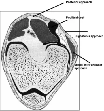
When surgical treatment of a popliteal cyst is indi-cated, three main surgical techniques are available, notably the common posterior approach, the postero-medial approach according to Hughston, and the postero-medial intra-articular approach.
The popliteal cyst usually lies between the medial head of the gastrocnemius muscle and the semimembr-anosus tendon. The posterior as well as the posterome-dial approach are suitable for removing the cyst. With a medial intra-articular approach only the communication of the cyst with the joint can be closed without removal of the cyst (Fig.4).
Common posterior approach
The patient is in prone position. The cyst is approached through the popliteal region by an S-shaped incision. The cyst is usually identified in the area between the medial head of the gastrocnemius and the semimembr-anosus tendon under the superficial fascia. The medial head of the gastrocnemius muscle is retracted to dem-onstrate the extent of the cyst and to identify the com-munication between the cyst and the joint. The synovial
Fig. 3 Magnetic resonance (MR) T2-weighted sagittal image of a popliteal cyst
sac is separated from the often adherent surrounding tissues and followed to its origin in the posterior capsule. The cyst, or pathologic enlargement of the bursa, is dissected free from the posterior capsule, separated from its loose, or sometimes adherent, attachment to the surrounding tissues, and then removed. The capsular defect is closed with sutures.
Posteromedial approach
Hughston’s posteromedial approach differs from the common posterior surgical technique only in the hockey stick incision and the supine position of the patient, which is necessary during the preceding arthroscopy. With the patient supine on the operating table, the hip is externally rotated and flexed to 45°, and the knee is flexed to 90°. A medial hockey stick incision is made. If an arthroscopic examination has been performed, only the posteromedial portion of the incision is used. Otherwise, a posteromedial capsular incision is made, and the intra-articular aspect of the posterior capsule is inspected. The popliteal cyst is then identified in the area between the medial head of the gastrocnemius muscle and the semimembranosus tendon.
Medial intra-articular approach
This approach allows the capsular opening to be closed without removing the cyst. The opening to the cyst is
localized with a 75° arthroscope through the intra-articular groove. Closure of the communication of the cyst with the joint can be achieved through a postero-medial approach after freshening of the cyst edges. A pedicled tendinous graft from the medial gastrocnemius can also be used to cover the opening.
The incision is approximately 5 cm in length just along the posterior border of the medial collateral liga-ment. The posteromedial joint compartment is opened and the capsular opening of the cyst identified. The edge of the capsular defect is freshened. The defect is closed by suture. The popliteal cyst is left alone and will eventually disappear.
The postoperative management includes elevation of the leg on a frame, nonweight-bearing for 4 days, partial weight-bearing for 10 days, full weight-bearing after 2 weeks, and return to full activities after 6 weeks [11].
Discussion
Sansone and De Ponti [9] proposed an interesting sur-gical attitude: 30 patients underwent arthroscopic treatment of their knee derangement and an opening-debridement of the capsular orifice connecting the knee joint to the popliteal cyst. This technique corrects the unidirectional flow of the articular fluid that fills the popliteal cyst. Closing the channel between the cyst and the joint appears to be unnecessary as is excision of the cyst. All patients, followed up with US, showed signifi-cant reduction of their cyst and were considered healed after 2 years. According to the literature, surgery is never advocated for children.
The popliteal cyst is almost never an isolated pathology in an adult knee. In rare cases, it is impos-sible to find its origin. Surgical excision cannot be considered as a definitive solution in a majority of patients.
European questionnaire
A European questionnaire was sent to all the ESSKA members (>1,000). More than 400 replies were col-lected. Several issues were addressed: (1) What is the term you usually use? (2) Do patients in your country consider Baker cyst as a problem? (3) How do you diagnose a Baker cyst? (4) Are the patients referred to you? (5) What is your first approach when treating adults? (6) What is your first approach when treating children? (7) Have you become more aggressive as to the treatment and (8) What are the treatment costs?
When addressing the first question: ‘What is the term you use when confronted with a popliteal cyst?’, more than 60% of the European orthopaedic surgeons men-tioned the term Baker’s cyst, 38% menmen-tioned popliteal
cyst, and 2% popliteal ganglion. None referred to it as bursitis.
In Europe, 83% of the patients consider a popliteal cyst to be a clinical problem. When they are asked: ‘How do you diagnose a popliteal cyst ?’, 40% of the surgeons today request a US, 20% suggest MRI. Twenty-four percent still insist on obtaining X-rays. Seven percent would immediately perform an arthros-copy. Only 3% would use a CT-scan to obtain a cor-rect diagnosis. Most of the patients (92%) are referred to the surgeon.
When orthopaedic surgeons are confronted with Baker’s cyst in adults, masterly neglect appears to be the general approach (37%). Arthroscopy is performed by 25%, aspiration is the first approach by 16% and sur-gery is only by 6%. Sixteen percent adopt various other approaches.
When confronted with a popliteal cyst in children, almost half of the orthopaedic surgeons suggest a mas-terly neglect approach (44%). In 13% of the cases, aspiration is performed first. Ten percent would perform arthroscopy, whereas 8% would opt for surgery. Twenty-five percent use various other treatment modalities.
When asked about their treatment policy, more than 80% of the orthopaedic surgeons have not changed their approach over the last 10 years. Seventeen percent confirmed they have become more aggressive in their treatment.
A huge variation in the reimbursement of the treat-ment costs exists in different countries. Thirty-five per-cent of the orthopaedic surgeons rate the costs at about 100 euros, while almost 40% rate it at equal or more than 300 euros.
References
1. Adams R (1840) Chronic rheumatic arthritis of the knee joint. Dublin J Med Sci 17:520–522
2. Baker WM (1877) On the formation of the synovial cysts in the leg in connec-tion with disease of the knee joint. St Barth Hosp Rep 13:245–261 3. Bui-Mansfield LT, Younberg RA
(1997) Intra-articular ganglion of the knee: prevalence, presentation, etiology and management. Am J Roentgenol 168:123–127
4. De Maeseneer M, Debaere C, Despre-chins B, Osteaux M (1999) Popliteal cysts in children: prevalence, appear-ance and associated findings at MR imaging. Pediatr Radiol 29(8):605–609
5. Malghem J, Vandenberg BC, Lebon C, Lecouvet F, Maldague BE (1998) Gan-glion cysts of the knee: articular com-munication revealed by delayed radiography and CT after arthrogra-phy. Am J Roentgenol 170:1579–1583 6. Miller TT, Staron RB, Koenigsberg T,
Levin TL, Feldman F (1996) MR imaging of Baker cysts: association with internal derangement, effusion and degenerative arthropathy. Radiology 201(1):247–250
7. Rauschning W, Lindgren PG (1979) Popliteal cysts (Baker’s cysts) in adults. Clinical and roentgenological results of operative excision. Acta Orthop Scand 50:583–591
8. Rauschning W (1980) Anatomy and function of the communication between knee joint and popliteal bursa. Ann Rheum Dis 39:354–358
9. Sansone V, De Ponti A (1999) Arthro-scopic treatment of popliteal cyst and associated intra-articular knee disorders in adults. Arthroscopy 15(4):368–372 10. Stone KR, Stoller D, De Carli A, Day
R, Richnak J (1996) The frequency of Baker’s cysts associated with meniscal tears. Am J Sports Med 24(5):670–671 11. Wirth CJ, Rose C (1996) Der
intra-artikula¨re Verschluss der Poplitealzyste. Operat Orthop Traumatol 8(3):232–238