HAL Id: hal-01952238
https://hal.archives-ouvertes.fr/hal-01952238
Submitted on 12 Dec 2018HAL is a multi-disciplinary open access archive for the deposit and dissemination of sci-entific research documents, whether they are pub-lished or not. The documents may come from teaching and research institutions in France or abroad, or from public or private research centers.
L’archive ouverte pluridisciplinaire HAL, est destinée au dépôt et à la diffusion de documents scientifiques de niveau recherche, publiés ou non, émanant des établissements d’enseignement et de recherche français ou étrangers, des laboratoires publics ou privés.
Targeting a G-protein coupled receptor overexpressed in
endocrine tumors by magnetic nanoparticles to induce
cell death
Claire Sanchez, Darine El Hajj Diab, Vincent Connord, Pascal Clerc, Etienne
Meunier, Bernard Pipy, Bruno Payré, Reasmey Phary Tan, Michel Gougeon,
Julian Carrey, et al.
To cite this version:
Claire Sanchez, Darine El Hajj Diab, Vincent Connord, Pascal Clerc, Etienne Meunier, et al.. Tar-geting a G-protein coupled receptor overexpressed in endocrine tumors by magnetic nanoparticles to induce cell death. ACS Nano, American Chemical Society, 2014, 8 (2), pp.1350-1363. �hal-01952238�
Targeting a G-protein coupled receptor overexpressed in endocrine tumors by magnetic nanoparticles to induce cell death
Claire Sanchez1, Darine El Hajj Diab≠1, Vincent Connord≠2, Pascal Clerc1, Etienne Meunier3, Bernard Pipy3, Bruno Payré4, Reasmey P. Tan2, Michel Gougeon5, Julian Carrey2, Véronique Gigoux1 and Daniel Fourmy1*
1 Université de Toulouse 3, EA 4552, Toulouse, France
2 INSA/CNRS, Laboratoire de Physique et Chimie des Nano-Objets, Toulouse 3 Université de Toulouse 3, EA2405, Toulouse, France
4
Université de Toulouse 3, CMEAB, Faculté de Médecine, Toulouse, France 5 Université de Toulouse 3/CNRS, CIRIMAT, Toulouse, France
≠ authors equally contributed to the work
*corresponding authors: Daniel Fourmy, EA 4552 Inserm U1048/I2MC, 1 avenue Jean Poulhès, BP 84225, 31432 Toulouse Cedex 4, France. Email: Daniel.Fourmy@inserm.fr
Abstract
Nanotherapy using targeted magnetic nanoparticles grafted with peptidic ligands of receptors over-expressed in cancers is a promising therapeutic strategy. However, nanoconjugation of peptides can dramatically affect their properties with respect to receptor recognition, mechanism of internalization, intracellular trafficking and fate. Furthermore, investigations are needed to better understand the mechanism whereby application of an alternating magnetic field to cells containing targeted nanoparticles induces cell death. Here, we designed a nanoplatform (termed MG-IONP-DY647) composed of an iron oxide nanocrystal decorated with a ligand of a G-protein coupled receptor, the cholecystokinin-2 receptor (CCK2R) that is over-expressed in several malignant cancers. MG-IONP-DY647 did not stimulate inflammasome of Raw 264.7 macrophages. They recognized cells expressing CCK2R with a high specificity, subsequently internalized via a mechanism involving recruitment of β-arrestins, clathrin-coated pits and dynamin, and were directed to lysosomes. Binding and internalization of MG-IONP-DY647 were dependent on the density of the ligand at the nanoparticle surface and were slowed down relative to free ligand. Trafficking of CCK2R internalized with the nanoparticles was slightly modified relative to CCK2R internalized in response to free ligand. Application of an alternating magnetic field to cells containing MG-IONP-DY647 induced apoptosis and cell death through a lysosomal death pathway, demonstrating that cell death is triggered even though nanoparticules of low thermal power are internalized in minute amounts by the cells. Together with pioneer findings using iron oxide nanoparticles targeting tumoral cells expressing Epidermal Growth Factor (EGF) receptor (Creixell M., et al. ACS Nano, 2011, 5, 7124; Domenech, M., ACS Nano, 2013, 7, 5091-101), these data represent a solid basis for future studies aiming at establishing the proof-of-concept of nanotherapy of cancers using ligand-grafted magnetic nanoparticles specifically internalized via cell surface receptors.
Cancer is a leading cause of death, with millions of new people diagnosed with cancer every year, making the development of new methods for efficient and specific delivery of diagnostic and therapeutic agents to tumors a permanent challenge. Over the two past decades, the introduction of targeted cancer therapeutics in clinic has been a major breakthrough in cancer therapy. Currently, targeted delivery of diagnosis and/or therapeutic agents to tumoral cells uses biological markers selectively expressed, or at least over-expressed in tumors relative to healthy surrounding and distant tissues 1, 2.
In this context, much attention is focused towards the development of strategies combining molecular targeting and nanoparticle delivery 1, 3. Among the wide range of available nanoparticles, magnetic nanoparticles have emerged as potential biocompatible systems for cancer detection by magnetic resonance imaging (MRI) and for targeted cancer therapy 3-7. Indeed, magnetic nanoparticles offer the theoretical potential to be driven to the tumors by grafting the nanoparticles with ligands or antibodies recognizing receptors or antigens over-expressed in tumors 1. In vivo, targeted magnetic nanoparticles injected intravenously accumulate within the tumor where they can play as a thermoablative agent upon application of an alternating magnetic field. In addition, targeted magnetic nanoparticles can serve as nanocarriers of cytotoxic agents through the body, enabling their preferred delivery to primary and metastatic tumors 1. Interestingly, targeted magnetic nanoparticles offer the opportunity of combining targeted hyperthermia therapy and chemiotherapy. Using such therapeutic strategies, exposure of tumoral tissues to the therapeutic agents should be optimized whereas that of healthy tissues should be minimized.
Iron oxide nanoparticles (IONPs), which display low toxicity in humans, are the subject of renewed interest in the perspective of nanotherapy of cancers 8. A first field of investigations aims at increasing our knowledge about engineering targeted iron oxide nanoparticles to enhance their ability to efficiently reach solid tumors after systemic administration. Escape of the nanoparticles from the reticuloendothelial system, their extravascular transport to the tumor and subsequent tumor penetration and accumulation still represent important challenges, although key breakthroughs were recently made 9, 10
. Another important point concerns the specificity of targeted nanoparticles which is often far from expected, including on cultured cells in vitro. In fact, targeting is usually obtained through the chemical grafting of ligands (in the case of receptor targeting) to the surface of the nanoparticles. Yet, this nanoconjugation can dramatically modify properties of the ligands with respect to recognition parameters (kinetics, specificity, affinity), mechanism of internalization, intracellular trafficking and fate 11-13. A second field of active research is related to magnetic hyperthermia and mechanisms at the origin of cancer cell death 13-15. In vivo studies with transplanted tumors in animals and even clinical trials with prostate cancer or brain tumor patients have provided the proof-of-concept of hyperthermia therapy of cancers with IONPs 16, 17. However, in most of the in vivo investigations so far reported, high amounts of IONPs were injected intravenously or even directly injected into the tumors.
Therefore, results obtained in these studies cannot be generalized to targeted IONPs which are actively and specifically internalized in lower amounts by tumoral cells through their cell-surface target.
Interestingly, magnetic iron oxide nanoparticules targeting Epidermal Growth Factor (EGF) receptor were recently shown to be internalized by tumoral cells and to kill the cells upon application of an alternating magnetic field13, 14, 18. Cell death was achieved without a perceptible temperature rise and occurred through a mechanism involving lysosomal death pathways13, 14, 18. Such data stimulate research aimed at developing strategies of targeted nanotherapy of cancers overexpressing other cell surface receptors.
The aim of our study was to design and evaluate a hybrid system (termed MG-IONP-DY647) composed of an iron oxide nanocrystal decorated with a peptidic ligand of the cholecystokinin-2 receptor (CCK2R). The CCK2R was chosen as an example of targeted receptor because this seven-transmembrane domain peptide receptor is expressed at high incidence and density in cancers such as medullary thyroid carcinomas, small cell lung cancers (SCLC), gastrointestinal stromal tumors (GIST), and insulinomas 19-21. Moreover, the CCK2R undergoes rapid internalization and subsequent trafficking to lysosomes following stimulation by its equally potent natural ligands, gastrin and cholecystokinin (CCK) 22, 23. In the recent period, a number of radio-labeled replicates of CCK or gastrin have been developed, with the indication of receptor targeted tumor imaging and therapy, on the example of imaging probes which are currently used in clinic to detect neuroendocrine tumors over-expressing somatostatin receptors 19, 20. However, so far, none of these CCK or gastrin radiolabels has been approved for clinical use as their renal retention and expected radio-toxicity remains a major drawback.
We demonstrated the ability of MG-IONP-DY647 to specifically target cells expressing CCK2R, characterized internalization and intracellular trafficking of the nanoparticles and finally used them as a targeted therapeutic agent to induce tumoral cell death through a cell death lysosomal pathway. Thus, we here confirm that a small amount of nanoparticles displaying a small heating power is sufficient to induce cell death upon an alternating magnetic field. These data are very promising in light to the recent concept that lysosomal membrane permeabilization could be an effective way to kill apoptosis resistant cancer cells24.
Results
Preparation of gastrin-decorated iron oxide magnetic nanoparticles (MG-IONP-DY647)
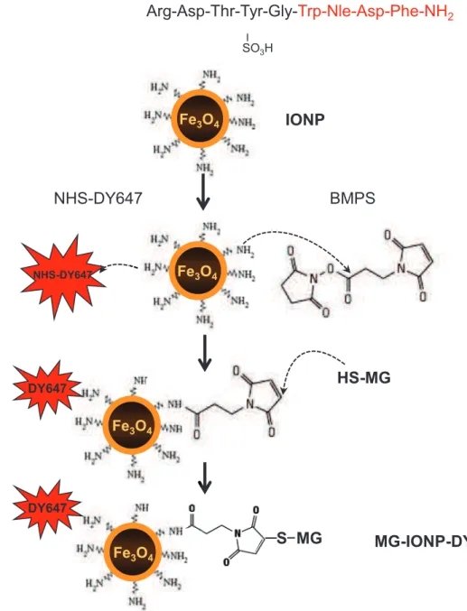
In order to selectively target CCK2R with minor cross-recognition of the CCK1R which is also frequently expressed in tumors 19, gastrin, a selective CCK2R ligand, was chosen as a cross-linked peptide to IONPs. Previous data showing that radiolabeled gastrin injected to animals is abundantly retained by the kidney due to the presence of 5 glutamic acids in the middle of the peptidic sequence were taken into account for the design of the gastrin replicate 25, 26 (Fig. 1a). Thus, a synthetic replicate of gastrin, termed MG, which contains the pharmacophore of natural gastrin but has
its Glu-Glu-Glu-Glu-Glu sequence replaced by Cys-Lys-Ser-Ser-Glu was synthesized. Importantly, this peptide shares its C-terminal bioactive sequence with the natural agonist CCK which is used as reference peptide for CCK2R internalization studies (Fig. 1a). Control experiments established that MG bound to the CCK2R with only 7.3-fold lower affinity than CCK and stimulated production of inositol phosphates through phospholipase-C activation with the same efficacy as CCK, and only 1.3-fold lower potency (Supplementary information, Fig. 1a, b). Finally, MG triggered CCK2R internalization as demonstrated by endocytosis of GFP-tagged CCK2R and trapping of MG-DY647 within endocytosic vesicules (Supplementary information, Fig. 1c).
The strategy illustrated in Fig. 1b was used to obtain magnetic nanoparticles decorated with both gastrin replicate (MG) and fluorescent label (DY647). These were termed MG-IONP-DY647. Batches of MG-IONP-DY647 decorated with 24, 100 or 240 molecules of MG per nanoparticle were routinely prepared.
Starting nanoparticles were commercial iron oxide nanoparticles having characteristics shown in Supplementary information table 1. Heating properties of these nanoparticules were measured in a magnetic field of frequency 275 kHz, with an amplitude ranging from 24 to 40 mT (Fig. 2a). At the largest applied magnetic field, specific absorption rate was 13 W/g. The size of MG-IONP-DY647 determined by transmission electron microscopy was 8.7 ± 1.6 nm (Fig. 2c) and analysis by dynamic light scattering in a biological medium enriched with 0.5% fetal calf serum showed a distribution around a size of 40 nm, with a significant amount of objects reaching 100 nm size. Size distribution was not significantly affected by the density of MG grafted at the nanoparticle surface (Fig. 2b). Hence, in the biological medium, MG-IONP-DY647 behaved either as clusters of several individual nanoparticles and/or likely bound serum proteins.
Absence of effect of MG-IONP-DY647 nanoparticles on macrophage inflammasome
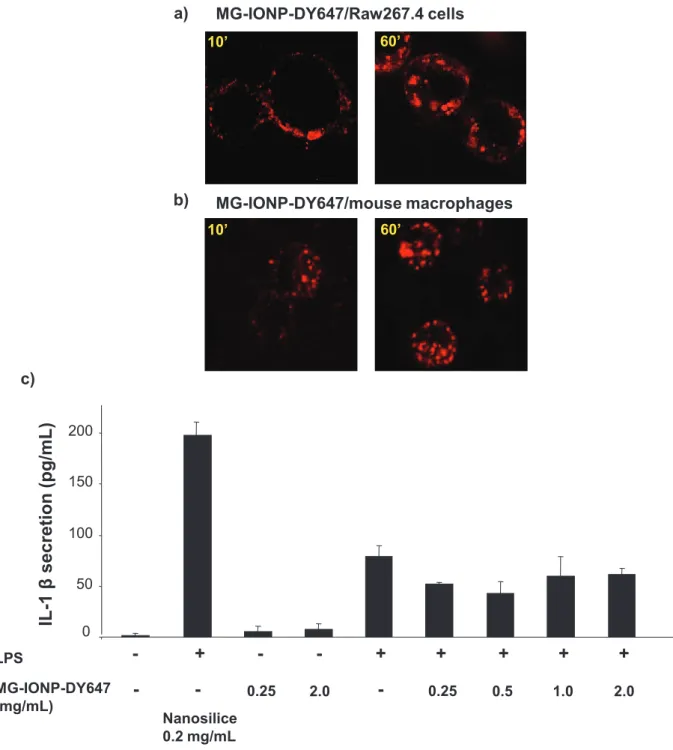
To evaluate biocompatibility of MG-IONP-DY647, we determined whether the nanoparticles could activate macrophages. Indeed, side-targets of nanoparticles in vivo are resident macrophages of liver and spleen as well as monocytes and circulating macrophages involved in the inflammation processes 4, 27, 28. Incubation of MG-IONP-DY647 in vitro with Raw 264.7 mouse leukaemic monocyte macrophages or with mouse peritoneal macrophages resulted in rapid and abundant uptake of nanoparticles (Fig. 3a-b). Then, activation of macrophages was tested on both untreated and LPS-stimulated Raw 264.7 cells by measuring the secretion of the cytokine IL-1β (Fig. 3c). We found that in contrast to nanosilice nanoparticles recognized as potent inducer of inflammasome and which elicit release of the cytokine IL-1β 29, MG-IONP-DY647 at concentrations up to 2mg/mL did not trigger significant release of IL-1β from both untreated and LPS-primed Raw 264.7 cells (Fig. 3c). These first results are in favor of biocompatibility of MG-IONP-DY647.
Specific targeting of HEK293 cells expressing CCK2R by MG-IONP-DY647 and intracellular trafficking of the nanoparticles
Our targeting strategy using MG-IONP-DY647 requires specific recognition of nanoconjugated MG molecules by the CCK2R expressed at the cell surface and internalization of the nanoparticles through the endocytosic machinery usually involved in CCK2R endocytosis. Yet, nanoconjugated MG molecules might modify its pharmacological properties. We therefore characterized binding and internalization of MG-IONP-DY647 in HEK293 cells expressing the CCK2R, namely Flp-InTMCCK2R-293 previously used to precisely delineate agonist-induced CCK2R internalization 22.
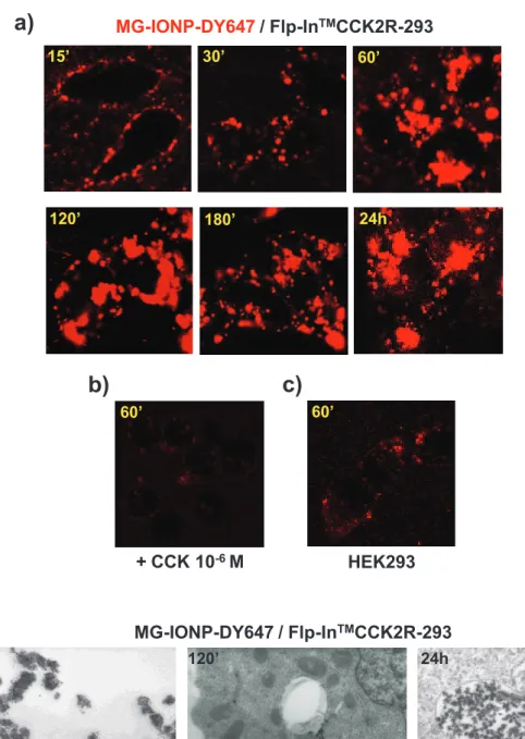
Confocal microscopy images show slow binding of MG-IONP-DY647 to the membrane of Flp-InTMCCK2R-293 cells which were progressively illuminated at their surface in the contact of MG-IONP-DY647 (Fig. 4a). This binding of MG-MG-IONP-DY647 to the cell surface was slower that previously appreciated with unconjugated ligands 22. Following binding, MG-IONP-DY647 abundantly internalized over the time (Fig. 4a). Only minor non specific binding/uptake of red fluorescence was seen on HEK293 cells lacking the CCK2R (Fig. 4c), or on Flp-InTMCCK2R-293 cells incubated in the presence of a saturating concentration (1 µM) of CCK (Fig. 4b).
Sub-cellular localization of MG-IONP-DY647 was assessed by transmission electron microscopy. As illustrated on Fig. 4d, MG-IONP-DY647 were located in endocytosic vesicles. The number of nanoparticles in small vesicles, as well as the number of vesicles containing nanoparticles, increased within the first 2-3 h of incubation. Then, the size of vesicles containing MG-IONP-DY647 clearly increased showing intracellular accumulation and storage of highly concentrated nanoparticles (Fig. 4d).
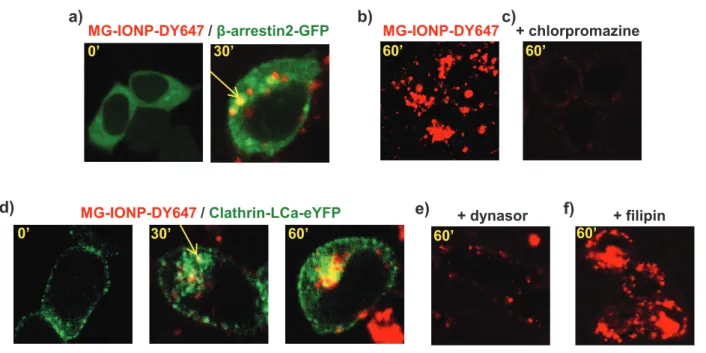
Given that nanoparticles can enter cells through several ways, the initial mechanism of MG-IONP-DY647 internalization was investigated. We previously showed that the molecular mechanism of agonist-induced internalization of CCK2R involves recruitment of non visual arrestins, clathrin-coated pits, and dynamin 22. On Fig. 5a, confocal microscopy imaging of cells transiently expressing GFP-tagged β-arrestin2 incubated with MG-IONP-DY647 shows fluorescent nanoparticles in intracellular vesicles containing β-arrestin2. Furthermore, internalization of MG-IONP-DY647 was blocked in the presence of chlorpromazine, a clathrin inhibitor, at 75µM (Fig 5c, dose-response effect shown on supplementary information, Fig. 2). In line with this result, incubation of MG-IONP-DY647 with cells transiently expressing eYFP-tagged clathrin polypeptide showed abundant intracellular co-localization (Fig. 5d). Furthermore, MG-IONP-DY647 internalization was inhibited in the presence of the dynamin inhibitor, dynasore (Fig. 5e). The possibility of uptake through caveoles was also examined by testing the effect of filipin, an inhibitor of caveoles. However, abundant internalization of MG-IONP-DY647 in the presence of filipin was still observed, supporting that MG-IONP-DY647 endocytosis did not significantly occur through caveoles in Flp-InTMCCK2R-293 cells (Fig. 5f).
Intracellular trafficking of internalized MG-IONP-DY647 was then investigated. First, co-localization of MG-IONP-DY647 with the CCK2R during intracellular trafficking were studied and quantified in cells expressing GFP-tagged CCK2R. As shown on Fig. 6a and 6b, internalized MG-IONP-DY647, as well as CCK-DY647, co-localized with the CCK2R in endocytosic vesicles. However, the time-courses of co-localization differed. Indeed, comparing Fig. 6a and Fig. 6b shows that co-localization between CCK-DY647 and the CCK2R was persistent whereas progressive and partial dissociation between MG-IONP-DY647 and GFP-tagged CCK2R was noted at earlier times. These results suggest that nanoconjugation of MG to magnetic nanoparticles affects the trafficking properties of internalized CCK2R and perhaps enables the CCK2R to recycle faster than when stimulated with unconjugated ligand. To examine this possibility, we evaluated co-localization of CCK2R stimulated with MG-IONP-DY647 versus CCK-DY647 in vesicles expressing Rab11, a small GTPase of recycling vesicles 30. As represented on Fig. 7a, localization of CCK2R-GFP in Rab11-DsRed containing vesicles was maximal after 4 h of incubation with MG-IONP-DY647 and then slightly decreased. When stimulated with CCK-DY647 (or with MG-DY647, not shown), localization profile of CCK2R in DsRed-tagged Rab-11 positive vesicles was significantly different, with a slower increase to a maximum after 5 h of incubation. Thus, CCK2R stimulated with MG-IONP-DY647 seems to recycle faster than when stimulated with the non-conjugated ligands, however, the recycling takes several hours, as previously demonstrated 22.
At last, the intracellular fate of MG-IONP-DY647 was analyzed using the lysosome staining reagent LysoTraker® and by incubating MG-IONP-DY647 with cells transiently expressing DsRed-tagged Rab7, a small GTPase of late endosomes and lysosomes. As illustrated on Fig. 7b, a high proportion of LysoTracker® stained lysosomes contained MG-IONP-DY647 after time longer than 1h of incubation, and co-localisation images between MG-IONP-DY647 and Rab7-Ds-Red were observed in most cells (Fig. 7c).
Together, this set of results demonstrates that nanoconjugation of MG to IONPs enables active endocytosis of the nanoparticles and, by facilitating separation of the receptor from endocytosic vesicles, nanoconjugation slightly accelerates the presence of the targeted receptor in slow recycling vesicles. Importantly, MG-IONP-DY647 uptake by cells remains fully dependent on CCK2R internalization and involves recruitment of β-arrestin2, clathrin-coated pits and dynamin. Furthermore, internalized MG-IONP-DY647 are directed to lysosomes where they are trapped for times up to 24 h.
Targeting of InR1G9-CCK2R tumoral endocrine cells by MG-IONP-DY647 nanoparticles Tumoral cells display profound modifications relative to “normal” cells which affect plasma membrane, intracellular trafficking as well as lysosomal volume, composition and cellular distribution31. Therefore, we next characterized the behavior of MG-IONP-DY647 towards tumoral cells expressing or not the CCK2R. Specificity and kinetics of MG-IONP-DY647 binding, accumulation in lysosomes and dependency on the density of MG at the nanoparticle surface were
precisely investigated on the tumoral endocrine cell line expressing the CCK2R, namely InR1G9-CCK2R 32.
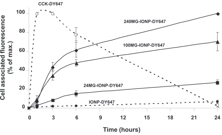
Confocal microscopy observations confirmed specific binding of MG-IONP-DY647 to the tumoral cells and subsequent internalization, as no significant uptake was detected in InR1G9 lacking the CCK2R or InR1G9-CCK2R incubated in the presence of 1 µM CCK (supplementary information, Fig. 3a, b, c). MG-IONP-DY647 binding and internalization determined by FACS indicates that the amount of MG-IONP-DY647 uptaken by the cells increased with the time of incubation but much slower than that of CCK-DY647, which was maximum between 1h and 3h of incubation and then decreased (Fig. 8). Furthermore, the amount of bound and internalized MG-IONP-DY647 by the cells increased proportionally to MG density at the surface of the nanoparticles. After 24h of incubation, magnetic measurements showed an amount of 2.2 ± 0.2 pg of iron per cell incubated with nanoparticles grafted with 100 molecules of MG (termed 100MG-IONP-DY647 on Fig. 8 and 9). FACS quantifications also confirmed high specificity of targeting since nanoparticles without grafted MG (termed IONP-DY647 on Fig. 8) were uptaken in very low amount by InR1G9-CCK2 tumoral cells and MG-IONP-DY647 were poorly uptaken by InR1G9 tumoral cells lacking CCK2R at their surface (9.1 ± 2.1% of non specific binding after 24h of incubation, not shown). Finally, lysosome occupancy by MG-IONP-DY647 (illustrated in supplementary information, Fig. 3d) was quantified by confocal microscopy. Kinetics of accumulation of MG-IONP-DY647 in lysosomes were in agreement with that of binding/internalization (Fig. 9). After 24h of incubation, lysosome occupancy by nanoparticles grafted with 24, 100 or 240 MG molecules reached 31.6 ± 1.9 %, 46.8 ± 0.7 % or 49.8 ± 1.8 %, respectively. No significant presence of nanoparticules without MG (termed IONP-DY647) could be noticed in lysosomes after 24h incubation (Fig. 9). On the other hand, lysosome occupancy by unconjugated CCK-DY647 was maximal at 1h and 3h of incubation and then dramatically decreased to almost disappear at 24h, suggesting ligand degradation in lysosomes.
Internalized MG-IONP-DY647 by tumoral cells induce apoptosis and cause cell death upon an alternating magnetic field
Having assessed the ability of MG-IONP-DY647 to specifically target endocrine tumoral cells expressing the CCK2R and to abundantly accumulate in lysosomes of these cells, the next step was to test the ability of the nanoparticles to induce cell death upon exposure to an alternating magnetic field. For this purpose, InR1G9-CCK2R tumoral cells incubated with MG-IONP-DY647 for 24h were maintained at 37°C and exposed to an alternating magnetic field (275 kHz, 40 mT or 52 mT for 2h). Controls were composed of cells having internalized ligand without nanoparticles (CCK-DY647) and cells having internalized MG-IONP-DY647 but which were not exposed to the alternating magnetic field. In preliminary experiments, we also checked that cells incubated with nanoparticles without grafted gastrin and which did not internalize these nanoparticles (Fig. 9) behaved similarly as cells having internalized CCK-DY647 (not illustrated).
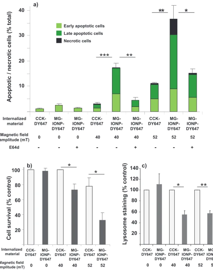
The impact of magnetic field treatment was determined by counting cells labeled by FITC-tagged annexinV and/or propidium iodure which identified early apoptotic, late apoptotic and necrotic cells, respectively. As shown on Fig. 10, application of the alternating magnetic field during 2h caused the cells to enter in apoptosis. The total number of apoptotic cells reached 17.1 ± 1.6 % at 40 mT amplitude and 36.4 ± 5.5% at 52 mT amplitude. In control cells having internalized unconjugated CCK-DY647 and exposed to the same magnetic field amplitudes, apoptotic cells represented 3.0 ± 0.7 % and 11.2 ± 0.4 % of total cell population. The number of necrotic cells, namely cells which became positive for propidium iodure labeling only, was not significant with magnetic field of 40 mT whereas it increased up to 5.9 % at 52 mT. Determination of cell survival 24h after magnetic field exposure using MTT assay indicated that magnetic field of 40 or 52 mT killed 26.9 ± 7.6 and 67.1 ± 9.9 % of tumoral cells, respectively. It is worthy to note that magnetic field of 52 mT affected viability of cells that had not accumulated nanoparticles whereas an amplitude of 40 mT had a minor effect (Fig. 10a, b).
Since a recent study with EGF-grafted nanoparticles documented that alternating magnetic field caused lysosome membrane permeabilization, we determined whether it was also the case with MG-IONP-DY647 nanoparticles. First, effect of magnetic field was evaluated on lysosome staining with LysoTracker® Red. As evidenced on Fig. 10c, exposure of cells containing MG-IONP-DY647 to magnetic field of 40 or 52 mT induced drops of lysosome staining suggesting leaking of lysosome content (see Supporting information, Fig. 4 for illustration). Furthermore, the effect of the magnetic field on apoptosis was almost totally reversed by E64d, an inhibitor of cysteine proteases such as lysosomal cathepsins B, L C, H P (Fig. 10a). Pepstatin A, an inhibitor of aspartyl peptidases such as lysosomal cathepsin D, did not reverse cell death (not shown). The effect of the magnetic field on apoptosis was also significantly diminished by chloroquine, a weak base affecting activity of lysosomes and autophagy (at 40 mT, amount of apoptotic cells: 6.8 ± 0.5 % in the presence of chloroquine versus 17.1± 1.6 % in the absence of chloroquine, not illustrated).
This set of results shows that application of an alternating magnetic field to InR1G9 tumoral cells containing low amounts of MG-IONP-DY647 nanoparticules internalized specifically through cell surface CCK2R cause apoptosis and cell death through a mechanism involving lysosome leaking and lysosomal cysteine proteases.
Discussion
In the context of increasing success of targeted therapy of cancers and accumulation of data showing overexpression of peptide receptors in several types of tumors as well as their contributing role in cancer development 33, 34, a synthetic analogue of gastrin was conjugated to iron oxide nanoparticles in order to produce a magnetic nanoplatform for targeted therapy of tumors overexpressing the CCK2R. The synthetic analogue of gastrin (MG) pharmacologically behaved similarly to CCK, the most efficient and potent natural agonist of the CCK2R 35.
We first showed that MG-IONP-DY647 nanoparticles, even at high concentrations, do not promote inflammation as evidenced by the absence of IL-1β secretion by cultured macrophages. This result confirms recognized good biocompatibility of iron oxide nanoparticles. Binding, uptake, intracellular trafficking of MG-IONP-DY647 nanoparticles and the underlying cellular and molecular mechanisms were characterized on HEK293 cells, a reference cell model for characterization of membrane receptor internalization, as well as on tumoral cell lines (InR1G9) expressing or not the CCK2R32. We found that MG-IONP-DY647 nanoparticles binding and internalization were entirely dependent on the presence of the CCK2R at the cell surface. Nevertheless, kinetics of binding and internalization of MG-IONP-DY647 were much slower than that of fluorescent unconjugated CCK (CCK-DY647), although MG-IONP-DY647 nanoparticles internalized together with the CCK2R through the initial molecular mechanism described for CCK- or gastrin-induced internalization of the CCK2R22. Indeed, key steps in the internalization process such as β-arrestin2 recruitment, involvement of clathrin-coated pits and of dynamin were observed with both MG-IONP-DY647 (this study) and CCK-DY647 22.
Analysis of CCK2R intracellular trafficking in HEK293 cells expressing either GFP-tagged CCK2R or DsRed-tagged Rab11, a small GTPase of slow recycling vesicles, strongly supports that nanoconjugation of MG affected both stability of the endocytosic complexes and CCK2R trafficking. Indeed, after stimulation with MG-IONP-DY647, CCK2R was found in vesicles distinct from that containing the nanoparticles, and was targeted to recycling vesicles containing Rab11 earlier than CCK2R stimulated with unconjugated CCK or gastrin (Fig. 6 and 7). These results are consistent with data showing that internalization and trafficking of a G-protein coupled receptors is initially dictated by its ligand 36, 37.
Our data are in agreement with a report showing that nanoconjugation of an antibody directed against EGF receptor modulates antibody-induced internalization of this receptor 12. Furthermore, it has been also shown that ligand dissociation from G-protein coupled receptors (GPCRs) and subsequent degradation by endosomal peptidases controls trafficking and endosomal signaling of peptide receptors. For example, endothelin-converting enzyme 1 (ECE-1) rapidly degrades substance P, calcitonin gene-related peptide (CGRP) and somatostatin peptide in endosomes, disrupting the peptide-receptor-β-arrestin complex and then allowing β-arrestins to return to the cytoplasm and receptors, to recycle 38-40. For class B GPCRs such as the CCK2R, dissociation from β-arrestin in endosomes is necessary for recycling and resensitization 37. As an alternative hypothesis to explain the increased time-course of CCK2R localization in slow recycling vesicles, CCK2R internalization as oligomers due to simultaneous binding of MG-IONP-DY647 to several receptor molecules at a time must be also considered. Such multi-ligand binding was described for neuropeptide Y1 receptor when targeted by neuropeptide Y-decorated quantum dots 11. Importantly, besides modifications of CCK2R trafficking, internalized MG-IONP-DY647 nanoparticles are directed to lysosomes where they accumulate both in HEK293 and InR1G9 tumoral cells. Of interest, internalized MG-IONP-DY647
were much stable in lysosomes than unconjugated fluorescent CCK which became almost non-detectable after 24h of contact with the cells.
At last, we started to explore the opportunity offered by magnetic nanoparticles accumulated into lysosomes of tumoral cells to act as a therapeutic agent inducing cell death upon exposure to an alternating magnetic field. The results that were obtained clearly demonstrate that exposure of tumoral cells containing actively internalized MG-IONP-DY647 to an alternating magnetic field of moderate strength causes apoptosis followed by cell death. Moreover, first data on the mechanisms at the origin of cell death support the involvement of lysosomes and cysteine proteases, since staining of lysosomes with pH-independent ysoTracker®Red was diminished by exposure of cells containing targeted nanoparticles to the magnetic field. Moreover, E64d inhibitor was able to reverse the effect of the magnetic field on apoptosis. On this basis, and according to our current knowledge, lysosome membrane permeabilization followed by leaking of cathepsin B is likely at the origin of tumoral cell death. Thus, MG-IONP-DY647 exposed to an alternating magnetic field behaved as a lysosomotropic agent in tumoral endocrine cells. Several other agents have been shown to induce lysosome membrane permeabilization and subsequent activation of lysosomal death pathways 24. EGF-grafted iron nanoparticles internalized by tumoral cells and exposed to an alternating magnetic field were previously reported to cause lysosomal membrane permeabilization, together with production of reactive oxygen species (ROS) and subsequent cell death13. It is worthy to note that ROS formation, lysosomal membrane permeabilization and cell death occurred in absence of any perceptible temperature rise in the incubation medium of the cells (13, 14, 18 and this study). These experimental data, as well as basic calculations41, 42, support the view that minute amounts of nanoparticles located in lysosomes are unable to elicit temperature rise, including inside the cells. Alternatively, local temperature increase 15, 43, nanoparticle rotation13 or nanoparticle high-frequency motion44 have been proposed.
Whatever the initial mechanism at the origin of lysosomal membrane permeabilization, data from the current study highlight the feasibility of cell death induction with very small quantities of actively and specifically internalized nanoparticles. Basic calculation using the model presented in 45 indicates that inducing a temperature increase of 5 °C in a 3-mm size tumor with MG-IONP-DY647 would require approximately 165 pg Fe/cell, which is a completely unrealistic objective using targeted nanoparticles which are internalized only via cell surface receptors. Here, a maximum of 2.2 pg Fe/cell could be specifically internalized via cell surface CCK2R receptors. Although this low amount of nanoparticles was sufficient to induce cell death, it likely explains, together with the low heating power of the iron oxide nanoparticles, the relatively modest level of cell death achieved in this study using a magnetic field of 40 mT. Our results show that increasing the amplitude of the magnetic field from 40 mT to 52 mT augmented death rate of cells containing nanoparticules. However, such a treatment also caused damage on cells which had not accumulated nanoparticles. Therefore,
optimization of the nanoparticles will be required in order to enhance their killing efficiency. Their size and composition could be changed to increase their heating power.
In conclusion, grafting of gastrin to iron oxide nanoparticles enabled highly specific targeting of HEK293 and tumoral endocrine cells INR1G9 expressing the CCK2R, a G-protein coupled receptor overexpressed in endocrine tumors. Subsequently, nanoparticles undergo internalization with the CCK2R through a mechanism involving β-arrestins, clathrin-coated pits and dynamin composing the endocytosic machinery of the CCK2R. Internalized nanoparticles are then directed to lysosomes where they accumulate in minute amount (2.2 pg Fe/cell). Application of an alternating magnetic field to cells containing MG-IONP-DY647 induced apoptosis and by cell death through a lysosomal death pathway demonstrating that cell death is triggered even though nanoparticles of low thermal power are internalized in minute amounts by the cells. These data are very promising in light to the recent concept that lysosomal membrane permeabilization could be an effective way to kill apoptosis resistant cancer cells24. Together with previous pioneer findings13, 14, they represent a solid basis for future studies aimed at establishing the proof-of-concept of nanotherapy of cancers using ligand-grafted magnetic nanoparticles specifically internalized via their cell surface receptors.
Materials and Methods
Chemical and biological materials.
Iron oxide nanoparticles were purchased from Genovis (Lund, Sweeden). The characteristics of these IONP provided by the manufacturer are given in Supplementary information table 1. Stable replicate of CCK, sulfated [Thr28,Nle31]-CCK 25-33 was kindly given by Prof. L. Moroder 46 and is referred to as CCK. 125I-Sodium (2000 Ci/mmol) and [myo-3H]inositol (5 μCi/ml) were from from Perkin Elmer Life Sciences. CCK was conjugated with Bolton-Hunter reagent, purified, and radioiodinated as described previously 47 and is referred to as 125I-CCK. Gastrin replicate termed MG (Fig. 1) was synthesized by Covalab (Villeurbanne, France). DY647 tagged CCK or MG were obtained according to the procedure described for Glucose Insulinotropic Polypeptide by coupling the peptides to NHS-DY647-PEG1 (Dyomics GmbH, Jena, Germany) 48 and were referred to as CCK-DY647 or MG-DY647. The following producted were supplied as follows: 3-Maleimidopropionic acid N-hydroxysuccinimide ester (BMPS) (Thermo Scientific), magnetic columns (Miltenyi, Berisch, Gladback, Germany), specific inhibitor of dynamin, dynasore (Calbiochem), an inhibitor of clathrin dependent uptake, chlorpromazine (Sigma-Aldrich), an inhibitor of caveoles, filipin (Sigma), LysoTracker® (Invitrogen).
The cDNAs encoding CCK2R was subcloning in pcDNA3 vector, and green fluorescent protein (GFP) tagged CCK2R was generated by subcloning the CCK2R cDNA in pEGFP-N1 (BD biosciences clontech). DsRed tagged Rab11, DsRed tagged Rab7 and clathrin-LCa-eYFP were obtained from Addgene (www.addgene.org). GFP tagged β-arrestin2 was kindly given by Robert Lefkowitz (Duke University Medical Center, Durham, USA). Annexin V Binding Apoptosis Assay Kit was from AAT
Bioquest, Inc. (Sunnyvale, CA, USA). Cell lines were: HEK 293 cells stably expressing the CCK2R (Flp-InTM CCK2R-293) obtained using the Flp-InTM system (HEK293) (Invitrogen) as previously described 22; the glucagon-producing hamster tumoral cell line InR1G9 stably expressing CCK2R obtained as previously described (InR1G9-CCK2R) 32 and Raw 264.7 Mouse leukaemic monocyte macrophage cell line. Lab-Tek II chambers were from Thermo Fisher (Rochester, NY, USA), GlassBotton Cellview were from Greiner Bio-one, Courtaboeuf, France.
Preparation of gastrin-decorated magnetic nanoparticles (MG-IONP-DY647)
The protocol used for grafting 100 molecules of MG per IONP was as follows: an aliquot of IONP (80 µL IONP at concentration 12 mg Fe3O4/mL) was added to 120 µL of sodium phosphate buffer (0.2 M Na2HPO4 pH: 7.3, 150 mM NaCl) and sonicated for 10 min on melting ice (Bioblock Scientific 88154). NHS-DY647-PEG1 (12 µg) in solution in dimethyl formamid (24 µL) was added and let to react for 2h at room temperature. Then, 53 µg of 3-Maleimidopropionic acid N-hydroxysuccinimide ester (BMPS) in solution in 10 µl DMF was added and incubated at room temperature for 1h. MG (100 µL of a solution 30%DMF, 70% H2O containing 20 µg peptide) was added to IONP and let to react for 2h. Finally, free maleimide functions were saturated by addition of an excess of cystein (62 µg in 50 µL of reaction buffer). Grafted nanoparticles were recovered through a magnet column in H2O. MG-IONP-DY647 were stored at 4°C. Before use, MG-IONP-DY647 were always sonicated on ice for 10 min. The amount of recovered nanoparticules was determined by magnetic measurement (see below) and density of grafted MG at the nanoparticle surface was assessed by measuring the ability of different dilutions of MG-IONP-DY647 to stimulate production of inositol phosphates in Flp-InTM CCK2R-293 cells. Density of MG was calculated from dose-response curve with free MG (Supplementary Fig. 1b).
Dynamic light scattering measurements of hydrodynamic diameter
Suspension of iron nanoparticles at 0.1 mg/ml was prepared in RPMI 1640 medium containing 0.5% FBS. The iron nanoparticle suspension was sonicated for 10 min on ice. After temperature equilibrium to 20°C, particle size was measured by dynamic light scattering using Nanotrac 250 (Microtrac, York, PA).
Specific absorption rate measurements
A 300 µl solution of IONPs at 5.9 mg Fe/mL was used for specific absorption rate measurements. The temperature increase of the sample placed in a 275 kHz alternating magnetic field delivered by a commercial magnetic inductor (Fives Celes, Lautenback, France) during 100s was measured. A blank sample containing water was used in parallel in order to remove contribution of eddy current to the temperature increase. Temperature measurements were performed using a thermal probe (Reflex,
Neoptix, Canada). Specific absorption rate was calculated using standard formula and taking into account the specific heat of water.
Determination of iron concentrations and amounts
Amounts of iron were determined by magnetic measurements using a Vibrating Sample Magnetometer (PPMS, Quantum Design, USA). First, we checked the value of concentration and saturation magnetization provided by the manufacturer by performing magnetic measurements on a dried powder of MG-IONP-DY647. Hysteresis cycles at 270 K were measured and the linear slope corresponding to the diamagnetic signal was removed from raw measurement. The remaining signal was fitted with a distribution of Langevin function in parallel with a fit of the NP size distribution obtained by TEM. The magnetization and concentration values determined from this fit are 73 ± 10 Am2/kg Fe304 and 2.1 ± 0.2 mg/ml, respectively, when 80 Am2/kg Fe304 and 2 mg/ml were expected from manufacturer data. We thus considered manufacturer data as reliable and used them in the following. To determine iron content in cells, a suspension containing a known number of dried cells (2.5 ± 0.2 ´106) was put into a magnetic measurement capsule, the excess of liquid removed, and the remaining liquid let to evaporate. Hysteresis loops were measured at 300 K upon a magnetic field of 5 T. The obtained magnetization was divided by the saturation magnetization of the nanoparticles provided by the nanoparticle supplier (107 Am2/kg Fe) and the number of cells to get the iron content per cell.
Transmission Electron Microscopy
Flp-InTM CCK2R-293 or HEK293 cells were grown onto poly-L-lysine coated wells and incubated with MG-IONP-DY647 (8µg Fe/mL) for indicated times in PBS 0.2% BSA. After 2 washes, cells were fixed with 4% glutaraldehyde in Sorensen buffer for 4h at 4°C. After washes, cells were postfixed in 1% osmium tetra oxide (osmium 2%, saccharose 0.25 M, Sorensen 0.05 M) for 1h at 20°C, followed by washings with distilled water and uranyl acetate 2% for 12h at 4°C. After dehydratation, 70 nm sections of cells embedded in EMBed 812 resin were stained with uranyl acetate and lead citrate and examined with a TEM (Hitachi HU12A, Japan) operating at 75 kV.
Cell culture and transfections
Flp-InTM CCK2R-293 and HEK293 cells were cultured in Dulbecco’s modified Eagle’s medium (DMEM) containing 10% FBS and 1% Penicillin-Streptomycin (Invitrogen). InR1G9 and Raw cells were cultured in RPMI 1640 MEDIUM containing 10% fetal bovine serum (FBS) and 1% Penicillin-Streptomycin (Invitrogen). Cells were grown in a humidified atmosphere at 95% air and 5% CO2. Transfections of HEK293 cells were carried out using plyethylenimine (PEI) (Polyscience) on 10-cm dishes containing 2.0x10⁶ cells which were seeded 24h before transfection. Plasmids containing cDNA encoding GFP tagged CCK2R, β-arrestin2-GFP, Rab7-DsRed, Rab11-DsRed or clathrin-LCa-eYFP were transfected.
Analysis of macrophage inflammasome activation
To evaluate IL-1β secretion, Raw 264.7 macrophages were primed with ultrapure lipopolysaccharide (LPS, 500 ng/ml) (Invivogen, Toulouse, France), for 4 hours. This step of LPS stimulation triggers synthesis and intracellular accumulation of pro-IL-1β 49. Medium was removed and cells were washed twice with PBS. Then, Raw 264.7 were stimulated with IONP-MG-DY647 or silice nanoparticles for 6 hours at 37°C. This second step of stimulation is expected to assemble multiprotein complex termed inflammasome, leading to enzymatic cleavage of pro-IL1β by caspase-1 and release of its active form IL1β49. Supernatants were recovered and assayed for IL-1β secretion with ELISA kit according to the manufacturer instructions.
Receptor binding and inositol phosphate assays
Cells were plated onto 24-well plates and grown for 24h. Binding assays were performed on attached cells using 125I-CCK according to the protocol previously described in detail 50. IC50 were calculated from homologous 125I-CCK competition binding experiments using the non-linear curve fitting software GraphPad Prism (San Diego, CA). Inositol Phosphate production was determined as previously described after 24h incubation in DMEM containing 2 µCi/ml of myo-[2-3H]inositol (specific activity: 10–25 Ci/mmol)50.
Quantification of MG-IONP-DY647 binding and uptake by flow cytometry
Cells were seeded onto poly-lysine-coated 12-well plates at a density of 150.103 cells/well and grown for 24h RPMI 1640 MEDIUM containing 2% FBS. MG-IONP-DY647 (4 to 8 µg Fe) or CCK-DY647 (0.1 µM) in 250 µL medium were added and incubated at 37°C in 5% CO2 atmosphere for desired times. Then, cells were rinsed twice with iced-cold PBS containing 0.5% BSA and once with PBS alone. Cells were recovered and transferred to FACS tubes. Cell-associated fluorescence was determined using a BD FACSCalibur™ flow cytometer. In parallel, samples of cells were collected for magnetic determination of iron.
Internalization and trafficking studies
Internalization and trafficking of nanoparticles were characterized using Zeiss Laser Scanning Microscope (LSM-510). Routinely, cells which had been eventually transfected 24h earlier, were plated (~80-100x103 cells/compartment) onto poly-L-lysine coated 4-compartment Cellview or Lab-Tek chambered coverglass. After overnight growth, cells were rinsed with PBS and incubated with MG-IONP-DY647 (4 to 16 µg Fe/mL) or CCK-DY647 (0.1 or 0.01µM) in 250 µL PBS containing 0.5% BSA (for HEK cells) or RPMI 1640 MEDIUM containing 10% FBS and 1% Penicillin-Streptomycin (for INR1G9 cells) completed or not with inhibitors. For lysosome staining, cells were incubated for 30 min in the presence of 75 nM LysoTracker® Red DND-99 prior stimulation with
nanoparticles or CCK-DY647. Collected images were analyzed using ImageJ softaware and Jacop Plugin which provided cytofluorograms, Pearson’s and overlap coefficients that account for colocalization.
Cell treatment by an alternating magnetic field
Cells were seeded 24h before the experiments onto 4-compartment Cellview at a density of ~80x 103 cells/compartment in RPMI 1640 medium containing 10% FBS and 1% Penicillin-Streptomycin. Cells were rinsed with PBS and incubated with MG-IONP-DY647 (16 µg Fe/mL) or DY647 (0.1µM) for 24h at 37°C in RPMI 1640 buffered with 10mM Hepes buffer pH:7.4 and containing 0.5% FBS and 1% Penicillin-Streptomycin with or without protease inhibitors E64d (10 µM), Peptatin A(100 µM) or chloroquine (25µM). Incubation medium was withdrawn and cells were rinsed twice with incubation medium. Attached cells were exposed to an alternating magnetic field (275 kHz, 40 or 52 mT) delivered by a commercial magnetic inductor (Fives Celes, Lautenback, France) for 2h. The magnetic field amplitude was measured by a home-made pick-up coil. The latter was calibrated by measuring the high-frequency magnetic field on an electromagnet in which the current/magnetic field relationship was calibrated in dc and the ac current could be accurately measured using a non-contact ac current probe (Tektronix,P6021)51. The inner diameter of the coil was 3.5 cm so that the Cellview fitted in its middle. The magnetic field was applied in the plane of the Cellview. The temperature of the Cellview was controlled using a thermal probe (Reflex, Neoptix, Canada) placed in the incubation medium of the cells. Temperature of the Cellview was maintained at 37.0 ± 0.4°C thanks to an air gun placed below. Control samples were also maintained out of the cell incubator during hyperthermia experiments and their temperature was regulated similarly to the assay samples. At the end of experiments, cells were placed in humidified atmosphere at 5% CO2 at 37°C for 4h.
Determination of apoptosis and cell death
The effects on magnetic field treatment were investigated as following: first, apoptotic and necrotic cells labeled with FITC-annexinV and/or propidum idodure (Cell Meter AnnexinV apoptosis assay kit) were numbered 4h after the end of magnetic field exposure. Numbering of labelled cells was carried out by analysing confocal microscopy images representing populations of 2-3000 cells/experiment using imageJ software. Lysosome integrity was determined using LysoTracker®-Red staining reagent which was added (at 75 nM) to cells at the beginning of magnetic field runs. Quantification of staining was performed 30 min later by analyzing 2-3000 cells/experiment using Morpho ExpertTM Software (Explora Nova, La Rochelle, France). Cell death was determined 24h post-magnetic field treatment using a MTT viability assay (MTT:[(3-(4,5-dimethylthiazol-2-yl)-2,5-diphenyltetrazolium Bromide], Sigma-Aldrich).
Acknowledgements
We thank Cellular Imaging Facility Rangueil and Flow Cytometry platform of I2MC/INSERM for the excellent technical support.This research was partly funded by Ligue Nationale Contre le Cancer, the European Community’s Seventh Framework Programm under grant agreement no. 262943 “MULTIFUN” and INSERM grant N° PC201310.
References
1. Peer, D.; Karp, J. M.; Hong, S.; Farokhzad, O. C.; Margalit, R.; Langer, R., Nanocarriers as an emerging platform for cancer therapy. Nat Nanotechnol 2007, 2, (12), 751-60.
2. Yoo, D.; Lee, J. H.; Shin, T. H.; Cheon, J., Theranostic magnetic nanoparticles. Acc Chem Res 2011, 44, (10), 863-74.
3. Schutz, C. A.; Juillerat-Jeanneret, L.; Mueller, H.; Lynch, I.; Riediker, M., Therapeutic nanoparticles in clinics and under clinical evaluation. Nanomedicine (Lond) 2013, 8, (3), 449-67. 4. Corot, C.; Robert, P.; Idee, J. M.; Port, M., Recent advances in iron oxide nanocrystal technology for medical imaging. Adv Drug Deliv Rev 2006, 58, (14), 1471-504.
5. Ferrari, M., Cancer nanotechnology: opportunities and challenges. Nat Rev Cancer 2005, 5, (3), 161-71.
6. Murthy, S. K., Nanoparticles in modern medicine: state of the art and future challenges. Int J Nanomedicine 2007, 2, (2), 129-41.
7. Figuerola, A.; Di Corato, R.; Manna, L.; Pellegrino, T., From iron oxide nanoparticles towards advanced iron-based inorganic materials designed for biomedical applications. Pharmacol Res 2010, 62, (2), 126-43.
8. Hilger, I.; Kaiser, W. A., Iron oxide-based nanostructures for MRI and magnetic hyperthermia. Nanomedicine (Lond) 2012, 7, (9), 1443-59.
9. Mahon, E.; Salvati, A.; Baldelli Bombelli, F.; Lynch, I.; Dawson, K. A., Designing the nanoparticle-biomolecule interface for "targeting and therapeutic delivery". J Control Release 2012, 161, (2), 164-74.
10. Almeida, J. P.; Chen, A. L.; Foster, A.; Drezek, R., In vivo biodistribution of nanoparticles. Nanomedicine (Lond) 2011, 6, (5), 815-35.
11. Hild, W.; Pollinger, K.; Caporale, A.; Cabrele, C.; Keller, M.; Pluym, N.; Buschauer, A.; Rachel, R.; Tessmar, J.; Breunig, M.; Goepferich, A., G protein-coupled receptors function as logic gates for nanoparticle binding and cell uptake. Proc Natl Acad Sci U S A 2010, 107, (23), 10667-72. 12. Bhattacharyya, S.; Bhattacharya, R.; Curley, S.; McNiven, M. A.; Mukherjee, P., Nanoconjugation modulates the trafficking and mechanism of antibody induced receptor endocytosis. Proc Natl Acad Sci U S A 107, (33), 14541-6.
13. Domenech, M.; Marrero-Berrios, I.; Torres-Lugo, M.; Rinaldi, C., Lysosomal membrane permeabilization by targeted magnetic nanoparticles in alternating magnetic fields. ACS Nano 2013, 7, (6), 5091-101.
14. Creixell, M.; Bohorquez, A. C.; Torres-Lugo, M.; Rinaldi, C., EGFR-targeted magnetic nanoparticle heaters kill cancer cells without a perceptible temperature rise. ACS Nano 2011, 5, (9), 7124-9.
15. Riedinger, A.; Guardia, P.; Curcio, A.; Garcia, M. A.; Cingolani, R.; Manna, L.; Pellegrino, T., Subnanometer local temperature probing and remotely controlled drug release based on azo-functionalized iron oxide nanoparticles. Nano Lett 2013, 13, (6), 2399-406.
16. Johannsen, M.; Thiesen, B.; Wust, P.; Jordan, A., Magnetic nanoparticle hyperthermia for prostate cancer. Int J Hyperthermia 2010, 26, (8), 790-5.
17. Silva, A. C.; Oliveira, T. R.; Mamani, J. B.; Malheiros, S. M.; Malavolta, L.; Pavon, L. F.; Sibov, T. T.; Amaro, E., Jr.; Tannus, A.; Vidoto, E. L.; Martins, M. J.; Santos, R. S.; Gamarra, L. F., Application of hyperthermia induced by superparamagnetic iron oxide nanoparticles in glioma treatment. Int J Nanomedicine 2011, 6, 591-603.
18. Villanueva, A.; de la Presa, P.; Alonso, J. M.; Rueda, T.; Martinez, A.; Crespo, P.; Morales, M. P.; Gonzalez-Fernandez, M. A.; Valdes, J.; Rivero, G., Hyperthermia HeLa Cell Treatment with Silica-Coated Manganese Oxide Nanoparticles. Journal of Physical Chemistry C 2010, 114, (5), 1976-1981.
19. Reubi, J. C., Targeting CCK receptors in human cancers. Curr Top Med Chem 2007, 7, (12), 1239-42.
20. Reubi, J. C.; Maecke, H. R., Peptide-based probes for cancer imaging. J Nucl Med 2008, 49, (11), 1735-8.
21. Reubi, J. C.; Waser, B., Unexpected high incidence of cholecystokinin-B/gastrin receptors in human medullary thyroid carcinomas. Int J Cancer 1996, 67, (5), 644-7.
22. Magnan, R.; Masri, B.; Escrieut, C.; Foucaud, M.; Cordelier, P.; Fourmy, D., Regulation of Membrane Cholecystokinin-2 Receptor by Agonists Enables Classification of Partial Agonists as Biased Agonists. Journal of Biological Chemistry 2011, 286, (8), 6707-6719.
23. Magnan, R.; Escrieut, C.; Gigoux, V.; De, K.; Clerc, P.; Niu, F.; Azema, J.; Masri, B.; Cordomi, A.; Baltas, M.; Tikhonova, I. G.; Fourmy, D., Distinct CCK-2 Receptor Conformations Associated with beta-Arrestin-2 Recruitment or Phospholipase-C Activation Revealed by a Biased Antagonist. Journal of the American Chemical Society 2013, 135, (7), 2560-2573.
24. Groth-Pedersen, L.; Jaattela, M., Combating apoptosis and multidrug resistant cancers by targeting lysosomes. Cancer Lett 2010, 332, (2), 265-74.
25. Behe, M.; Kluge, G.; Becker, W.; Gotthardt, M.; Behr, T. M., Use of polyglutamic acids to reduce uptake of radiometal-labeled minigastrin in the kidneys. J Nucl Med 2005, 46, (6), 1012-5. 26. Laverman, P.; Joosten, L.; Eek, A.; Roosenburg, S.; Peitl, P. K.; Maina, T.; Macke, H.; Aloj, L.; von Guggenberg, E.; Sosabowski, J. K.; de Jong, M.; Reubi, J. C.; Oyen, W. J.; Boerman, O. C., Comparative biodistribution of 12 (1)(1)(1)In-labelled gastrin/CCK2 receptor-targeting peptides. Eur J Nucl Med Mol Imaging 2011, 38, (8), 1410-6.
27. Daldrup-Link, H. E.; Golovko, D.; Ruffell, B.; Denardo, D. G.; Castaneda, R.; Ansari, C.; Rao, J.; Tikhomirov, G. A.; Wendland, M. F.; Corot, C.; Coussens, L. M., MRI of tumor-associated macrophages with clinically applicable iron oxide nanoparticles. Clin Cancer Res 17, (17), 5695-704. 28. Levy, M.; Luciani, N.; Alloyeau, D.; Elgrabli, D.; Deveaux, V.; Pechoux, C.; Chat, S.; Wang, G.; Vats, N.; Gendron, F.; Factor, C.; Lotersztajn, S.; Luciani, A.; Wilhelm, C.; Gazeau, F., Long term in vivo biotransformation of iron oxide nanoparticles. Biomaterials 32, (16), 3988-99.
29. Yazdi, A. S.; Guarda, G.; Riteau, N.; Drexler, S. K.; Tardivel, A.; Couillin, I.; Tschopp, J., Nanoparticles activate the NLR pyrin domain containing 3 (Nlrp3) inflammasome and cause pulmonary inflammation through release of IL-1alpha and IL-1beta. Proc Natl Acad Sci U S A 107, (45), 19449-54.
30. Stenmark, H., Rab GTPases as coordinators of vesicle traffic. Nat Rev Mol Cell Biol 2009, 10, (8), 513-25.
31. Kallunki, T.; Olsen, O. D.; Jaattela, M., Cancer-associated lysosomal changes: friends or foes? Oncogene 32, (16), 1995-2004.
32. Leung-Theung-Long, S.; Roulet, E.; Clerc, P.; Escrieut, C.; Marchal-Victorion, S.; Ritz-Laser, B.; Philippe, J.; Pradayrol, L.; Seva, C.; Fourmy, D.; Dufresne, M., Essential interaction of Egr-1 at an islet-specific response element for basal and gastrin-dependent glucagon gene transactivation in pancreatic alpha-cells. J Biol Chem 2005, 280, (9), 7976-84.
33. Reubi, J. C., Peptide receptors as molecular targets for cancer diagnosis and therapy. Endocr Rev 2003, 24, (4), 389-427.
34. Dorsam, R. T.; Gutkind, J. S., G-protein-coupled receptors and cancer. Nat Rev Cancer 2007, 7, (2), 79-94.
35. Dufresne, M.; Seva, C.; Fourmy, D., Cholecystokinin and gastrin receptors. Physiol Rev 2006, 86, (3), 805-47.
36. Violin, J. D.; Lefkowitz, R. J., Beta-arrestin-biased ligands at seven-transmembrane receptors. Trends Pharmacol Sci 2007, 28, (8), 416-22.
37. Murphy, J. E.; Padilla, B. E.; Hasdemir, B.; Cottrell, G. S.; Bunnett, N. W., Endosomes: a legitimate platform for the signaling train. Proc Natl Acad Sci U S A 2009, 106, (42), 17615-22.
38. Padilla, B. E.; Cottrell, G. S.; Roosterman, D.; Pikios, S.; Muller, L.; Steinhoff, M.; Bunnett, N. W., Endothelin-converting enzyme-1 regulates endosomal sorting of calcitonin receptor-like receptor and beta-arrestins. J Cell Biol 2007, 179, (5), 981-97.
39. Roosterman, D.; Cottrell, G. S.; Padilla, B. E.; Muller, L.; Eckman, C. B.; Bunnett, N. W.; Steinhoff, M., Endothelin-converting enzyme 1 degrades neuropeptides in endosomes to control receptor recycling. Proc Natl Acad Sci U S A 2007, 104, (28), 11838-43.
40. Roosterman, D.; Kempkes, C.; Cottrell, G. S.; Padilla, B. E.; Bunnett, N. W.; Turck, C. W.; Steinhoff, M., Endothelin-converting enzyme-1 degrades internalized somatostatin-14. Endocrinology 2008, 149, (5), 2200-7.
41. Carrey, J.; Mehdaoui, B.; Respaud, M., Simple models for dynamic hysteresis loop calculations of magnetic single-domain nanoparticles: Application to magnetic hyperthermia optimization. Journal of Applied Physics 2011, 109, (8).
42. Mehdaoui, B.; Meffre, A.; Carrey, J.; Lachaize, S.; Lacroix, L. M.; Gougeon, M.; Chaudret, B.; Respaud, M., Optimal Size of Nanoparticles for Magnetic Hyperthermia: A Combined Theoretical and Experimental Study. Advanced Functional Materials 2011, 21, (23), 4573-4581.
43. Huang, H.; Delikanli, S.; Zeng, H.; Ferkey, D. M.; Pralle, A., Remote control of ion channels and neurons through magnetic-field heating of nanoparticles. Nat Nanotechnol 2010, 5, (8), 602-6. 44. Carrey, J.; Connord, V.; Respaud, M., Ultrasound generation and high-frequency motion of magnetic nanoparticles in an alternating magnetic field: Toward intracellular ultrasound therapy? Applied Physics Letters 2013, 102, (23), 5.
45. Hergt, R.; Dutz, S., Magnetic particle hyperthermia-biophysical limitations of a visionary tumour therapy. Journal of Magnetism and Magnetic Materials 2007, 311, (1), 187-192.
46. Moroder, L.; Wilschowitz, L.; Gemeiner, M.; Gohring, W.; Knof, S.; Scharf, R.; Thamm, P.; Gardner, J. D.; Solomon, T. E.; Wunsch, E., [Cholecystokinin-pancreozymin synthesis. Synthesis of [28-threonine,31-norleucine]- and [28-threonine,31-leucine]cholecystokinin-pancreozymin-(25-33)-nonapeptide]. Hoppe Seylers Z Physiol Chem 1981, 362, (7), 929-42.
47. Fourmy, D.; Lopez, P.; Poirot, S.; Jimenez, J.; Dufresne, M.; Moroder, L.; Powers, S. P.; Vaysse, N., A new probe for affinity labelling pancreatic cholecystokinin receptor with minor modification of its structure. Eur J Biochem 1989, 185, (2), 397-403.
48. Yaqub, T.; Tikhonova, I. G.; Lattig, J.; Magnan, R.; Laval, M.; Escrieut, C.; Boulegue, C.; Hewage, C.; Fourmy, D., Identification of determinants of glucose-dependent insulinotropic polypeptide receptor that interact with N-terminal biologically active region of the natural ligand. Mol Pharmacol 2010, 77, (4), 547-58.
49. Martinon, F.; Mayor, A.; Tschopp, J., The inflammasomes: guardians of the body. Annu Rev Immunol 2009, 27, 229-65.
50. Foucaud, M.; Marco, E.; Escrieut, C.; Low, C.; Kalindjian, B.; Fourmy, D., Linking non-peptide ligand binding mode to activity at the human cholecystokinin-2 receptor. J Biol Chem 2008, 283, (51), 35860-8.
51. Lacroix, L. M.; Carrey, J.; Respaud, M., A frequency-adjustable electromagnet for hyperthermia measurements on magnetic nanoparticles. Rev Sci Instrum 2008, 79, (9), 093909.
Figure captions
Fig. 1: Schematic representation of nanoconjugation method to magnetic iron oxide nanoparticles. a: Primary structure of gastrin (G17), synthetic replicate of gastrin (MG) and cholecystokinin (CCK) used in the study. Amino-acids in red are common to CCK and gastrin (two natural ligands of the CCK2R) and correspond to the biologically active region of the two regulatory peptides. b: Scheme for the conjugation reaction of MG to magnetic iron nanoparticles yielding MG-IONP-DY647. Details of the protocols are given in “Materials and methods” section. The layer colored in yellow represents coating by polyethylene glycol 2000.
Fig. 2: Physico-chemical properties of MG-IONP-DY647. a: Specific absorption rate of a mother solution of magnetic nanoparticles measured at a frequency of 275 kHz as a function of the magnetic field amplitude. Solution concentration was 5.9 mg Fe/mL. b: Dynamic light scattering analysis of MG-IONP-DY647 in solution (0.1 mg Fe/mL) in RPMI 1640 medium containing 0.5% FBS. The iron nanoparticle suspension was sonicated for 10 min on ice before analysis. c: Transmission electron microscopy analysis of MG-IONP-DY647 showing size homogeneity of IONP nanaparticles. An average size of 8.7 ± 1.6 nm was calculated.
Fig. 3: Absence of inflammasome activation of macrophage by MG-IONP-DY647. a: Uptake of nanoparticles by Raw 264.7 mouse leukaemic monocyte macrophages. Confocal microscopy images illustrating Raw 264.7 cells incubated with MG-IONP-DY647 (8 µg Fe/mL) for indicated times at 37°C. b: Uptake of nanoparticles by mouse peritoneal macrophages. Resident peritoneal cells were harvested by washing the peritoneal cavity of female Swiss mice with sterile NaCl 0,9%. Two hours after seeding, macrophages were incubated with MG-IONP-DY647 (8 µg Fe/mL) for indicated times at 37°C. c: Assay of IL-1β secretion. Untreated or LPS-primed Raw 264.7 cells were stimulated with increasing concentrations of MG-IONP-DY647 or with nanoSilica (0.2 mg/mL) for 6h at 37°C. IL-1β release in culture supernatant was assayed with ELISA kit. Results are mean ± SEM of 3 separated experiments.
Fig. 4: MG-IONP-DY647 specifically bind to Flp-InTMCCK2R-293 cells and are subsequently internalized and trapped within the cells. a, b, c: Confocal microscopy images illustrating specificity of uptake of MG-IONP-DY647. (a) HEK293 cells expressing CCK2R (Flp-InTM CCK2R-293) were incubated at 37°C with MG-IONP-DY647 alone (8 µg Fe/ml) or (b) in the presence of CCK 1µM. (c) HEK293 lacking CCK2R were incubated with MG-IONP-DY647. d: Electron microscopy analysis of MG-IONP-DY647 internalization in Flp-InTMCCK2R-293 cells. Cells were incubated with nanoparticles (8 µg Fe/ml) for indicated times at 37°C. Arrows depict presence of nanoparticles in endocytosic vesicles.
Fig. 5: Internalization of MG-IONP-DY647 in Flp-InTMCCK2R-293 cells occurs through CCK2R endocytosic machinery. a: MG-IONP-DY647 internalize through recruitment of β-arrestin2. Flp-InTMCCK2R-293 cells transiently expressing β-arrestin-2-GFP were incubated with MG-IONP-DY647. At initial time, β-arrestin-2 (in green) was uniformely scattered in the cytoplasm whereas after 30 min of stimulation with nanoparticles, it was also seen in endocytosic vesicules together with MG-IONP-DY647 (in yellow). b, c, d : MG-IONP-DY647 internalize via clathrin coated-pits. Flp-InTMCCK2R-293 cells were incubated at 37°C for 60 min with (b) MG-IONP-DY467 alone or (c) in the presence of the clathrin coated-pits inhibitor chlorpromazine (75 µM). Dose-response effect of chlorpromazine is shown in supplementary information Fig. 2. d: Flp-InTM
CCK2R-293 cells transiently expressing clathrin-LCa-eYFP were incubated with MG-IONP-DY647 at 37°C. Co-localization between DY647 and clathrin-LCa-eYFP is seen in yellow. e: MG-IONP-DY647 internalization requires dynamin. Flp-InTMCCK2R-293 cells were incubated min with MG-IONP-DY467 in the presence of dynasor (160 µM). Dynasor efficiently inhibited internalization. f: MG-IONP-DY647 internalization is not sensitive to filipin, an inhibitor of caveoles. Flp-InTMCCK2R-293 cells were incubated with MG-IONP-DY467 in the presence of filipin (10 µM). No significant inhibition of internalization could be observed.
Fig. 6: MG-IONP-DY647 dissociate faster from internalized CCK2R than unconjugated fluorescent ligand. Confocal microscopy images illustrating changes in co-localization between MG-IONP-DY647 and CCK2R-GFP over the time. HEK293 cells transiently expressing CCK2R-GFP were incubated with (a) MG-IONP-DY647 (8 µg Fe/ml) or (b) CCK-DY647 (50 nM). Merge images show that CCK2R-GFP does not co-localize with MG-IONP-DY647 as long as CCK-DY647 does.
Fig. 7: MG-IONP-DY647 accelerate slow recycling of internalized CCK2R and accumulate in lysosomes. a: MG-IONP-DY647 modulates CCK2R recycling. HEK293 cells transiently co-expressing CCK2R-GFP and Rab11-dsRed were incubated with MG-IONP-DY647 nanoparticles (8 µg Fe/ml) or CCK-DY647 (50 nM). Cells expressing both CCK2R-GFP and Rab11-dsRed were counted and the percentage of cell presenting co-localization between the CCK2R-GFP and Rab11-dsRed was determined. Results are the mean ± SEM of 3 distinct experiments with degree of confidence * 0.01<p<0.05. In the insert, an example of confocal microscopy images of co-localization between CCK2R-GFP and Rab-11-DsRed is shown. b, c: Internalized nanoconjugated MG-IONP-DY647 are directed to lysosomes. b: Flp-InTMCCK2R-293 cells were pre-incubated with 75 µM LysoTracker® (green) for 30 min before incubation with MG-IONP-DY647. c: Flp-InTMCCK2R-293 cells transiently expressing Rab7-DsRed (green) were incubated with MG-IONP-DY647.
Fig. 8: Time-course and specificity of uptake of MG-IONP-DY647 by tumoral endocrine cells InR1G9-CCK2R. InR1G9-CCK2R cells were incubated with MG-IONP-DY647 (16 µg Fe/mL) or CCK-DY647 (0.1 µM) for indicated times at 37°C. Kinetics are shown for nanoparticles without grafted MG (labeled as IONP-DY647) as well as for nanoparticles grafted with 24, 100 and 240 MG molecules per nanoparticle. Fluorescence was measured by flow cytometry and results are expressed as % of maximum fluorescence associated with the cells and are the mean ± SEM of 3 separated experiments.
Fig. 9: MG-IONP-DY647 trafficking to lysosomes of InR1G9 tumoral endocrine cells. InR1G9-CCK2R cells were incubated at 37°C with MG-IONP-DY647 (16 µg Fe/mL), IONP-DY647 (16 µg Fe/mL) or CCK-DY647 (0.1 µM). LysoTracker (75nM) was added 30 min before analysis on confocal
microscope. Presence of nanoparticles in lysosomes was quantified on the basis of colocalisation between DY-647 and LysoTracker fluorescences using ImageJ softaware and Jacop Plugin. In average 2-3000 cells/experiment were analyzed. Results are the mean ± SEM of 3 separated experiments.
Fig. 10: Internalized MG-IONP-DY647 by tumoral cells exposed to an alternating magnetic field induce apoptosis and cause cell death by affecting lysosome integrity. InR1G9-CCK2R cells were incubated with MG-IONP-DY647 (16 µg Fe/mL) or CCK-DY647 (0.1 µM) for 24h at 37°C. When indicated, E64d (10 µM) was added in the medium. Cells were then exposed for 2h to an alternating magnetic field (275 kHz, 40 mT or 52 mT) at 37°C. a: apoptotic and necrotic cells were numbered 4h after the end of magnetic field exposure by confocal microscopy analysis of cells labeled with FITC-annexinV and/or propidum idodure. Cells only labeled with FITC-annexinV were regarded as early apoptotic cells, those doubled-labeled with FITC-annexin/propidium iodure were regarded as late apoptotic cells and those only labeled with propidium iodure were necrotic cells. b: Cell survival was determined using MTT assay 24h after after the end of magnetic field exposure. c: Lysosome staining with LysoTracker®-Red was determined 30 min after the end of magnetic field exposure. Results are the mean ± SEM of 3-5 separated experiments. * 0.01<p<0.05; ** 0.001<p<0.01; ***p<0.001.
Legends to supplementary information table and figures
Supplementary information, Table 1: Characteristics of iron oxide nanoparticles (IONP) provided by the manufacturer.
Supplementary information, Fig. 1: MG is a high affinity full agonist of the CCK2R which stimulates CCK2R internalization. a: Competitive binding of MG to CCK2R. Inhibition binding was carried out by incubating radio-iodinated CCK (125I-CCK) with HEK293 cells transiently expressing CCK2R in the presence of increasing concentrations of competitor as described in experimental procedure section. Calculated IC50 was 6.0 ± 1.4 nM for CCK and 44.0 ± 1.0 nM for MG. Results are the mean ± SEM of 3 distinct experiments. b: MG-induced inositol phosphate production. Inositol phosphate production (Ins-P) was measured on HEK293 cells transiently expressing CCK2R cell following 1h of stimulation with CCK or MG. Concentrations giving half-maximal production (EC50) for CCK was 1.4 ± 0.5 nM and for MG was 1.8 ± 0.3 nM. Results are the mean ± SEM of 3 experiments. c: MG-induced CCK2R internalization. HEK293 cells transfected with CCK2R-GFP were incubated at 37°C with MG-DY647 (50 nM). The images illustrate initial binding of MG-DY647 at the cell surface and internalization of CCK2R-GFP following stimulation with MG-DY647 after 30 min.