Design of a portable wide field of view
GPU-accelerated multiphoton imaging system for
real-time imaging of breast surgical specimens
The MIT Faculty has made this article openly available.
Please share
how this access benefits you. Your story matters.
Citation
Giacomelli, Michael G. et al. “Design of a Portable Wide Field of
View GPU-Accelerated Multiphoton Imaging System for Real-Time
Imaging of Breast Surgical Specimens.” Multiphoton Microscopy
in the Biomedical Sciences XVI, February 2016, San Francisco,
California, USA, edited by Ammasi Periasamy, Peter T. C. So, and
Karsten König, SPIE, March 2016
As Published
http://dx.doi.org/10.1117/12.2209241
Publisher
SPIE
Version
Final published version
Citable link
http://hdl.handle.net/1721.1/114749
Terms of Use
Article is made available in accordance with the publisher's
policy and may be subject to US copyright law. Please refer to the
publisher's site for terms of use.
Design of a portable, wide field of view, GPU-accelerated multiphoton
imaging system for real-time imaging of breast surgical specimens
Michael G. Giacomelli
a, Tadayuki Yoshitake
a, Lennart Husvogt
b, Lucas Cahill
a, Osman Ahsen
a,
Hilde Vardeh
c, Yury Sheykin
c#, Beverly E. Faulkner-Jones
c, Joachim Hornegger
d, Jeff Brooker
e,
Alex Cable
e, James L. Connolly
c, and James G. Fujimoto
a*a
Department of Electrical Engineering and Computer Science and Research Laboratory of
Electronics, Massachusetts Institute of Technology, Cambridge, MA 02139, USA;
bPattern
Recognition Lab, Friedrich-Alexander University Erlangen-Nürnberg (FAU), Erlangen-Nürnberg,
Germany;
cDepartment of Pathology, Beth Israel Deaconess Medical Center, Harvard Medical
School, Boston, MA, USA
d
Graduate School in Advanced Optical Technologies, Friedrich-Alexander University
Erlangen-Nürnberg (FAU),Erlangen-Erlangen-Nürnberg, Germany.
eImaging Systems, Thorlabs, Inc., Sterling, VA
20166.
#Alternative spelling of this author's name is Yury Sheykin.
*jgfuji@mit.edu
Abstract
We present a portable multiphoton system designed for evaluating centimeter-scale surgical margins on surgical breast specimens in a clinical setting. The system is designed to produce large field of view images at a high frame rate, while using GPU processing to render low latency, video-rate virtual H&E images for real-time assessment. The imaging system and virtual H&E rendering algorithm are demonstrated by imaging unfixed human breast tissue in a clinical setting.
Keywords: multiphoton, histology, surgical pathology, H&E
1. INTRODUCTION
Paraffin-embedded histopathology is the gold standard for the evaluation of many neoplasms. In histopathology, tissue is fixed, dehydrated, infiltrated with paraffin, sectioned into several micron thin sections, rehydrated, stained with dyes that label specific tissue components, and then mounted on a glass slide for visualization in a transmission microscope. The most commonly used stains, hematoxylin and eosin (H&E) respectively stain the cell nucleus blue and stroma, collagen, muscle and cytoplasm pink. Because of the complex preparation, preparation of histology slides typically requires overnight processing, which prevents the use of conventional paraffin-embedded histopathology for intrasurgical guidance [1]. The lack of histopathology guidance during procedures may lead to incomplete resection of pathology and so necessitate a secondary surgery [2-4]. Unfortunately, repeat surgeries pose additional risks to patients, delay subsequent therapy, and contribute substantially to healthcare costs [5, 6]. To address this problem, many epi-illumination optical imaging techniques have been applied to surgical pathology including confocal microscopy [7, 8], multiphoton microscopy [9, 10], and structured illumination microscopy [11]. These techniques enable real-time imaging of thick, unfixed tissue specimens without the lengthy delay associated with fixation and physical sectioning. To facilitate interpretation of epi-illumination images by pathologies trained in traditional transmission optical microscopy of thin sections, various groups have demonstrated virtual H&E rendering, in which epi-reflectance mode images are color coded to approximate transmission microscopy images [7, 9, 12]. Using this technique, we previously demonstrated that multiphoton fluorescence images of breast surgical specimens can be read by trained pathologists with 95.4% sensitivity and 93.3% specificity as compared to corresponding histopathology [9]. This result suggests that multiphoton microscopy combined with virtual H&E rendering can provide intrasurgical evaluation of breast margins comparable to paraffin embedded histopathology.
A limitation of our previous studies was the use of postoperative imaging and review of specimen images because of the slow imaging speed, inability to perform to real-time virtual H&E rendering, and lack of a sufficiently compact system
to operate in a clinical setting. In this manuscript, we present multiphoton virtual H&E images and a new clinical multiphoton system designed for real-time operation using GPU accelerated rendering.
2. METHODS
2.1 GPU H&E Rendering
In order to implement H&E rendering in real-time, it is essential that processing latency per image frame be reduced to an absolute minimum, preferably below the frame period of the computer display being used. In the case of a 60 Hz monitor, this is 16.66 milliseconds. Unfortunately, generating a usable virtual H&E image requires a number of steps, including parsing of fluorescence data, rendering virtual H&E images using data from 2 or more spectral channels, and then resizing the virtual H&E image from the microscope resolution (frequently 1024x1024 or 2048x2048 pixels) to the display resolution (frequently some fraction of a 1920x1080 or 4096x2048 pixel screen). These steps require considerable computational time due to the large number of pixels per frame, resulting in sub-real-time performance. It is widely recognized that performing such operations purely on a central processing unit (CPU) is typically impractical. Consequently, most modern PCs incorporate graphics processing units (GPUs) that are optimized for performing very large numbers of pixel operations in parallel such that displays can be driven at 60, 75, 100 or even 120 Hz without noticeable processing delay. To accomplish this, GPUs incorporate large numbers of simple processors (often thousands), each capable of performing elementary floating point multiply/add operations at a rate of about 1 billion per second per processor. Therefore modern GPUs possess the ability to process tens or even hundreds of billions of pixels per second, orders of magnitude faster than even the fastest fluorescent imaging systems can generate pixels.
To use this capability, we implemented virtual H&E processing in OpenGL using two concepts, the vertex shader and pixel shader. In our implementation, the vertex shader is used to position a quad (rectangular surface suitable for rendering images) on the screen. Using the vertex shader, the vertices of the quad are associated with the vertices of a texture containing multiphoton image data. By manipulating the association from the texture to quad vertices, operations such as rotation or mirroring can be implemented such that the geometry of the quad exactly matches the physical geometry of the specimen holder. The texture is then loaded with a frame of fluorescence data from a multiphoton microscope at a resolution of 16 bits per fluorescence channel. Using the association between the fluorescence data and the quad defined by the vertex shader, as well as the size of the quad (in pixels) on the screen, a pixel shader is used to perform virtual H&E rendering for each output pixel, interpolating between texture pixels as needed to render at the correct screen resolution. The result is a virtual H&E display of the tissue specimen generated at the output screen resolution without intervention by the CPU other than to re-initialize the texture data when new frames are generated by the microscope. Because of the extremely high pixel rate of the GPU, all rendering happens in a very small fraction of one frame period, meaning that the screen is updated instantaneously from the user’s perspective.
2.2 Tissue preparation and staining
To demonstrate our rendering method, we obtained discarded breast surgical specimens. All tissue was imaged under a protocol approved by the Massachusetts Institute of Technology Committee on the Use of Humans as Experimental Subjects (COUHES) and the Beth Israel Deaconess Medical Center (BIDMC) Committee on Clinical Investigations (CCI). Specimens were stored in chilled RPMI solution, then dissected into small segments, stained with a solution of DAPI and eosin for 2 minutes, rinsed in HBSS, and finally imaged. To generate images for comparison, a user panned around the specimen in real-time imaging mode, selected relevant areas, and then saved multiple frames to disk. Following imaging, saved frames were stitched together using photograph stitching software (Image Composite Editor, Microsoft Research).
3. RESULTS
3.1 Multiphoton Imaging
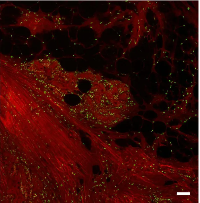
An image of a region of normal stroma from a discarded breast specimen is depicted in Figure 1 shown at approximately 10x magnification by stitching several frame together. The specimen is shown with the DAPI (nuclear) channel shown in green and the eosin (stroma) channel shown in red. Various constituent parts of the normal breast stroma are readily visible, including normal collagen-rich stroma with fibers oriented diagonally from top left to bottom right in the lower left portion of the image. Mixed in with the collagen are isolated normal stroma cells with easily visible nuclei. In the center of the image a glandular breast duct is shown with dense stroma surrounding tight, nuclei-rich glands and small
tubules. On the top of the image a large region of adipocytes is present, with characteristic nuclei folded compressed against the edges of large, unstained fat droplets.
3.2 Virtual H&E Color Mapping
Next we demonstrate color remapping of an individual 20x magnification image field using our GPU implementation. A region of normal breast stroma with a large number of nuclei and readily apparent collagen fiber structure was selected. The region was imaged and then both raw fluorescent data (Figure 2, left frame) and rendered as a virtual H&E image. In comparison to the raw fluorescent image, the virtual H&E image is much brighter because of the inverted (virtual transmission) color scale, making identification of fine features such as fine collagen fiber and weakly staining nuclei much easier.
Figure 1. Region of normal breast tissue imaged with multiphoton microscopy with the eosin channel shown in red, and nuclear channel shown in green. Scale bar: 100 microns.
: . 5 Gr
.
! r '',
.^a dp <0i
^
Ús
. t Ì %' }/
e 9 ,',.
a'e0.I. .}
rf
.f
w
°f1R3ñ
`.a Sl1.;
.
' o/
o.tt r
.;
`
:4t
j
s,
fN Ea 'It,. ',C.i
:'°'s
:
9ed
<,ti It
-Pe y//
E a4,,DO: r hÿ
, If
6 af
!,
0,
.s
0'
-.: .+'
,
e. .,. s
, s.t. ,
-.
'.+.. °¡
` w,
111, 4.'*
-r 7 ../
-'g,
_a s°
AI ^ I . m ir
>% a,'. y,
.
.._
-, ® t4.
4
¡
,
,,
,,;.
r
v. 11^ .
....
!
f.
`v4
AM. . e
.i, ...-Figure 2. Region of normal breast tissue imaged with multiphoton microscopy with the eosin and DAPI channels rendered as a virtual H&E image. Image size: 460x460 microns.
4. CLINICAL MULTIPHOTON SYSTEM
4.1 System Design
Operation in an intrasurgical scenario imposes strict constraints on system size and imaging speed while requiring that the system be portable. To overcome these limitations, the clinical multiphoton system was designed on a wheeled, air-floated vibration isolating table (PFP6090-8, Thorlabs). The table supports a 36”x30” steel optical breadboard on which is mounted a titanium sapphire laser (Chameleon, Coherent), and digitally variable beam attenuator, and a scan head with scan/tube lens (Thorlabs Imaging Systems). To facilitate rapid loading and unloading of specimens, the system uses an inverted geometry such that tissue is held to the imaging surface by gravity. This enables multiple specimens to be loaded into the system in parallel without having to mount each individually into imaging cassettes. The entire specimen holder is then covered with a single removable light-shielding lid that incorporates a magnetic interlock that can be used to disable the system if the lid is removed during operation. The specimen holder is a removable glass surface with handles inserted into a two axis translation stage (MLS203, Thorlabs). Z actuation is accomplished using a motorized objective translator. The entire specimen holder is large enough to accommodate all of the sampled margins of a typical surgically-excised ductal carcinoma lumpectomy. In the event that a larger number of margins need to be evaluated, specimen holders are interchangeable, enabling a technician to prepare multiple holders worth of specimens in parallel with the evaluation by a pathologist. A computer aided design (CAD) model of the system as assembled is presented in Figure 3. Laser drive and cooling electronics are vibration isolated from the imaging optics on platforms below the optical bread board.
4.2 System Performance
Using a 10x objective (Nikon Plan Apo Lambda 0.45 NA), the system obtains an axial resolution comparable to paraffin embedded sectioning and a field of view of 2.3 mm at full scan. In addition, digital zoom is used to implement 10/20/40x magnification imaging in real-time, closely mimicking the operation of a typical pathology microscope. At 2048x2048 resolution, the system can sample the entire 10x equivalent field at 1 micron intervals, or a 20x field at 500 nm interval at a rate of 8 frames per second. The use of GPU rendering enables real-time virtual H&E rendering.
.10
Chameleon
Figure 3. CAD model showing the top view of the clinical multiphoton system. The largest fraction of the system area is taken up by the titanium sapphire laser on the right side of the image. From the laser aperture (top right), the beam is expanded and then attenuated as it passed towards the top left. A scan head incorporating a resonant scanner fast axis and a galvanometer slow axis is integrated in scan/tube lens assembly (grey, vertically oriented rectangle). A lid (black rectangle) sits on top of the specimen holder (grey rectangle on the lower left).
5. CONCLUSION
We have developed a clinical multiphoton system optimized for real-time virtual H&E imaging in a clinical setting. The system is designed to be portable, and is built onto a vibration isolated, wheeled cart that can be moved around a pathology or radiology laboratory. The system incorporates GPU-accelerated rendering of multiphoton fluorescence images as virtual H&E slides with negligible latency while obtaining a 2.3 mm FOV with a 10x objective. The wide field of view enables variable 10/20/40x magnification imaging, analogously to a conventional histopathology microscope.
REFERENCES
[1] J. D. Bancroft, and A. Stevens, [Theory and practice of histological techniques] Churchill Livingstone, Edinburgh ; New York(2008).
[2] B. Fisher, S. Anderson, J. Bryant et al., “Twenty-Year Follow-up of a Randomized Trial Comparing Total Mastectomy, Lumpectomy, and Lumpectomy plus Irradiation for the Treatment of Invasive Breast Cancer,” New England Journal of Medicine, 347(16), 1233-1241 (2002).
[3] J. A. van Dongen, A. C. Voogd, I. S. Fentiman et al., “Long-term results of a randomized trial comparing breast-conserving therapy with mastectomy: European Organization for Research and Treatment of Cancer 10801 trial,” J Natl Cancer Inst, 92(14), 1143-50 (2000).
[4] J. Engel, J. Kerr, A. Schlesinger-Raab et al., “Quality of life following breast-conserving therapy or mastectomy: results of a 5-year prospective study,” Breast Journal, 10(3), 223-31 (2004).
[5] G. A. Cefaro, D. Genovesi, R. Marchese et al., “Predictors of local recurrence after conservative surgery and whole-breast irradiation,” Breast Cancer Res Treat, 98(3), 329-35 (2006).
[6] J. L. Connolly, J. Boyages, A. J. Nixon et al., “Predictors of breast recurrence after conservative surgery and radiation therapy for invasive breast cancer,” Mod Pathol, 11(2), 134-9 (1998).
[7] J. Dobbs, S. Krishnamurthy, M. Kyrish et al., “Confocal fluorescence microscopy for rapid evaluation of invasive tumor cellularity of inflammatory breast carcinoma core needle biopsies,” Breast Cancer Research and Treatment, 149, 303-310 (2014).
[8] D. S. Gareau, Y. Li, B. Huang et al., “Confocal mosaicing microscopy in Mohs skin excisions: feasibility of rapid surgical pathology,” Journal of Biomedical Optics, 13, 054001 (2008).
[9] Y. K. Tao, D. Shen, Y. Sheikine et al., “Assessment of breast pathologies using nonlinear microscopy,” Proceedings of the National Academy of Sciences, 111, 15304-15309 (2014).
[10] I. Pavlova, K. R. Hume, S. a. Yazinski et al., “Multiphoton microscopy and microspectroscopy for diagnostics of inflammatory and neoplastic lung.,” Journal of biomedical optics, 17, 036014 (2012).
[11] M. Wang, H. Z. Kimbrell, a. B. Sholl et al., “High-Resolution Rapid Diagnostic Imaging of Whole Prostate Biopsies Using Video-Rate Fluorescence Structured Illumination Microscopy,” Cancer Research, 75, 4032-4041 (2015).
[12] D. S. Gareau, “Feasibility of digitally stained multimodal confocal mosaics to simulate histopathology,” Journal of biomedical optics, 14, 034050 (2009).