HAL Id: hal-02998150
https://hal.archives-ouvertes.fr/hal-02998150
Submitted on 9 Dec 2020
HAL is a multi-disciplinary open access
archive for the deposit and dissemination of
sci-entific research documents, whether they are
pub-lished or not. The documents may come from
teaching and research institutions in France or
abroad, or from public or private research centers.
L’archive ouverte pluridisciplinaire HAL, est
destinée au dépôt et à la diffusion de documents
scientifiques de niveau recherche, publiés ou non,
émanant des établissements d’enseignement et de
recherche français ou étrangers, des laboratoires
publics ou privés.
by Desulfovibrio sp.86 with a switch from ring-opening
dechlorination to reductive sulfidation activity
Oriane Della-Negra, Sébastien Chaussonnerie, Nuria Fonknechten, Agnès
Barbance, Delphine Muselet, Déborah Martin, Stéphanie Fouteau, Cécile
Fischer, Pierre-Loïc Saaidi, Denis Le Paslier
To cite this version:
Oriane Della-Negra, Sébastien Chaussonnerie, Nuria Fonknechten, Agnès Barbance, Delphine
Muse-let, et al.. Transformation of the recalcitrant pesticide chlordecone by Desulfovibrio sp.86 with a
switch from ring-opening dechlorination to reductive sulfidation activity. Scientific Reports, Nature
Publishing Group, 2020, 10 (1), �10.1038/s41598-020-70124-9�. �hal-02998150�
transformation of the recalcitrant
pesticide chlordecone
by Desulfovibrio sp.86 with a switch
from ring‑opening dechlorination
to reductive sulfidation activity
oriane Della‑negra
1,2, Sébastien Chaussonnerie
1,2, Nuria Fonknechten
1, Agnès Barbance
1,
Delphine Muselet
1, Déborah E. Martin
1, Stéphanie Fouteau
1, Cécile Fischer
1,
pierre‑Loïc Saaidi
1,3*& Denis Le paslier
1,3*The insecticide chlordecone has been used in the French West Indies for decades, resulting in long term pollution, human health problems and social crisis. In addition to bacterial consortia and
Citrobacter sp.86 previously described to transform chlordecone into three families of transformation
products (A: hydrochlordecones, B: polychloroindenes and C: polychloroindenecarboxylic acids), another bacterium Desulfovibrio sp.86, showing the same abilities has been isolated and its genome was sequenced. Ring-opening dechlorination, leading to A, B and C families, was observed as previously described. changing operating conditions in the presence of chlordecone gave rise to the formation of an unknown sulfur-containing transformation product instead of the aforementioned ones. Its structural elucidation enabled to conclude to a thiol derivative, which corresponds to an undocumented bacterial reductive sulfidation. Microbial experiments pointed out that the chlordecone thiol derivative was observed in anaerobiosis, and required the presence of an electron acceptor containing sulfur or hydrogen sulfide, in a confined atmosphere. It seems that this new reaction is also active on hydrochlordecones, as the 10-monohydrochlordecone A1 was transformed the same way. Moreover, the chlordecone thiol derivative called F1 was detected in several chlordecone contaminated mangrove bed sediments from Martinique Island, highlighting the environmental relevance of these results.
Chlordecone is a highly recalcitrant organochlorine pesticide that has been added to the Stockholm convention list of persistent organic pollutants in 2009. Several pest control products containing chlordecone (Kepone, Curlone) or a functionalized derivative (Kelevan) have been manufactured in the US, Brazil and France and applied in the Caribbean, Central America, West Africa, and Europe over an extended period of forty years1. To
date, chlordecone has caused two major environmental disasters: (1) acute exposure of workers at the Hopewell (US) chlordecone production plant in 1975 and massive pollution of the James River, extending over 100 miles that lasted for decades2, (2) ongoing impregnation of the French West Indies (FWI) population due to extensive
agricultural use of chlordecone from 1972 to 1993 that has resulted in long-term pollution of environmental compartments (soils, water resources, coastal areas) and subsequently of some local food production (vegeta-bles, farmed animals, and seafood). Until now, no remediation strategy has been proven satisfactory in the FWI environment whereas the population has to deal with health troubles (increased risk of cancer, motor and cognitive development disorders in young children, premature births) and social difficulties (disappearance of local economy activities)1.
open
1Génomique Métabolique, Genoscope, Institut François Jacob, CEA, CNRS, Univ Evry, Université Paris-Saclay,
91057 Evry, France. 2These authors contributed equally: Oriane Della-Negra and Sébastien Chaussonnerie. 3These
authors jointly supervised this work: Pierre-Loïc Saaidi and Denis Le Paslier. *email: plsaaidi@genoscope.cns.fr;
In this context, chlordecone microbial degradation has been the focus of several studies: some Pseudomonas
aeruginosa sp. as well as mixed cultures isolated from the Hopewell area in the 1980s exhibited significant aerobic
dechlorination ability leading to mono- and dihydrochlordecones transformation products (TPs)3. Later on,
the conversion of chlordecone into unknown polar and nonpolar TPs in the presence of the anaerobic archaea
Methanosarcina thermophila at 50 °C was reported4. More recently, two bacterial consortia (86 and 92), isolated
from microbial enrichment cultures from chlordecone-contaminated soils and wastewater treatment plant sludge, were able to transform chlordecone, under anoxic conditions, at room temperature. Several TPs were identified and grouped into three families: hydrochlordecones (family A), polychloroindenes (family B) and polychloroin-denecarboxylic acids (family C)5,6. TPs from families B and C that were predominant arose from a ring-opening
dechlorination of the chlordecone bishomocubane cage. Metagenomic analysis of these consortia indicated the prevalence of Citrobacter and Desulfovibrio sp. genomes among the dozen of bacterial genomes sequenced. Two members of these consortia, Citrobacter sp.86 and Citrobacter sp.92 that were isolated, both degraded chlordecone resulting in the same TP profile. Reductive dehalogenases are key enzymes in halorespiration7,8 and could be
indicative of a dehalogenation process. Here no gene sequences related to these enzymes were detected neither in the metagenomic data nor in the isolated Citrobacter genomes5.
The mechanistic degradation pathways and the exact role of each bacterium remains to be elucidated. In the past, several Desulfovibrio sp. or other sulfate-reducing bacteria have been reported in microbial cultures degrading chlorinated compounds9–14.
Herein, we report the isolation of Desulfovibrio sp.86 previously identified in chlordecone-degrading consor-tium 865 and explore its capacity to transform chlordecone and other chlorinated derivatives. TPs from the B and
C families as well TP A1 were detected in pure cultures of Desulfovibrio sp.86. Unexpectedly, other incubation conditions in presence of chlordecone gave rise to the formation an unknown sulfur-containing TP instead of the aforementioned TPs. The elucidation of its structure allowed us to conclude to a thiol derivative. To the best of our knowledge, it corresponds to an unprecedented bacterial reductive sulfidation. This thiol derivative was detected in several chlordecone-contaminated mangrove bed sediments from the Martinique Island.
Results
isolation of Desulfovibrio sp.86.
Isolation of Desulfovibrio sp.86 was achieved from the chlordecone-degrading consortium 865 using sulfate-reducing conditions. As bacterial consortium 86, able to transformchlordecone, was obtained from an enriched mineral medium (MM) named MM + 5, supplemented with
chlor-decone, the main chemical composition was kept but electron donors and acceptors were modified. Several media formulations used to enrich sulfate-reducing bacteria utilize organic acids e.g. lactate, as carbon and energy sources (electron donor) and sulfate that is used as eletron-acceptor for growth15–18. In this context,
pyruvate in the MM + liquid medium was replaced by lactate and sulfate was added (MMD liquid medium, see “Methods” section). The enrichment was spread on MMD agar and the brown vibrio-like bacterial colonies (observed under optical microscope) were purified further through two additional plate streakings (Fig. S1). An isolated bacterial strain was found to be identical to Desulfovibrio sp.86 from consortium 86 based on 100% identities of their 16S rRNA genes (1538 bp each).
Genome analysis of Desulfovibrio sp.86.
The whole genome consists of a single 3,464,070 bp circu-lar chromosome. CheckM analysis19 performed with 61 genomes and 284 lineages indicates that the genomebelongs to the Deltaproteobacteria and CheckM Completeness is 100% (zero essential marker missing). The average G + C content for the DNA is 58.06%. A total of 3,342 coding DNA sequences (CDSs) were predicted for the chromosome, 4 pseudogenes and 10 miscellaneous RNAs (misc-RNA), 3 rRNA operons, and 54 tRNA genes.
The three 16S rRNA genes of Desulfovibrio sp.86 are identical. Their best BLAST hits (NCBI) were from uncultured Desulfovibrio sp. clones from Microbial Fuel Cells such as MFC63A04 (Genbank accession number : FJ823865; coverage 98%; identity 99,87%)20. A phylogenetic tree using the available 16S rRNA of cultivable
bacteria confirmed the similarities between Desulfovibrio sp.86 and Desulfovibrio simplex DSM4141 (Genbank 16S rRNA accession number: NR_117110; coverage 99%; identity 99, 22%; genomic sequence unavailable) (Fig. S2)21. At the genomic level, Desulfovibrio sp.86 ranks first with Desulfovibrio desulfuricans subsp.
desulfu-ricans str. ATCC 27,774 genome (GenBank:NC_011883). Nevertheless, Desulfovibrio sp.86 shares only 1,408
genes (43%) with D. desulfuricans (over 80% amino acids identity and 80% alignment coverage). In addition, the average nucleotide identities (ANI) between Desulfovibrio sp.86 and sequenced Desulfovibrio genomes are lower than the 95% ANI cut-off value generally accepted for species delineation (Fig. S3)22. These results indicate that
Desulfovibrio sp.86 is most probably a new species of the Desulfovibrio phylum. The presence of two superoxide
dismutases and one catalase gene accounts for its relative oxygen tolerance. As expected, the Desulfovibrio sp.86 genome exhibits an extensive gene complement for sulfur metabolism, encompassing the sulfur respiration pathways with inorganic sources like sulfate, sulfite, bisulfite and tetrathionate as electron acceptors. Organic sulfur sources may be provided by fermentation products of sulfur biomass (sulfoquinovose of sulfoquinovosyl lipids) sulfoacetate, the sulfur non-proteogenic amino acid taurine via sulfoacetaldehyde, or alkanesulfonates23.
Desulfovibrio sp.86 degrades chlordecone into known transformation products as well as an
unexpected sulfur-containing compound.
The chlordecone-degrading ability of the isolatedDesulfo-vibrio sp.86 strain was investigated using GC–MS (Gas Chromatography Mass Spectrometry) and LC-HRMS
(Liquid Chromatography High Resolution Mass Spectrometry) techniques in growth conditions successfully applied for Desulfovibrio sp.86 isolation (MMD liquid medium). Na2S was used as reductant and an anaerobic
N2/H2 (98/2; V/V) atmosphere was applied using a glove box system. These incubation conditions led to
LC-HRMS TP profiles as those obtained with Citrobacter sp.866: monohydrochlordecone A1, pentachloroindene
B1, tetrachloroindenes B3-B4 and polychloroindenecarboxylic acids C1-C2 and C3-C4 (Fig. 1a,b). In the glove box system, bacterial cultures were performed using 100 mL glass vials with a hydrophobic porous film to enable gas exchange and avoid contamination. This incubation condition was considered as “renewed atmosphere” condition (RA). In this system, the atmosphere was regularly renewed with N2/H2 (98/2; V/V) and the box was
not thermostatically controlled so the temperature varied between 25 and 33 °C. To better control growth and degradation conditions, Desulfovibrio sp.86 cultures were placed in MMD medium using 100 mL glass vials, sealed with butyl rubber septa, in an oven at 30 °C. Sealed vials were initially purged with N2/H2 (98/2; V/V) gas
to insure anaerobiosis. This incubation condition was considered as “confined atmosphere” conditions (CA). After six-week incubation with chlordecone, the pesticide was no longer detectable by the same analytical techniques. Concomitantly, a single unknown chlorinated compound named F1 appeared. It was detectable only through GC–MS (25.6 min) (Fig. 1c). Like in the previously reported chlordecone degradation cultures5,6, the
main chlordecone transformation appeared during the stationary phase (Fig. 1e). Since no visible chlorinated peak could be observed in LC-HRMS (Fig. 1d), we based the structural elucidation of F1 on the interpreta-tion of GC–MS data (Fig. 1f). Assuming that the higher isotopic pattern centered at m/z 507.8 contained the molecular ion, we hypothesized two possible neutral formulae for F1, C10Cl10O2H2 and C10Cl10SH2. In-source
fragments were highly similar to those observed in polychlorinated bishomocubane-based structures including chlordecone, hydrochlordecones, chlordecol and mirex5,6,24. Indeed, the isotopic patterns at m/z 201.0, 235.9 and
271.9 presumably arose from the known in-source bishomocubane retrocyclodimerization and corresponded to positively charged C5-fragments. Comparison with isotopic simulations limited the possibility to the
follow-ing radical ions: [C5Cl3+nSH2]+· [C5Cl5+n]+· and [C5Cl3+nO2H2]+·, with n = 0,1. The latter bis-oxygenated generic
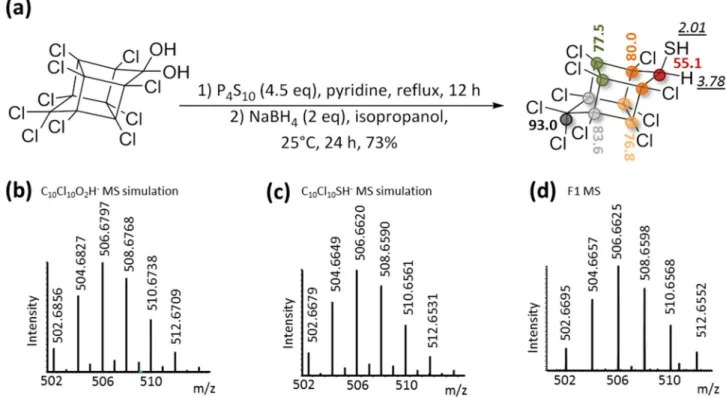
formula turned to be highly unlikely since it required the presence of a gem-diol function or a gem-chlorohydrin moiety on the cyclopentenyl ring that had to resist the harsh GC chromatographic conditions (> 200 °C). Instead, we concluded that a sulfur moiety, i.e. a thiol function, was probably present on the bishomocubane polycycle. We proposed for F1 the structure depicted in Fig. 2a and suggested the name chlordecthiol, the sulfur analogue of chlordecol (chlordecone alcohol).
Synthesis of the chlordecthiol standard and confirmation that transformation product F1 is
identical to chlordecthiol.
In order to confirm the structure of F1, the standard chlordecthiol was chemi-cally synthesized and fully characterized. To achieve chemical reductive sulfidation of chlordecone, two steps were needed. The first one consisted in the conversion of the gem-diol function of chlordecone in equilibrium with the corresponding ketone form20,25 into the sulfur analog, i.e. gem-thiol/thiocarbonyl moiety. To performthis step, phosphorus-containing sulfur reagents are generally applied26,27. Here we used phosphorus decasulfide
(P4S10) also called Berzelius reagent27,28 according to the protocol of Zaidi and coworkers who successfully
syn-thesized camphorthiol29 (Fig. 2a). The second step consisted in the reduction of the gem-thiol intermediate using
NaBH4. After purification, a white solid was obtained in an overall yield of 73% (Fig. 2a). 1D- and 2D-NMR
analyses confirmed the chlordecthiol structure: (i) two 1H signals integrating each for one proton and coupled
together (δ 3.78 ppm, d, J = 5.5 Hz and δ 2.01 ppm, d, J = 5.5 Hz), with the one at δ 3.78 ppm correlated to a
13C signal shifted downfield (δ 55.1 ppm) in HSQC experiment that perfectly account for the CH moiety next
to the thiol function and (ii) a total of six visible 13C signals reflecting the plane of symmetry conserved in the
present bishomocubane structure (Fig. 2a, Figs. S19–23). Although in microbial transformation, F1 could not be detected using LC-HRMS tool in the developed conditions, a concentrated sample of the synthetic standard chlordecthiol provided a significant signal. The presence of a sulfur atom and the expected neutral formula C10Cl10SH2 were confirmed (Fig. 2c,d), and the raw formulae C10Cl10O2H2 was ruled out (Fig. 2b). Lastly, GC–
MS analysis demonstrated the perfect match (i.e., the same retention time of 25.6 min and same in-source mass spectra Fig. S6) between the synthetic standard and TP F1, thus definitely assigning it as chlordecthiol. Indeed, F1 represents the first member of a new family of chlordecone TPs possessing for the first time a sulfur atom.
the ability of Desulfovibrio sp.86 to degrade chlordecone transformation products.
Com-pounds A1, B1, C1-C2 and F1 representative of the four families of TPs possibly formed in presence of
Desul-fovibrio sp.86 were synthesized according to chemical protocols previously reported6 and herein developed for
F1. Each of them was incubated in presence of Desulfovibrio sp.86 cultures in conditions that were shown to promote reductive sulfidation (sealed vial; CA conditions) or dechlorination with ring-opening (open vial; RA conditions). Dual GC–MS and LC-HRMS monitoring of all samples was performed.
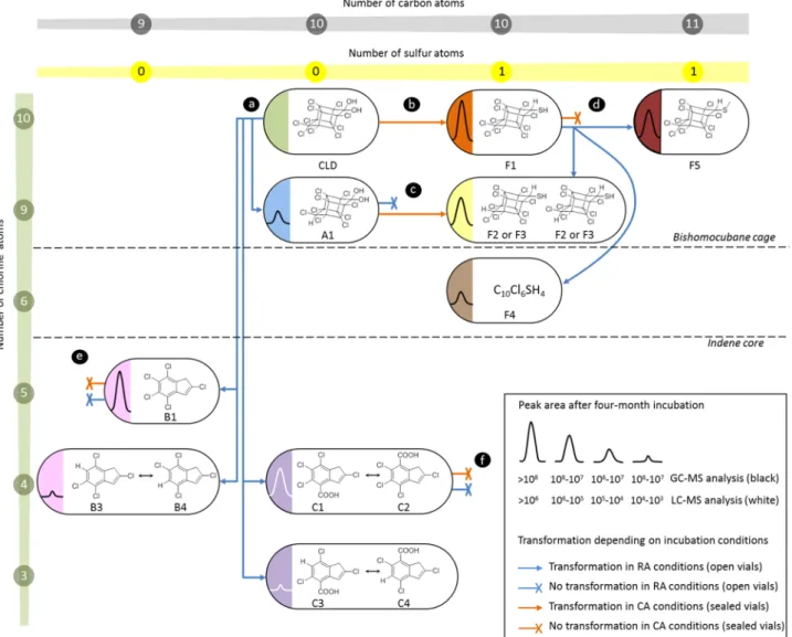
After one-month incubation under CA conditions, 10-monohydrochlordecone A1 was completely trans-formed into two unknown chlorinated compounds barely separated using GC–MS (retention times: 24.3 min F2 and 24.5 min F3, Fig. 3, Figs. S7–S11). They showed an identical in-source mass spectrum that was highly similar to the F1 fragmentation pattern (Fig. S8). All detected in-source fragments turned to possess one chlorine atom less than their analogues in F1 mass spectrum. Compounds F2 and F3 were thus assumed to be diastereoisomers of 10-monohydrochlordecthiol (Fig. 3c). This was confirmed by the chemical synthesis of the two 10-mono-hydrochlordecthiol standards from 10-monohydrochlordecone A1 using the procedure previously applied for the synthesis of chlordecthiol F1 (Figs. S24–27). These chemical standards had the same retention times (24.3 and 24.5 min) and the same mass spectra compared to the biological F2 and F3 (Fig. S8). TPs B1, C1-C2 and F1 remained unchanged even after six-month incubation with Desulfovibrio sp.86 under CA conditions.
After six-month incubation under RA conditions, chlordecthiol F1 was partially converted into 10-mono-hydrochlordecthiols F2 and F3, and two other unknown chlorinated species detected using GC–MS, named F4 (retention time: 17.9 min) and F5 (retention time: 26.6 min) (Fig. 3d, Fig. S12). None of these two compounds gave any detectable signal in LC-HRMS. GC–MS analysis enabled to propose C10Cl6SH4 as raw formula for F4
(Fig. S14), and C11Cl10SH4 for F5 (Fig. S15). Methyl chlordecsulfide was chemically synthesized and fully
char-acterized by NMR (Fig. S28–31). The perfect correspondence of GC retention times and GC mass spectra of methyl chlordecsulfide and biological F5 (Fig. S13) confirms its postulated identity. Noteworthy, the structure of F5 (Fig. 3d) represents a S-methylated form of F1. The exact nature of F4 remains to clear (see SI-Supplementary Text). All other TPs remained unchanged under RA conditions.
In summary, A1 (formed in RA conditions) underwent a reductive sulfidation just as chlordecone; under both incubation conditions polychloroindenes (B1 and C1-C2) did not seem to be degradable by Desulfovibrio sp.86, whereas F1 (produced under CA conditions) could be transformed under RA conditions (Fig. 3).
Investigation of the incubation conditions leading to bacterial reductive sulfidation.
Toques-tion the physico-chemical or physiological parameters influencing the transformaques-tions pathways of chlordecone in the cultures of Desulfovibrio sp.86, GC–MS monitoring (B1 and F1 presence as evidences of RA and CA conditions, respectively) was chosen.
Two sulfur-containing inorganic compounds, the reductant Na2S and the electron acceptor Na2SO4, were
present in the original MMD liquid medium. A first series of experiments in sealed vials with substitution of Na2S by other reducing agents, sulfured or not, i.e. cysteine and titanium(III) citrate (TiCi) led to a comparable
level of chlordecthiol F1 (Table 1). Traces of TP B1 were also observed in all experiments, including Na2S, the
positive control (Table 1).
A second set of experiments was designed using Na2S as reducing agent and alternative sulfur-based electron
acceptors in sealed vials (Table 2). Use of 90% 34S-enriched inorganic sulfate resulted in a chlordecthiol product
showing similar 34S-enrichment level (90% 34S-F1/10% F1, Fig. S16). GC–MS analysis of culture headspace also
revealed the presence of H234S and H3C34SH (Fig. S16), thus showing that Desulfovbibrio sp.86 produced H2S
from sulfate. These gas were detected in the culture headspace in CA conditions, using MMD medium, whereas they were absent from the culture headspace in RA condition that corresponded to the glove box atmosphere (Fig. S17). Absence of sulfate did not inhibit Desulfovibrio sp.86 growth, however it resulted in formation of B1 (Table 2)5. The remplacement of sulfate by sulfite, bisulfite or thiosulfate led in all cases to the formation of
chlordecthiol F1.
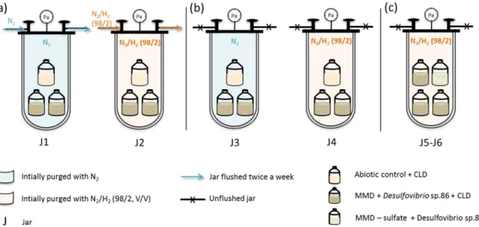
A third series of experiments using jars investigated the effect of the nature of the gas phase in contact with the culture. Between the RA condition using open vials in glove box and CA condition using sealed vials in oven, several parameters differed (atmosphere nature and volume, temperature). Using a jar system including several vials, we selectively study the effect of atmosphere renewal on chlordecone transformation by Desulfovibrio sp.86 in MMD medium. In each case, open vials with hydrophobic porous films were placed in jars initially purged with a selected gas. All the jars were incubated at 30 °C. Some of them were flushed several times, to mimic the glove box system, whereas others were kept closed.
Jars 1–4 contained two identical open vials filled with MMD, chlordecone and Desulfovibrio sp.86 inoculum, and one negative abiotic control. Among them, two jars were flushed twice a week, jar 1 with (N2/H2 (98/2; V/V)),
jar 2 with N2 (Fig. 4a), whereas jars 3 and 4, initially purged with N2 and (N2/H2 (98/2; V/V)) respectively were
left unflushed (Fig. 4b).
The last two jars (jar 5 and 6) contained three open vials filled with MMD, chlordecone and
Desulfovi-brio sp.86, plus one culture without sulfate. These jars were initially flushed with (N2/H2 (98/2; V/V)) and left
unflushed for 2 months (Fig. 4c).
In parallel to the jars, two sealed vials filled with MMD without sulfate, chlordecone and Desulfovibrio sp.86 were flushed with extemporarily synthesized H2S (Fig. S4).
After two-month incubation at 30 °C, TP B1 was detected in vials placed in the flushed jars 1 and 2 (Table 3a), whereas TP F1 was only found in vials placed in unflushed jars 3 and 4 (Table 3b). No TP were present in the abiotic controls. In jars 5 and 6, F1 was present in both sulfate and sulfate-free Desulfovibrio sp.86 open cultures (Table 3c). In the same way, Desulfovibrio sp.86 sulfate-free cultures, flushed with H2S showed significant levels
of F1 TP, whereas no transformation was observed in the negative control (Table 3d).
An additional chemical experiment was carried out to assess the influence of H2S on chlordecone
transfor-mation. Classic chemical protocol enabling B and C TPs formation, was applied (chlordecone, titanium citrate, vitamin B12, water). Under N2 atmosphere, B and C TPs were obtained, however under H2S atmosphere, only
A1 was produced (Fig. S18).
Figure 1. GC–MS and LC-HRMS monitoring, in full-scan mode (arbitrary unit), of chlordecone
transformation by Desulfovibrio sp.86 in RA conditions (in glove box, anaerobiosis (N2/H2, 98/2, V/V), room
temperature, open vials with a porous film, in MMD medium supplemented with 40 mg/L of chlordecone), and CA conditions (in oven, anaerobiosis (initially purged with N2/H2, 98/2, V/V), 30 °C, sealed vials, in MMD
medium supplemented with 40 mg/L of chlordecone); and identification of TP F1. (a) GC–MS monitoring of chlordecone transformation by Desulfovibrio sp.86 in RA conditions. (b) LC-HRMS monitoring of chlordecone transformation by Desulfovibrio sp.86 in RA conditions. (c) GC–MS monitoring of chlordecone transformation by Desulfovibrio sp.86 in CA conditions. (d) LC-HRMS monitoring of chlordecone transformation by
Desulfovibrio sp.86 in CA conditions. In each graph, green circles represent chlordecone, red circles F1, blue
triangles 10-monohydrochlordecone A1, pink squares 2,4,5,6,7-pentachloroindene B1 and purple diamonds 2,5,6,7-tetrachloroindenecarboxylic acid and 2,4,5,6-tetrachloroindenecarboxylic acid C1–C2. In RA conditions traces of B3-B4 and C3–C4 TPs were also detected. (e) Chlordecone transformation over the time in CA conditions, OD600 corresponds to the optical density at 600 nm in biotic conditions. (f) GC mass spectrum of F1
TP and its interpretation.
These results clearly show that the formation of chlordecthiol F1 requires a closed incubation system with a reducing atmosphere. However H2 as the initial gas atmosphere is not mandatory whereas the presence of H2S
is required. Chemically, the presence of H2S prevents from ring-opening dechlorination process.
Environmental relevance of chlordecone reductive sulfidation.
We reinvestigated our previous collection of the Martinique Island chlordecone-contaminated environmental samples (eight soils and two bed sediments) in which various levels of chlordecone TPs had been reported6. Among the novel sulfur-containingTPs herein reported, we found appreciable levels of F1 in the two bed sediments samples (927 and 928) at a con-centration estimated around 50 μg/kg and 20 μg/kg of wet sediment, respectively (Table S1). In parallel, bacterial population diversity data issued from metabarcoding analysis of these samples (Fig. 5, Fig. S5) were processed to recover a hierarchical clustering of environmental samples according to their taxonomic composition6. It was
noticed that sulfate-reducing bacteria were much more present in bed sediments than in the other compart-ments, as previously reported30–32.
Discussion
To date, the mechanism of chlordecone microbial transformation by ring-opening dechlorination process has not been reported, nor has any associated enzyme activity been described. Bacteria from the genus
Desulfovi-brio are indeed known to be part of consortia able to dechlorinate molecules like aromatic hydrocarbons32–35.
Desulfovibrio sp.86 is able to produce the same TPs of chlordecone as previously described5,6. However, the only
other complete genome of a chlordecone degrading organism is from a Citrobacter species. The large divergence between these two bacterial species at the level of genus, lifestyle and metabolism precludes a straightforward differential genomic approach searching for common genes to understand ring-opening dechlorination process at the level of the pathway. Even if a parsimonious hypothesis could invoke the same mechanism for chlordecone ring-opening dechlorination process in both species, additional isolated species and genomes are welcomed to give clues to this question.
In this work, we also report on the identification, in the presence of Desulfovibrio sp.86, of another decone transformation product issued from a reduction process, the sulfured molecule which we named chlor-decthiol and which had not been described so far. The sulfur metabolism of bacteria from the genus
Desulfovi-brio is versatile, and encompasses the use of various inorganic sulfur sources that eventually can be reduced to
H2S. The Desulfovibrio sp.86 gene content is consistent with the production of sulfide. In particular, under the
experimental incubation conditions we used, reduction of sulfate from the mineral medium can be achieved by the sulfate and sulfite reductase genes.
Figure 2. Synthesis of the chemical standard chlordecthiol and its complete characterization. (a) Chemical
reaction scheme of chlordecthiol synthesis and NMR shifts in CD2Cl2: 1H NMR shift, in italic and underlined
and 13C NMR shift, in bold; similarly colored circles indicate equivalent carbon atoms. (b) m/z simulation
of C10Cl10O2H-, (c) m/z simulation of C10Cl10SH-, (d) extracted high resolution mass spectrum of synthetic
The reductive sulfidation was also observed with the monohydrochlordecone A1 and then could be extrapo-lated to other hydrochlordecones. Remarkably, putting F1 in renewed atmosphere conditions essentially leads to sulfured analogs of F1 (F2 and F3), and the methylated derivative F5. This process generates a greater diversity of chlordecone TPs and extends the list of chlordecone derivatives that could be present in the environment. According to LC-HRMS analyses, F1 appeared less polar than chlordecone and chlordecol (Table S2). Indeed, its physico-chemical properties are likely to be different from those of chlordecone due to the replacement of the gem-diol fraction (chlordecone) by the thiol function (F1). The fate of F1 in the environment is therefore
Figure 3. Fates of chlordecone transformation products with Desulfovibrio sp.86 depending on incubation
conditions. (a) Transformation of chlordecone under RA conditions and (b) under CA conditions. Transformation of (c) A1, (d) F1, (e) B1 and (f) C1–C2.
Table 1. Influence of the reducing agent on chlordecone TP profile. GC–MS analysis of the two-month
incubation extract: each TP peak area (full scan) was normalized to the sum of all peak areas (chlordecone and TPs).
Culture parameters Desulfovibrio sp.86
TPs (relative GC– MS peak area ratio TP/TPs + CLD)
Atmosphere Reducing agent Electron acceptor CLD B1 F1
N2/H2 (98/2) Na2S Na2SO4 X – 0.06 0.94 1.00 – – Cysteine X 0.17 0.03 0.79 TiCi (III) X 0.17 0.03 0.79
Table 2. Influence of the sulfur-based electron acceptor on chlordecone TP profile. GC–MS analysis of the
two-month incubation extract: each TP peak area (full scan) was normalized to the sum of all peak areas (chlordecone and TPs).
Culture parameters
Desulfovibrio sp.86
TPs (relative GC– MS peak area ratio TP/TPs + CLD)
Atmosphere Reducing agent Electron acceptor CLD B1 F1
N2/H2 (98/2) Na2S Ø X 0.51 0.49 – Na2SO4 X 0.05 – 0.95 Na2SO4 1.00 – – Na234SO4 X 0.16 – 0.84 Na2SO3 X 0.09 – 0.91 NaHSO3 X 0.46 – 0.54 Na2S2O3 X 0.10 – 0.90
Figure 4. Schematic representation of gas-controlled experiments. (a) Flushed jars, (b) unflushed jars, (c)
unflushed jars with sulfate-free Desulfovibrio sp.86 cultures.
Table 3. Influence of gas atmosphere on chlordecone TP profile. GC–MS analysis of the two-month
incubation extract: each TP peak area (full scan) was normalized to the sum of all peak areas (chlordecone and TPs).
Experiment no.
Culture parameters
Desulfovibrio sp.86
TPs (relative GC– MS peak area ratio TP/TPs + CLD)
Atmosphere Reducing agent Electron acceptor CLD B1 F1
(a) J1 N2 flush Na2S Na2SO4 X 0.86 0.14 – (a) J2 N2/H2 (98/2, V/V) flush X 0.63 0.37 – 1.00 – – (b) J3 N2 X 0.49 – 0.51 (b) J4 N2/H2 (98/2, V/V) X 0.45 – 0.55 1.00 – – (c) J5 N2/H2 (98/2, V/V) Na2SO4 X 0.24 – 0.76 Ø X 0.55 – 0.45 (d) H2S Ø X 0.36 – 0.64 1.00 – –
likely to be different in terms of biodegradability, stability, mobility to other environmental compartments and/ or biomagnification along the food chain.
Sulfate reducing bacteria are frequently encountered in anaerobic environments like sediments where Fe(III) and sulfate dominate the pool of electron acceptor34. Thus, the environmental relevance of chlordecthiol detection
that we report herein in chlordecone-contaminated sediments from the French West Indies may originate from microbial processes. Moreover, our work in laboratory condition indicates that at least some TPs of chlordecone are also prone to reductive sulfidation (hydrochlordecones). Even if at this stage a large set of questions related to their appearance, persistence, trophic food-chain transfer and toxicological profile can only be envisioned, our results open a new window on the secondary contamination by chlordecone TPs as well as possible clues to its natural elimination.
Additionally, the change in the TP profile observed in our experiments with Desulfovibrio sp.86 under vari-ous conditions of sulfur donor and atmosphere (confined or renewed in anaerobic conditions) could indicate biochemical pathways blockade and/or promotion triggered by H2S accumulation, leading to re-routing of
chlordecone into this newly described reductive sulfidation instead of the previously reported ring-opening dechlorination TPs. At this stage, it is worth mentioning that all transformations of chlordecone with the con-sortium 86 had been conducted in open vials conditions, i.e. renewed atmosphere conditions. In a study related to dechlorination of trichlorofluoromethane by sulfate reducing bacteria, sulfide was proved to be inhibitory to the dehalogenation process35.
Some papers mentioned that H2S or HS- can bind to metal centers and have an inhibitor effect on zinc
and cobalt γ-class enzymes36,37. It was suggested that corrinoids could play a role in chlordecone ring-opening
dechlorination conditions5 and broadly, vitamin B
12, known to be a cofactor of reductive dehalogenases, can also
intervene in the reductive dehalogenation processes as protein-free corrinoid38–40, in both biotic41 and abiotic
settings6,42. In this work, we showed that vitamin B
12 combined with titanium citrate and chlordecone under
N2 atmosphere gave B and C families, whereas the same combination under H2S atmosphere only led to A1
formation (Fig. S18). The weakness of the bond strength of cobalt with H2O inducing a spontaneous reductive
substitution of aquo − cobalamin(III) (Cbl(III) − H2O) by sulfide − cobalamin(III) (Cbl(III) − S2−) in the presence
of S2− was reported43. In the case of corrinoid implication in B and C families formation, it could be assessed that
the presence of H2S could also be inhibitory and thus favored F1 by another mechanism.
Reductive sulfidation of ketones and aldehydes are possible using a large variety of chemical protocols, very distant from physiological conditions27. Several C-S bond formation are described, some mechanisms involve
a glutathion-ester intermediate or a cysteine precursor to form thiol derivatives via nucleophilic reactions44,45,
others use radical SAM enzymes45. Several enzymes known to catalyze sulfur addition reactions could take part in
the reductive sulfidation of chlordecone. Among them thiolase, sulfurtransferase, sulfotransferase, sulfhydrylase and numerous Fe-S proteins are predicted in Desulfovibrio sp.86 genome. However, at this stage there is no clue enabling to suggest a mechanism and even to confirm an enzymatic activity toward chlordecone or TPs that were
Figure 5. Dendrogram generated from hierarchical clustering of environmental samples according to the
bacterial population diversity (from R package pvclust v. 1.3-2, https ://doi.org/10.1093/bioin forma tics/btl11 7). 200,000 sequences were taken for each environmental sample and normalized. The percentage of detected phyla was represented here with a focus on sulfate reducing bacteria from the Deltaproteobacteria class, Firmicutes and Nitrospirae phyla. In red is represented the unbiased (au) p-value and in green the bootstrap probability value (bp). SRB = Sulfate-reducing bacteria.
shown to undergo reductive sulfidation in Desulfovibrio sp.86 cultures. Further experiments will be necessary to consider if any of these processes are indeed operating with Desulfovibrio sp.86.
Desulfovibrio sp.86 is the first sulfate-reducing bacterium known to date that is able to transform chlordecone,
and only the second isolated and sequenced bacterial species coping with this pesticide. In this work, we reported for the first time on microbial reductive sulfidation, which occured on substrates like chlordecone or hydro-chlordecone. This raises the question of whether the microbial sulfidation process is restricted or not to these peculiar substrates, and whether it can be ascribed to the intrinsic sulfur-reducing ability of Desulfovibrio sp.86. In this respect, this new microbial strain, cultivated under our experimental conditions that promote reductive sulfidation, could provide a framework to investigate further this process with other natural or synthetic com-pounds. Finally, this study demonstrates that the way anaerobiosis is managed in the laboratory (e.g., renewed atmosphere in the glove box or atmosphere confined in sealed vials) is of critical importance for microbiological cultures as illustrated here by the change in the transformation pathways of chlordecone.
Methods
chemicals and media.
Chlordecone was obtained from Azur Isotopes (purity 98%). Na2S (≥ 98%),pyri-dine (99.8%), iodomethane (> 99%) and phosphorus pentasulfide (99%) were purchased from Sigma Aldrich. Titanium (III) citrate was prepared from titanium (III) chloride (> 12% in HCl) (Sigma Aldrich) and sodium citrate and neutralized with Na2CO3. Dichloromethane (DCM) and pentane (HPLC grade) were obtained from
Fisher Chemical; cyclohexane (99.8%) and NMR solvents from Acros Organics; and acetonitrile (MeCN, LC-HRMS grade) and acetone (> 99.9%) from VWR Chemicals. Chemical products used for microbiological media were obtained from Sigma Aldrich. Chemicals and media used for chlordecone TPs are described elsewhere6.
Analytics.
GC–MS analyses were carried out using a Thermo Fisher Focus GC coupled to asingle-quadrupole mass spectrometer (Thermo Fisher DSQ II). The instrument was equipped with a non-polar 30 m × 0.25 mm × 0.25 µm DB-5MS column (Agilent J&W) and a split/splitless injector. Ionization conditions and GC program were described elsewhere5,6.
HS-GC–MS (Head Space Gas Chromatography Mass Spectrometry) analyses were performed on a Thermo Fisher Trace 1300 coupled to ISQ 7000 VPI single quadrupole mass spectrometer. The instrument was equipped with a 30 m × 0.25 mm × 0.25 µm DB-624-UI column (Agilent J&W), a split/splitless injector and an automatic sampler TriPlus RSH coupled to a HeadSpace tool. For mass spectrometry (MS) analyses, the following standard working conditions were applied: electronic impact ionization, positive mode detection, ion source at 220 °C, detector voltage 70 eV, full scan mode m/z 33–300 (scan time 0.20 s). Injection and transfer line temperatures were set up at 200 °C, respectively 280 °C. 1 mL was injected each time with a filling speed of 10 min/mL, an injection speed of 10 mL/min and a penetration speed of 10 mL/s.
For H2S detection, vials were incubated for 1 min at 40 °C and sampled with a syringe at 40 °C. The split
mode was applied (30 °C, flow rate at 16.7 mL/min, with a ratio of 33.4). The carrier gas was helium at 0.5 mL/ min for 1 min followed by a gradient of 0.05 mL/min until reaching 1 mL/min (hold time 9 min). GC program was an isocratic at 24 °C for 20 min.
LC-HRMS analyses were carried out using a Dionex Ultimate 3000 LC system coupled to an LTQ-Orbitrap Elite mass spectrometer (Thermo Fisher Scientific) fitted with a heated electrospray ionization source (HESI) operating in negative ionization mode. Voltage optimization was described elsewhere5. Chromatographic
sepa-ration was achieved using a Thermo Fisher Syncronis C18 column (50 mm length, 2.1 mm inner diameter, 1.7 µm particle size) and carried out at 30 °C with a flow rate of 0.5 mL/min using NH4OAc buffer (10 mM; pH
7 adjusted with NH4OH) as solvent A and MeCN as solvent B. The gradient started at 20% B for 1 min, followed
by a linear gradient at 100% B for 7 min and remained 2 min at 100% B. The system returned to the initial solvent composition in 2 min and was re-equilibrated under these conditions for 2 min.
Organochlorine extraction for microbiological culture monitoring.
After homogenization of theliquid culture, 500 μL of the turbid solution were collected and extracted twice using 250 μL isooctane. The com-bined organic layers were then analysed through GC–MS analysis. When HS-GC–MS was required, 700 μL of cultures were sampled and put into a Chromacol 10-HSV vial of 10 mL (Agilent). For LC-HRMS analysis, 50 μL of the turbid solution were collected, mixed with 200 μL of acetonitrile/NH4OAc buffer (10 mM; pH 7 adjusted
with NH4OH) (V/V, ¼) and filtered on 0.22 μm filters.
Genomic DNA extraction, sequencing and analysis.
DNA extraction was performed as describedpreviously5.
Genome sequencing was performed mixing short and long reads (Illumina technology and Oxford Nanopore technology (ONT)). The Illumina and ONT data, around 280- and 20-fold coverage, respectively, were assem-bled using Unicycler (version: v0.4.6) (https ://cab.spbu.ru/softw are/spade s/) as described46. The annotation and
research of putative orthologous relations between genomes defined as gene couples satisfying the bi-directional best hit (BBH) criterion or a blastP alignment threshold with a minimum of 35% sequence identity on 80% of the length of the smallest protein was performed using the Microscope platform22.
The final annotated genome of Desulfovibrio sp.86 is available in GenBank/ENA under the accession number LR738849 and project number PRJEB35237.
Anoxic microbial incubations.
In “renewed atmosphere” conditions, Desulfovibrio sp.86 cultures were incubated at 25–33 °C (room temperature) in a glove box (Unilab mBraun) under anaerobic conditions (N2/H2previ-ously described5 supplemented with lactate as carbon source (20 mM), yeast extract (1 g/L), Na
2SO4 (7 mM) and
Na2S as reducing agent (0.4 g/L). In the glove box, culture vials were open, equipped with a hydrophobic porous
film (VWR, 114 μm thick, medical grade).
In “confined atmosphere” conditions, Desulfovibrio sp.86 cultures were incubated at 30 °C, in oven, under anaerobic conditions (N2/H2 (98/2; V/V)). They were carried out in MMD medium, in sealed culture vials,
closed with butyl rubber septa.
Desulfovibrio sp.86 cultures were inoculated at the onset of an experiment with 0.5 mL pre-culture pre-grown
in oven for 24 h.
During degradation reactions, the MMD medium was supplemented with 40 mg/L of chlordecone (from a solution of chlordecone at 200 mg/mL in dimethylformamide). Each experiment, tested condition or chlor-decone degradation was done in duplicate, plus a negative control (without bacteria) in 100 mL glass serum vials containing 50 mL of mineral medium.
Anoxic microbial degradation of chlordecone with different reducing agents.
Desulfovibriosp.86 cultures were incubated in CA conditions with chlordecone. They were carried out in MMD medium. And when indicated the reducing agent (Na2S, 0.4 g/L) was replaced by cysteine (1 g/L) or titanium citrate (0.38 mM).
Anoxic microbial degradation of chlordecone with different electron acceptors.
Desulfovibriosp.86 cultures were incubated in CA conditions with chlordecone. They were carried out in MMD medium. And when indicated the electron acceptor (Na2SO4, 7 mM) was replaced by Na234SO4, Na2SO3, NaHSO3 or Na2S2O3
(7 mM). Na234SO4 was 34S enriched at 90%. For cultures containing Na2SO3 or Na234SO4, the gas volume
con-tained in the vial was analysed after two-month incubation by HS-GC–MS analysis.
Gas-controlled chlordecone microbial degradation experiments.
Six homemade anaerobic jarscombined with a flushing system were used. Four jars contained two identical open 100 mL flasks filled with
Desulfovibrio sp.86, chlordecone 40 mg/L, and MMD (50 mL), and one negative control flask with chlordecone
(40 mg/L) and MMD (50 mL). Among these jars, two were flushed twice a week for 10 min, jar 1 with (N2/
H2 (98/2; V/V)), jar 2 with N2, whereas jar 3 initially purged with N2 and 4, with (N2/H2 (98/2; V/V)) were
left unflushed. Two extra jars (jars 5 and 6) contained three identical open 100 mL flasks filled with
Desulfovi-brio sp.86, chlordecone 40 mg/L, and MMD (50 mL), and one flask filled with DesulfoviDesulfovi-brio sp.86, chlordecone
(40 mg/L) and MMD pulling sulfate out (50 mL). These jars were initially purged with (N2/H2 (98/2; V/V)) and
left unflushed. After two months incubation at 30 °C, all flasks were analyzed through GC–MS.
Degradation of chlordecone tps by Desulfovibrio sp.86.
All degradation experiments were carriedin duplicate 100 mL vials filled with 50 mL MMD inoculated with an actively growing Desulfovibrio sp.86 culture (1/100; V/V) in duplicate and an abiotic control was included. Chlordecone TPs were added to a final concentra-tion of 80 μM in each vials from a stock soluconcentra-tion. Cultures were incubated in a glove box or/and in the oven and monitored by GC–MS and/or LC-HRMS.
Environmental samples extraction and analysis.
Environmental samples origin and treatment werealready described elsewhere6. They were collected on Martinique Island. Soil (2 andosol, 2 nitisol, 2 ferralsol soils
and 2 ashes and 2 pumice stones) from the vicinity of the “Montagne Pelée” volcano and 2 bed sediment samples from Galion bay were taken from the 0–30 cm layer and conserved in glass boxes.
Dual chemical analysis of these samples (using GC–MS and LC–MS analysis) was already published elsewhere but without searching and taking into account the possible presence of sulfured TPs6. Samples were re-analysed
and re-extracted according to the previously reported procedure6, to focus on sulfured chlordecone TPs. Each
sample was processed in duplicate. For soils and sediments (10 g), 50 mL of milliQ water was added, followed by acidification to pH 1 with HCl (1 M) and vortexing. After decanting, the supernatant was extracted with DCM (10 × 50 mL) and the pellet washed twice with DCM (50 mL). Organic layers were pooled, concentrated
in vacuo, and analyzed in duplicate injections by GC–MS (in dichloromethane). Soil sample from Martinique
taken at a location known not to be contaminated with chlordecone (Nitisol_neg) was used as negative control, and was prepared and treated as mentioned above. GC–MS calibration curve was done using pure F1 compound in dichloromethane.
Soil biodiversity analysis was done on the previously described soil and bed sediment samples.The extrac-tion protocol and the 16S rRNA gene pyrosequencing and analysis is described in Supplementary Informaextrac-tion. Putative sulfate-reducing bacteria were assessed according to 16S rRNA sequences and according to the known sulfate-reducing bacteria already reported17. The list of families found in this analysis is given in Fig. S5. The
dendrogram generated from hierarchical clustering of environmental samples was done using R package pvclust v. 1.3–247.
Chemical synthesis of F1 TP.
Phosphorus pentasulfide (400 mg, 9.0 10–4 mol, 4.5 eq) was added to asolu-tion of chlordecone (100 mg, 2.0 10–4, 1 eq) in pyridine (20 mL). The reaction mixture was stirred under N 2,
at reflux for 12 h. After cooling, 100 mL of pentane was added and the organic layer was successively washed with hydrochloric acid (2 M, 3 × 50 mL), distilled water and brine. Organic phase was dried over MgSO4, and
concentrated under reduced pressure to give rise to an orange crude solid. To the crude solid, NaBH4 (22 mg,
6.0 10–4 mol, 3 eq) was added in 2-propanol (10 mL). The reaction mixture was stirred under N
2, at room
solution was extracted with pentane (3 × 50 mL). The organic layer was then evaporated under reduced pressure and F1 TP was purified by PLC (Preparative Layer Chromatography; Merck, PLC Silica gel, 2 mm, F254, 20 × 20)
(cyclohexane/acetone 10:1; F1 retardation factor = 0.38). It was obtained as a white solid (73 mg; 1.4 10–4 mol;
73%).
Chemical synthesis of F2–F3 TPs.
Phosphorus pentasulfide (200 mg, 4.5 10–4 mol, 4.5 eq) was added toa solution of A1 (50 mg, 1.1 10–5, 1 eq) in pyridine (10 mL). The reaction mixture was stirred under N
2, at reflux
for 12 h. After cooling, 50 mL of pentane was added and the organic layer was successively washed with hydro-chloric acid (2 M, 3 × 25 mL), distilled water and brine. Organic phase was dried over MgSO4, and concentrated
under reduced pressure to give rise to an orange crude solid. F2-F3 TP were purified by PLC (Preparative Layer Chromatography; Merck, PLC Silica gel, 2 mm, F254, 20 × 20) (cyclohexane/acetone 10:1; F2-F3 retardation
fac-tors = 0.40); and obtained as white solids (13 mg; 2.8 10–6 mol; 26%).
Chemical synthesis of F5 TP.
To a solution of F1 (20 mg, 4.0 10–5 mol, 1 eq) and K2CO3 (52 mg, 4.0
10–4 mol, 10 eq) in acetone (50 mL), iodomethane (24 μL, 3.9 10–4 mol, 10 eq) was added. The reaction mixture
was stirred under N2, in the dark, at room temperature, for 30 min. It was quenched with 50 mL of methanol and
stirred for 15 min. After concentration under reduced pressure, the crude solid was purified by PLC (Preparative Layer Chromatography; Merck, PLC Silica gel, 2 mm, F254, 20 × 20) (cyclohexane/acetone 10:1; F5 retardation
factor = 0.71); and F5 was obtained as a white solid (18 mg; 3.5 10–5 mol; 53%).
Received: 16 March 2020; Accepted: 21 July 2020
References
1. Le Déaut, J. Y. & Procaccia, C. Les impacts de l’utilisation de la chlordécone et des pesticides aux Antilles: bilan et perspectives d’évolution. OPECST 487 (2009).
2. Epstein, S. S. Kepone-hazard evaluation. Sci. Total Environ. 9, 1–62. https ://doi.org/10.1016/0048-9697(78)90002 -5 (1978). 3. Orndorff, S. A. & Colwell, R. R. Microbial transformation of kepone. Appl. Environ. Microbiol. 39, 398–406 (1980).
4. Jablonski, P. E., Pheasant, D. J. & Ferry, J. G. Conversion of Kepone by Methanosarcina thermophila. FEMS Microbiol. Lett. 139, 169–173. https ://doi.org/10.1111/j.1574-6968.1996.tb081 98.x (1996).
5. Chaussonnerie, S. et al. Microbial Degradation of a Recalcitrant Pesticide: Chlordecone. Front. Microbiol. 7, 2025. https ://doi. org/10.3389/fmicb .2016.02025 (2016).
6. Chevallier, M. L. et al. Natural chlordecone degradation revealed by numerous transformation products characterized in key French West Indies environmental compartments. Environ. Sci. Technol. 53, 6133–6143. https ://doi.org/10.1021/acs.est.8b063 05 (2019). 7. Maymo-Gatell, X., Chien, Y., Gossett, J. M. & Zinder, S. H. Isolation of a bacterium that reductively dechlorinates tetrachloroethene
to ethene. Science 276, 1568–1571. https ://doi.org/10.1126/scien ce.276.5318.1568 (1997).
8. Hug, L. A. et al. (2013) Overview of organohalide-respiring bacteria and a proposal for a classification system for reductive dehalogenases. Philos. Trans. R. Soc. Lond. Ser. B Proc. Biol. Sci. 368, 20120322, https ://doi.org/10.1098/rstb.2012.0322. 9. Sun, B., Cole, J. R., Sanford, R. A. & Tiedje, J. M. Isolation and characterization of Desulfovibrio dechloracetivorans sp. nov., a
marine dechlorinating bacterium growing by coupling the oxidation of acetate to the reductive dechlorination of 2-chlorophenol. Appl. Environ. Microbiol. 66, 2408–2413. https ://doi.org/10.1128/aem.66.6.2408-2413.2000 (2000).
10. Boyle, A. W., Phelps, C. D. & Young, L. Y. Isolation from estuarine sediments of a Desulfovibrio strain which can grow on lactate coupled to the reductive dehalogenation of 2,4,6-tribromophenol. Appl. Environ. Microbiol. 65, 1133–1140 (1999).
11. Lin, X. Q. et al. Accelerated microbial reductive dechlorination of 2,4,6-trichlorophenol by weak electrical stimulation. Water Res. 162, 236–245. https ://doi.org/10.1016/j.watre s.2019.06.068 (2019).
12. Liu, J. & Haggblom, M. M. Genome-guided identification of organohalide-respiring deltaproteobacteria from the marine environ-ment. mBio https ://doi.org/10.1128/mBio.02471 -18 (2018).
13. Trueba-Santiso, A. et al. Interspecies interaction and effect of co-contaminants in an anaerobic dichloromethane-degrading culture. Chemosphere 240, 124877. https ://doi.org/10.1016/j.chemo spher e.2019.12487 7 (2020).
14. Wang, Q., Yang, M., Song, X., Tang, S. & Yu, L. Aerobic and Anaerobic Biodegradation of 1,2-Dibromoethane by a Microbial Consortium under Simulated Groundwater Conditions. Int. Journal Environ. Rese. Public Health https ://doi.org/10.3390/ijerp h1619 3775 (2019).
15. Postgate, J. R. Versatile medium for the enumeration of sulfate-reducing bacteria. App. Microbiol. 11(3), 265–267 (1963). 16. So, C. M. & Young, L. Y. Isolation and characterization of a sulfate-reducing bacterium that anaerobically degrades alkanes. Appl.
Environ. Microbiol. 65(7), 2969–2976. https ://doi.org/10.1128/AEM.65.7.2969-2976.1999 (1999).
17. Muyzer, G. & Stams, A. J. M. The ecology and biotechnology of sulphate-reducing bacteria. Nature Rev. Microbiol. 6(6), 441–454.
https ://doi.org/10.1038/nrmic ro189 2 (2008).
18. Ňancucheo, I. et al. Solid and liquid media for isolating and cultivating acidophilic and acid-tolerant sulfate-reducing bacteria. FEMS Microbiol. Lett. https ://doi.org/10.1093/femsl e/fnw08 3 (2016).
19. Parks, D. H. et al. CheckM: assessing the quality of microbial genomes recovered from isolates, single cells, and metagenomes. Genome Res. 25, 1043–1055 (2015).
20. Borole, A. P., Mielenz, J. R., Vishnivetskaya, T. A. & Hamilton, C. Y. Controlling accumulation of fermentation inhibitors in biore-finery recycle water using microbial fuel cells. Biotechnol. Biofuels 2, 7. https ://doi.org/10.1186/1754-6834-2-7 (2009).
21. Yarza, P. et al. Sequencing orphan species initiative (SOS): Filling the gaps in the 16S rRNA gene sequence database for all species with validly published names. Syst. Appl. Microbiol. 36, 69–73. https ://doi.org/10.1016/j.syapm .2012.12.006 (2013).
22. Vallenet, D. et al. MicroScope: an integrated platform for the annotation and exploration of microbial gene functions through genomic, pangenomic and metabolic comparative analysis. Nucl. Acids Res. 48, D579–D589. https ://doi.org/10.1093/nar/gkz92 6
(2020).
23. Cook, A. M. & Denger, K. Dissimilation of the C2 sulfonates. Arch. Microbiol. 179, 1–6. https ://doi.org/10.1007/s0020 3-002-0497-0
(2002).
24. Alley, E. G., Layton, B. R. & Minyard, J. P. Jr. Identification of the photoproducts of the insecticides Mirex and Kepone. J. Agr. Food Chem. 22, 442–445. https ://doi.org/10.1021/jf601 93a05 4 (1974).
25. Wilson, N. K. & Zehr, R. D. Structures of some Kepone photoproducts and related chlorinated pentacyclodecanes by carbon-13 and proton nuclear magnetic resonance. The J. Org. Chem. 44, 1278–1282. https ://doi.org/10.1021/jo013 22a02 0 (1979). 26. Cortes-Santiago, A., Navarrete-Lopez, A. M., Vargas, R. & Garza, J. Dissociation energy for the P2S2 ring in a family of thionation
reagents and the corresponding chemical reactivity of separated species: a density functional theory analysis. J. Phys. Org. 30, e3624. https ://doi.org/10.1002/poc.3624 (2017).
27. Murai, T. The Construction and Application of C=S Bonds. Top. Curr. Chem. (Cham) 376, 31, https ://doi.org/10.1007/s4106 1-018-0209-0 (2018).
28. Ozturk, T., Ertas, E. & Mert, O. A Berzelius reagent, phosphorus decasulfide (P4S10), in organic syntheses. Chem. Rev. 110, 3419–3478. https ://doi.org/10.1021/cr900 243d (2010).
29. Zaidi, J. H., Naeem, F., Khan, K. M., Iqbal, R. & Ullah, Z. Synthesis of dithioacetals and oxathioacetals with chiral auxiliaries. Synth. Commun. 34, 2641–2653. https ://doi.org/10.1081/Scc-20002 5627 (2004).
30. Sanchez-Andrea, I., Stams, A. J., Hedrich, S., Nancucheo, I. & Johnson, D. B. Desulfosporosinus acididurans sp. nov.: an acidophilic sulfate-reducing bacterium isolated from acidic sediments. Extremophiles 19, 39–47, https ://doi.org/10.1007/s0079 2-014-0701-6
(2015)
31. Luek, J. L., Thompson, K. E., Larsen, R. K., Heyes, A. & Gonsior, M. Sulfate reduction in sediments produces high levels of chromo-phoric dissolved organic matter. Sci. Rep. 7, 8829. https ://doi.org/10.1038/s4159 8-017-09223 -z (2017).
32. Jaekel, U., Zedelius, J., Wilkes, H. & Musat, F. Anaerobic degradation of cyclohexane by sulfate-reducing bacteria from hydrocarbon-contaminated marine sediments. Front. Microbiol. 6, 116. https ://doi.org/10.3389/fmicb .2015.00116 (2015).
33. Song, J. et al. The microbial community responsible for dechlorination and benzene ring opening during anaerobic degradation of 2,4,6-trichlorophenol. Sci. Total Environ. 651, 1368–1376. https ://doi.org/10.1016/j.scito tenv.2018.09.300 (2019).
34. Kenneke, J. F. & Weber, E. I. Reductive dehalogenation of halomethanes in iron- and sulfate-reducing sediments. 1. Reactivity pattern analysis. Environ. Sci. Technol. 37, 713–720, https ://doi.org/10.1021/es020 5941 (2003).
35. Sonier, D. N., Duran, N. L. & Smith, G. B. Dechlorination of trichlorofluoromethane (CFC-11) by sulfate-reducing bacteria from an aquifer contaminated with halogenated aliphatic compounds. Appl. Environ. Microbiol. 60, 4567–4572 (1994).
36. Innocenti, A., Zimmerman, S., Ferry, J. G., Scozzafava, A. & Supuran, C. T. Carbonic anhydrase inhibitors. Inhibition of the zinc and cobalt γ-class enzyme from the archaeon Methanosarcina thermophila with anions. Bioorg. Med. Chem. Lett. 14, 3327–3331,
https ://doi.org/10.1016/j.bmcl.2004.03.101 (2004).
37. Cuevasanta, E., Möller, M. N. & Alvarez, B. Biological chemistry of hydrogen sulfide and persulfides. Arch. Biochem. Biophys. 617, 9–25. https ://doi.org/10.1016/j.abb.2016.09.018 (2017).
38. Bommer, M. et al. Structural basis for organohalide respiration. Science 346, 455–458. https ://doi.org/10.1126/scien ce.12581 18
(2014).
39. Renpenning, J. et al. Combined C and Cl isotope effects indicate differences between corrinoids and enzyme (Sulfurospirillum mul-tivorans PceA) in reductive dehalogenation of tetrachloroethene, but not trichloroethene. Environ. Sci. Technol. 48, 11837–11845.
https ://doi.org/10.1021/es503 306g (2014).
40. Payne, K. A. et al. Reductive dehalogenase structure suggests a mechanism for B12-dependent dehalogenation. Nature 517, 513–516. https ://doi.org/10.1038/natur e1390 1 (2015).
41. Fetzner, S. & Lingens, F. Bacterial dehalogenases: biochemistry, genetics, and biotechnological applications. Microbiol. Rev. 58, 641–685 (1994).
42. Schrauzer, G. N. & Katz, R. N. Reductive dechlorination and degradation of mirex and kepone with Vitamin B12. Bioinorg. Chem. 9, 123–143. https ://doi.org/10.1016/s0006 -3061(00)80285 -9 (1978).
43. Kyung, D., Amir, A., Choi, K. & Lee, W. Reductive Transformation of Tetrachloroethene Catalyzed by Sulfide-Cobalamin in Nano-Mackinawite Suspension. Ind. Eng. Chem. Res. 54, 1439–1446. https ://doi.org/10.1021/ie503 605n (2015).
44. Roland, A., Schneider, R., Razungles, A. & Cavelier, F. Varietal thiols in wine: discovery, analysis and applications. Chem. Rev. 111, 7355–7376. https ://doi.org/10.1021/cr100 205b (2011).
45. Naowarojna, N. et al. Mini-review: ergothioneine and ovothiol biosyntheses, an unprecedented trans-sulfur strategy in natural product biosynthesis. Biochemistry 57, 3309–3325. https ://doi.org/10.1021/acs.bioch em.8b002 39 (2018).
46. Fouteau, S. et al. Genome Sequence of Piezophilic Bacterium Desulfovibrio profundus Strain 500–1, Isolated from a Deep Sedi-ment Layer in the Japan Sea. Genome Announc. https ://doi.org/10.1128/genom eA.01181 -17 (2017).
47. Suzuki, R. & Shimodaira, H. Pvclust: an R package for assessing the uncertainty in hierarchical clustering. Bioinformatics 22, 1540–1542. https ://doi.org/10.1093/bioin forma tics/btl11 7 (2006).
Acknowledgements
Support was provided by Commissariat à l’Energie Atomique et aux Energies Alternatives (CEA), the Centre National de la Recherche Scientifique (CNRS) and the University Evry Val d’Essonne (UEVE). ODN work was supported by the "IDI 2017" project funded by the IDEX Paris-Saclay, ANR-11-IDEX-0003-02 and DEM work by a PhD studentship from the University Paris-Saclay (UEVE). We also thank Olek Maciejak for assistance in NMR data collection. UEVE is acknowledged for NMR and GC-MS facilities, and The Region Ile de France and CNRS for their contributions to NMR equipment. The LABGeM (CEA/Genoscope & CNRS UMR8030), the France Génomique and French Bioinformatics Institute national infrastructures (funded as part of Investissement d’Avenir program managed by Agence Nationale pour la Recherche, contracts ANR-10-INBS-09 and ANR-11-INBS-0013) are acknowledged for support within the MicroScope annotation platform.
Author contributions
O.D.-N., S.C., P.-L.S. and D.L.P. conceived the project. O.D.N., S.C., A.B., D.E.M., D.M. performed the experi-ments. O.D.N., S.C., N.F., S.F., C.F., P.-L.S., and D.L.P. analysed the results. O.D.N., S.C., C.F., P.-L.S. and D.L.P. wrote the paper. All authors read and approved the final manuscript.
competing interests
The authors declare no competing interests.
Additional information
Supplementary information is available for this paper at https ://doi.org/10.1038/s4159 8-020-70124 -9.
Correspondence and requests for materials should be addressed to P.-L.S. or D.L.P. Reprints and permissions information is available at www.nature.com/reprints.
Publisher’s note Springer Nature remains neutral with regard to jurisdictional claims in published maps and
institutional affiliations.
Open Access This article is licensed under a Creative Commons Attribution 4.0 International
License, which permits use, sharing, adaptation, distribution and reproduction in any medium or format, as long as you give appropriate credit to the original author(s) and the source, provide a link to the Creative Commons license, and indicate if changes were made. The images or other third party material in this article are included in the article’s Creative Commons license, unless indicated otherwise in a credit line to the material. If material is not included in the article’s Creative Commons license and your intended use is not permitted by statutory regulation or exceeds the permitted use, you will need to obtain permission directly from the copyright holder. To view a copy of this license, visit http://creat iveco mmons .org/licen ses/by/4.0/.


