CALCIPHYLAXIS : A successful outcome whilst on peritoneal dialysis
(CALCIPHYLAXIE : une issue favorable lors d’une dialyse péritonéale)
Ana Gaspar1, Luís Falcão2, Sofia Correia3, Maria João Carvalho3, Anabela Rodrigues3, António Cabrita3 1Département de néphrologie de l’hôpital Docteur Fernando da Fonseca, Amadora, Portugal
2Département de néphrologie de l’hôpital Beatriz Ângelo, Loures, Portugal 3Département de néphrologie de l’Hôpital général Santo António, Porto, Portugal
Résumé
La calciphylaxie est une maladie potentiellement mortelle pour laquelle les patients atteints d’insuffisance rénale chronique, en particulier dialysés, courent un risque accru. Comme il s’agit d’une maladie rare, les options thérapeutiques sont limitées et la durée du traitement n’est pas claire, surtout pour les patients traités par dialyse péritonéale.
Nous rapportons le cas d’une femme de 55 ans, en insuffisance rénale traitée par dialyse péritonéale depuis cinq mois, qui présentait une calciphylaxie sévère des deux jambes. Elle était sous traitement par warfarine pour une fibrillation auriculaire. L’approche thérapeutique a été multimodale, comprenant un traitement par thiosulfate de sodium et oxygène hyperbare, la suspension de la warfarine et le maintien d’une prescription optimisée de dialyse péritonéale. Les pansements étaient faits tous les deux jours et une antibiothérapie prescrite à chaque signe d’infection. La douleur était maîtrisée par des doses élevées d’opiacés et d’adjuvants. La patiente a présenté une amélioration significative, la fonction rénale résiduelle a été préservée et le traitement par dialyse péritonéale a pu être maintenu.
Cette observation soulève la question du transfert en hémodialyse des patients traités par dialyse péritonéale atteints de calciphylaxie ; il est en effet possible d’assurer un programme de dialyse à haute efficacité par dialyse péritonéale automatisée.
Le
B
ulletin de la
D
ialyse à
D
omicile
Mots clés : calciphylaxie, oxygène hyperbare, dialyse péritonéale, thiosulfate de sodium
journal officiel du Registr e de D ialyse Péritonéale de Langue Française RDPLF www .rdplf.or g Summary
Calciphylaxis is a life threatening disease for which patients with chronic kidney disease, particular those on dialysis, are at increased risk. As it is a rare condition, treatment options are limited and duration of treatment is not clear, especially when it comes to patients on renal replacement treatment with peritoneal dialysis.
We report a case of a 55 year-old woman with end stage renal disease on renal replacement treatment with peritoneal dialysis for five months that presented with severe calciphylaxis of both legs. She had started haemodialysis at the age of 31 and 3 later she received a cadaveric renal transplant that lasted for 20 years. She was treated with warfarin due to atrial fibrillation diagnosis. The treatment approach was multimodal and multidisciplinary and included treatment with sodium thiosulfate and hyperbaric oxygen, warfarin suspension and maintenance of optimized peritoneal dialysis prescription. Additionally, wound dressings were done every other day and antibiotics started whenever there were signs of infection and pain was managed with high doses of opiates and pain adjuvants. There was significant improvement and the patient kept peritoneal dialysis treatment with preserved residual renal function. This raises the question, if there is a need to transition calciphylaxis patients on peritoneal dialysis to haemodialysis, as it is possible to ensure a high efficiency dialysis program with automated peritoneal dialysis.
Keywords : Calciphylaxis, Hyperbaric Oxygen, Peritoneal Dialysis, Sodium Thiosulfate
INTRODUCTION
Calciphylaxis is a rare and life threatening condition that results from calcification and thrombosis of small and medium size vessels from subcutaneous adipose tissue and dermis. This leads to tissue ischemia and necrosis resulting in extremely painful and extensive ulcers that can result in infection, sepsis and death [1]. The diagnosis can be made through skin biopsy, although high clinical suspicion can be enough [2-3]. On the initial stages the skin can present with discoloration, induration or livedo, progressing to reticulate areas of erythema and ultimately to extensive and rapidly progressing ulcers with black eschars. This incapacitating condition renders 50% of the patients wheelchair-bound or bedridden and more than 70% of patients require hospitalization [1].
It is most frequently diagnosed in patients with end stage renal disease, particularly those on dialysis, but it can also affect patients in early stages of chronic kidney disease (CKD), with acute kidney injury, transplant recipients or with normal kidney function [1,4-5]. Of all, dialysis patients have the highest mortality, with a 1-year mortality that can go from 45 to 80% [1, 6-7].
Other risk factors include female gender, diabetes and obesity. The mean age is 50 years. On dialysis-dependent patients, high calcium and phosphorus blood levels also increase the risk of calciphylaxis, although 80% of these patients have normal or low calcium blood level and 40% have normal or low phosphorus blood levels [1,8]. High PTH blood levels are related to the development of calciphylaxis but about 45% of patients have lower than recommended blood PTH levels at the time of diagnosis (possibly related to the use of vitamin D and calcium-based phosphate binding drugs). Also the use of vitamin K antagonist anticoagulants increases the risk by a factor of 3 to 13 times and about 50% of patients with uremic calciphylaxis have been treated with these drugs [1,8]. In spite of this, many patients do not develop calciphylaxis, suggesting that a triggering events on high risk patients leads to the appearance of this condition [1].
As it is a rare condition, there are no prospective studies on its management to date [2,9]. Also, most of the research has been done on haemodialysis patients and data on patients on peritoneal dialysis is scarse [10]. Also in some reports, it appears the there is a higher incidence and risk for calciphylaxis on peritoneal dialysis patients compared to haemodialysis patients (9 versus 3.5 cases per 1000 patient-years in one of the few case reviews on this matter) [1,8], possibly related to the higher incidence of hypoparathyroidism and hyperphosphatemia and a positive calcium balance related with peritoneal dialysis
solutions. However PD prescription has improved and previous reports may not match updated protocols [11, 12]. Thus the best treatment options for patients on peritoneal dialysis with calciphylaxis and whether they should be transitioned to haemodialysis for optimized phosphorus and calcium control remain unclear [9].
Case Report
A 55-year-old woman with end stage renal disease (ESRD) on peritoneal dialysis was admitted at the nephrology department for extensive ulcerated lesions of the lower limbs.
She had a previous history of ureteral-vesical reflux and recurrent urinary tract infections since the age of 12, which lead to bilateral nephrectomy. She started haemodialysis by the age of 31 and 3 years later she was transplanted with a cadaveric renal graft. Ten years after transplantation she was diagnosed with atrial fibrillation and started oral anticoagulation with warfarin.
After 20 years of transplant, due to recurrent pyelonephritis and chronic graft dysfunction, she progressed to renal graft failure and secondary hyperparathyroidism, which was managed with vitamin D. As she kept good residual graft function and diuresis, peritoneal dialysis was planned and she had a Tenckhoff catheter implanted using the Moncrief-Popovich method. At this time, pre dialysis CKD stage, she was diagnosed with a calciphylaxis lesion of the lower right limb, confirmed by skin biopsy. The blood analysis showed slightly low PTH level (136 pg/ml) and normal phosphorus (4,3 mg/dl) and calcium (10mg/dl) (Table I). She was successfully treated with vitamin D analogues suspension and dressing of the wounds. One year later she developed congestive heart failure due to volume overload. The Tenckhoff catheter was externalized and she started continuous ambulatory peritoneal dialysis (CAPD), with 3 exchanges a day and using low calcium solutions. She showed rapid improvement and optimal volume status. Year 2014 2015(*) 2016 2017(1) 2018 PTH (pg/ml) 134 136 165 35 245 Total calcium (mg/dl) 9.5 10.0 9.4 8.7 10.1 Phos-phorus (mg/dl) 3.5 4.3 4.5 5.5 4.6
(*) First calciphylaxis diagnosis on pre dialysis CKD stage; (1) Se-cond calciphylaxis diagnosis, 5 months after starting treatment with
journal officiel du Registr e de D ialyse Péritonéale de Langue Française RDPLF www .rdplf.or g
After five months, she presented two indurated lesions on her right leg and painful livedo reticularis on both lower limbs (Figure 1). There were no lesions on her feet and the dorsalis pedis artery pulse was palpable and symmetric. At this time the blood analysis showed poparathyroidism (PTH level of 35pg/ml) and mild hy-perphosphatemia (phosphorus level of 5,5mg/dl) (Table I). At this time anticoagulation with warfarin was tran-sitioned to low-molecular-weight-heparin and low dose vitamin D analogues were withdrawn.
Over two months it progressed to several and bilateral lesions with plaques and induration of the skin (Figure 2) and severe nociceptive and neuropathic pain. The patient started treatment with intravenous sodium thio-sulfate 25g, 3 times a week. Due to severe pain she was referred to pain management consultations and started on opioid drugs.
However after two months, in spite of treatment and risk factor control, the lesions progressed to extensive cutaneous necrosis on both legs, with black eschars and exposure of both Achilles tendons (Figure 3), with dif-ficult symptomatic control and deeming the patient to
be bedridden. She was then admitted in the nephrology ward.
During her hospital stay, she maintained treatment with intravenous sodium thiosulfate and started hyperbaric oxygen treatment. The peritoneal dialysis prescription was transitioned from CAPD to automated peritoneal dialysis (APD) with an extra diurnal exchange in order to increase peritoneal dialysis dose. In the presence of hyperphosphatemia she started non-calcic phosphate binding drugs, which were prescribed as needed. She also kept regular follow up with a nutritionist in order to optimize phosphorus control and nutritional status. The wound dressings were done every other day with mechanical and chemical debridement of necrotic tis-sue; antibiotics were started whenever there were signs of infection of the skin lesions. Pain was managed through high doses of opiates and pain adjuvants. She maintained physical therapy and psychology consulta-tions throughout the admission.
She completed 8 months of treatment with sodium thiosulfate and 60 sessions in the hyperbaric oxygen chamber. There was significant improvement (Figure 4b and 5b) and the patient was discharged after 5 months, maintaining peritoneal dialysis treatment and residual renal function. Three months after the discharged she presented with two small lesions on the second and third toes of the right foot for which she was successfully treated with a one month course of sodium thiosulfate. At her one year follow-up appointment she had
com-journal officiel du Registr e de D ialyse Péritonéale de Langue Française RDPLF www .rdplf.or g
Figure 1. Right leg lesions and livedo reticularis of both legs
Figure 2. Lesions with plaques and induration on external aspect of right leg
Figure 3. Extensive necrotic calciphylaxis lesions after two month treatment with sodium thiosulfate. Red arrow: Achilles tendon exposure
9]. Traditionally calciphylaxis has been associated with high PTH blood levels and hyperparathyroidism, but about 45% of patients have lower than recommended blood PTH levels at the time of diagnosis, suggesting that the development of hypoparathyroidism can act as a trigger in the appearance of this condition [9, 13] . However this profile of CKD bone disease in PD might have changed, since dialysis solutions in updated sche-dules allow lower glucose exposition and lower calcium balance, but we are lacking studies with bone biopsy in contemporary PD. Also, on this case review, most of the patients on peritoneal dialysis who developed cal-ciphylaxis were previously treated with haemodialysis, suggesting that more than the modality of dialysis, the transition between modalities carried the most risk for developing calciphylaxis [10].
On the first Consensus Conference on uremic calciphy-laxis [9] stated a suggestion to transition patients with calciphylaxis on peritoneal dialysis to haemodialysis in order to achieve better phosphorus control. This again is debatable since there is no evidence of better phosphate management in standard haemodialysis schedules in comparison with PD [14, 15]. In the case described the authors opted to maintain renal replacement treatment with peritoneal dialysis. Through transitioning from CAPD to APD with an extra diurnal exchange it was possible to increase dialysis dose which in association with treatment with a non-calcic phosphate binder kept phosphorus blood levels under the recommended range. As the rationale for transitioning peritoneal dialysis pa-tients to haemodialysis is to achieve better calcium and phosphorus control [8], there was no benefit in switching modality. This raises the question, if there is a need to transition calciphylaxis patients on peritoneal dialysis to haemodialysis, as it is possible to ensure an adequate dialysis dose and phosphorus control.
As there are no guidelines or prospective studies to date regarding treatment of calciphylaxis, especially when it comes to peritoneal dialysis, managing patients such as the one described on this case report becomes extremely challenging [2, 9-10]. In clinical practice patient mana-gement is based primarily on the physician´s clinical experience and retrospective reports and a multimodal and multidisciplinary approach is taken with several treatment options. However they are largely based on expert opinion and lack high-level clinical evidence with regards to the treatment protocol and relevant outcomes such as wound healing and survival [1-2, 9, 16-17]. A re-cent meta-analysis publish in 2018, which reviewed 147 articles, including case reports, single centre case series and cohort studies failed to show survival improvement plete resolution of the lesions and functional recovery
on physical rehabilitation program (Figure 4c and 5c).
DISCUSSION
This case report illustrates a highly complex case of cal-ciphylaxis. The patient has ESRD on replacement treat-ment for over twenty years and has been treated with all the renal function replacement modalities. Although her main lesions appeared 5 months after starting peritoneal dialysis, this patient had her first diagnosis of calciphy-laxis on the final year of her transplant and she had also had been treated previously with haemodialysis. Thus it is not possible to establish a relationship between the treatment modality and the appearance of calciphylaxis in this case. She also presented with other risk factors such as treatment with warfarin and vitamin analogues that might have contributed to the development of the condition.
Moreover there are several speculations as to why it ap-pears to be a higher incidence of calciphylaxis in peri-toneal dialysis. On a case series it was hypothesized that peritoneal dialysis patients shared several risk factors common to the risk factors for calciphylaxis develop-ment such mean age of 50, female gender and obesity [10]. Also the fact that these patients tend to have worse phosphorus control and higher incidence of hypopa-rathyroidism, which is associated with adynamic bone disease and vascular calcification, could play a role [5,
journal officiel du Registr e de D ialyse Péritonéale de Langue Française RDPLF www .rdplf.or g
Figure 4. Evolution of the left Achilles tendon lesion: a) Two months after admission at Nephrology department; b) At discharge, five months after admission; c) One year follow-up
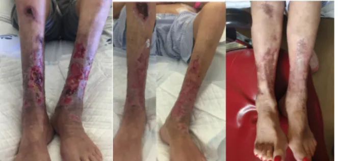
Figure 5. Evolution of calciphylaxis lesion of both legs: a) Two months after admission at Nephrology department; b) At discharge, five months after admission; c) One year follow-up
with the most frequently used treatment modalities, in-cluding sodium thiosulfate [18-19] and hyperbaric oxy-gen treatment [20], as the use of multimodal approach rendered impossible to assess the benefit of each moda-lity individually [2].
In the case described, besides increasing dialysis dose and phosphorus control, all other risk inducing factors such as treatment with vitamin D and warfarin were suspended. As the patient did not respond favourably to the initial treatment with sodium thiosulfate, the patient started second line treatment with hyperbaric oxygen in addition to dialysis dose optimization. As there are no guidelines as to the optimal treatment protocols for sodium thiosulfate and hyperbaric oxygen treatment, treatment duration was guided by the clinical improve-ment of the patient. In addition the medical treatimprove-ment the remaining multidisciplinary interventions, including wound dressing and debridement, early treatment of in-fection, nutritional support, pain management, physical therapy and psychological support played a crucial role on the a favourable outcome.
Other treatment choices, such as vitamin K, have shown to reduce and possibly reverse vascular calcifications in in vitro and in vivo studies. Vitamin K2 was proven to reduce inactive vitamin K-dependent matrix GIa protein (MPG), a powerful inhibitor of calcification, in a group of patients undergoing haemodialysis [8]. Also, the role of direct oral anticoagulants (DOACs) has been studied based on the fact that calciphylaxis is associated with a hypercoagulable state that leads to thrombosis of small and medium size vessels. A retrospective study of 18 CKD patients with calciphylaxis showed clinical im-provement in those patients whose treatment included DOACs, with no major bleeding events [21]. Other op-tions such as the use of double filtration rheopheresis, have been described in single case reports of patients with calciphylaxis undergoing haemodialysis with fa-vourable outcome [22]. However, based on current ex-pert opinion, the efficacy of these treatments options remains to be further studied.
CONCLUSION
Calciphylaxis is a rare and challenging condition. In this case, through a multimodal approach it was possible to achieve complete healing of the lesions and obtain func-tional recovery of the patient whilst maintaining peri-toneal dialysis.
As this is a single case report there is an absolute need for prospective, multicentre, randomized studies to as-sess not only the role of each treatment intervention but
also their applicability on peritoneal dialysis patients and whether there is a true role of this technique on the pathophysiology of calciphylaxis development and pro-gression.
Promising prospective studies are under development. The orfan drug SNF 472, a selective calcification inhi-bitor approved for investigational treatment of calciphy-laxis, was shown to improve wound healing, pain and quality of life of calciphylaxis patients on heamodialysis over the course of a 12 weeks on an open lable phase 2 trial [23]. This was the first prospective, interventio-nal and multicenter study on calciphylaxis treatment. A phase 3 randomized controlled trial of SNF 472 on cal-ciphylaxis treatment is in preparation.
Also in preparation is the Better Evidence And Transla-tion in Calciphylaxis (BEAT-Calci) Trial (AKTN 17.01), a prospective and randomized trial starting on 2020, that will evaluate therapeutic intervention on prevalent hae-modialysis patients suffering from calciphylaxis through the course of 6 months. Hopefully these trials will pro-vide us with more information as to better manage these patients in the future.
CONFLITS D’INTERET
The authors declare that they have no conflict of interest for this article
BIBLIOGRAPHIE
(1) Nigwekar SU, Thadhani R, Brandenburg MD. Cal-ciphylaxis. N Engl J Med 2018;378:1704-14.; https://doi. org/10.1056/NEJMra1505292
(2) Udomkarnjananun S, Kongnatthasate K, Praditporn-silpa K, et al. Treatment of Calciphylaxis in CKD: A Systema-tic Review and Meta-Analysis. Kidney Int Rep 2018. https:// doi.org/10.1016/j.ekir.2018.10.002
(3) Ellis CL, O´Neill WC. Questionable specificity of histologic findings in calcific uremic arteriolopathy. 2018; 94(2): 390-395. https://doi.org/10.1016/j.kint.2018.03.016 (4) Kalajian AH, Malhotra PS, Callen JP, Parker LP. Calciphylaxis with normal renal and parathyroid function: not as rare as previously believed. Arch Dermatol 2009; 145: 451-8. https://doi.org/10.1681/ASN.2015091065
(5) Vanparys J, Sprangers B, Sagaert X, Kuypers DR. Chronic wounds in a kidney transplant recipient with moderate renal impairment. Acta Clin Belg 2013; 68: 128-31. https:// doi.org/10.2143/acb.3191
(6) Nigwekar SU, Zhao S, Wenger J, et al. A national-ly representative study of calcific uremic arteriolopathy risk factors. J Am Soc Nephrol 2016; 27: 3421-9. https://doi. org/10.1681/ASN.2015091065
(7) McCarthy JT, El-Azhary RA, Patzelt MT, et al. Sur-vival, risk factors, and effect of treatment in 101 patients with
journal officiel du Registr e de D ialyse Péritonéale de Langue Française RDPLF www .rdplf.or g
calciphylaxis. Mayo Clin Proc 2016; 91: 1384-94. https://doi. org/10.1016/j.mayocp.2016.06.025
(8) Brandenburg VM, Kramann R, Rothe H, et al. Cal-cific uraemic arteriolopathy (calciphylaxis): data from a large nationwide registry. Nephrol Dial Transplant 2017; 32: 126-32. https://doi.org/10.1093/ndt/gfv438
(9) 4. Brandenburg VM, Evenepoel P, Floege J, et al. Lack of evidence does not justify neglect: how can we address unmet medical needs in calciphylaxis? Nephrol Dial Transplant 2016; 31: 1211-9. https://doi.org/10.1093/ndt/gfw025
(10) Zhang Y, Corapi KM, Luongo M, Thadhani R, Ni-gwekar SU. Calciphylaxis in peritoneal dialysis patients: a single center cohort study. Int J Nephrol Renovasc Dis 2016; 9: 235-41. https://doi.org/10.2147/IJNRD.S115701
(11) Heaf JG. Chronic Kidney Disease-Mineral Bone Di-sorder in the Elderly Peritoneal Dialysis Patient. Perit Dial Int 2015; 35(6):640-4. http://doi.org/10.3747/pdi.2014.00339 (12) Wang AY .Calcium Balance and Negative Impact of Calcium Load in Peritoneal Dialysis Patients. Perit Dial Int. 2014;34(4):345-52. http://doi.org/10.3747/pdi.2013.00177 (13) Brandenburg VM, Cozzolino M, Ketteler M. Calci-phylaxis: a still unmet challenge. J Nephrol. 2011;24(2):142– 148. https://doi.org/10.5301/JN.2011.6366
(14) Nicholas J, Evans R, Shaw C, et alc.UK Renal Re-gistry 18th Annual Report: Chapter 9 Biochemical Variables amongst UK Adult Dialysis Patients in 2014: National and Centre-specific Analyses. Nephron. 2016;132 Suppl 1:195-236. https://doi.org/10.1159/000444823
(15) Kuhlmman MK. Phosphate Elimination in Moda-lities of Hemodialysis and Peritoneal Dialysis. Blood Purif. 2010;29(2):137-44. http://doi.org/10.1159/000245640 (16) Zitt E, König M, Vychytil A, et al. Use of sodium thiosulphate in a multi-interventional setting for the treatment of calciphylaxis in dialysis patients. Nephrol Dial Transplant 2013; 28: 1232-40. https://doi.org/10.1093/ndt/gfs548 (17) Baldwin C, Farah M, Leung M, et al. Multi-inter-vention management of calciphylaxis: a report of 7 cases. Am J Kidney Dis 2011; 58: 988-91. https://doi.org/10.1053/j. ajkd.2011.06.002
(18) Nigwekar SU, Brunelli SM, Meade D, Wang W, Hymes J, Lacson E Jr. Sodium thiosulfate therapy for calcific
uremic arteriolopathy. Clin J Am Soc Nephrol 2013; 8: 1162-70. https://doi.org/10.2215/CJN.09880912
(19) Singh RP, Derendorf H, Ross EA. Simulation-based sodium thiosulfate dosing strategies for the treatment of calci-phylaxis. Clin J Am Soc Nephrol 2011; 6: 11559. https://doi. org/10.2215/CJN.09671010
(20) An J, Devaney B, Ooi KY, Ford S, Frawley G, Me-nahem S. Hyperbaric oxygen in the treatment of calciphylaxis: a case series and literature review. Nephrology (Carlton) 2015; 20: 444-50. https://doi.org/10.1111/nep.12433
(21) King BJ, El-Azhary RA, McEvoy MT, et al. Direct oral anticoagulant medications in calciphylaxis. Int J Dermatol 2017;56:1065-70. https://doi.org/10.1111/ijd.13685
(22) Bouderlique E, Provot F, Lionet A. Rheophere-sis for Adjuvant Treatment in ReRheophere-sistant Calciphylaxis. Ther Apher Dial 2018;22(4):413-414. https://doi.org/10.1111/1744-9987.12666
(23) Brandenburg VM, Sinha S, Terregrosa JV, et al. Im-provement in wound healing, pain, and quality of life after 12 weeks of SNF 472 treatment: a phase 2 open-lable study of patients with calciphylaxis. J Nephrol 2019;32(5):811-822. https://doi.org/10.1007/s40620-019-00631-0
Received 19/08/15, accepted after revision 19/10/07, published 19/12/15 journal officiel du Registr e de D ialyse Péritonéale de Langue Française RDPLF www .rdplf.or g
Open Access This article is licensed under a Creative Commons Attribution 4.0 International License, which permits use, sharing, adaptation, distribution and reproduction in any medium or format, as long as you give appropriate credit to the original author(s) and the source, provide a link to the Creative Commons license, and indicate if changes were made. The images or other third party material in this article are included in the article’s Creative Commons license, unless indicated otherwise in a credit line to the material. If material is not included in the article’s Creative Commons license and your intended use is not permitted by statutory regulation or exceeds the permitted use, you will need to ob-tain permission directly from the copyright holder. To view a copy of this license, visit http://creativecommons.org/licenses/by/4.0/.