FUNGAL PERITONITIS IN PERITONEAL DIALYSIS PATIENTS,
A MOROCCAN SINGLE CENTER EXPERIENCE
Prise en charge des péritonites fongiques,
expérience d’une unité de dialyse péritonéale au Maroc
Moussokoro Hadja Koné1, Tarik Bouattar1, Ibtissam Fares1, Meryem Benbella1, Naima Ouzeddoun1, Rabia Bayahia1, Loubna Benamar1
1Service de Néphrologie, Dialyse et Transplantation Rénale, CHU Ibn Sina Rabat, Faculté de Médecine et de Pharmacie Rabat, Université Mohamed V, Rabat, (Maroc)
Résumé
Introduction : La péritonite fongique (PF) en dialyse péritonéale (DP) est une infection grave qui met en jeu le pronostic fonctionnel du péritoine et le pronostic vital du patient. Elle doit bénéficier d’une prise en charge rapide mais néanmoins peu codifiée. Chaque centre assure donc une prise en charge individualisée de ses patients.
Matériels et méthode : Le but de notre étude est de décrire notre expérience de 10 ans à travers nos patients qui ont présenté une PF. Nous avons réalisé une étude rétrospective descriptive des cas de péritonites et extrait les PF documentées dans l’unité de DP.
Résultats : La prévalence de la PF était de 5,1%, soit 9 cas. Les signes cliniques prédominant étaient la turbidité du dialysat et les douleurs abdominales. La PF était primitive pour 3 patients. Le traitement antifongique utilisé en majorité était le Fluconazole, associé à une augmentation du nombre des échanges péritonéaux. L’ablation du cathéter de DP a été faite chez 8 patients avec un délai moyen de 5,5 jours. L’évolution globale était favorable et 3 patients ont repris une épuration extrarénale par DP. Nous n’avons déploré aucun décès ni péritonite encapsulante imputable à la DP.
Discussion et conclusion : La PF est une complication infectieuse de fréquence variable et un taux de mortalité élevé. Le taux de sortie de la technique de DP est aussi important. L’évolution favorable de nos patients restés en DP laisse présager la possibilité d’un plus grand nombre de patients maintenus en DP après une PF
Le
B
ulletin de la
D
ialyse à
D
omicile
Mots clés : dialyse péritonéale, péritonite fongique,
périto-nite médicale. journal officiel du Registr e de D ialyse Péritonéale de Langue Française RDPLF www .rdplf.or g Abstract
Introduction: Fungal peritonitis (PF) in peritoneal dialysis (PD) is a serious infection that involves the functional prognosis of the peritoneum and the patient’s vital prognosis. It must benefit from a fast handling but nevertheless not very codified. Each center therefore ensures an individual care of its patients.
Materiel and method: The purpose of our study is to describe our 10-year experience through our patients who presented FP. We performed a descriptive retrospective study of FP cases documented in the PD unit.
Results: the prevalence of FP was 5,1%, which represent 9 cases. Predominant clinical signs were dialysat turbidity and abdominal pain. FP was primitive for 3 patients. The antifungal therapy used was Fluconazole, which was combined with an increased number of peritoneal exchanges. DP catheter ablation was done for 8 patients with an average delay of 5.5 days. The overall outcome was favorable and 3 patients continued PD. No death or encapsulating peritonitis was a consequence of FP.
Discussion and conclusion: FP is an infectious complication in PD. Its’ death rate is elevated; dropping-out of PD rate too is elevated. The favorable evolution of our patients that stayed in PD let us think that it may be possible to maintain more patients in PD after FP.
Keywords : fungal peritonitis, medical peritonitis,
peritoneal dialysis.
INTRODUCTION
Peritoneal dialysis (PD) is one the modalities of chronic renal replacement therapy in the therapeutic arsenal available to treat patients with end stage renal disease. The principles of PD are based on exchanges between blood and dialysate through the peritoneum. A PD catheter allows access to the peritoneal cavity. In order to prevent contamination of the peritoneal fluid and infections, PD exchanges must be performed using strict aseptic technique.
Most of these infections are of bacterial origin, but in 1 to 15% of cases the infection is caused by a fungal organism. Fungal peritonitis is a serious infection requiring discontinuation of PD in the majority of cases according to literature [1].
.
The purpose of our study is to determine the prevalence of fungal peritonitis (FP) in the PD unit of the Ibn Sina University Hospital in Rabat (CHUIS-R), describe our FP management and compare our results with literature. MATERIALS AND METHODS
This descriptive retrospective study conducted from 2008 to 2018 in the PD unit, includes all episodes of infectious peritonitis in PD patients. All our patients are registered on the French-language PD Registry (RDPLF). We studied peritonitis episodes of fungal origin during this period. Patients treated without microbiological evidence were excluded from the study. FP was defined as primitive in patients who had never had prior episodes of infectious peritonitis.
We collected epidemiological and clinical data from patients’ medical records and obtained the list of their signs and symptoms, the delay between start of PD and FP and the delay between last bacterial peritonitis (if any) and FP.
Microbiologically, peritonitis was defined as the presence of cloudy effluent, white blood cell count greater than 100 / mm3 on direct examination. The diagnosis of FP was retained when the mycological examination revealed yeast on direct examination and / or dialysate culture.
We also analyzed the therapeutic parameters such as changes in dialysis prescription at the time of FP diagnosis, antifungal therapy, and surgical treatment.
Data analysis was done using MicroSoft Excel. RESULTS
Between 2008 and 2018, we recorded 186 cases of infectious peritonitis in our PD unit distributed as follows: 67.5% bacterial peritonitis, 28% negative culture peritonitis and 5.1% of fungal peritonitis. We had 9 cases of FPs occurring in 9 different patients with a sex ratio of 0.8 M / F and an average age of 42.11 +/- 16.21 years. The average duration of PD treatment before FP was 26.19±19 months.
FP was primary in 3 patients, 4 patients had broad-spectrum antibiotic therapy 6 months prior to FP. The most common clinical manifestations were cloudy fluid in all patients and abdominal pain in 8 patients. Table 1 describes the clinical characteristics of patients diagnosed with FP.
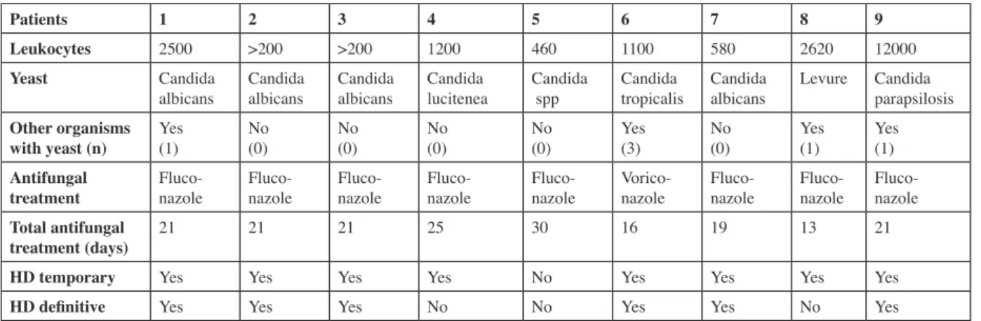
Bacteriologically, candida was the most common organism identified in the fluid cultures of 8 patients (88.8%), and one unidentified yeast in the fluid culture of one patient. Peritonitis was multimicrobial in 4 patients (44.4%), one of which had 3 yeast-associated bacteria. The bacteriological, therapeutic and follow up characteristics of the patients are summarized in Table II. Therapeutically, all patients benefited from more frequent exchanges (6 or more) for CAPD patients and a decreased dwell time for APD patients. Isotonic concentration dialysate was used, Eight patients were treated with oral Fluconazole, starting on day 3 of the onset of peritonitis, with an initial dose of 400 mg on the first day, then 200 mg daily. One patient was treated with Voriconazole IV at a dose of 6 mg/kg on the first day and then 4 mg/kg daily because of the severity of the clinical presentation. Antifungal therapy was maintained for an average of 20.7 days in all patients. Eight out of 9 (89,9%) patients had their PD catheter removed after an average time of 5.5 days from the onset of symptomatology. It was maintained in one patient because of the impossibility to establish a hemodialysis access. However, the patient’s PD catheter extension had been changed. The patient was able to continue PD, without the quality of dialysis being affected, and without another onset of peritonitis for the following 16 months.
journal officiel du Registr e de D ialyse Péritonéale de Langue Française RDPLF www .rdplf.or g
microbiologists to avoid undetected FP due to false-negative results in the initial culture [2]. In our unit, the incidence of FPs is 5.1%. Not only, this serious infectious complication is a significant factor of PD drop out but it also involves the vital prognosis of PD patients. In fact, more than 40% of FP patients transfer permanently to hemodialysis. The mortality rate ranges from 15 to 50% [3]. In our study, the 66.6% transfer rate from PD to hemodialysis is consistent with data from the literature, though with a zero mortality rate.
Primary FP is rare, found only in 17.9% of cases as shown in an Indian study [1] and in 21.4% of cases in an Italian study [4]. In our serie, they represented 33.3% of cases, occurring in the context of multi microbial peritonitis, in patients without prior digestive conditions. The overall patients outcomes were favorable: Six patients
transferred permanently to hemodialysis, 1 patient remained on PD, 2 patients resumed PD. For the latter, the PD catheter was removed and reinserted respectively 4 and 20 days later. One of these patients is still on PD with the same catheter after 2 years, while the second one died 2 years later as a result of a surgical operation. We did not deplore any deaths in this serie nor any encapsulating peritonitis secondary to FP.
DISCUSSION
Fungal peritonitis is a rare occurrence in peritoneal dialysis, 1 to 15% of all cases of peritonitis [1]. Accurate diagnosis requires a close collaboration with
journal officiel du Registr e de D ialyse Péritonéale de Langue Française RDPLF www .rdplf.or g
Tableau I : clinical characteristics of patients diagnosed with FP
PD : peritoneal dialysis ; FP :fungal peritonitis ; ATB : Antibiotic ; UF : Ultrafiltration; ND : non détermined. * : IV at day 1 thenIP
Tableau II : bacteriological, therapeutic and follow up characteristics of the patients
Patients 1 2 3 4 5 6 7 8 9
Age (years) 17 41 57 56 57 35 27 28 61
Nephropathy Glomerular Unknown Tubular Vascular Vascular Glomerular Unknown Unknown Diabetic
Time on PD (months) 7 19 16 11 8 52 50 52 22
Number of prior peritonitis
episodes before FP 0 2 1 1 2 2 2 0 0
Antibiotherapy (ATB) Duration (days))
Administration route* No ATB NDIP NDIP 17IP 13 et 21IP 18 et 20IP 14 et 20IP No ATB No ATB Time elapsed between
onset of FP and last ATB (in months)
No ATB 0,7 11 0,5 01 08 1,5 No ATB No ATB
Clinical signs :
Cloudy peritoneal effluent Abdominal pain Fever
GI Symptoms Decreased UF
Yes Yes Yes Yes Yes Yes Yes Yes Yes
Yes Yes Yes Yes No Yes Yes Yes Yes
Yes No Yes No No Yes Yes Yes Yes
No Yes Yes No No No Yes Yes No
No No Yes No No No No No No
Patients 1 2 3 4 5 6 7 8 9
Leukocytes 2500 >200 >200 1200 460 1100 580 2620 12000
Yeast Candida
albicans Candidaalbicans Candidaalbicans Candidalucitenea Candida spp Candidatropicalis Candidaalbicans Levure Candidaparapsilosis Other organisms
with yeast (n) Yes (1) No (0) No (0) No (0) No (0) Yes (3) No (0) Yes (1) Yes (1)
Antifungal
treatment Fluco-nazole Fluco-nazole Fluco-nazole Fluco-nazole Fluco-nazole Vorico-nazole Fluco-nazole Fluco-nazole Fluco-nazole
Total antifungal
treatment (days) 21 21 21 25 30 16 19 13 21
HD temporary Yes Yes Yes Yes No Yes Yes Yes Yes
General broad-spectrum antibiotherapy is a common contributing factor to FP, found in 6 of our patients. Godie et al. reported that in patients who had received broad-spectrum antibiotic therapy over the previous 3 months, the frequency of onset of FP was 74%, and 87% in patients who had received antibiotic therapy over the previous 6 months.
Other risk factors for FP are immunosuppression and diverticulitis.
After analyzing 216 cases of peritonitis [6], Chou et al. also found a higher frequency of FP following gram negative bacillus peritonitis or multiple-organisms peritonitis Medication management consists of intravenous (IV), intraperitoneal (IP) or oral administration of antifungal agents according to the spectrum of local resistance and antifungigram. The International Society for Peritoneal Dialysis (ISPD) discusses the use of Amphotericin B and Flucytosine [7]. These antifungals present some disadvantages if used daily such as the development of resistance to Flucytosine, severe abdominal pain in the case of Amphotericin B IP along with poor IP diffusion in IV administration [3]. ]. In a North American study, Fluconazole is the most commonly used antifungal, after confirmation by antifungigram [8]. However, the spectrum of resistance can vary from one region of the world to another. We treated our patients with Fluconazole per os at an initial dose of 400 mg then 200 mg daily, with a favorable outcome in 8 patients. We treated another patient with Voriconazole IV immediately due to the severity of the initial clinical presentation. The duration of Antifungal therapy should be continued at least 10 days following the removal of the PD catheter [3]. In our PD unit, treatment is maintained for a total of 20 days in average including the 15 days after removal of the PD catheter. PD catheter removal is the rule as soon as the diagnosis of FP is made [7]. In a retrospective study of 140 episodes of polymicrobial peritonitis, Sczeto et al. demonstrate, that yeast peritonitis is an independent risk factor with poor initial response to treatment in the absence of PD catheter removal[9]. However, if there is no other alternative renal replacement therapy possible, PD catheter removal is then not an option, as it was the case for our patient whose catheter was left in place. Peritoneal lavage, in the presence of intraperitoneal filaments that could contribute to the inflammation of the peritoneum and thus alter its subsequent use, is only
When we suspect a case of FP in our center, a series of rapid peritoneal exchanges (sometimes with APD to avoid too many manipulations) for peritoneal lavage purposes is set up. Upon confirmation of the diagnosis, the catheter is removed after an abundant peritoneal lavage over the course of 3 to 5 days.
FP prevention always requires patient education, patient compliance with hygiene technique and proper exit site care. Another pillar of care seems to be the rational use of antibiotics, apart from formal indications. Finally, in the case of confirmed bacterial peritonitis, the indication of a systematic prophylactic antifungal treatment is not clearly established [10]. In our unit, patients with relapse or recurrence of bacterial peritonitis and patients for whom long term antibiotic therapy is required, are prescribed Fluconazole 200 mg/day for the duration of antibiotic therapy as a prophylactic antifungal therapy,
The value of this series of FPs lies in the low prevalence of FP and the possibility of maintaining the PD technique, without affecting the quality of the
dialysis treatment. However, this is a retrospective study of a limited number of patients, which reduces
the impact of this series on the issue of peritoneal lavage.
CONCLUSION
Fungal peritonitis is a very serious complication of peritoneal dialysis. The non-drug part of the treatment is not well coded and depends on the experience of each center. The multiplication of exchanges prior to removal of the PD catheter should be evaluated and offered to patients.
Permanent discontinuation of PD should not be systematic and it would be interesting to evaluate the outcome of patients who remained on PD after FP.
DISCLOSURES
The authors declare that they have no conflict of interest in this article.
REFERENCES
1. Prasad and Gupta. Fungal peritonitis in peritoneal dialysis patients. Perit Dial Int. 2005; 25 (3):207– 222.
2. Antoine Grillon, Pierre-Hugues Boyer et Françoise Heibel.Bacteriological sampling in peritoneal dia-lysis fluid : decreasing the number of sterile
peri-journal officiel du Registr e de D ialyse Péritonéale de Langue Française RDPLF www .rdplf.or g
1 (1):15-19. DOI: https://doi.org/10.25796/bdd. v1i1.20
3. Joanna Matuszkiewicz–Rowinska. Update on fun-gal peritonitis and its treatment. Peritoneal Dialysis International, 2009; 29 (2):S161–5.
4. Auricchio, S., Giovenzana, M. E., Pozzi, M., Ga-lassi, A., Santorelli, G., Dozio, B., &Scanziani, R. Fungal peritonitis in peritoneal dialysis: a 34-year single centre evaluation. Clinical Kidney Journal. 2018: 11(6):874-880.
5. Goldie SJ et al. Fungal peritonitis in a large chronic peritoneal dialysis population: a report of 55 epi-sodes. Am J Kidney Dis 1996; 28 (1):86–91. 6. Chou CY, Kao MT, Kuo HL, Liu JS, Liu YL, Huang
CC. Gram-negative and polymicrobial peritonitis are associated with subsequent fungal peritonitis in CAPD patients. Perit Dial Int. 2006; 26 (5):607–8. 7. Philip Kam-Tao Li, David W. Johnson et al. ISPD
Peritonitis Recommendations: 2016 Update on Prevention and Treatment. Perit Dial Int. Septem-ber-October 2016 ; 36 (5) : 481-508.
8. J. Levallois et al. Ten-year experience with fungal peritonitis in peritoneal dialysis patients: antifungal
susceptibility patterns in a North-American center. International Journal Of Infectious Diseases, 2012; 16 (1): e41–3.
9. Sczeto et al. Conservative management of polymi-crobial peritonitis complicating peritoneal dialysis: a series of 140 consecutive cases. Am J Med. 2002; 113 (9):728-33.
10. Davenport A. Wellsted D. On behalf of the Pan Thames Renal Audit Peritoneal Dialysis Group. Does Antifungal Prophylaxis with Daily Oral Flu-conazole Reduce the Risk of Fungal Peritonitis in Peritoneal Dialysis Patients? The Pan Thames Re-nal Audit. Blood Purif. 2011 ; 32:181–185.
Received 2019:05/18, accepted after revision 2019/05/29 published 2019/06/18 journal officiel du Registr e de D ialyse Péritonéale de Langue Française RDPLF www .rdplf.or g
Open Access This article is licensed under a Creative Commons Attribution 4.0 International License, which permits use, sharing, adaptation, distribution and reproduction in any medium or
format, as long as you give appropriate credit to the original author(s) and the source, provide a link to the Creative Commons license, and indicate if changes were made. The images or other third party material in this
article are included in the article’s Creative Commons license, unless indicated otherwise in a credit line to the material. If material is not included in the article’s Creative Commons license and your intended use is not permitted by statutory regulation or exceeds the permitted use, you will need to obtain permission directly from the