https://doi.org/10.1021/la0494831
READ THESE TERMS AND CONDITIONS CAREFULLY BEFORE USING THIS WEBSITE. https://nrc-publications.canada.ca/eng/copyright
Vous avez des questions? Nous pouvons vous aider. Pour communiquer directement avec un auteur, consultez la première page de la revue dans laquelle son article a été publié afin de trouver ses coordonnées. Si vous n’arrivez pas à les repérer, communiquez avec nous à PublicationsArchive-ArchivesPublications@nrc-cnrc.gc.ca.
Questions? Contact the NRC Publications Archive team at
PublicationsArchive-ArchivesPublications@nrc-cnrc.gc.ca. If you wish to email the authors directly, please see the first page of the publication for their contact information.
NRC Publications Archive
Archives des publications du CNRC
This publication could be one of several versions: author’s original, accepted manuscript or the publisher’s version. / La version de cette publication peut être l’une des suivantes : la version prépublication de l’auteur, la version acceptée du manuscrit ou la version de l’éditeur.
For the publisher’s version, please access the DOI link below./ Pour consulter la version de l’éditeur, utilisez le lien DOI ci-dessous.
Access and use of this website and the material on it are subject to the Terms and Conditions set forth at
Effect of the hydrophilic size on the structural phases of aqueous
nonionic Gemini surfactant solutions
Nieh, Mu-Ping; Kumar, Sanat K.; Fernando, Raymond H.; Colby, Ralph H.;
Katsaras, John
https://publications-cnrc.canada.ca/fra/droits
L’accès à ce site Web et l’utilisation de son contenu sont assujettis aux conditions présentées dans le site LISEZ CES CONDITIONS ATTENTIVEMENT AVANT D’UTILISER CE SITE WEB.
NRC Publications Record / Notice d'Archives des publications de CNRC:
https://nrc-publications.canada.ca/eng/view/object/?id=04a572f7-4c45-4735-8cab-71f367722f99
https://publications-cnrc.canada.ca/fra/voir/objet/?id=04a572f7-4c45-4735-8cab-71f367722f99
Effect of the Hydrophilic Size on the Structural Phases of
Aqueous Nonionic Gemini Surfactant Solutions
Mu-Ping Nieh,
†Sanat K. Kumar,
‡Raymond H. Fernando,
§Ralph H. Colby,
|and
John Katsaras*
,†Neutron Program for Material Research, National Research Council, Chalk River Laboratory, Chalk River, Ontario, Canada K0J 1J0, Department of Chemical Engineering, Rensselaer
Polytechnic Institute, Troy, New York 12210, Department of Chemistry and Biochemistry, California Polytechnic State University, San Luis Obispo, California 93407, and Department
of Materials Science and Engineering, Pennsylvania State University, University Park, Pennsylvania 16802
Received February 27, 2004. In Final Form: August 3, 2004
Aggregate structures of aqueous nonionic Gemini surfactant solutions, R,R′ -[2,4,7,9-tetramethyl-5-decyne-4,7-diyl]bis[ω-hydroxyl-polyoxyethylene] with three different length polyoxyethylenes (i.e., 10, 20, and 30 ethylene oxide monomers, denoted from now on as S-10, S-20, and S-30, respectively), are investigated using small angle neutron scattering, dynamic light scattering, and fluorescence spectroscopy. For S-10 at low surfactant concentrations (Cs< 0.9 wt %), large “clusters”, with an average hydrodynamic radius
〈RH〉> 40 nm, are found to coexist with monomers. At intermediate Cs(0.9 < Cs< 2 wt %), some clusters break down forming micelles, with an〈RH〉∼2-3 nm, while the remaining clusters coexist with micelles. Increasing Csfurther (>2 wt %) results in a pure micellar phase with little or no clusters present. S-20 and S-30 mixtures, on the other hand, differ from S-10 in that irrespective of surfactant concentration, large clusters and small monomers/dimers are found to coexist, while there is no direct evidence for the presence of micelles.
Introduction
Gemini surfactants are composed of two or more pairs of hydrophilic and hydrophobic groups connected to each other with a spacer.1 Usually, their critical micellar concentration (cmc) in aqueous solutions is much lower than the cmc of conventional surfactants with the same hydrophilic and hydrophobic groups. As a result, smaller amounts of Gemini surfactants are needed to modify the surface tension of the solution.
In the past decade, many aqueous solutions of ionic Gemini surfactants2-11 and their mixtures with other surfactants12-17 have been investigated to understand the various parameters that affect their aggregate
struc-tures. Some of these parameters are hydrophobic tails asymmetry,2-4temperature, concentration,5-7,12tail hy-drophobicity,8,13 salt concentration,9,15 properties of the spacers,10,14-17 and the asymmetry of the counterpart surfactant.11These studies have shown that ionic Gemini surfactants composed of small hydrophilic headgroups and long hydrocarbon tails (>12 carbons) form a variety of structures (e.g., spherical, discoidal, threadlike micelles, vesicles, and lamellae).
Recently, nonionic Gemini surfactants have been syn-thesized.18,19 However, morphological studies of these surfactants are rare. A recent transmission electron microscopy study reported that the phase behavior of nonionic glycosylated Gemini surfactants in aqueous solutions was strongly dependent on the rigidity and length of the spacers.20 Menger and Mbadugha also reported on the rich structural behavior of nonionic Gemini surfactants having different tail lengths.10To our knowl-edge, the effect of the hydrophilic group’s size on the resultant structure has not been systematically investi-gated.
The nonionic Gemini surfactants in our study, R,R′ - [2,4,7,9-tetramethyl-5-decyne-4,7-diyl]bis[ω-hydroxyl-poly-oxyethylene] (Figure 1) with m + n ethylene oxide (EO) segments, are commonly used to lower the dynamic surface tension of solutions and as defoaming agents, metalwork lubricants, and pressure-sensitive adhesives. The mol-ecules have two hydrophilic poly(ethylene oxide) (PEO) †National Research Council.
‡Rensselaer Polytechnic Institute. §California Polytechnic State University. |Pennsylvania State University.
(1) Hait, S. K.; Moulik, S. P. Curr. Sci. 2002, 82, 1101-1111. (2) Oda, R.; Huc, I.; Candau, S. J. Chem. Commun. 1997, 21, 2105-2106.
(3) Oda, R.; Huc, I.; Homo, J.-C.; Heinrich, B.; Schmutz, M.; Candau, S. Langmuir 1999, 15, 2384-2390.
(4) Sikiric´, M.; Primozˇicˇ, I.; Filipovic´-Vincekovic´, N.; Heinrich, B. J.
Colloid Interface Sci. 2002, 250, 221-229.
(5) Aswal, V. K.; De, S.; Goyal, P. S.; Bhattacharya, S.; Heenan, R. K. J. Chem. Soc., Faraday Trans. 1998, 94, 2965-2967.
(6) Bernheim-Groswasser, A.; Zana, R.; Talmon, Y. J. Phys. Chem.
B 2000, 104, 4005-4009.
(7) Oelschlaeger, Cl.; Waton, G.; Candau, S. J.; Cates, M. E. Langmuir
2002, 18, 7265-7271.
(8) Huc, I.; Oda, R. Chem. Commun. 1999, 20, 2025-2026. (9) Knaebel, A.; Oda, R.; Mendes, E.; Candau, S. Langmuir 2000, 16, 2489-2494.
(10) Menger, F. M.; Mbadugha, B. N. A. J. Am. Chem. Soc. 2001, 123, 875-885.
(11) Menger, F. M.; Peresypkin, V. J. Am. Chem. Soc. 2001, 123, 5614-5615.
(12) Zana, R.; Le´vy, H.; Kwetkat, K. J. Colloid Interface Sci. 1998,
197, 370-376.
(13) Oda, R.; Huc, I.; Danino, D.; Talmon, Y. Langmuir 2000, 16, 9759-9769.
(14) Bernheim-Groswasser, A.; Zana, R.; Talmon, Y. J. Phys. Chem.
B 2000, 104, 12192-12201.
(15) Buhler, E.; Mendes, E.; Boltenhagen, P.; Munch, J. P.; Zana, R.; Candau, S. J. Langmuir 1997, 13, 3096-3102.
(16) Alargova, R. G.; Kochijashky, I. I.; Sierra, M. L.; Kwetkat, K. J.; Zana, R. J. Colloid Interface Sci. 2001, 235, 119-129.
(17) Danino, D.; Talmon, Y.; Zana, R. J. Colloid Interface Sci. 1997,
185, 84-93.
(18) Castro, M. J. L.; Kovenski, J.; Cirelli, A. F. Tetrahedron 1999,
55, 12711-12722.
(19) Bertho, J.-N.; Coue´, A.; Ewing, D. F.; Goodby, J. W.; Letellier, P.; Mackenzie, G.; Plusquellec, D. Carbohydr. Res. 1997, 300, 341-346. (20) Wathier, M.; Polidori, A.; Ruiz, K.; Fabiano, A.-S.; Pucci, B. New
J. Chem. 2001, 25, 1588-1599. 10.1021/la0494831 CCC: $27.50 © 2004 American Chemical Society
chains connected to a 14-carbon hydrophobic “hub”; the segmental lengths (m and n) of these two PEO chains are not necessarily identical. In this paper, the surfactants will be named as follows: S-m + n, (e.g., S-10 has a total of 10 EO monomers per molecule, m + n ) 10). More than a decade ago, Sato and Kishimoto first investigated aqueous S-10 solutions using vapor pressure depression, viscosity measurements,21enthalpy of micellation,22 os-motic pressure,23and NMR spectroscopy.24They observed two cmc values (0.9 and 2 wt %) but could not conclusively ascribe a morphology to either of these two transitions. Nevertheless, Sato and Kishimoto assumed the presence of monomers (below the first cmc) and micelles (beyond the first and second cmc’s) even though the monomer morphology was inconsistent with their viscosity data.21 Using small angle neutron scattering (SANS), dynamic light scattering (DLS), and fluorescence spectroscopy (FS), the present study reports on the structural phases of aqueous S-10, S-20, and S-30 solutions. Specifically, the effect of the hydrophilic group’s size on the aggregate structure is thoroughly investigated. We have found that, as a function of increasing surfactant concentration, an unusual structural phase transition takes place, that is, large clusters f micelles, which to the best of our knowledge has not been previously observed in other single-component surfactant systems and possibly ex-plains Sato and Kishimoto’s viscosity data.21In addition, concentration- and time-dependent studies are conducted to understand the formation and stability of the aggregate structures.
Experimental Methods
Sample Preparation. S-10, S-20, and S-30 surfactants were
obtained from Air Products & Chemicals, Inc., and used without further purification. S-10 and S-20 have the consistency of a viscous fluid (S-10 is transparent, while S-20 has a brown color), while S-30 is a brown gel. All three surfactants are solvated using water. For SANS measurements, D2O instead of H2O was used (99%, Cambridge Isotope, Inc.) to better contrast the surfactant from the solvent. The surfactant concentration, Cs, was varied from 0.2 to 5 wt %. For DLS measurements, samples were dissolved in distilled deionized H2O, which was filtered (pore size ) 100 nm) prior to mixing with the surfactants. Surfactant concentrations between 0.2 and 3 wt % were used to prevent the possibility of strong interparticle interactions, thus, allowing us to use the Stokes-Einstein equation in calculating the hydrodynamic radius (RH). In the case of FS experiments, pyrene (Py; Sigma Chemical Co.) was predissolved in distilled deionized water at a concentration of 2 ppm and then mixed with the surfactants to obtain the desired concentrations. All mea-surements, irrespective of the technique used, were carried out at room temperature (∼23 °C).
Technique and Data Reduction. SANS. Experiments were
performed using the 30m NG7 SANS instrument located at the National Institute of Standards and Technology (Gaithersburg, MD). The 6-Å wavelength neutrons were detected using a two-dimensional (2-D) detector with a 20-cm offset. Two sample-to-detector distances (SDD) were employed (SDD ) 1.5 and 14 m), covering a q range between 0.004 and 0.3 Å-1. The scattering vector, q, is defined as (4π/λ) sin(θ/2), where θ is the scattering angle and λ is the wavelength.
Samples were placed in circular quartz cells with path lengths varying from 2 to 4 mm, depending on the scattered intensity. The 2-D raw data were corrected for both ambient background and empty cell scattering and then put on an absolute scale (cross section per unit volume) through a procedure that estimates the neutron flux impinging on the sample.25The data were then circularly averaged to yield the one-dimensional intensity profile, I(q). The incoherent scattering was approximated from the high-q intensity plateau data for each sample and then subtracted from the corresponding reduced data. The corrected I(q) is, thus, proportional to the product of the form factor, P(q), which accounts for the morphology of the aggregates, and the structure factor, S(q), which describes the interaggregate interactions. In our case, because only dilute samples were studied, only P(q) is of consequence [i.e., I(q) ∼ P(q)].
DLS.The Stokes-Einstein formula [eq 1] is commonly used to describe the relationship between the diffusion coefficient D and the hydrodynamic radius RHof dilute, neutral monodisperse spherical particles in solution and can be written as follows:
where k, T, and ηware the Boltzmann constant, the absolute temperature, and the viscosity of water, respectively. For particles of different shapes (e.g., disk, cylinder, etc.), RHrepresents an equivalent hydrodynamic radius. For DLS measurements, the intensity time correlation function, G(τ), is defined as follows:
where τ is the delay time. From the Siegert relation, G(τ) can be expressed in terms of the field autocorrelation function, g(τ), as
where γ is the instrument-dependent coherence factor. For a polydisperse system, g(τ) is the sum of the exponential decays contributed from all particles and for a specific delay time, τ, and can be written as
where q is the scattering vector and Airepresents the amplitude of the ith particle with diffusion coefficient Di. Using eq 1, one can relate Dito the corresponding hydrodynamic radius RHi.
CONTIN is used in analyzing the DLS data solving for a group of g(τ) through eigenvalue decomposition in combination with regularization, a smoothing technique used to overcome the ill-posed nature of a Laplace transform inversion. The size distribution function expressed in terms of intensity can, thus, be resolved as a function of Di(or RHi).〈RH〉is, thus, obtained using intensity as a weighting factor.
DLS experiments were conducted using a DynaPro/MS-X (Proteinsolutions, Inc.) light scattering setup, which measures the scattered intensity at a fixed scattering angle (θ) of 90°. The correlator has 256 channels with delay times between 1 and 105 µs. The instrument is equipped with a power-adjustable laser source (λ ) 782.8 nm) and a temperature-controlled sample cell. The intensity overflow limit for the detector is ∼7 × 10⁶counts/s. The normalized intensity correlation function, Gh (τ), was obtained by averaging over a given acquisition time (∼10 s in our case). Prior to experimentation, the instrument was calibrated using standard polystyrene microbead aqueous solutions with particle sizes ranging between 1 nm and 1 µm ( 2%. DLS measurements (21) Sato, S.; Kishimoto, H. Bull. Chem. Soc. Jpn. 1985, 58, 282-287.
(22) Sato, S.; Kishimoto, H. J. Colloid Interface Sci. 1988, 123, 216-223.
(23) Sato, S.; Kishimoto, H. J. Colloid Interface Sci. 1988, 126, 108-113.
(24) Sato, S. J. Phys. Chem. 1989, 93, 4829-4833. J. J.; Orts, W. J. J. Appl. Crystallogr. 1998, 31, 430-445.(25) Glinka, C. J.; Barker, J. G.; Hammouda, B.; Krueger, S.; Moyer, Figure 1. Chemical structure ofR,R′
-[2,4,7,9-tetramethyl-5-decyne-4,7-diyl]bis[ω-hydroxyl-polyoxyethylene]. D)6πηkT wRH (1) G(τ) )
∫
0∞I(t) I(t + τ) dt (2) G(τ) ) 1 + γg(τ)2 (3) g(τ) )∑
i Aie -Diq2τ (4)were also taken at various time intervals after preparation, to investigate the time dependence of the surfactant systems.
FS. The SPEX Fluorolog 3 fluorimeter (Analytical Chemistry Division, NIST) has a double monochromator for selecting both the excitation and emission wavelengths. The excitation source is a 450-W Xe lamp. The emission intensity is measured using a Hamamatsu R928 photon counting photomultiplier tube and was collected at an angle of 90° from the incident excitation light. The λ of the excitation beam is 335 nm, and the detected wavelength of the emission beam ranged from 360 to 460 nm. Py was used as the fluorescent probe and was predissovled in the solvent (H2O) at a concentration of 2 × 10⁻⁶g Py/g total. The surfactants were then prepared to the desired Csusing the Py-doped H2O. The emission spectrum of Py contains five peaks in the range of 360-400 nm. The intensity ratio of the first peak (I1) to the third peak (I3), I1/I3, varies when the polarity of its surroundings changes.26
Results
S-10sPhase Behavior. Figure 2a shows SANS data
for S-10 samples with Cs between 0.5 and 5 wt %. The scattering pattern of the 0.5 wt % sample is very different from those of the other samples, indicating that a structural phase transition occurs at 0.5 < Cs< 1.1 wt %. This result is consistent with the first cmc (0.9 wt %)
reported by Sato and Kishimoto;21,23,24however, the q-4 dependence of the scattered intensity at low-q values (up to q ∼ 0.02 Å-1) for the 0.5 wt % sample indicates the existence of structures exceeding 50 nm (e.g., “clusters”). This morphology is different than the expected monomers that one usually finds in surfactant solutions at concen-trations below the cmc. Beyond the q-4region, the scatted intensity plateaus (0.03 < q < 0.1 Å-1) and exhibits a q-2 behavior indicative of small particles of size ∼ 1 nm. Because the intensity contribution from large structures, such as clusters, is mainly in the low-q regime and close to the probing limit of our instrument, it is not possible for us to obtain detailed structural information from the present data. Nevertheless, it is reasonable to assume that, for q > 0.03 Å-1, the major contribution to the scattered intensity is from small particles whose size can be precisely obtained from SANS data. For this case, two methods are used to evaluate the size of the small particles, assuming that for the 0.5 wt % sample interparticle interactions are negligible.
The Debye function is commonly employed in describing the scattering curve of dilute polymer solutions and can be written as follows:
(26) Dong, D. C.; Winnik, M. A. Can. J. Chem. 1984, 62, 2560-2565.
Figure 2. (a) SANS data of aqueous S-10 solutions (0.5 e Cse5 wt %). The solid lines are the best fit using the Debye relationship [eq 5] for the Cs) 0.5 wt % sample and a polydisperse spherical model with a hard sphere structure factor for the samples 1.1 eCse5 wt %. (b) 1/I versus q2plot constructed from the SANS data of the Cs) 0.5 wt % samples using a q range between 0.04 and 0.2 Å-1. The slope from the plot is 1225 cm‚Å-2. (c) The intensity plateau, I
0, as a function of Csfor S-10 samples with 1.1 e Cse5 wt %. The intercept, Cs0, is in agreement with the first cmc (e.g., 0.9 wt %) value, reported by Sato and Kishimoto.21(d) Zimm plots constructed from SANS data using the 1.1 e Cse5 wt % samples and intensity data from the q range between 0.008 and 0.1 Å-1.
where f(x) ) (2/x2)(e-x- 1 + x); φ
s,nis the volume fraction of the surfactant aggregates; ∆F is the scattering length density difference, or so-called contrast factor between the surfactant and D2O (∼5.92 × 1010cm-2for S-10); vs is the molecular volume of the surfactant (1.06 × 10⁻²¹ cm3for S-10); nsis the number of aggregation particles; and RGis average radius of gyration. In the range of q > 0.04 Å-1the data are fitted using the Debye function [solid line in Figure 2a], and an RGof ∼7.8 ( 1.0 Å is obtained. A more general approach for determining the RGof large aggregates (i.e., RGq< 1) from SANS data is
where A2 is the second virial coefficient related to the interparticle interaction, whose sign indicates the net interaction between aggregates (+ ) repulsive, - ) attractive). By plotting φs,n/I(q) versus q2, a straight line is obtained in the regime where RGq< 1, corresponding to a q range of 0.04 < q < 0.2 Å-1. Assuming that interparticle interactions are negligible for this dilute sample (i.e., A2∼0), the slope of the fitted line is then proportional to [1/(∆F2v
sns)](RG2/3). The resultant RGvalue of 8.3 ( 1.0 Å from the graph shown in Figure 2b is consistent with the RGobtained from our previous fit using the Debye function.
Kinugasa et al. reported experimental data on the relationship between MW(molecular weight) and RGfor PEO in water.27
Because the major component of S-10 is PEO and there is no predicted or experimental value for RGof S-10, we can roughly estimate its value, using eq 7. It turns out that RGfor S-10 is 7.2 Å/monomer and is in good agreement with the value obtained from the Debye fit [Figure 2a] and the fit to the plot in Figure 2b. It, therefore, seems that the small particles coexisting with clusters in solution are monomers.
Using the Debye function, the best fit result of φs,nfor
ns) 1 is 0.0051, which translates into 0.48 wt %. This means that >95% of the 0.5 wt % S-10 sample is comprised of monomers, although the few clusters that do exist
contribute, because of their size, strongly to the scattered intensity at low q.
For Csg1.1 wt %, the strong scattering intensity at low
qvanishes [Figure 2a], indicating the absence of clusters.
All SANS curves are characterized by an intensity plateau,
I0, at low q followed by a monotonic decay at higher q, typical of scattering from micelles. Given the fact that in the high-q regime the scattered intensity decays as q-4, instead of q-2[Figure 2a], the Debye function [eq 5] cannot be applied to fit these data. Because I0is proportional to the micellar number density, one can obtain the onset concentration for micellation, Cs0(∼0.9 wt %), from I0) 0, as shown in Figure 2c. This result is consistent with the previously reported cmc.21,23,24 Therefore, the micelle concentration can be obtained from the expression Cs
-Cs0, while RG, ns, and A2 (related to the interparticle interaction) of the micelles can be obtained from a Zimm plot of φm/I versus (q2+ bφm). φmis the volume fraction of micelles and is equivalent (Cs - Cs0), where b is a multiplier used to separate the various φmdata for better visualization.
Figure 2d shows Zimm plots for four S-10 sample concentrations (1.1 and 5 wt %) obtained from SANS data in the q range of between 0.01 and 0.04 Å-1. The lower q limit is determined from the deviation of the plateau resulting from large clusters, while the higher q limit is chosen so that qRG< 1. The slopes of the extrapolated lines at φm) 0 and q ) 0 are [1/(∆F2vsns)](RG2/3) and 2A2/ (∆F2b), respectively, and their intercepts yield, from eq 6, 1/(∆F2vsns). The values for RG, ns, and A
2obtained from the Zimm plots are 14.7 ( 3.5 Å, 17.2 ( 0.2, and (-2 ( 6) × 10-5mol/cm3and are summarized in Table 1. The aggregation number, ns, for micelles is comparable to values reported in the literature.21-23Because the error for A2is comparable to its experimental value, the sign of A2 cannot be determined from the present result. However, the interparticle interaction, if any, is weak. The data are also fitted using a model of polydisperse spheres in combination with a hard sphere structure factor (to a first order approximation), where only φm, the sphere’s radius, and the polydispersity are allowed to vary. The best fit result (Table 1) yields an R comparable to RGand a polydispersity ∆R/〈R〉(∆R is the standard deviation of Rand〈R〉is the mean value of R) of ∼0.28 ( 0.02.
DLS results for various CsS-10 samples equilibrated over 1 week are in agreement with the SANS data and provide further detailed information about the various morphologies (Figure 3). A single population of clusters (27) Kinugasa, S.; Nakahara, H.; Fudagawa, N.; Koga, Y.
Macro-molecules 1994, 27, 6889-6892.
Table 1. Best Fit Results of the Various Analyses Used for the Presence of Small Aggregates in S-10, S-20, and S-30 Solutionsa-c S-10 S-20 S-30 Cs(wt %) 0.5 1.1 2.3 3.4 5 0.2 0.5 1 2.5 4 0.2 0.5 1 2.5 5 Debye Fit Cm(wt %) 0.48 0.15 0.5 0.86 2.47 3.97 0.2 0.45 0.82 2.48 4.94 RG(Å) 7.8 13.6 12.2 12.9 13.2 15.1 13.5 12.9 12.8 14.2 13.1 ns 1 1.6 1.8 1.5 1.6 3 1.2 1.2 1.1 1.2 0.8
Zimm Plot or 1/(I - q2) Plot
RG(Å) 8.4 14.7 ( 3.5 14.7 ( 3.5 14.7 ( 3.5 14.7 ( 3.5 13.3 ( 2.1 13.3 ( 2.1 13.3 ( 2.1 13.3 ( 2.1 13.5 ( 1.2 13.5 ( 1.2 13.5 ( 1.2 13.5 ( 1.2 ns 1 17.2 ( 0.2 17.2 ( 0.2 17.2 ( 0.2 17.2 ( 0.2 1.6 ( 0.2 1.6 ( 0.2 1.6 ( 0.2 1.6 ( 0.2 1.16 ( 0.14 1.16 ( 0.14 1.16 ( 0.14 1.16 ( 0.14 Polydisperse Sphere Modeld
R(Å) 14.1 14.3 15.3 14.3
aEmpty cells represent that the analysis is not available.bThe contrast factor ∆F is calculated to be 5.92 × 10¹⁰, 5.89 × 10¹⁰, and 5.76 × 1010cm-2for S645, S480, and S485, respectively.cMolecular volumes v
sfor S465, S480, and S485 are 1.06 × 10⁻²¹, 1.53 × 10⁻²¹, and 2.14 × 10⁻²¹cm3, respectively.dUsing the spherical model to fit the polydispersity of S-10 samples yields a value ∼0.28 ( 0.02. With the exception of sample S-10 Cs) 0.5 wt %, RGand nsvalues are obtained from Zimm plots by extrapolating to Cs) 0.
φs,n I(q)) 1 ∆F2[1 - (RG2/3)q2+ ...)
(
1 vsns+ 2A2φs,n+ ...)
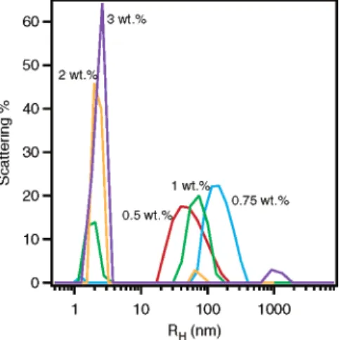
(6) 〈RG2〉) 4.08 × 10⁻¹⁸×MW1.1 (7)with an〈RH〉∼40 nm is observed for the 0.5 wt % S-10 solution (Figure 3). As a result of inadequate instrumental resolution, S-10 monomers are too small to be seen. However, as Csincreases to 0.75 wt %, the cluster〈RH〉 grows to >100 nm, while a small population of aggregates with an〈RH〉∼1.3 nm is also observed. A dramatic change in the size distribution function is observed as Csis further increased to 1 wt %. At this wt %, two〈RH〉populations appear, one with a value between 1.8 and 2.3 nm and another with〈RH〉∼100 nm, a good indication of micelle and cluster coexistence. As Csis increased to g2 wt %, most of the clusters disappear, and〈RH〉is found centered ∼2.3 nm. This phase transition is consistent with the second cmc (2 wt %) reported by Sato and Kishimoto.21It is reasonable to assume that, for micelles,〈RH〉will be slightly larger than RGfor the following reasons: (a) RG is defined as the second moment average length from each point of the object to the mass center, while〈RH〉is related to the hydrodynamic behavior of the particles. In the case of a sphere with radius R, RGis equal to (3/5)1/2R, but〈RH〉 ) R. (b) The measured 〈RH〉 includes H2O molecules incorporated with the hydrophilic EO groups, while RG, obtained from Zimm plots, is based on the contrast between the D2O and the surfactant molecules and does not account for the volume of associated D2O. (c) The〈RH〉is normalized using the scattered intensity (volume2), instead of the volume, as is the case for〈RG〉. As a result, a higher〈RH〉 value for particles of the same size distribution function is obtained.
From SANS and DLS data of S-10 solutions, we conclude that a sharp transition takes place at Cs∼0.9 wt % where micelles first form in a dilute solution of large clusters. These clusters already exist at Cs< 0.9 wt %. For Cs> 2 wt %, micelles predominate, while coexisting clusters are few in number or nonexistent.
Fluorescent spectroscopy using Py molecules as probes was employed to investigate the detailed structure of the clusters. Because Py molecules prefer to remain in a hydrophobic environment and the first-to-third peak intensity ratio (I1/I3) is sensitive to the environment of Py molecules, the location of Py molecules in solution can be deduced. Figure 4a shows the emission spectra of Py-doped pure water and S-10 samples at concentrations e1 wt %. Figure 4c depicts the relationship of I1/I3as a function of Cs. For a pure water/Py solution, the I1/I3 ratio was found to be 1.78 ( 0.08, similar to the 1.87 value reported in the literature.26 As Cs increases, the ratio remains unaltered until ∼0.8 wt %, the approximate value of the first cmc, where it suddenly drops. This indicates that
most Py molecules are no longer in water but instead go into the hydrophobic region of micelles [Figure 4c]. Two possibilities may explain the high I1/I3values in the case of clusters (i.e., Py existing in the water phase). First, the fact that the cluster population density is low means that only a small fraction of Py molecules can reside in the cluster’s hydrophobic region. Second, the cluster hydro-phobic region may not be large enough to accommodate the Py molecules, so they remain in water.
S-10sTime Dependence. The stability of S-10 clusters
and micelles is also investigated as a function of time after sample preparation. Figure 5a shows the size distribution functions for S-10 clusters at Cs) 0.75 wt % over a period of 26 days. Although, in some cases (1 h and 11 days), micelles are also observed, here we will focus on the size evolution of clusters. At Cs ) 0.75 wt %, S-10 clusters have an initial RHvalue of 50 nm, which increases over time to ∼170 nm. Figure 5b illustrates that clusters reach their equilibrium RHvalue after ∼100 h. A similar trend is observed for 0.5 wt % sample clusters. The final cluster size is Cs-dependent (higher Csresults in larger clusters) as shown in Figure 5b. Continuous growth of surfactant aggregates over a period of several months was previously observed in some conventional ionic surfactant mixtures which yielded unilamellar vesicles (ULVs).28,29 This growth was attributed to the slow process of vesicular formation, the limiting factor being the partitioning of surfactant molecules between inner and outer leaflets of the bilayer making up the ULV.29
The same time-dependent study was carried out using a 1 wt % mixture (Figure 6). Clusters are not observed until 1 h after sample preparation. After 20 min there is a broad micellar size distribution, which decreases with time. After 1 h the micelles reach a size distribution that remains practically unaltered over a period of 13 days. As for the clusters, not only does their population increase with time but their size also grows continuously, in the same manner as that of the low Cssamples, indicating that some micelles transform into clusters. Moreover, SANS data of 1 wt % samples 3 h and 17 days after preparation (data not shown) are virtually identical, the exception being the very low-q regime (q < 0.004 Å-1) where the intensity of the 17-day sample is higher, indicating that the clusters are growing both in size and in number. For the Csg2 wt % samples the micelles are stable over a period of 2 weeks. Occasionally clusters are found in small populations (data not shown).
S-20 and S-30sPhase Behavior. S-20 and S-30
samples yielded, at corresponding Cs values, SANS patterns [Figure 7a,b] resembling those of 0.5 wt % S-10 samples, indicating that similar structures are present in all three mixtures. As a function of q, the scattering pattern is characterized by a monotonic decay, followed by a plateau region, and then another decay, again implying the coexistence of clusters and small particles. For the Cs e2.5 wt % sample, the intensity decay at q > 0.1 Å-1 follows a q-2 dependence indicating that the Debye relationship [eq 5] is appropriate in describing the data. The plateau intensity, I0, is determined by fitting the data in the regime of q > 0.04 Å-1. Plotting I0versus Csyields a straight line and the onset concentration, Cs0, for small particles [insets to Figure 7a,b]. However, unlike S-10 solutions, Cs0 is practically 0, indicating that either micelles are not forming or the cmc is extremely small in these two systems. Zimm plots (Figure 8) of these two systems were constructed with the Cse2.5 wt % samples (28) Verbrugghe, S.; Laukkanen, A.; Aseyev, V.; Tenhu, H.; Winnik, F. M.; Prez, F. E. D. Polymer 2003, 44, 6807-6814.
(29) Iampietro, D. J.; Kaler, E. W. Langmuir 1999, 15, 8590-8601.
Figure 3. Size distribution functions from DLS measurements
of S-10 samples with Cs) 0.5, 0.75, 1.0, 2.0, and 3.0 wt %. The data were taken at least 1 week after equilibration.
in the same manner as discussed previously. For S-20, RG and ns are found to be 13.3 ( 2.1 Å and 1.6 ( 0.2, respectively, and representative of dimers. On the other hand, the obtained RGand nsvalues of S-30 solutions are 13.5 ( 1.2 Å and 1.2 ( 0.14, respectively, indicating the predominance of monomers. The A2value for S-20 is (2.2 ( 6.4) × 10-4mol/cm3. Again as in the case for S-10, the experimental error is comparable to the obtained value signifying weak interparticle interactions. The same can be said for the S-30 sample [A2) (2.6 ( 1.4) × 10-3mol/ cm3]. RGvalues from the Debye fits are similar to those obtained from the Zimm plots (Table 1).
For the Cs> 2.5 wt % samples, I0deviates from the fit for both S-20 and S-30 solutions [insets of Figure 7a,b]. On the other hand, the Debye relationship yields reason-able fits [Figure 7a,b]. In the case of S-20, the positive deviation of I0for the 4 wt % sample is possibly due to an increase of ns, which is found to be ∼3 from the Debye fit instead of 2 obtained from lower concentration samples. For S-30, the negative deviation of I0for the 5 wt % sample is most likely a structure factor effect arising from strong interparticle interactions.
The DLS data of S-20 solutions show a multimodal size distribution (data not shown). As a result, fluorescent
Figure 4. Fluorescent emission spectra for (a) S-10/water/Py systems with Csvarying from between 0 and 1 wt % and (b) S-30/ water/Py systems with Csvarying from between 0 and 2 wt %. (c) The ratios of I1/I3are plotted as a function of Csfor the data sets in parts a and b. A sharp transition for the S-10 samples is observed at ∼0.8 wt %.
Figure 5. (a) Size distribution function from DLS time-dependent studies of a 0.75 wt % S-10 solution. The peak corresponding
to the cluster morphology continuously shifts, with time, to a larger size distribution. (b) The evolution of S-10 clusters, with time, at two different Csvalues (e.g., 0.75 and 0.5 wt %).
spectroscopy experiments were performed on S-30 samples with differing Cs[Figure 4b]. Unlike the S-10 system, the intensity ratio of I1/I3 remains constant at 1.7 ( 0.1 throughout a range of concentrations e2 wt % [Figure 4c], lending support to our conclusions derived from SANS data that the small particles found in S-30 solutions are monomers and not micelles.
Discussion
Clusters. Clusters are found in S-10 solutions with Cs < 2 wt % and all of the S-20 and S-30 solutions, regardless of Cs. The rheological behavior of S-10 solutions containing such clusters has been previously reported.21Moreover, the reduced viscosity exhibited by samples below 0.9 wt % was found to be ∼4, higher than the theoretical value of 2.5.21The previous interpretation by Sato and Kishimoto was that the highly hydrophilic EO groups contributed to the increase in the apparent volume fraction of the solute.21 In fact, the unexpected high value of the reduced viscosity may be due to the presence of such clusters. Of note is that large cluster formation has been observed in some low CsPEO-grafted polymers and disappeared at higher
Csin the same manner as S-10 solutions.28Although we do not present a detailed structure of the cluster mor-phology, judging by the slow kinetics in reaching an equilibrium phase,29,30it is likely that the clusters resemble a vesicular structure, consistent with the viscosity ex-hibited by this phase. Because the two EO branches of S-10 molecules are most likely not of equal length, the flip-flop of S-10 molecules taking place to minimize the curvature free energy results in a continuous size evolution and is consistent with the explanation given by Yatcilla et al.30The system, however, does not necessarily form bilayers because the trans configuration of the two EO branches, with respect to the triple bond, presumably results in a lower steric energy. Such a planar structure formed by the trans configuration of the two EO branches also rationalizes the possible insufficient hydrophobic volume needed for accommodating the Py molecules. Moreover, vesicular structures (e.g., unilamellar or multi-lamellar) were also found in other Gemini surfactants solutions.11,13,14,17
Another possible cluster morphology could be “micro-bubbles”. They have recently been observed in water and can be removed via centrifugation.31SANS data from a degassed S-10 solution (∼0.7 wt %) continues to exhibit
strong low-q intensity (data not shown), further solidifying the notion of clusters. Another possibility is that the surfactant molecules stabilize the microbubbles in solu-tion. However, presently there is no experimental evidence supporting this assumption, except that these surfactants are commonly used for defoaming agents and the “sta-bilized” microbubbles might be able to prevent the formation of the larger bubbles that could lead to foaming. The manner in which the scattered intensity decays as a function of q provides us with evidence of the gross morphologies present in various sample mixtures. For example, a q-4dependence is indicative of spherical or nearly spherical objects or scattering media with a sharp interfacial boundary (two-phase coexistence). On the other hand, a q-1describes rodlike particles, while extended sheets and random walk coils are characterized by a q-2 dependence. In Figure 7 the intensity decay, as exhibited by several samples (Cse1 wt % for both S-20 and S-30 solutions), does not follow a q-4dependence. This may be ascribed to one of three possibilities or any combination of these. First, the structure of the above-mentioned clusters is far from isotropic (e.g., nonspherical). This argues against the possibility of “microbubbles”, whose shape is most probably spherical. Another possibility could be that there is an insufficient amount of separation between the various contributions from the different morphologies in a mixture that the expected q-4decay from the clusters could be buried in the signal arising from smaller particles. Finally, the continuous evolution of the clusters over the period of time it takes to collect the SANS data could result in a variable low-q slope.
Effect of the Size of the Hydrophilic Groups.
Comparing the experimental results from the different surfactant samples, the size of the hydrophilic group is found to strongly affect the structural phase behavior. A cmc of ∼0.9 wt % was observed for S-10 solutions with an
ns ∼17.3, while the ns and RGvalues of S-20 samples indicate the formation of dimers. S-30 with very long PEO chains form monomers as a result of the PEO branches being long enough to shield the hydrocarbon chains. The micellation of S-30 would result in a higher free energy than that of monomers due to a reduction in entropy. As a result, a trend of forming smaller micelles at a higher cmc is found as the hydrophilic groups become larger. This is also expected of surfactants with increased spontaneous curvature as a result of large-sized hydro-philic groups.32
Cluster f Micelle. The cluster f micelle transition
as a function of increasing Csis only clearly observed in S-10 solutions. This transition has previously been observed in some surfactant mixtures,33-37but to the best of our knowledge, not in single surfactant systems. Compared to micelles, clusters are much larger structures having a lower entropy. In addition, they are more stable at smaller Cs(<0.9 wt %), indicating a lower enthalpy. Therefore, the cluster f micelle transition can presumably be attributed to a decrease in the total free energy of the system by increasing the entropy. In the case of S-20 or S-30, the cluster f dimer or monomer transition is not
(30) Yatcilla, M. T.; Herrington, K. T.; Brasher, L. L.; Kaler, E. W.; Chiruvolu, S.; Zasadzinski, J. A. J. Phys. Chem. 1996, 100, 5874-5879.
(31) Glinka, C. (NCNR, NIST). Personal communication.
(32) Israelachvili, J. Intermolecular and Surface Forces, 2nd ed.; Academic Press: New York, 2000.
(33) Schurtenberger, P.; Mazer, N.; Waldvogel, S.; Ka¨nzig, W.
Biochim. Biophys. Acta 1984, 775, 111-114.
(34) Caria, A.; Khan, A. Langmuir 1996, 12, 6282-6290. (35) Marques, E. F.; Regev, O.; Khan, A.; da Graca Miguel, M.; Lindman, B. J. Phys. Chem. B 1998, 102, 6746-6758.
(36) Villeneuve, M.; Kaneshina, S.; Imae, T.; Aratono, M. Langmuir
1999, 15, 2029-2036.
(37) Egelhaaf, S. U.; Schurtenberger, P. Phys. Rev. Lett. 1999, 82, 2804-2807.
Figure 6. Size distribution function from DLS as a function
of time for a 1.0 wt % S-10 solution. It is clear that both the size and the population density of clusters increase with time.
observed, implying that to break up these clusters requires more enthalpic energy than entropic energy gained from the formation of dimers or monomers. One possibility may be that clusters are a more stable morphology for molecules having longer PEO groups (e.g., S-20 and S-30), because PEO is known to form large aggregates in aqueous solutions.38,39Moreover, the asymmetry of the two PEO chains, which is most likely larger for S-20 and S-30, is not well characterized, and may affect the molecular packing.32A detailed study on the effect of PEO asymmetry on aggregate morphologies is needed to conclusively resolve this issue.
Our time-dependent study of the 1 wt % S-10 sample seems to suggest that the pathway to cluster formation for samples with coexisting clusters/micelles may undergo a micellar phase because no clusters are observed within the first hour after sample preparation. Moreover, this transition has proved to be reversible (i.e., micelles f clusters) upon diluting the sample from g2 wt % to below 1 wt %.
Conclusion
Aqueous solutions of nonionic Gemini surfactants (S-10, S-20, and S-30) were studied using various techniques, SANS, DLS, and FS, to characterize their various mor-phologies. In contrast to the monomer f micelle (first cmc) and micelle f micelle (second cmc) transitions
previously reported by Sato and Kishimoto,21,22,24 the present work shows that S-10 does in fact undergo cluster/ monomer f cluster/micelle f micelle transitions. The two critical surfactant concentrations corresponding to the above-mentioned transitions are consistent with the so-called “cmc” presented by Sato and Kishimoto.21Moreover, the cluster/monomer f micelle transition is similar to the vesicle f micelle transition observed in some other surfactant mixtures. To the best of our knowledge, the cluster/monomer f micelle transition has not been observed in any single-component surfactant solution. With time (∼100 h), the clusters grow slowly in size, and their population density increases while in the cluster/ micelle phase. Above 2 wt %, the cluster morphology is practically nonexistent. For S-20 samples, clusters and dimers/trimers coexist over the entire Csregime studied (e4 wt %). As for S-30 samples, large clusters are also observed for all samples studied and coexist with mono-mers up to 5 wt % without any micelle formation.
Acknowledgment. We benefited from many
discus-sions with Kevin R. Lassila and Christine M. Kretz of Air Products and Chemicals, Inc. We would like to thank Paul C. DeRose (National Institute of Standards and Technol-ogy) for his help with the fluorescent spectroscopy experi-ments and SAMCOM funding from Air Products and Chemicals, Inc. We also thank Charles J. Glinka for SANS experiments on the degassed S-10 solution. This work utilized facilities supported in part by the National Science Foundation under Agreement No. DMR-9986442.
LA0494831 (38) Cuniberti, C. Polymer 1972, 13, 379-384; Polymer 1975, 16,
306-307.
(39) Layec, Y.; Layec-Raphalen, M. N. J. Phys. Lett. 1985, 44, 121-128.
Figure 7. SANS results of (a) S-20 solutions (0.2 e Cse4 wt %) and (b) S-30 solutions (0.2 e Cse5 wt %). The solid curves are the Debye fits for q-range data g 0.04 Å-1. For all samples, the intensity decay at high q follows a q-2dependence. The insets to parts a and b are of I0versus Csplots used to determine the Cs0for the S-20 and S-30 mixtures. A positive deviation of I0is observed for S-20 at 4 wt % while a negative deviation of I0is found for the 5 wt % S-30 samples.
Figure 8. Zimm plots for (a) S-20 and (b) S-30 solutions at 0.2 e Cse2.5 wt %.
![Figure 1. Chemical structure of R,R′ -[2,4,7,9-tetramethyl-5- -[2,4,7,9-tetramethyl-5-decyne-4,7-diyl]bis[ω-hydroxyl-polyoxyethylene]](https://thumb-eu.123doks.com/thumbv2/123doknet/14188963.477641/3.918.170.350.70.225/figure-chemical-structure-tetramethyl-tetramethyl-decyne-hydroxyl-polyoxyethylene.webp)
![Figure 2. (a) SANS data of aqueous S-10 solutions (0.5 e C s e 5 wt %). The solid lines are the best fit using the Debye relationship [eq 5] for the C s ) 0.5 wt % sample and a polydisperse spherical model with a hard sphere structure factor for the sample](https://thumb-eu.123doks.com/thumbv2/123doknet/14188963.477641/4.918.164.743.71.630/figure-aqueous-solutions-debye-relationship-polydisperse-spherical-structure.webp)