HAL Id: hal-00830731
https://hal.archives-ouvertes.fr/hal-00830731
Submitted on 8 Jan 2021
HAL is a multi-disciplinary open access
archive for the deposit and dissemination of
sci-entific research documents, whether they are
pub-lished or not. The documents may come from
teaching and research institutions in France or
abroad, or from public or private research centers.
L’archive ouverte pluridisciplinaire HAL, est
destinée au dépôt et à la diffusion de documents
scientifiques de niveau recherche, publiés ou non,
émanant des établissements d’enseignement et de
recherche français ou étrangers, des laboratoires
publics ou privés.
Compressive sensing in medical ultrasound
H. Liebgott, A. Basarab, Denis Kouamé, Olivier Bernard
To cite this version:
H. Liebgott, A. Basarab, Denis Kouamé, Olivier Bernard. Compressive sensing in medical
ultra-sound. IEEE International Ultrasonics Symposium 2012, IEEE, Oct 2012, Dresden, Germany. pp.1-6,
�10.1109/ULTSYM.2012.0486�. �hal-00830731�
Compressive sensing in medical ultrasound
1
Hervé Liebgott, 2 Adrian Basarab, 2 Denis Kouame, 1Olivier Bernard and 1Denis Friboulet
1Université de Lyon, CREATIS,
CNRS UMR 5220, INSERM U630, Université de Lyon,
1Université Lyon 1, 2INSA Lyon
Lyon, France
e-mail: name@creatis.insa-lyon.fr
2
Université de Toulouse, IRIT CNRS UMR 5505, Université Paul Sabatier
Toulouse, France e-mail: namet@irit.fr
Abstract — One of the fundamental theorem in information theory is the so-called sampling theorem also known as Shannon-Nyquist theorem. This theorem aims at giving the minimal frequency needed to sample and reconstruct perfectly an analog band-limited signal. Compressive sensing (or compressed sensing, compressive sampling) or CS in short is a recent theory that allows, if the signal to be reconstructed satisfies a number of conditions, to decrease the amount of data needed to reconstruct the signal. As a result this theory can be used for at least two purposes: i) accelerate the acquisition rate without decreasing the reconstructed signal quality (e.g. in terms of resolution, SNR, contrast …) ii) improve the image quality without increasing the quantity of needed data.
Even if medical ultrasound is a domain where several potential applications can be highlighted, the use of this theory in this domain is extremely recent.
In this paper we review the basic theory of compressive sensing. Then, a review of the existing CS studies in the field of medical ultrasound is given: reconstruction of sparse scattering maps, pre-beamforming channel data, post-beamforming signals and slow time Doppler data. Finally the open problems and challenges to be tackled in order to make the application of CS to medical US a reality will be given.
Keywords: compressive sensing, sparse, beamforming, ultrasound
I. INTRODUCTION
Whereas the Shannon theorem fixes the limit for the sampling frequency of a signal to twice its maximum frequency component fmax, the recently introduced compressed
sensing (CS) theory allows – under certain assumptions – to recover a signal sampled far below this limit [1-3]. Compressed sensing (also known as compressive sensing or compressive sampling) can be applied for two main purposes. i) it can lower the amount of data needed and thus allows to speed up acquisition. An example in the field of medical imaging of
such application is dynamic MRI [4] ii) it can improve the reconstruction of signals/images in fields where constraints on the physical acquisition set up yields very sparse data sets. A typical example is seismic data recovery in geophysics [5]. The application of this theory to the field of medical ultrasound is extremely recent even though there are applications that are excellent candidates such as e.g. triplex acquisitions [6] for CFM/B mode/Doppler or 3D imaging using matrix arrays. The objective of this paper is to give the reader an overview of the different attempts to show the feasibility of CS in medical ultrasound. The classification of the studies is done according to the data that are considered to be sparse (the scatterer distribution itself, the pre-beamforming channel data, the beamformed RF signal and even Dopple data). For each example the potential of using CS is highlighted as well as the specificity in terms of modeling or numerical implementation. The paper is organized as follows: in section II the CS theory is recalled, section III presents different applications of CS in medical ultrasound and finally section IV gives a short discussion and conclusion
II. COMPRESSIVE SENSING THEORY
Compressive sensing (CS) [2] allows the reconstruction of a signal x \∈ n from a linear combination of a small number of random measurements y∈\ m < n. m
In a general setting, the measurements y may be acquired in the so-called "sensing basis" Φ , which depends on the acquisition device. As an example, in MRI, Φ is the Fourier basis.
= Φ
y x (1)
where Φ is an m x n matrix. The columns of Φ have an entry one at random positions and zero elsewhere, thereby modeling the random selection of the measurements.
At the heart of CS is the assumption that x has a sparse representation in some model orthonormal basis Ψ , i.e.:
= Ψ
x v (2)
where vhas only s < m < n non zero coefficients. The signal
vis called s-sparse. CS theory shows that this sparsity allows an exact recovering ofvwith overwhelming probability for a certain class of matrices ΦΨ [1]. In particular, the sensing basis has to be incoherent with the model basis Ψ [7], which is ensured by the randomness of the non-zeros components of Φ . Finally the problem can be written as follows:
= ΦΨ =
y v Av (3)
where A is an m x n full rank matrix (i. e. the m raws of A are independent).
In these settings, the CS problem thus amounts to solve (3) for
v, under the constraint that v is sparse. Once v is estimated, the signal x, can then be computed from (2).
For matrices A with a specified isometry constant of the so-called "restricted isometry property" (RIP), Candès et al. [1] showed that the CS problem may be solved through the following 0-minimization problem P0:
P0 0 ˆ arg min n subject to ∈ = = v v vA y Av \ (4)
where the 0 norm is
{
}
0:= i v, i ≠0
vA
Equation (4) thus implies that from all the possible solutions of (3), we seek the sparsest one. In general, solving (4) is NP hard. Sub-optimal greedy algorithm attempt to solve this problem by successively adding non zero components to a sparse approximation of v. (see [8]).
By imposing a more restrictive bounds on the isometry constant, the sparsest solutionˆxof (3) can be found by solving the following basis pursuit (BP) problem P1 [9, 10]:
P1 1 ˆ arg min n subject to ∈ = = v v vA y Av \ (5)
where the 1 norm is
1 1 n i i= v =
¦
v A .The 0- 1 equivalence, using the RIP, was presented by Candès
in [9] (see also [1]).
The framework described above assumes that we are given exact samples of the signal to be recovered. This is seldom the case in practice, since the measurements are very often corrupted by noise. In the case of measurements with additive noise, we have:
= +
y Av e (6)
where e represent a noise term of bounded energy
2 ≤ ε
eA ,
P1 can be then recast as [9, 11]:
P2 1 2 ˆ arg min n subject to ∈ = = − ≤ ε v v v A y y AxA \ (7)
In practical applications the signal is generally not exactly sparse but most of its coefficients in (2) are small. When signal coefficientsvdecays exponentially in absolute value, the signal is called ‘compressible’. The solution found by P1 (5) or P2 (7) gives the approximation of vby keeping its S largest entries. For solving P1 and P2, mainly two classes of methods have been employed. The first one consists in deterministic
optimization algorithms. The second one uses stochastic (Bayesian) algorithms. With this approach, statistical a priori laws are associated to each of the unknown variables, further combined using the Bayes law to find the a posteriori probability to be maximized.
ሺ࢜ȁ࢟ሻ ן ሺ࢟ȁ࢜ሻሺ࢜ሻ (8) In (8), p(y|v) represents the likelihood and p(v) contains prior information about the unknown v. Note that if the noise is considered Gaussian and the prior law on v is a Laplacian, the maximization of the posterior probability in (8) is equivalent to the classical LASSO optimization.
III. APPLICATION TO ULTRASOUND IMAGING
A central concern in CS is that the data under consideration should have sparse expansion in some dictionaries (e.g., Fourier basis, wavelet basis, dictionary learned from data, etc...), i.e., the number of non-zero coefficients of the image or signal in this representation basis should be as small as possible. Moreover, US echographic data may be considered at various stages of the image formation pipeline. In a conventional configuration, the set of raw radio-frequency (RF) signals received at each transducer element (i.e. channel RF data) is beamformed to build the beamformed RF image. Detection applied to the latter yields the envelope-detected image, and finally the log-envelope image corresponding (possibly after some interpolation) to the conventional B-mode display is obtained. As a consequence, one of the main features of the existing studies is the type of signal/image to be reconstructed and the choice of the representation where the US data are assumed to be sparse. Another important characteristic of these studies corresponds to the formalization of the reconstruction problem and related algorithm (e.g., Bayesian modeling, deterministic basis pursuit, or matching pursuit).
A. Sparse diffusion map
Several groups of authors [12-18] have chosen to model the medium under investigation itself as a sparse distribution of scatterers. The discrete scattering model is commonly used in the literature and is the basis for the most used ultrasound simulation program (Field II [19, 20]). However considering that most of the scatterers have an echogenecity close to zero is more unusual.
The approach proposed by [12, 13] aims at producing, with only one single plane wave transmission, an image having equivalent spatial resolutions as with conventional imaging. The potential of the approach is to provide fast imaging. Indeed in such a situation the highest possible temporal resolution (1 image per transmitted pulse) would be reached. The basic idea is to write the direct scattering problem and solve the inverse problem under the constraint that the scatterer distribution is sparse. This can be written as follows
(
)
(
)
sc
p
e
θ=
G e
θγ
κ (9)with psc(eθ) the scattered pressure received by the transducer elements after transmission of a plane wave in direction θ ,
G e
(
θ)
.represents propagation and interactionwith the scatterers and
γ
κ is the scatterer dis a Nz times Nx regular grid. Solving theproblem is an ill posed linear problem equ problem described in equation (4) or (5) if be sparse. As a result it can be solved by
Figure 1shows a result from a simple phan four isolated scatterers obtained with this ap sparsity assumption is obviously perfectly result outperforms even the synthetic apertur
Figure 1. Result from Shiffner 2012 obtained consisting in four isolated scatterers. The CS res with synthetic aperture (a), delay and sum (b) propagation (c)
Figure 2. Result from Wagnerr 2012(a) origina image obtained by CS. The edges are well re speckle is close to be completely lost
With the same assumption [14, 15] proposed based on finite rate of innovation and technique is used to reconstruct cardiac ima Figure 2. In these images the edges are whereas the speckle patterns are much redu in some parts of the images. This is co assumption of scatterer map sparsity. hypothesis tends to keep only the highest ech the small ones which are however extremely construction of the speckle patterns.
stribution lying on inverse scattering uivalent to the CS κ
γ
is assumed to y 1 minimization. ntom consisting in pproach. Here the y verified and there approach.
d from a phantom sult (d) is compared ) and Fourier back
al cardiac image (b) econstructed but the
d another approach d Xampling. The ages as reported in clearly preserved uced and even lost onsistent with the Indeed such an hoes and to reduce y important for the
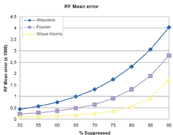
Figure 3. Result from [21] obtai compares the reconstruction error of data and sparsifying basis. Wav
B. Sparse Raw
Another group of authors channel data gathered at ea receive have a sparse decom objective of such an approach beamformed data acquired an approach to reconstruct B-mod three sparsifying bases Ψ hav and the recently introduced acquisition of the raw data m spatially uniform random samp using 1 minimisation. The s
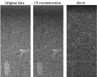
simulations using Field II and the Ultrasonix MDP scanner. Figure 3 shows a comparison o as a function of removed d expected, for all bases the er when more data is removed. Th that wave atoms lead to an erro three times as small as wavelets In order to evaluate from a vis this approach to reconstruct Figure 4 shows experimental r of the original data using CS an According to this result one m the ability of CS to provide the image reconstruction. Howeve not been mentioned here is obtained results indeed corre random acquisition of the raw data this way is probably as d channel data set. An interesting entire columns of the raw data s disconnecting some elements extremely interesting for 3D order to reduce the typical hug arrays. Unfortunately the initia are not given here show that t much lower quality.
ned from simulated data. The plot r as a function of removed quantity ve atoms lead to the best result
RF
[21-24] consider that the raw ach transducer element during mposition in some basis. The is to reduce the quantity of pre-nd evaluate the ability of this
e images of good quality. In [21]
ve been tested: wavelets, Fourier wave-atoms basis [25]. The measurements was done using a pling. Reconstruction was done study was carried out through experimental data acquired with
of the mean reconstruction error data and sparsifying basis. As rror of reconstruction increases he most interesting thing here is or twice as small as Fourier and s.
sual point of view the ability of good quality B-mode images, results obtained from only 20% nd wave atoms.
might be extremely confident on e right framework for ultrasound er one important point that has the measurement setup. The espond to a spatially uniform channel data-set. Acquiring the difficult as just acquiring all the g approach would be to remove set. This would indeed consist in of the probe. This could be imaging with matrix arrays in ge number of elements of such al results of this approach which this strategy leads to images of
Figure 4. Experimental results from [21] showing the feasibility of the proposed technique to reconstruct pre-beamformed data able to lead to good quality B-mode images. The error is spread evenly over the entire image and both the strong structures and the speckle are reconstructed.
C. Sparse assumption of the RF images Fourier transform
In this section the reconstruction of post-beamforming 2D RF images via CS technique is discussed. The sparsity assumption is related here to the assumption of bandlimited RF signal acquisition. Thus, the 2D Fourier transform of RF images is assumed to be sparse. With the notations introduced in section II, Φ stands in this section for the spatial acquisition mask (showing the random positions where spatial samples are measured), Ψ is the 2D inverse Fourier transform, y are the randomly measured post-beamformed RF samples and v is the 2D Fourier transform of the RF image to be reconstructed. Thus, considering an additive Gaussian noise Ș with zero-mean and variance "2, the acquisition model is:
࢟ ൌ ܣ࢜ ࣁ (10) With ࢟ ൌ ሺݕଵǡ ǥ ǡ ݕெሻ א ԧெ, ࢜ ൌ ሺݒଵǡ ǥ ǡ ݒேሻ א ԧேand ࣁ ൌ ሺߟଵǡ ǥ ǡ ߟெሻ א ԧெ representing the vectorized forms of the corresponding images.
From a theoretical point of view, the ideal sampling mask Φ (the most incoherent with the Fourier transform) is a uniform random pattern in the two spatial directions. As previously mentioned, it has however only a limited interest in US imaging. For this reason, a more suitable random sampling mask has been proposed in [26-28]. It considers that randomly chosen beamformed RF lines are not sampled at all, and is thus less incoherent with the Fourier transform. Thanks to this sampling scheme, the number of US pulses emitted during the acquisition is potentially reduced and as a consequence the acquisition time reduced. An example of such a 2D sampling mask is shown in Figure 5(for its extension in 3D, see [26, 28]).
Figure 5. Random post-beamforming RF sampling mask. The white pixels correspond to the measured samples locations.
The resulting inverse problem in (10) can be solved using a conjugate gradient descent method, as shown by the results obtained in [28]. However, more accurate results are provided in [29, 30] using a Bayesian framework. We give hereafter the main details of the Bayesian model proposed in [30] and its extension to a regularized form in [29]. Given the inverse problem in (10) and keeping in mind that Ș is a zero-mean Gaussian noise, the associated likelihood is:
ሺ࢟ȁ࢜ǡ ߪଶሻ ൌ ͳ
ሺߨߪଶሻே݁ݔ ൬ߪͳଶԡ࢟ െ ܣ࢜ԡଶଶ൰ (11) Where ԡכԡଶ stands for the standard l2 norm.
At this stage, the inverse problem has two unknowns, the variance of the noise and v, the 2D Fourier transform of the RF image. For the latter, two assumptions are taken into account in order to construct the prior distribution: the Gaussian property of the RF images and image sparsity in the Fourier domain. Thus, each pixel vi of the Fourier domain has been assigned a
mixture of a Gaussian statistical law and a mass at the origin. ሺݒȁݓǡ ߪ௩ଶሻ ൌ ሺͳ െ ݓሻߜሺȁݒȁሻ ߨߪݓ ௩ଶ݁ݔ ቆെ ȁݒȁ ߪ௩ଶቇ (12)
Where w is the prior probability of having a non-zero in the Fourier domain. In [30], w was considered a priori uniform random. In [29], assuming that the non-zero pixels in the Fourier domain of a RF image are located in compact regions around the central frequency of the probe, a further spatial regularization is taken into account by the prior on w.
Note that the Gaussian characteristics may be replaced by other laws reported in the literature. For example, in the context of the application of CS in US imaging, Achim et al. used in [31] an #-stable distribution.
The posterior distribution is maximized using a MCMC method (also known as Gibbs sampler). The details concerning the sampling of the unknown variables over the iterations are given in [29, 30]. We give hereafter a simulation result extracted from [29], showing the original simulated image, the samples used for reconstruction and the results obtained using a gradient descent algorithm and with the Bayesian framework. In this case, 10% of the original image pixels are used for reconstruction (30% randomly located samples from 30% randomly chosen RF lines).
(a)
(c)
Figure 6. B-mode images corresponding to: (a) or image, (b) RF samples used for reconstruction, ( image using a reweighted conjugate gradien reconstructed RF image using the Bayesian fram [20].
D. Doppler
CS has also been proposed for Doppler im [32], the authors applied CS on duplex ultra is a mode of medical ultrasonography that all the same time the inner structure of the body blood flow at a particular point in the body This mode requires a strategy for alternating emissions. Traditional strategies either hal measurable velocity or introduce gaps in the f
Figure 7: In vivo Doppler result from a fem sonogram; b) reconstructed sonogram based on C the ratio of the number of Doppler samples retaine total number of samples.
In [32], CS is used to reconstruct the Doppl by segment. More precisely, the authors ma that the Fourier transform of the Doppler Using the notation introduced in section II section for the temporal acquisition mask (de positions where temporal Doppler samples the inverse Fourier transform, y are the ra Doppler samples and v is the Fourier transfo signal to be reconstructed. One particularity decomposition of the signal to be reconstru segments of same length. 1 minimisation i
each segment separately. By doing so, t (b) (d) riginal simulated RF (c) reconstructed RF nt optimization, (d) mework proposed in maging [32, 33]. In asonography which lows to visualize at y (B-mode) and the y (Doppler mode). g B-mode and flow lve the maximum flow data.
moral artery. a) real CS. r corresponds to ed to the
ler signal segment ade the assumption r signal is sparse. , Φ stands in this efining the random are measured), is andomly measured orm of the Doppler of this work is the ucted y in several is then applied on the reconstruction
algorithm, which is by far the m on a reduced number of samp applicability. The proposed me simulated and real sonograms Doppler signals with acceptabl obtained using 50%
IV. DISCUSSIO
As presented in this paper, ultrasound is a very recent fie drastic modifications in the developed. The technique i technological applicability. Th 1) a sparsifying basis Ψ , incoherent with Ψ , 3) d (electronics) and 4) fast and ro Improvements are necessary compressive sensing medical problem. In particular, efforts maintain the real-time charac which still makes it a particular
V. ACK
This work has been partiall US-3D
VI. REFERE
[1] E. J. Candes, J. Ro uncertainty principles from highly incomp
IEEE Transactions o pp. 489-509, 2006. [2] E. J. Candes and M. B Compressive Samplin Magazine, vol. 25, pp [3] D. L. Donoho, " Transactions on Info 1289-1306, 2006. [4] M. Lustig, D. Dono
MRI: The applicatio rapid MR imaging
Medicine, vol. 58, pp.
[5] F. J. Herrmann and G seismic data recov
Geophysical Journal I
248, 2008.
[6] J. A. Jensen, "Spe ultrasound using spars
Acoustical Society of A
2006.
[7] E. Candès and J. Rom in compressive sampli p. 969, 2007. [8] J. A. Tropp and A. From Random Me Matching Pursuit," Information Theory, v
most consuming step, is applied ples allowing to reach real-time thod has been evaluated on both s. In both cases, reconstructed le SNR (higher than 10 dB) are
ON AND CONCLUSION
compressed sensing medical eld of research that can lead to
way ultrasound scanners are is feasible but far from its e key points for CS to work are 2) a measurement basis Φ dedicated acquisition material
obust reconstruction algorithms. for all of these points and l ultrasound remains an open
s should be made in order to cteristic of medical ultrasound r imaging modality.
KOWLEDGMENT
ly financed by CNRS PEPS
CS-ENCES
omberg, and T. Tao, "Robust s: exact signal reconstruction plete frequency information,"
n Information Theory, vol. 52,
B. Wakin, "An Introduction To ng," IEEE Signal Processing . 21-30, 2008.
Compressed sensing," IEEE
ormation Theory, vol. 52, pp.
ho, and J. M. Pauly, "Sparse on of compressed sensing for g," Magnetic Resonance in
1182-1195, 2007.
G. Hennenfent, "Non-parametric very with curvelet frames,"
International, vol. 173, pp.
233-ectral velocity estimation in se data sets," The Journal of the
America, vol. 120, pp. 211-220,
mberg, "Sparsity and incoherence ing," Inverse Problems, vol. 23,
C. Gilbert, "Signal Recovery easurements Via Orthogonal
IEEE Transactions on
[9] E. J. Candès, "The restricted isometry property and its implications for compressed sensing," Compte
Rendus de l'Academie des Sciences, vol. 346, pp.
589-592, 2008.
[10] E. J. Candes and T. Tao, "Decoding by linear programming," IEEE Transactions on Information
Theory, vol. 51, pp. 4203-4215, 2005.
[11] A. Austeng and S. Holm, "Sparse 2-D arrays for 3-D phased array imaging - design methods," IEEE
Transactions on Ultrasonics, Ferroelectricity and Frequency Control, vol. 49, pp. 1073-1086, 2002.
[12] M. Schiffner, T. Jansen, and G. Schmitz, "Compressed Sensing for Fast Image Acquisition in Pulse-Echo Ultrasound," Biomedical Engineering, vol. 57, pp. 192-195, 2012.
[13] M. Schiffner and G. Schmitz, "Fast Pulse-Echo Ultrasound Imaging Employing Compressive Sensing," in IEEE International Ultrasonics Symposium, Orlando, Florida, USA, 2011, p. in press.
[14] N. Wagner, Y. C. Eldar, A. Feuer, G. Danin, and Z. Friedman, "Xampling in Ultrasound Imaging," in
SPIE Medical Imaging, 2011.
[15] N. Wagner, Y. C. Eldar, A. Feuer, and Z. Friedman, "Compressed beamforming applied to B-mode ultrasound imaging," in IEEE International
Symposium on Biomedical Imaging (ISBI),
Barcelona, Spain, 2012, p. in press.
[16] M. Shen, Q. Zhang, and J. Yang, "A novel receive beamforming approach of ultrasound signals based on distributed compressed sensing," in
Instrumentation and Measurement Technology Conference (I2MTC), 2011, pp. 1-5.
[17] Q. Zhang, B. Li, and M. Shen, "A measurement-domain adaptive beamforming approach for ultrasound instrument based on distributed compressed sensing: Initial development,"
Ultrasonics, p. in press, 2012.
[18] X. Zhuang, Y. Zhao, Z. Dai, H. Wang, and L. Wang, "Ultrasonic signal compressive detection with sub-Nyquist sampling rate," Journal of Scientific and
Industrial Research, vol. 71, pp. 195-199, 2012.
[19] J. A. Jensen, "Field: A Program for Simulating Ultrasound Systems," Medical & Biological Engineering & Computing, vol. 34, pp. 351-353,
1996.
[20] J. A. Jensen and N. B. Svendsen, "Calculation of pressure fields from arbitrarily shaped, apodized and excited ultrasound transducers," IEEE Transactions
on Ultrasonics, Ferroelectrics and Frequency Control, vol. 39, pp. 262-267, March 1992 1992.
[21] H. Liebgott, R. Prost, and D. Friboulet, "Pre-beamformed RF signal reconstruction in medical ultrasound using compressive sensing," Ultrasonics, p. accepted, 2012.
[22] D. Friboulet, H. Liebgott, and R. Prost, "Reconstruction de données RF ultrasonores par
compressive sensing," in GRETSI, Bordeaux, France, 2011, p. Accepted.
[23] D. Friboulet, H. Liebgott, and R. Prost, "Compressive sensing for raw RF signals reconstruction in ultrasound," in IEEE International Ultrasonics
Symposium, San Diego, California, USA, 2010, pp.
367-370.
[24] H. Shaoyan, Y. Ming, and D. Mingyue, "Compressed Sensing for RF Signal Reconstruction in B-model Ultrasound Imaging," in International Conference on
Intelligent Computation and Bio-Medical
Instrumentation (ICBMI), 2011, pp. 19-22.
[25] L. Demanet and L. Ying, "Wave atoms and sparsity of oscillatory patterns," Applied and Computational
Harmonic Analysis, vol. 23, pp. 368-387, 2007.
[26] C. Quinsac, A. Basarab, and D. Kouamé, "Frequency domain compressive sampling for ultrasound imaging," Advances in Acoustics and Vibration,
Advances in Acoustic Sensing, Imaging, and Signal Processing, vol. 12, pp. 1-16, 2012.
[27] C. Quinsac, A. Basarab, J. M. Girault, and D. Kouamé, "Compressed sensing of ultrasound images: sampling of spatial and frequency domains," in IEEE
Workshop on Signal Processing Systems, San
Francisco, California, USA, 2010, pp. 231-236. [28] C. Quinsac, A. Basarab, J.-M. Gregoire, and D.
Kouamé, "3D compressed sensing ultrasound," in
IEEE International Ultrasonics Symposium, San
Diego, California, USA, 2010, pp. 363-366.
[29] N. Dobigeon, A. Basarab, D. Kouamé, and J.-Y. Tourneret, "Regularized Bayesian compressed sensing in ultrasound imaging," in European Signal
and Image Processing Conference (EUSIPCO),
Bucharest, Romania, 2011, p. in press.
[30] C. Quinsac, N. Dobigeon, A. Basarab, D. Kouamé, and J.-Y. Tourneret, "Bayesian compressed sensing in ultrasound imaging " in IEEE International
Workshop on Computational Advances in Multi-Sensor Adaptive Processing (CAMSAP), Puerto Rico,
2011, pp. 101-104.
[31] A. Achim, B. Buxton, G. Tzagkarakis, and P. Tsakalides, "Compressive sensing for ultrasound RF echoes using a-Stable Distributions," in International
Conference of the IEEE Engineering in Medicine and Biology Society (EMBC 2010), Buenos Aires
(Argentina), 2010, pp. 4304-4307.
[32] J. Richy, H. Liebgott, R. Prost, and D. Friboulet, "Spectral doppler using compressive sensing," in
IEEE International Ultrasonics Symposium, Orlando,
Fl, USA, 2011.
[33] S. M. S. Zobly and Y. M. Kakah, "Compressed sensing: Doppler ultrasound signal recovery by using non-uniform sampling & random sampling," in 28th
National Radio Science Conference (NRSC), 2011,
![Figure 6. B-mode images corresponding to: (a) or image, (b) RF samples used for reconstruction, ( image using a reweighted conjugate gradien reconstructed RF image using the Bayesian fram [20]](https://thumb-eu.123doks.com/thumbv2/123doknet/14275975.490983/6.892.81.406.135.438/figure-corresponding-samples-reconstruction-reweighted-conjugate-reconstructed-bayesian.webp)
