HAL Id: hal-02335516
https://hal.archives-ouvertes.fr/hal-02335516
Submitted on 19 Mar 2021
HAL is a multi-disciplinary open access
archive for the deposit and dissemination of
sci-entific research documents, whether they are
pub-lished or not. The documents may come from
teaching and research institutions in France or
abroad, or from public or private research centers.
L’archive ouverte pluridisciplinaire HAL, est
destinée au dépôt et à la diffusion de documents
scientifiques de niveau recherche, publiés ou non,
émanant des établissements d’enseignement et de
recherche français ou étrangers, des laboratoires
publics ou privés.
Plasminogen activation triggers transthyretin
amyloidogenesis in vitro
P. Patrizia Mangione, Guglielmo Verona, Alessandra Corazza, Julien
Marcoux, Diana Canetti, Sofia Giorgetti, Sara Raimondi, Monica Stoppini,
Marilena Esposito, Annalisa Relini, et al.
To cite this version:
P. Patrizia Mangione, Guglielmo Verona, Alessandra Corazza, Julien Marcoux, Diana Canetti, et
al.. Plasminogen activation triggers transthyretin amyloidogenesis in vitro. Journal of Biological
Chemistry, American Society for Biochemistry and Molecular Biology, 2018, 293 (37), pp.14192-14199.
�10.1074/jbc.RA118.003990�. �hal-02335516�
Plasminogen activation triggers transthyretin
amyloidogenesis in vitro
Received for publication, May 15, 2018, and in revised form, July 6, 2018 Published, Papers in Press, July 17, 2018, DOI 10.1074/jbc.RA118.003990 P. Patrizia Mangione‡§1, Guglielmo Verona‡1, Alessandra Corazza‡¶储1,X Julien Marcoux**, Diana Canetti‡, Sofia Giorgetti§, Sara Raimondi§, Monica Stoppini§, Marilena Esposito‡2, Annalisa Relini‡‡, Claudio Canale§§, Maurizia Valli§, Loredana Marchese§,X Giulia Faravelli§, Laura Obici¶¶, Philip N. Hawkins储储, Graham W. Taylor‡, Julian D. Gillmore储储, Mark B. Pepys‡储储, and Vittorio Bellotti‡§3
From the‡Wolfson Drug Discovery Unit, Centre for Amyloidosis and Acute Phase Proteins, Division of Medicine, University College London, London NW3 2PF, United Kingdom,§Department of Molecular Medicine, Institute of Biochemistry, University of Pavia, 27100 Pavia, Italy,¶Department of Medicine (DAME), University of Udine, 33100 Udine, Italy,储Istituto Nazionale Biostrutture e Biosistemi, 00136 Roma, Italy, **Institut de Pharmacologie et de Biologie Structurale, Université de Toulouse, CNRS, UPS, 31000 Toulouse, France,‡‡Department of Chemistry and Industrial Chemistry, University of Genoa, 16146 Genoa, Italy,§§Department of Physics, University of Genoa, 16146 Genoa, Italy,¶¶Amyloidosis Research and Treatment Center, Foundation IRCCS Policlinico San Matteo, 27100 Pavia, Italy,储储National Amyloidosis Centre, University College London and Royal Free Hospital,
London NW3 2PF, United Kingdom Edited by Paul E. Fraser
Systemic amyloidosis is a usually fatal disease caused by extra-cellular accumulation of abnormal protein fibers, amyloid fibrils, derived by misfolding and aggregation of soluble globu-lar plasma protein precursors. Both WT and genetic variants of the normal plasma protein transthyretin (TTR) form amyloid, but neither the misfolding leading to fibrillogenesis nor the ana-tomical localization of TTR amyloid deposition are understood. We have previously shown that, under physiological conditions, trypsin cleaves human TTR in a mechano-enzymatic mecha-nism that generates abundant amyloid fibrils in vitro. In sharp contrast, the widely used in vitro model of denaturation and aggregation of TTR by prolonged exposure to pH 4.0 yields almost no clearly defined amyloid fibrils. However, the exclusive duodenal location of trypsin means that this enzyme cannot contribute to systemic extracellular TTR amyloid deposition in
vivo. Here, we therefore conducted a bioinformatics search for
systemically active tryptic proteases with appropriate tissue dis-tribution, which unexpectedly identified plasmin as the leading
candidate. We confirmed that plasmin, just as trypsin, selec-tively cleaves human TTR between residues 48 and 49 under physiological conditions in vitro. Truncated and full-length protomers are then released from the native homotetramer and rapidly aggregate into abundant fibrils indistinguishable from
ex vivo TTR amyloid. Our findings suggest that physiological
fibrinolysis is likely to play a critical role in TTR amyloid for-mation in vivo. Identification of this surprising intersection between two hitherto unrelated pathways opens new avenues for elucidating the mechanisms of TTR amyloidosis, for seeking susceptibility risk factors, and for therapeutic innovation.
The in vivo processes responsible for misfolding of native precursors, for formation of amyloid fibrils, and for the ana-tomical localization of amyloid deposition are not known either for transthyretin (TTR)4or for other types of systemic
amyloi-dosis (1). The late onset of TTR amyloiamyloi-dosis, despite the abun-dance of circulating TTR from birth, is also mysterious.
In vitrostudies suggest that TTR fibrillogenesis requires dis-sociation of the native tetramer, which is favored by the desta-bilizing mutations that are known to be amyloidogenic. Indeed the most aggressive, earlier onset forms of the disease are caused by highly destabilizing mutations whereas mutations that increase tetramer stability prevent amyloidosis (2). A sin-gle, selective, proteolytic cleavage in the loop interconnecting strands C and D dramatically destabilizes the native tetramer in the most unstable amyloidogenic S52P TTR (3) and the unusual Glu-51_Ser-52 duplicate variant (4) leading to abundant amy-loid formation. In addition, mechanical forces, generated by a combination of physiological fluid flow and contact with hydro-phobic surfaces, enhance susceptibility to this cleavage and thus uniquely promote formation of unequivocal amyloid
This work was supported by investment from the University College London Technology Fund and by the University College London Amyloidosis Research Fund, the United Kingdom Medical Research Council Grant MR/K000187/1, the Rosetrees Trust/Royal Free Charity PhD programme M427, the Cariplo Foundation Projects 2013 0964 and 2014 0700, the Ital-ian Ministry of Health Ricerca Finalizzata RF 2013 02355259, the ItalItal-ian Ministry of Research and University Dipartimenti di Eccellenza 2018-2022 grant to the Molecular Medicine Department (University of Pavia), and the Istituto Nazionale di Biostrutture e Biosistemi. Core support for the Wolf-son Drug Discovery Unit is provided by the United Kingdom National Insti-tute for Health Research Biomedical Research Centre and Unit Funding Scheme via the UCLH/UCL Biomedical Research Centre. The authors declare that they have no conflicts of interest with the contents of this article.
Author’s Choice—Final version open access under the terms of the Creative Commons CC-BY license.
This article containsFigs. S1–S3.
1These authors contributed equally to this work.
2Present address: Dept. of Chemical Sciences, Federico II University, 80126
Naples, Italy.
3To whom correspondence should be addressed: Rowland Hill St., London
NW3 2PF, United Kingdom. Tel.: 44 20 7433 2773; Fax: 44 20 7433 2803; E-mail:v.bellotti@ucl.ac.uk.
4The abbreviations used are: TTR, transthyretin; w/w, weight for weight; ThT,
thioflavin T; tPA, tissue plasminogen activator; uPA, urokinase plasmino-gen activator; AFM, atomic force microscopy; buffer A, 20 mMTris-HCl, pH 7.5 containing 150 mMNaCl and 5 mMCaCl2.
Author’s Choice
14192
J. Biol. Chem. (2018) 293(37) 14192–14199 This is an open access article under the CC BY license.fibrils, both by other amyloidogenic variants that are more sta-ble than S52P and by WT TTR (5).
Trypsin, which we have previously used to trigger TTR amy-loid fibril formation in vitro, is synthesized only by the exocrine pancreas and secreted exclusively into the small bowel lumen. It is therefore unlikely to be involved in pathogenesis of systemic TTR amyloidosis. However, we show here that plasmin, identi-fied in our comprehensive bioinformatics search for patho-physiologically plausible candidate proteases, effectively repli-cates the role of trypsin in in vitro TTR amyloidogenesis. Furthermore, the normal, ubiquitous, continuous, physiologi-cal activation of plasminogen is fully consistent with a key role of plasmin in TTR amyloidogenesis.
Results
Search for candidate tryptic proteases in the MEROPS database
There were 344 peptidases in the MEROPS database (6) able to cleave substrates with tryptic specificity, that is C-terminal to lysine (position P1), and with relevantly wider tissue distribu-tion than trypsin itself. Seventy-five of them were both human and extracellular according to the curated UniProt protein database, the majority being either serine chymotrypsin–like or metallopeptidase types (Table 1). Among the four enzymes with specificity higher than 30% for lysine at P1 (Table 2), tryp-sin was excluded because of its exocrine location. Tryptase alpha did not trigger TTR amyloid formation in our fibrillogen-esis assay (3) and kallikrein-related peptidase 12 had very mod-est activity (Fig. S1). In contrast, plasmin not only fulfilled our search criteria but its active site is also strikingly similar to that of trypsin (Fig. 1).
Amyloidogenic cleavage of TTR by plasmin
Consistent with its known structure and proteolytic specific-ity, plasmin did indeed trigger TTR amyloid formation in vitro,
although it was slightly less active than trypsin (Fig. 2). With S52P TTR in solution, stirred at physiological pH and ionic strength, and the same enzyme:TTR w/w ratio, the thioflavin T (ThT) signal increased more rapidly in the presence of trypsin than plasmin and reached a higher final value (Fig. 2A). Never-theless, both samples contained abundant amyloid fibrils with the pathognomonic amyloid red-green birefringence after Congo Red staining when viewed in strong cross-polarized light, and showing typical fibrillar morphology in negative staining EM (Fig. 2, B and C). The crucial residue 49 –127 frag-ment produced by the specific amyloidogenic cleavage was present after fibrillogenesis induced by plasmin but was slightly less abundant than with trypsin (Fig. 2, D and E), consistent with the longer lag phase and lower yield of fibrils (Fig. 2A). However, as in our previous studies with trypsin, TTR amyloid fibrillogenesis mediated by plasmin in the mechano-enzymatic process was accelerated by seeding with preformed TTR amy-loid fibrils, which eliminated the lag phase and produced a
Figure 1. Structural and functional similarities between trypsin and plas-min in complex with the peptide P3-P3ⴕ corresponding to sequence 46 –51 of TTR. The backbones of trypsin (magenta; PDB ID:3D65) and plasmin
(green; PDB ID: 3UIR) are overlaid; the catalytic triad, in ball and stick, with the Asp residue, in sticks, that lead to the correct orientation of the Lys-substrate (Lys-48 in TTR) are specifically highlighted. The numbering refers to trypsin residues. The P3-P3⬘ peptide backbone of textilinin-1 in the complex with plasmin is shown in cyan. The side chain of Lys in position P1 is also repre-sented in sticks with the distances from Asp-189. For clarity the corresponding peptide complexed to trypsin is not shown.
Table 1
Bioinformatics search for trypsin like protease(s)
Summary of the human extracellular proteases identified in the MEROPS database with lysine in position P1 of the substrate.
Clan Family Type Number
A A01 Asp_pepsin_like 3
C C01 Cys_papain_like 1
MA M01 Aminopeptidase_like 2
MA M10 Metallopeptidase 14
MA M12 Astacin_like 7
MA–MC M13–M43 Neprilysin_like; carboxypeptidase 7
PA S01 Ser_chymotrypsin_like 38
SB S08 Ser_subtilisin_like 2
SR S60 Ser_lactoferrin 1
Total 75
Table 2
Secreted peptidases with specificity for lysine in position P1 higher than 30%
Plasmin and tryptase have structural similarities with trypsin; the structure of kallikrein-related peptidase 12 is not known.
Enzymes
Specificity for
Lys at P1 Primary localization (%)
S01.151: trypsin 1 60 Intestinal tract
S01.143: tryptase alpha 56 Lung, stomach, spleen, heart, and skin
S01.020: kallikrein-related peptidase 12 55 Salivary glands, stomach, uterus, trachea, prostate, thymus, lung, colon, brain, breast, and thyroid
S01.233: plasmin 45 Plasma and many other extracellular fluids
Plasmin primes TTR amyloidogenesis
higher final yield (Fig. S2). Plasmin-induced fibrillogenesis was inhibited by␣2-antiplasmin, the natural inhibitor of the enzyme (Fig. 2F).
The critical importance of protease specificity for TTR amy-loid formation was exemplified by the failure of three different, potent, proteolytic enzymes, thrombin, chymotrypsin, and pro-teinase K, to trigger any amyloidogenesis (Fig. 2A). On the other hand, all the amyloidogenic TTR variants tested so far, as well as WT TTR, were cleaved by plasmin in our in vitro mechano-enzymatic system. They all formed unequivocal amyloid fibrils, although the yields were lower with V30M, L55P, and V122I TTR than with S52P and were lowest with WT TTR (Fig. 3). Crucially, the known, superstable, T119M TTR variant was not cleaved at all (Fig. 3). These observations are fully consistent with the usually earlier onset and more aggressive phenotypes in carriers of amyloidogenic TTR mutations, compared with the late onset of WT TTR amyloidosis, and with the protection against TTR amyloidosis in carriers of amyloidogenic TTR gene mutations afforded by co-inheritance of the gene for the T119M variant.
In contrast to the susceptibility of native TTR to cleavage by plasmin, which was greatly enhanced by mechanical forces, preformed TTR amyloid fibrils were completely resistant to degradation by plasmin (Fig. S3). This differs from A-amyloid fibrils that are digested by plasmin, which has been suggested to be a putative protective mechanism against amyloid formation in Alzheimer’s disease (7).
From fibrin to fibril formation
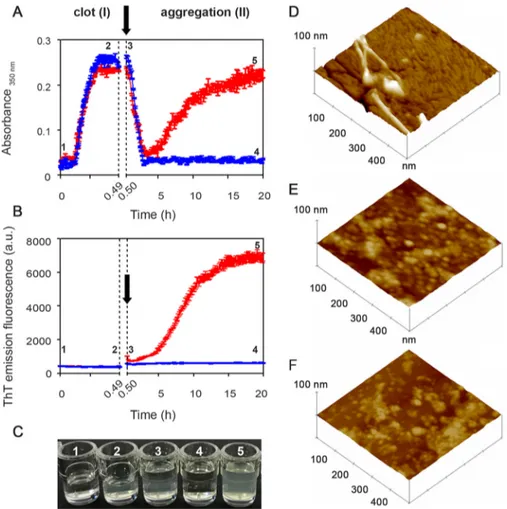
To study the amyloidogenicity of plasmin in a more physio-logical environment, we created a model fibrin clot on which fibrinolysis was initiated in the presence of either the highly amyloidogenic unstable S52P TTR variant or the superstable nonamyloidogenic T119M variant. Polymerization and depoly-merization were monitored by nonspecific light scattering at 350 nm (Fig. 4A) and by the specific spectrofluorimetric signal of ThT binding to amyloid fibrils (Fig. 4B).
In phase I, fibrinogen was converted into fibrin by addition of thrombin, monitored by the rapid increase in turbidity. Once the clot was formed, tissue plasminogen activator (tPA),
plas-Figure 2. Plasmin-mediated amyloid fibrillogenesis of S52P TTR. A, increase in ThT emission fluorescence for S52P TTR incubated in the presence of plasmin
compared with trypsin. No amyloid-specific ThT signal was seen after incubation of S52P TTR with thrombin, chymotrypsin, or proteinase K. B and C, negatively stained transmission electron micrographs of S52P TTR amyloid fibrils formed in the presence of trypsin (B) or plasmin (C). Scale bar, 100 nm. D, 15% SDS-PAGE under reducing conditions. M, marker proteins (14.4, 20.1, 30.0, 45.0, 66.0, and 97.0 kDa); lane 1, S52P TTR at time 0; lane 2, S52P TTR fibrils formed in the presence of trypsin; and lane 3, S52P TTR fibrils formed in the presence of plasmin. E, immunoblot analysis of samples separated in 15% SDS-PAGE (see lanes 1, 2, and 3 in D). Position of marker proteins at 15 and 10 kDa are indicated. F, inhibition by␣2-antiplasmin of fibril formation by S52P TTR mediated by 20 ng/l plasmin. The data were normalized to the ThT signal plateau in the samples without␣2-antiplasmin. Mean ⫾ S.D. of three replicates is shown. a.u., arbitrary units.
minogen, and TTR were gently layered on the clot surface, at the time point shown (arrow) inFig. 4, A and B, producing physiological fibrinolysis of the clot as indicated by the rapid decline in turbidity in phase II. When S52P TTR was present, the initial fall in light scattering was swiftly followed by a sharp rise that correlated with the appearance and increase in the ThT amyloid fibril signal (Fig. 4B). In the presence of T119M TTR, which is not susceptible to cleavage by plasmin and does not form amyloid fibrils (5) (Fig. 3), there was no secondary rise in turbidity and no ThT signal (Fig. 4,A–C).
Atomic force microscopy analysis of the reactants at the end of the experiment with the stable T119M or the pathogenic S52P variant TTR (that is 4 and 5 in Fig. 4, A–C) showed remarkably different structures, consistent with the spectrom-etry results. S52P TTR produced morphologically typical mature amyloid fibrils, 4 –7 nm in height (Fig. 4D) emerging from a thick layer of short fibrils. No fibrillar material was seen either with T119M TTR (Fig. 4E) or in the absence of any added TTR (Fig. 4F). Only single globular structures and short beaded chains were observed.
Discussion
The spectrum of systemic TTR amyloidosis comprises the many very rare hereditary forms caused by different mutations (8), the cardiac amyloidosis caused by the V122I variant in indi-viduals of African origin (9) and cardiac amyloidosis, mostly in elderly men, caused by WT TTR (10). Recent advances in imag-ing have shown that the latter is substantially more prevalent than previously recognized (11). There are no licensed treat-ments that arrest disease progression and TTR amyloidosis is thus an important unmet medical need. Current trials of TTR gene expression knockdown by experimental siRNA (12) and antisense oligonucleotide (ASO) drugs (13) have shown prom-ising results. However, elucidation of the mechanism underly-ing the in vivo transition of native, soluble, globular, tetrameric TTR into insoluble, polymeric, amyloid fibrils is crucial for understanding the natural history of the disease and for design of other effective therapies.
The influential original model of TTR denaturation and aggregation at low pH (14) demonstrated that tetramer disas-sembly is crucial, and that analogues of thyroxine, the natural ligand of TTR, can inhibit this process (15). The observations led to design, development, and clinical testing of tafamidis (16) and diflunisal (17), compounds that stabilize TTR against acid denaturation, for use as inhibitors of TTR amyloidogenesis, mimicking the trans-suppressive effect of the TTR-stabilizing T119M variant (2). Despite the capacity of tafamidis to increase the stability of TTR in plasma through the occupancy of just one of the two binding sites (18), its clinical use does not halt disease progression in a substantial proportion of patients (19). The limited clinical efficacy probably reflects the fact that the low pH model does not represent the actual pathophysiological mechanism of TTR amyloid fibrillogenesis. Indeed there is no relevant in vivo location in which TTR could be exposed to the acid conditions used in vitro.
We have recently demonstrated that specific proteolytic cleavage of the residue 48 – 49 bond in the flexible loop con-necting strands C and D, in just a single TTR protomer within the native tetrameric TTR assembly, causes rapid dissociation into cleaved and uncleaved protomers. Under physiological conditions in vitro, these then swiftly form abundant TTR fibrils, which are indistinguishable from ex vivo TTR amyloid fibrils (3, 5). The whole process occurs in the presence of phys-iological scale mechanical forces provided by stirring and by exposure to hydrophobic surfaces. Discovery of the critical role of proteolysis explains the almost universal presence of the TTR residue 49 –127 fragment in ex vivo TTR amyloid fibrils (20). Other features consistent with the mechano-enzymatic mechanism operating in vivo include the presence of a lag-phase preceding fibrillogenesis, and acceleration of fibril for-mation when preformed fibril seeds are present. We have also shown that binding of small ligands by the intact TTR tetramer significantly reduces its susceptibility to cleavage and aggrega-tion. However, maximum inhibition is only achieved by ligands that simultaneously occupy both the two binding sites and the central channel between them in the core of the TTR molecule (21).
A crucial question about the mechano-enzymatic mecha-nism has hitherto been the identity of the tryptic protease responsible for TTR amyloidosis in vivo. The present demon-stration of the efficacy of plasmin in vitro highlights it as an extremely plausible candidate. Other potent proteolytic enzymes were completely inactive in triggering TTR amyloid formation in vitro. Kallikrein-related peptidase 12, which has only very transient activity in vivo, did produce a small ThT signal of amyloid formation with S52P TTR, the most unstable and amyloidogenic TTR variant, but there was a long lag phase and very modest yield. Plasmin mediates the essential specific cleavage in TTR much more potently and, with clas-sical kinetic phases of nucleation and elongation, it generates abundant fibrils that are identical to ex vivo TTR amyloid fibrils. The relative lower activity of plasmin compared with trypsin cannot be easily explained. The remarkable self-di-gestion of plasmin, once activated, may reduce its activity and therefore delay the formation of the first fibrillar nuclei thus contributing to a reduced yield of fibrils. A complete
Figure 3. Plasmin-mediated fibrillogenesis. Relative ThT emission
fluores-cence intensities of TTR samples at 1 mg/ml after 25 h incubation with shak-ing in the presence of plasmin at an enzyme:substrate ratio of 1:50. Mean⫾ S.D. of three replicates is shown.
Plasmin primes TTR amyloidogenesis
characterization of the kinetics of all processes together with the determination of the TTR–plasmin structure should clarify the differences that we have observed. The several amyloidogenic TTR variants tested so far and the WT protein are all cleaved by plasmin, with varying efficiency replicating the findings with trypsin, whereas the stable, nonpathogenic, protective T119M variant is resistant. Fur-thermore, plasmin is ubiquitously and continuously acti-vated in vivo to provide for essential fibrinolysis on the vas-cular wall and also in the extracellular matrix, precisely where TTR amyloid is deposited.
The possible in vivo scenario of plasmin-mediated TTR fibrillogenesis is summarized inFig. 5. Plasminogen can be acti-vated by tPA within the clot and also by urokinase plasminogen activator (uPA) in the extracellular matrix. Sufficient proteoly-sis of the TTR tetramer by plasmin may then provide the critical concentration of both truncated and full-length TTR protom-ers required for nucleation of fibrils. Once nucleation has occurred, the elongation of fibrils can progress at lower concen-trations of monomers provided by either of the plasminogen activating pathways. Plasmin activity is finely regulated by
activators, including tPA and uPA, and inhibitors, including ␣2-plasmin inhibitor and plasminogen activator inhibitor. The conditions for critical TTR cleavage, sufficient to initiate amy-loidogenesis, may thus only arise rarely but there is certainly scope for variation in this complex system. For example, phys-iological fibrinolysis is notably affected by the intensity of nor-mal physical activity (22). All these features of plasmin, com-bined with the importance of mechanical forces, are consistent with the prevalence of TTR amyloid deposition in the heart and carpal tunnel, both of which are notable sites of continuous repetitive vigorous movement. The pathogenetic significance of plasmin also opens a broad and completely novel perspective for investigation of factors that may determine individual susceptibility and the natural history of the familial and acquired forms of TTR amyloidosis, including the initiation, progression, and tissue distribution of amyloid deposition. In addition, the wholly unexpected and surprising conflu-ence of the fibrinolysis pathway, the physiological remodel-ing of the extracellular matrix regulated by urokinase (23), and the pathogenesis of TTR amyloidosis are of considerable fundamental interest.
Figure 4. From fibrin to fibrils. A and B, spectrophotometric absorbance/light scattering at 350 nm (A) and amyloid-specific ThT emission fluorescence (B)
during clotting of fibrinogen to fibrin (phase I) followed by fibrinolysis in the presence of S52P TTR (red) or of the highly stable T119M TTR variant (blue) (phase II). Following fibrinolysis, increase in turbidity and ThT were observed in the presence of S52P TTR whereas neither of these signals increased when T119M TTR was present instead. Arrows indicate addition of tPA, plasminogen, and TTR. The results shown are the mean⫾ S.D. of three independent experiments. C, wells containing a solution of fibrinogen in the presence of 1) thrombin and 2) fibrin clot; 3) a solution of tPA, plasminogen, and TTR layered over the clot surface; 4) fibrinolysis with no further aggregation in the presence of T119M TTR; 5) fibrinolysis in the presence of S52P TTR showing the turbidity of amyloid fibril formation. D–F, surface plots of topographic tapping mode AFM images showing (D) the presence of fibrillar structures in the sample containing clot, tPA, plasminogen, and S52P TTR; (E and F) the presence of globular structures in samples containing clot, tPA, and plasminogen (E) in the presence of T119M TTR or (F) in the absence of any TTR isoform.
Experimental procedures
Reagents
Recombinant TTR variants were expressed and purified as described previously (21). Human fibrinogen was isolated from citrate-heparin–treated human plasma by affinity chroma-tography on recombinant clamping factor 221–559 frag-ment (24) and was absorbed with lysine-Sepharose 4B and gelatin-Sepharose 4B to remove traces of plasminogen and fibronectin, respectively. Enzymes purchased from Sigma-Aldrich were plasmin (P1867), proteinase K (P2308), chymo-trypsin (C2160000), tPA (T0831), plasminogen (SRP6518), thrombin (T7572), and tryptase (T7063). Trypsin was pur-chased from Promega (V5280) and recombinant human kal-likrein 12 from Biotechne (3095-S.E.). All the enzymes used were able to cleave the C-terminal end of Lys in theD
-Val-Leu-Lys 4-nitroanilide dihydrochloride peptide (Sigma-Al-drich, V0882) following the manufacturer’s instructions. All other reagents including ␣2-antiplasmin (SRP6313) were purchased from Sigma-Aldrich unless otherwise stated.
MEROPS database search
MEROPS (https://www.ebi.ac.uk/merops/) is a manually annotated database with information on more than 4000 pep-tidases classified according to families and clans. The residues of the proteolytic substrate are designated Pn—P4-P3-P2-P1- 储-P1⬘-P2⬘-P3⬘-P4⬘—Pm⬘ with 储 indicating the scissile bond. Substrate specificity was based on the frequency of Lys at position P1.
Proteolysis and fibrillogenesis of S52P TTR
Recombinant S52P TTR, 100l volumes at 0.5 mg/ml in 20 mMTris-HCl containing 150 mMNaCl, 5 mMCaCl2, pH 7.4,
containing 10MThT (25) was incubated at 37 °C in Costar 96-well black plates in the presence of a protease at an enzyme: substrate ratio of 1:50. Plasmin, trypsin, thrombin, proteinase K, chymotrypsin, tryptase alpha, and kallikrein 12 were tested. The plate was sealed with clear sealing film and subjected to 900 rpm double-orbital shaking. Bottom fluorescence was recorded at 500-s intervals (BMG LABTECH, FLUOstar Omega). Homogenous 15% SDS-PAGE (GE Healthcare) under reducing
Figure 5. From fibrin to fibrils. Shown is a cartoon of the putative flow of events leading to TTR amyloid fibril formation caused by plasmin cleavage within the
physiological scenario of fibrin formation and plasminogen activation. Circulating TTR can diffuse toward the extracellular compartment (blue arrow), be entrapped in the fibrin clot (green arrow), or escape from it (gray arrow). In the presence of activated plasminogen both in the presence of uPA (extracellular compartment) and tPA (within the clot), tetrameric TTR may be cleaved and then rapidly dissociate into a mixture of the truncated residue 49 –127 fragment (green) and full-length protomers (gray subunit). The released subunits may generate the fibrillar nuclei (highlighted within the red circle) that then aggregate into amyloid fibrils, which accumulate in the extracellular space. The legend at the bottom of the figure identifies all the TTR species.
Plasmin primes TTR amyloidogenesis
conditions was used to analyze protein composition before and after fibril formation. After electrophoretic separation, samples treated and untreated with trypsin or plasmin were blotted onto an activated PVDF membrane. Western blotting was developed with polyclonal sheep anti-human TTR (6g/ml, The Binding Site, United Kingdom/code AU066X) and poly-clonal rabbit anti-sheep peroxidase conjugate (1.3 g/ml, Dako, Denmark/code P0163) as primary and secondary anti-bodies, respectively. Peroxidase activity was visualized using a precipitating substrate containing 3,3⬘-diaminobenzidine and urea hydrogen peroxide (SigmaFAST DAB tablets, Sigma-Aldrich).
Effect of␣2-antiplasmin on TTR fibril formation
Recombinant S52P TTR in 200l volumes at 1 mg/ml in 20 mMTris-HCl at pH 7.5, containing 150 mMNaCl, 5 mMCaCl2,
10MThT, was incubated at 37 °C in sealed Costar 24-well
black-wall plates, together with 20 ng/l of plasmin while sub-jected to 900 rpm double-orbital shaking in the presence of 0.09, 0.18, 0.36, and 0.72M␣2-antiplasmin and in its absence.
Based on an average molecular mass of 55 kDa for plasmin, the selected inhibitor concentrations corresponded to molar ratios to plasmin of 0.25:1, 0.5:1, 1:1, and 2:1, respectively. ThT fluo-rescence emission was monitored using a BMG LABTECH FLUOstar Omega plate reader. Data were normalized to the ThT signal plateau reached in the samples without the plasmin inhibitor. All experiments were conducted in triplicate.
Fibrillogenesis of TTR variants and WT TTR
Recombinant S52P, V30M, L55P, V122I, WT and T119M TTR in 500l volumes at 1 mg/ml in 20 mMTris-HCl
contain-ing 150 mMNaCl, 5 mMCaCl2, 10MThT, pH 7.5 were
incu-bated at 37 °C in sealed Costar 24-well black-wall plates, together with 20 ng/l of plasmin while subjected to 900 rpm double-orbital shaking. ThT fluorescence emission was moni-tored until it reached a plateau. All experiments were con-ducted in triplicate.
Preparation of amyloid seeds from S52P TTR with plasmin
S52P TTR at 1 mg/ml in 20 mMTris-HCl, 150 mMNaCl, 5
mMCaCl2, 10 MThT, pH 7.5 was incubated at 37 °C with
plasmin at an enzyme:substrate ratio of 1:50 w/w in volume of 200l in a 96-well black-wall plate. The plate was sealed with clear sealing film, subjected to 900 rpm double-orbital shaking and bottom fluorescence was recorded (BMG LABTECH FLUOstar Omega). Aliquots of the final ThT positive material were stained with alkaline alcoholic Congo Red and viewed under high intensity cross-polarized light (26). Samples were also examined by EM (Joel1200EX) after negative staining with 2% uranyl acetate (3). After further blotting and drying in air, images were obtained at 80 kV.
Effect of seeds on plasmin-mediated S52P TTR fibrillogenesis
S52P TTR at 1 mg/ml in 20 mMTris-HCl, 150 mMNaCl, 5
mMCaCl2, 10 MThT, pH 7.5 was incubated at 37 °C with
plasmin at an enzyme:substrate ratio of 1:50 w/w in a volume of 200l in a 96-well black-wall plate. S52P TTR fibrils, prepared as described above, were added at the outset at 0.1 mg/ml to
three replicate wells; triplicate control well received addition of buffer alone. The plate was sealed with clear sealing film, sub-jected to 900 rpm double-orbital shaking, and bottom fluores-cence was recorded at 500-s intervals (BMG LABTECH FLUO-star Omega). Data were normalized to the highest value of ThT signal after subtraction of the fluorescence intensity attributa-ble to the added seeds alone.
Effect of plasmin on TTR amyloid fibrils
The concentration of S52P amyloid fibrils, produced as described above, was measured by bicinchoninic acid protein assay (Pierce). Fibrils at 0.1 mg/ml in 20 mMTris-HCl, 150 mM
NaCl, 5 mMCaCl2, 10MThT, pH 7.5 in 200l aliquots per
well were incubated at 37 °C in sealed Costar 96-well black-wall plates in the presence or absence of plasmin at an enzyme:sub-strate ratio of 1:50 w/w, with agitation as above. Bottom ThT fluorescence was monitored at 500-s intervals as before in three replicate test and control wells.
Fibrinolysis and/or fibril formation
The two-stage procedure comprised clot formation in phase I followed by fibrinolysis and potential amyloid fibrillogenesis in phase II. The experiments were conducted in Costar 96-well black plates at 37 °C using a multimode plate reader (BMG LABTECH FLUOstar Omega) to monitor either changes in tur-bidity at 350 nm or ThT emission fluorescence. Fibrin polymer-ization was initiated by adding thrombin (0.5 NIH units/ml) to 1Mhuman fibrinogen in 20 mMTris-HCl, pH 7.5 containing
150 mMNaCl and 5 mMCaCl2(buffer A) in a total volume of
100l per well. Clot formation was monitored by recording the turbidity at 350 nm at 20-s intervals. After turbidity had reached a stable level (usually within 30 min), 100l of a solu-tion containing tPA, plasminogen, and TTR in buffer A were gently layered on top of the fibrin clot to a final concentration of 0.027M, 1M, and 18M, respectively. The microtiter plate was then sealed with clear sealing film, subjected to 900 rpm double-orbital shaking and absorbance at 350 nm was mea-sured at 500-s intervals at 37 °C. Blank subtraction and a cor-rection based on the volume per well and the microplate dimensions were used to normalize all absorption values to 1-cm path length. ThT at 10Mwas present throughout.
Bot-tom fluorescence was recorded at 20-s intervals for 30 min in stage I; after addition of tPA, plasminogen, and TTR in stage II, the ThT signal was monitored at 500-s intervals for 20 h. Pellets harvested at the end of stage II by centrifugation at 10,600⫻ g for 20 min were further analyzed by negative staining EM, light microscopy after alkaline alcoholic Congo Red staining as described above (26), and atomic force microscopy (AFM).
Atomic force microscopy
Pellets harvested at the end of phase II were resuspended in water and 100-fold diluted; 10l aliquots of the diluted samples were deposited on freshly cleaved mica and dried under mild vacuum. Samples in which no pellet was present were diluted and deposited as described above, but after drying they were rinsed with water to remove excess salts. Tapping mode AFM images were acquired in air using a multimode scanning probe microscope equipped with an “E” scanning head (maximum
scan size, 10m) and driven by a Nanoscope V controller (Digital Instruments, Bruker). Single-beam uncoated silicon cantilevers (type OMCL-AC160TS, Olympus and TESPA_ V2, Bruker) were used. The drive frequency was between 260 and 310 kHz; the scan rate was 0.25– 0.5 Hz.
Author contributions—P. P. M., G. V., and A. C. data curation; P. P. M., G. V., A. C., J. M., D. C., S. G., S. R., M. S., M. E., A. R., C. C., M. V., L. M., G. F., L. O., G. W. T., and V. B. investigation; P. P. M., G. V., A. C., J. M., D. C., S. G., S. R., M. E., A. R., C. C., L. M., and G. F. methodology; P. P. M. and V. B. writing-original draft; G. V., A. C., J. M., D. C., M. V., P. N. H., G. W. T., J. D. G., and M. B. P. formal analysis; G. V. validation; A. C., M. S., L. O., P. N. H., J. D. G., M. B. P., and V. B. conceptualization; G. W. T. and M. B. P. writing-review and editing; M. B. P. and V. B. funding acquisition; V. B. supervision.
Acknowledgments—We thank Dr. Giampiero Pietrocola (Depart-ment of Molecular Medicine, University of Pavia) for providing human fibrinogen.
References
1. Pepys, M. B. (2009) A molecular correlate of clinicopathology in transthy-retin amyloidosis. J. Pathol. 217, 1–3CrossRef Medline
2. Hammarström, P., Schneider, F., and Kelly, J. W. (2001) Trans-suppres-sion of misfolding in an amyloid disease. Science 293, 2459 –2462
CrossRef Medline
3. Mangione, P. P., Porcari, R., Gillmore, J. D., Pucci, P., Monti, M., Porcari, M., Giorgetti, S., Marchese, L., Raimondi, S., Serpell, L. C., Chen, W., Relini, A., Marcoux, J., Clatworthy, I. R., Taylor, G. W., et al. (2014) Pro-teolytic cleavage of Ser52Pro variant transthyretin triggers its amyloid fibrillogenesis. Proc. Natl. Acad. Sci. U.S.A. 111, 1539 –1544CrossRef Medline
4. Klimtchuk, E. S., Prokaeva, T., Frame, N. M., Abdullahi, H. A., Spencer, B., Dasari, S., Cui, H., Berk, J. L., Kurtin, P. J., Connors, L. H., and Gursky, O. (2018) Unusual duplication mutation in a surface loop of human transthy-retin leads to an aggressive drug-resistant amyloid disease. Proc. Natl. Acad. Sci. U.S.A. 115,E6428 –E6436CrossRef Medline
5. Marcoux, J., Mangione, P. P., Porcari, R., Degiacomi, M. T., Verona, G., Taylor, G. W., Giorgetti, S., Raimondi, S., Sanglier-Cianferani, S., Benesch, J. L., Cecconi, C., Naqvi, M. M., Gillmore, J. D., Hawkins, P. N., Stoppini, M., Robinson, C. V., Pepys, M. B., and Bellotti, V. (2015) A novel mechano-enzymatic cleavage mechanism underlies transthyretin amyloidogenesis. EMBO Mol. Med. 7,1337–1349CrossRef Medline
6. Rawlings, N. D., Barrett, A. J., and Finn, R. (2016) Twenty years of the MEROPS database of proteolytic enzymes, their substrates and inhibitors. Nucleic Acids Res. 44,D343–D350CrossRef Medline
7. Tucker, H. M., Kihiko, M., Caldwell, J. N., Wright, S., Kawarabayashi, T., Price, D., Walker, D., Scheff, S., McGillis, J. P., Rydel, R. E., and Estus, S. (2000) The plasmin system is induced by and degrades amyloid-beta ag-gregates. J. Neurosci. 20, 3937–3946CrossRef Medline
8. Benson, M. D. (2003) The hereditary amyloidoses. Best Pract. Res. Clin. Rheumatol. 17,909 –927CrossRef Medline
9. Jacobson, D. R., Pastore, R. D., Yaghoubian, R., Kane, I., Gallo, G., Buck, F. S., and Buxbaum, J. N. (1997) Variant-sequence transthyretin (isoleu-cine 122) in late-onset cardiac amyloidosis in black Americans. N. Engl. J. Med. 336,466 – 473CrossRef Medline
10. Ruberg, F. L., and Berk, J. L. (2012) Transthyretin (TTR) cardiac amyloi-dosis. Circulation 126, 1286 –1300CrossRef Medline
11. Gillmore, J. D., Maurer, M. S., Falk, R. H., Merlini, G., Damy, T., Dispen-zieri, A., Wechalekar, A. D., Berk, J. L., Quarta, C. C., Grogan, M., Lach-mann, H. J., Bokhari, S., Castano, A., Dorbala, S., Johnson, G. B., et al.
(2016) Nonbiopsy diagnosis of cardiac transthyretin amyloidosis. Circu-lation 133,2404 –2412CrossRef Medline
12. Adams, D., Suhr, O. B., Dyck, P. J., Litchy, W. J., Leahy, R. G., Chen, J., Gollob, J., and Coelho, T. (2017) Trial design and rationale for APOLLO, a Phase 3, placebo-controlled study of patisiran in patients with hereditary ATTR amyloidosis with polyneuropathy. BMC Neurol. 17, 181CrossRef Medline
13. Benson, M. D., Waddington Cruz, M., Wang, A., Polydefkis, M., Plante-Bordeneuve, V., Berk, J., Barroso, F., Adams, D., Dyck, P., Brannagan, T., Whelan, C., Merlini, G., Scheinberg, M., Drachman, B., Heitner, S., et al. (2017) Safety and efficacy of inotersen in patients with hereditary tran-sthyretin amyloidosis with polyneuropathy (hATTR-PN). Orphanet J. Rare Dis. 12,Suppl. 1, 165
14. Colon, W., and Kelly, J. W. (1992) Partial denaturation of transthyretin is sufficient for amyloid fibril formation in vitro. Biochemistry 31, 8654 – 8660CrossRef Medline
15. Miroy, G. J., Lai, Z., Lashuel, H. A., Peterson, S. A., Strang, C., and Kelly, J. W. (1996) Inhibiting transthyretin amyloid fibril formation via protein stabilization. Proc. Natl. Acad. Sci. U.S.A. 93, 15051–15056CrossRef Medline
16. Coelho, T., Maia, L. F., Martins da Silva, A., Waddington Cruz, M., Planté-Bordeneuve, V., Lozeron, P., Suhr, O. B., Campistol, J. M., Conceição, I. M., Schmidt, H. H., Trigo, P., Kelly, J. W., Labaudinière, R., Chan, J., Packman, J., Wilson, A., and Grogan, D.R. (2012) Tafamidis for transthy-retin familial amyloid polyneuropathy: A randomized, controlled trial. Neurology 79,785–792CrossRef Medline
17. Berk, J. L., Suhr, O. B., Obici, L., Sekijima, Y., Zeldenrust, S. R., Yamashita, T., Heneghan, M. A., Gorevic, P. D., Litchy, W. J., Wiesman, J. F., Nordh, E., Corato, M., Lozza, A., Cortese, A., Robinson-Papp, J., et al. (2013) Repurposing diflunisal for familial amyloid polyneuropathy: A random-ized clinical trial. JAMA 310, 2658 –2667CrossRef Medline
18. Bulawa, C. E., Connelly, S., Devit, M., Wang, L., Weigel, C., Fleming, J. A., Packman, J., Powers, E. T., Wiseman, R. L., Foss, T. R., Wilson, I. A., Kelly, J. W., and Labaudinière, R. (2012) Tafamidis, a potent and selective tran-sthyretin kinetic stabilizer that inhibits the amyloid cascade. Proc. Natl. Acad. Sci. U.S.A. 109,9629 –9634CrossRef Medline
19. Planté-Bordeneuve, V., Gorram, F., Salhi, H., Nordine, T., Ayache, S. S., Le Corvoisier, P., Azoulay, D., Feray, C., Damy, T., and Lefaucheur, J. P. (2017) Long-term treatment of transthyretin familial amyloid polyneu-ropathy with tafamidis: A clinical and neurophysiological study. J. Neurol.
264,268 –276CrossRef Medline
20. Ihse, E., Rapezzi, C., Merlini, G., Benson, M. D., Ando, Y., Suhr, O. B., Ikeda, S., Lavatelli, F., Obici, L., Quarta, C. C., Leone, O., Jono, H., Ueda, M., Lorenzini, M., Liepnieks, J., et al. (2013) Amyloid fibrils containing fragmented ATTR may be the standard fibril composition in ATTR am-yloidosis. Amyloid 20, 142–150CrossRef Medline
21. Verona, G., Mangione, P. P., Raimondi, S., Giorgetti, S., Faravelli, G., Por-cari, R., Corazza, A., Gillmore, J. D., Hawkins, P. N., Pepys, M. B., Taylor, G. W., and Bellotti, V. (2017) Inhibition of the mechano-enzymatic amy-loidogenesis of transthyretin: Role of ligand affinity, binding cooperativity and occupancy of the inner channel. Sci. Rep. 7, 182CrossRef Medline
22. Weiss, C., Seitel, G., and Bärtsch, P. (1998) Coagulation and fibrinolysis after moderate and very heavy exercise in healthy male subjects. Med. Sci. Sports Exerc. 30,246 –251Medline
23. Smith, H. W., and Marshall, C. J. (2010) Regulation of cell signalling by uPAR. Nat. Rev. Mol. Cell Biol. 11, 23–36CrossRef Medline
24. Liu, C. Z., Cheng, H. J., and Chang, L. Y. (2008) A new feasible method for fibrinogen purification based on the affinity of Staphylococcus aureus clumping factor A to fibrinogen. Protein Expr. Purif. 61, 31–35CrossRef Medline
25. Naiki, H., Higuchi, K., Hosokawa, M., and Takeda, T. (1989) Fluorometric determination of amyloid fibrils in vitro using the fluorescent dye, thiofla-vin T1. Anal. Biochem. 177, 244 –249CrossRef Medline
26. Puchtler, H., Waldrop, F. S., and Meloan, S. N. (1985) A review of light, polarization and fluorescence microscopic methods for amyloid. Appl. Pathol. 3,5–17Medline