HAL Id: hal-01813882
https://hal.umontpellier.fr/hal-01813882
Submitted on 13 Jun 2018
HAL is a multi-disciplinary open access
archive for the deposit and dissemination of
sci-entific research documents, whether they are
pub-lished or not. The documents may come from
teaching and research institutions in France or
abroad, or from public or private research centers.
L’archive ouverte pluridisciplinaire HAL, est
destinée au dépôt et à la diffusion de documents
scientifiques de niveau recherche, publiés ou non,
émanant des établissements d’enseignement et de
recherche français ou étrangers, des laboratoires
publics ou privés.
Contamana, Peruvian Amazonia
Myriam Boivin, Laurent Marivaux, Maeva J. Orliac, François Pujos, Rodolfo
Salas-Gismondi, Julia Victoria Tejada-Lara, Pierre Olivier Antoine
To cite this version:
Myriam Boivin, Laurent Marivaux, Maeva J. Orliac, François Pujos, Rodolfo Salas-Gismondi, et
al.. Late middle Eocene caviomorph rodents from Contamana, Peruvian Amazonia. Palaeontologia
Electronica, Coquina Press, 2017, 20 (1), pp.19A. �10.26879/742�. �hal-01813882�
palaeo-electronica.org
http://zoobank.org/540D23AA-F705-4A05-8E10-FADAD3356D9C
Boivin, Myriam, Marivaux, Laurent, Orliac, Maëva J., Pujos, Francois, Salas-Gismondi, Rodolfo, Tejada-Lara, Julia V., and Antoine, Pierre-Olivier. 2017. Late middle Eocene caviomorph rodents from Contamana, Peruvian Amazonia. Palaeontologia Electronica 20.1.19A: 1-50
palaeo-electronica.org/content/2017/1822-eocene-amazonian-caviomorphs
Late middle Eocene caviomorph rodents from
Contamana, Peruvian Amazonia
Myriam Boivin, Laurent Marivaux, Maëva J. Orliac, Francois Pujos,
Rodolfo Salas-Gismondi, Julia V. Tejada-Lara, and Pierre-Olivier Antoine
ABSTRACT
Caviomorph rodents represent one of the most successful groups of placental mammals from South America. Despite their modern, Neogene and late Paleogene high diversity, their early evolutionary history has long remained obscure. Recent field expeditions in Peruvian Amazonia have yielded among the earliest representatives of that group, in deposits dating from the late middle Eocene (Contamana, CTA-27 local-ity, ~41 Ma). Here, we provide an exhaustive analysis of the rodent material from CTA-27 and from new sub-coeval localities discovered in the same area and geological for-mation (Pozo Forfor-mation): CTA-47, CTA-51, CTA-73, CTA-66, and CTA-29. A total of 20 rodent taxa are identified in these localities, among which one from CTA-29 (Pozomys
ucayaliensis gen. et sp. nov.) remains with uncertain suprafamilial affinities.
Addition-ally, the material of CTA-27 previously attributed to Eobranisamys sp. is assigned here to the new species Eobranisamys javierpradoi. In terms of taxonomic composition, Eocene localities from Contamana area have many taxa in common (Cachiyacuy,
Canaanimys, Eobranisamys, and Eoespina, or very close relatives). These Eocene
assemblages are clearly distinct from Oligocene ones, mostly documented at mid and high latitudes. In contrast, they share some affinities with the late Eocene-earliest Oli-gocene Santa Rosa locality (Peruvian Amazonia), from which the two genera
Eobrani-samys and Eoespina were originally described. This faunal closeness might more
reflect biogeographic affinities than contemporaneity. In addition, the occlusal pattern of some upper molars of Eosallamys from Santa Rosa recalls that of Cachiyacuy and
Canaanimys. These low-latitude caviomorph assemblages provide new insights into
the early evolutionary history, biogeography, and paleodiversity of that group.
Myriam Boivin. Laboratoire de Paléontologie, Institut des Sciences de l’Évolution de Montpellier (ISE-M, UMR 5554, CNRS/UM/IRD/EPHE), c.c. 064, Université de Montpellier, place Eugène Bataillon, F-34095 Montpellier Cedex 05, France. [email protected]
Laurent Marivaux. Laboratoire de Paléontologie, Institut des Sciences de l’Évolution de Montpellier (ISE-M, UMR 5554, CNRS/UM/IRD/EPHE), c.c. 064, Université de Montpellier, place Eugène Bataillon, F-34095 Montpellier Cedex 05, France. [email protected]
UMR 5554, CNRS/UM/IRD/EPHE), c.c. 064, Université de Montpellier, place Eugène Bataillon, F-34095 Montpellier Cedex 05, France. [email protected]
Francois Pujos. Instituto Argentino de Nivología, Glaciología y Ciencias Ambientales (IANIGLA), CCT– CONICET–Mendoza, Av. Ruiz Leal s/n, Parque Gral. San Martín, 5500 Mendoza, Argentina.
Rodolfo Salas-Gismondi. Departamento de Paleontología de Vertebrados, Museo de Historia Natural – Universidad Nacional Mayor San Marcos (MUSM), Av. Arenales 1256, Lima 11, Peru.
Julia V. Tejada-Lara. Departamento de Paleontología de Vertebrados, Museo de Historia Natural – Universidad Nacional Mayor San Marcos (MUSM), Av. Arenales 1256, Lima 11, Peru, and Department of Earth and Environmental Sciences, Columbia University in the City of New York and Division of
Paleontology, American Museum of Natural History, Central Park West at 79th Street, New York, NY 10024, USA. [email protected]
Pierre-Olivier Antoine. Laboratoire de Paléontologie, Institut des Sciences de l’Évolution de Montpellier (ISE-M, UMR 5554, CNRS/UM/IRD/EPHE), c.c. 064, Université de Montpellier, place Eugène Bataillon, F-34095 Montpellier Cedex 05, France. [email protected]
Keywords: Barrancan; South America; systematics; biostratigraphy; new genus; new species Submission: 3 November 2016. Acceptance: 5 April 2017
INTRODUCTION
The caviomorph rodents (Caviomorpha Wood, 1955) constitute one of the main placental mammal groups of South America. They are repre-sented nowadays by four superfamilies (Erethizon-toidea Bonaparte, 1845 [New World porcupines], Octodontoidea Waterhouse, 1839 [spiny rats and their kin], Chinchilloidea Bennett, 1833 [chinchillas and their kin], and Cavioidea Fischer de Waldheim, 1817 [guinea pig and their kin]), including a total of 13 families (e.g., Wilson and Reeder, 2005). Their modern specific richness amounts to ~11% of the rodent diversity (255/2277; Wilson and Reeder, 2005). Moreover, caviomorphs show a wide mor-phological disparity, many dietary and locomotory adaptations, and they occupy diverse environ-ments (e.g., Mares and Ojeda, 1982; Patton et al., 2015). This rodent group, documented in the fossil record of South America since the late middle Eocene (Antoine et al., 2012), also shows a great past diversity (e.g., Woods, 1984; Candela and Picasso, 2008; Rinderknecht and Blanco, 2008).
Until recently, the oldest fossil occurrence of caviomorphs in South America was recorded from one locality of Peruvian Amazonia: CTA-27 from Contamana (Loreto Department; Antoine et al., 2012). This locality was dated at ~41 Ma (late mid-dle Eocene, Barrancan South American Land Mammal Age [SALMA]) by radiometric Ar/Ar analy-ses (Antoine et al., 2012). On the basis of dental remains, five taxa were described from this locality,
three were new to science: Cachiyacuy
contaman-ensis Antoine et al., 2012, Cachiyacuy kummeli
Antoine et al., 2012, and Canaanimys maquiensis Antoine et al., 2012. These taxa are particularly interesting because they display a suite of primitive dental characters (for caviomorphs), which are oth-erwise found only in coeval African phiomorphs (Antoine et al., 2012; Barbière and Marivaux, 2015). This striking morphological affinity strength-ens support for the hypothesis of the African origin of the caviomorph clade. Another locality from Peruvian Amazonia as well, Santa Rosa (Ucayali Department), originally considered as ?late Eocene in age (Frailey and Campbell, 2004), has yielded a rich rodent assemblage (17 taxa), among which the earliest representatives of three cavio-morph superfamilies: Erethizontoidea, Octodon-toidea, and Cavioidea. However, the late Eocene age of the Santa Rosa locality proposed by Camp-bell (2004) is highly questioned, with some other authors advocating the possibility of a younger age (i.e., earliest Oligocene or later; Shockey et al., 2004; Croft et al., 2009; Antoine et al., 2012; Kay, 2015; Antoine et al., 2017).
Recently, Antoine et al. (2016) published a noteworthy paleontological and geological survey of the Cenozoic deposits along the Quebrada Cachiyacu near Contamana in Peruvian Amazo-nia. Of the 19 caviomorph-bearing localities identi-fied by these authors in a single section, 10 are Eocene in age (middle Eocene–late middle Eocene; including CTA-27). Three of these
locali-ties (CTA-47, CTA-51, and CTA-73; Figure 1.2–3) are stratigraphically below CTA-27, while the oth-ers (CTA-66, CTA-52, CTA-50, CTA-41, CTA-53, and 29; Figure 1.2-3) are located above CTA-27 in the same section. In this paper, we 1) provide an exhaustive analysis (description and compari-son) and a revision of the rodent material from CTA-27; and 2) describe the material from other late middle Eocene localities of Contamana (CTA-47, CTA-51, CTA-73, CTA-66, and CTA-29). The study of these caviomorph assemblages, which are
the oldest known at a South American scale, thus allows furthering our understanding of the early evolutionary history and paleodiversity of that group during its earliest adaptive radiation.
MATERIAL AND METHODS
The rodent fossils described in the present work come from six Eocene localities of Contam-ana (Loreto department, Peru; Figure 1.1): 47, 51, 73, 27, 66, and
CTA-FIGURE 1. Geographic location and Cenozoic stratigraphy of the Contamana area, Peru. 1, location map of the Con-tamana area in Peruvian Amazonia (Loreto Department). 2-3, synthetic stratigraphic units of the complete ConCon-tamana Cenozoic sequence along the Cachiyacu stream (modified after Antoine et al., 2016, figure 3), including fossil-bearing levels, among which Eocene rodent-yielding localities CTA-47, CTA-51, CTA-73, CTA-27, CTA-66, and CTA-29. Note also in the same section, the location of the other rodent-bearing localities, designated by an asterisk, in the Pozo, Chambira and Pebas Fm.; 2, NE Flank of the Maquía Anticline; 3, SW Flank of the Maquía Anticline. Modified from Antoine et al. (2012, 2016). Fm., Formation.
29. The four other Eocene caviomorph-bearing localities from the same section (CTA-52, CTA-50, CTA-41, and CTA-53; Antoine et al., 2016) have been excluded because they only yielded frag-ments of rodent incisors, unidentifiable at a family level or below. CTA-47 (7°19’52.1”S, 74°57’4.6”W), CTA-51 (7°19’46.1”S, 74°56’57.1”W), CTA-27 (7°19’48.4”S, 74°56’50.4”W), and CTA-29 (7°19’42.9”S, 74°56’44”W) are situated on the northeastern flank of the Maquía Anticline, whereas CTA-73 (7°20’29.8”S, 74°57’8.7”W) and CTA-66 (7°20’29.8”S, 74°57’15.5”W) are located on the southwestern flank of that anticline (Antoine et al., 2016, figure 2.A). Following Antoine et al. (2016, figure 3), all these localities are referred to the lower member of the Pozo Formation (“Pozo Sands”; Figure 1.2-3) and correspond to scattered inframetric channelized sandstone lenses (Antoine et al., 2016, figure 5.A-B). At CTA-51, CTA-27, CTA-29, and CTA-66, these sandstones are mas-sive, gray to yellowish, and unconsolidated. They include millimetric calcareous nodules of diage-netic origin (Antoine et al., 2016, figure 5.D), locally encrusting fossil elements (charophyte oogonia, crab claws, fish scales, and vertebrate teeth and bones). CTA-29 consists in a red-brown tuffaceous silty sand dated at 43.44 ± 2.5 Ma by Ar/Ar on bio-tites (late middle Eocene, Barrancan SALMA; Antoine et al., 2012, 2016; Figure 1.2). The Eocene localities of Contamana have yielded a wide array of aquatic and terrestrial organisms (plants, crabs, molluscs, chondrichthyans, osteich-thyans, frogs, turtles, snakes, and crocodylo-morphs), including numerous terrestrial mammals (allotherian, metatherians, xenarthrans, South American native ungulates [i.e., astrapothere, litop-tern, notungulates, and pyrotheres], and rodents). In addition, CTA-27 and CTA-66 have also yielded bat teeth (Antoine et al., 2016). In the Contamana section, fossil material of each locality was col-lected by screen-washing (1 mm mesh): ~205 kg of sediment for CTA-47 (in 2010 and 2011), 97 kg of sediment for CTA-51 (in 2010 and 2011), 11 kg of sediment for CTA-73 (2014), 665 kg of sediment for CTA-27 (from 2008 to 2010), 31 kg of sediment for CTA-66 (in 2013 and 2014), and 270 kg of sedi-ment for CTA-29 (from 2011 to 2013). During the last field expedition in the Contamana area (August 2016), our team regrettably noted the irremediable loss of several concerned localities (51, CTA-27, CTA-73, and CTA-29), either due to landslides, erosion, and silting caused by flooding events that have occurred since 2014 (Appendix 1).
All field missions were performed as part of an International Specific Agreement elaborated between the “Museo de Historia Natural, Universi-dad Nacional Mayor de San Marcos,” Lima, Peru (MUSM) and the “Université de Montpellier” (Insti-tut des Sciences de Montpellier [ISE-M]). The fossil material described in this paper is permanently housed in the paleontological collections of the MUSM.
The studied material was identified prelimi-narily in Antoine et al. (2016). As some determina-tions have evolved since then, synonymies are specified. The terminology for rodent dentition (Fig-ures 2, 3) follows the nomenclature of Boivin et al. (2017) based on Wood and Wilson (1936), Fields (1957), Marivaux et al. (2004, 2017), and Antoine et al. (2012). Lower case letters are used for the lower dentition (i.e., dp, for decidual premolar; p, for premolar; m, for molar) and upper case letters for the upper dentition (i.e., dP, for decidual premo-lar; P, for premopremo-lar; M, for molar). The caviomorph taxa used for comparison in this study are listed in Appendix 2. When fossils from the Eocene locali-ties of Contamana are compared with several taxa, the latter are primarily listed according to their chronostratigraphic order (from the oldest to the latest) and then alphabetically if they are coeval. All measurements are given in mm (Appendix 3). Two ratios, HIg and Hlb, deriving from the hypsodonty indice of Janis (1986), have been calculated from the maximum lingual crown height (Hg) and the labial height (Hb) of the less worn teeth available, respectively. As the hypsodonty indices of mea-sured teeth are inferior to 1, most specimens can be considered as brachydont. Depending on speci-mens, photographs were taken with two scanning electron microscopes (SEM): HITACHI S 4000 and HITACHI S 4800.
Institutional Abbreviations
LACM, Los Angeles County Museum, Los Ange-les, USA; MACN, Museo Argentino de Ciencias Naturales “Bernardino Rivadavia”, Buenos Aires, Argentina; MLP, Museo de Ciencias Naturales de La Plata, La Plata, Argentina; MNHN, Musée national d'Histoire naturelle, Paris, France; MUSM, Museo de Historia Natural de la Universidad Nacional Mayor San Marcos, Lima, Peru; UM, Uni-versité de Montpellier, Montpellier, France.
Other Abbreviations
Hb, maximum labial crown height; Hg, maximum lingual crown height; HIb, hypsodonty index calcu-lated from Hb; HIg, hypsodonty index calcucalcu-lated
from Hg; ML, maximum anteroposterior length; MW, maximum transversal width; S, south; SALMA, South American Land Mammal Age; W, west.
SYSTEMATIC PALEONTOLOGY Nomenclatural Remark
The new species and genera described below must be referred to Boivin, 2017, following the arti-cle 50.1 and the “recommendation 50A concerning
multiple authors” of the International Code of Zoo-logical Nomenclature (ICZN, 1999: 52, 182).
Order RODENTIA Bowdich, 1821 Infraorder HYSTRICOGNATHI Tullberg, 1899
Parvorder CAVIOMORPHA Wood, 1955 Superfamily CAVIOIDEA Fischer de Waldheim,
1817
Genus EOBRANISAMYS Frailey and Campbell, 2004
Type species. Eobranisamys romeropittmanae Frailey and Campbell, 2004.
FIGURE 2. Dental nomenclature for upper teeth in occlusal view. 1, upper molar; 2, P4; 3, dP4. 1, paracone; 2, proto-cone; 3, metaproto-cone; 4, hypoproto-cone; 5, parastyle; 6, mesostyle; 7, anteroloph; 8, anterior arm of the protoproto-cone; 9, poste-rior arm of the protocone (= lingual protoloph); 10, posteposte-rior outgrowth of the protocone; 11, protoloph (= labial protoloph); 12, mure; 13, third transverse crest (= central transverse crest); 14, mesolophule; 15, mesoloph; 16, ante-rior arm of the hypocone; 17, metaloph; 18, posteroloph; 19, paraflexus; 20, hypoflexus/hypofossette; 21, confluence of the paraflexus with the hypoflexus; 22, mesoflexus/mesofossette; 23, metaflexus; 24, confluence of the metaflexus with the posteroflexus; 25, posteroflexus. Based on observations made on the new material, the dental terminology is modified after Wood and Wilson (1936), Fields (1957), Marivaux et al. (2004, 2017) and Antoine et al. (2012).
Species content. The type species mentioned above and Eobranisamys riverai Frailey and Campbell, 2004; Eobranisamys javierpradoi nov. sp. (this work).
Geographic and stratigraphic distribution. Con-tamana (CTA-27 and CTA-66 localities), Pozo For-mation, lower member (late middle Eocene), Loreto department, Peru; Santa Rosa (LACM 6289 locality), “Yahuarango Formation” (?late Eocene/ early Oligocene), Ucayali department, Peru.
Emended generic diagnosis. Occlusal pattern similar to that of Branisamys by its tetralophodont and non-taeniodont lower molars; pentalophodont and taeniodont dP4s and upper molars with a strong third transverse crest and metaloph, which is lingually connected to the posteroloph.
Eobranis-amys differs from BranisEobranis-amys in having a lower
crown, thin loph(-id)s, and identifiable cusp(-id)s (modified after Frailey and Campbell, 2004, p. 79).
FIGURE 3. Dental nomenclature for lower teeth in occlusal view. 1, lower molar; 2, p4; 3, dp4. 1, protoconid; 2, metaconid; 3, mesoconid; 4, entoconid; 5, hypoconid; 6, mesostylid; 7, metalophulid I; 8, posterior arm of the metaco-nid; 9, posterior arm of the protocometaco-nid; 10, neomesolophid; 11, second transverse cristid; 12, mesolophid; 13, rest of the mesolophid?; 14, ectolophid; 15, mesial ectolophid; 16, distal ectolophid; 17, hypolophid; 18, anterior arm of the entoconid; 19, posterior arm of the entoconid; 20, anterior arm of the hypoconid; 21, posterior arm of the hypoconid; 22, anterior outgrowth of the hypoconid; 23, posterolophid; 24, anteroflexid/anterofossettid; 25, mesoflexid; 26, mesial mesoflexid; 27, distal mesoflexid; 28, confluence of the anteroflexid with the mesoflexid; 29, hypoflexid; 30, metaf-lexid; 31, confluence of the hypoflexid with the metaflexid. Based on observations made on the new material, the den-tal terminology is modified after Wood and Wilson (1936), Fields (1957), Marivaux et al. (2004, 2017) and Antoine et al. (2012).
Eobranisamys javierpradoi sp. nov.
Figure 4.7-8, Appendix 3
zoobank.org/54337122-7076-48E5-81CC-83D8A1435825
2012 Eobranisamys sp. Antoine et al., p. 1321–1322. 2016 Eobranisamys sp. Antoine et al.,
Supplemen-tary data, p. 7.
2017 Eobranisamys sp. Antoine et al., Supplemen-tary data, p. 9.
Etymology. In honour of Javier Prado, who founded the “Museo de Historia Natural, Universi-dad Mayor de San Marcos” in Lima, Peru, in 1918. Holotype. MUSM 1897, left M1 (in Antoine et al., 2012, figure 2b’). Deposited in the Museo de Histo-ria Natural de la Universidad Nacional Mayor de San Marcos”, Lima, Peru.
Referred material. In addition to the holotype (MUSM 1897) ̶̶ MUSM 2795, left m1 or m2; MUSM 1898, right fragmentary m2 (in Antoine et al., 2012, figure 2c’); MUSM 1899, left m3 (in Antoine et al., 2012, figure 2d’); MUSM 2796, right m3; MUSM 2797, right dP4 (Figure 4.7); MUSM 1896 (in Antoine et al., 2012, figure 2a’) and 2798, left P4s; MUSM 2799, right M1 or M2; MUSM 2800, left M1; MUSM 2801, left M3 (Figure 4.8).
Type locality. Contamana CTA-27, Loreto Depart-ment, Peru.
Formation and age. Pozo Formation, lower mem-ber, late middle Eocene (Antoine et al., 2012, 2016).
Diagnosis. Smallest species of Eobranisamys (about half the size of E. romeropittmanae and 20% smaller than E. riverai), showing sharper crests, more salient cusps, and longer M3 with a less rounded occlusal outline than other referred species.
Description. The lower molars are tetralophodont (in Antoine et al., 2012, figure 2c’–d’). Mesially, there is no trace of anterocingulid at the base of the crowns. On MUSM 1899 (m3; in Antoine et al., 2012, figure 2d’), the protoconid is the largest cus-pid of the tooth. On MUSM 1899 and 2795 (m1 or 2; in Antoine et al., 2012, figure 2c’), the metaconid is situated more mesially than the protoconid, with a strong and high posterior arm. The protoconid and metaconid are connected by a strong and complete metalophulid I. On m3, the second trans-verse cristid is strong and complete. Labially, this cristid starts from a well-defined and oblique (disto-lingually oriented) posterior arm of the protoconid, then it makes an angle and runs lingually to reach the distal extremity of a long posterior arm of the metaconid. The same is true on m1-2s. On MUSM 1898 (in Antoine et al., 2012, figure 2c’), a notice-able angulation in the middle of this cristid could
correspond to the junction of two cristids. Further-more, on this tooth, the lingual connection of this second cristid could be a mesostylid. So, the ques-tion remains as to whether this second cristid is a complete metalophulid II or a combination of two cristids, i.e., a short posterior arm of the protoconid and a lingual neomesolophid. The metalophulid I and second transverse cristid are well-separated, and isolate a broad and oval anterofossettid. The ectolophid is very short, medial (at the center of the tooth), and fully or roughly longitudinal. There is no mesoconid. The hypoconid and entoconid are labi-olingually opposed. The hypolophid, strong and oblique (slightly forwardly oriented), connects to both a thin, oblique and moderately long consists of two cristulids. The hypoconid is mesiodistally compressed. The posterolophid on m2 and m3 is massive but short. It is clearly separate from the entoconid on MUSM 1898 and 1899; it reaches the base of that cuspid on MUSM 2796. In any case, the posteroflexid remains open lingually.
Many upper teeth are referable to this taxon: one dP4, two P4s, and nine molars.
The dP4 (MUSM 2797; Figure 4.7) is dam-aged and highly worn. It is subrectangular, with a mesiodistal long axis. Only the mesiodistally elon-gated mesostyle is clearly distinct on the labial margin. The mesostyle is mesially separate from the paracone but distally connected to the metacone. The dP4 is pentalophodont and tae-niodont (absence of lingual protoloph). The anterol-oph is markedly curved, strongly connected to the protocone, and runs labially to reach and connect to the paracone mesiolabially. The mure, oblique and aligned with the anterior arm of the hypocone, reaches mesially the short and transverse protol-oph. The third transverse crest, interrupted labially and not reaching the mesostyle, is identified as a mesolophule. There are two accessory enamel crestules: one on the mesolophule and one on the anterior arm of the hypocone. The metacone is crestiform (mesiodistally elongated) and connected to the posteroloph distolabially. The metaloph, con-nected labially to the metacone, runs lingually and bends distally near the midline of the crown to link the posteroloph.
The P4s are oval in occlusal view, with a tetralophodont pattern (MUSM 1896 and 2798; in Antoine et al., 2012, figure 2a’). The paracone and metacone are twinned on MUSM 1896, whereas they are less connected on MUSM 2798. The pro-tocone is mesiodistally developed and lingually opposed to the labial paracone-metacone complex. There is a minute hypocone (distinct on MUSM
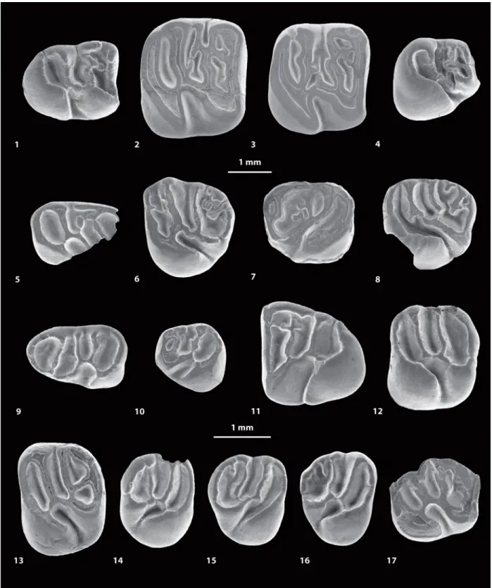
FIGURE 4. Scanning electron microscope images (in occlusal view) of fossil caviomorph teeth from CTA-27. Cachi-yacuy contamanensis (1-6), Eobranisamys javierpradoi sp. nov. (7-8), CachiCachi-yacuy kummeli (9-13), Canaanimys maquiensis (13-16) and cf. Eoespina sp. (17). 1, right m3 (MUSM 2713); 2, right m2 (MUSM 1914); 3, right m2 (MUSM 1915); 4, right p4 (MUSM 2678); 5, fragmentary right dp4 (MUSM 2670); 6, left M3 (MUSM 2758); 7, right dP4 (MUSM 2797); 8, fragmentary left M3 (MUSM 2801); 9, left dp4 (MUSM 2762); 10, left p4 (MUSM 2766); 11, right m3 (MUSM 2780, reversed); 12, left M1 (MUSM 2785); 13, left M2 (MUSM 2786); 14, fragmentary right M3 (MUSM 2794); 15, right M3 (MUSM 2793); 16, left M3 (MUSM 2792, reversed); 17, fragmentary left M2 (MUSM 2802). Top scale for 1-13, bottom scale for 14-17.
2798; broken on MUSM 1896), located distolabially to the protocone. These two lingual cusps, also twinned, form a protocone-hypocone complex. The paracone-metacone and protocone-hypocone complexes, associated with long and strongly developed posteroloph and anteroloph, design a circular enamel wall on the entire crown margin. On MUSM 1896 (in Antoine et al., 2012, figure 2a’), there is only a small depression on this wall, located mesially, at the level of the anteroloph-paracone junction. This depression is absent on MUSM 2798. The protoloph is short and limited to its labial part. There is no lingual protoloph: the tooth is taeniodont. The hypocone has an oblique anterior arm ending near the center of the tooth. This hypocone arm extends labially via a straight and long third transverse crest (mesolophule on MUSM 1896 and mesolophule/mesoloph? on MUSM 2798). There is a tiny enamel spur mesio-lingually directed at the junction between the hypo-cone arm and the third transverse crest.
The M1s referred here to Eobranisamys (MUSM 1897 and 2800; in Antoine et al., 2012, fig-ure 2b’) are subquadrate, while the M3 (MUSM 2801; Figure 4.8) is smaller and shows a more rounded occlusal outline. On all upper molars, the protocone appears mesiodistally opposed to the hypocone. However, the hypocone is more reduced with respect to the protocone on M3 than on M1. There is no significant difference of crown height with upper molars of other rodent species from CTA-27. All upper molars are fully pentalo-phodont. The main cusps are sometimes faintly visible, but still distinct. These upper molars are fully taeniodont. The paraflexus is merged with the hypoflexus to form a continuous labiolingual groove, without any vestige of lingual protoloph. The protocone is slightly labiolingually pinched and connected to a strong anteroloph. This mesial transverse crest runs labially and connects to a well-defined parastyle. There is no connection between the parastyle and the paracone, even if they are very close on MUSM 2799 (where the paraflexus is almost completely labially closed). The hypocone is slightly labiolingually pinched. Its anterior arm is strong and frequently runs labiome-sially to join the lingual extremity of the interrupted protoloph. Thus, the mure is present but rather indistinct except on MUSM 2800 where it is long and mesiolabially oriented. The third transverse crest is strongly connected to a well-defined and isolated mesostyle. This crest is a composite of either equally developed mesolophule and mesol-oph (MUSM 2800), or of a long mesolmesol-oph with a
short spur-like mesolophule (MUSM 1897 and 2801; in Antoine et al., 2012, figure 2b’). In addi-tion, the mesostyle is separate from the paracone and metacone by two notches (the posterior arm of the paracone and anterior arm of the metacone are absent or weakly developed). On all upper molars, the metacone is labiolingually compressed and oblique. The metaloph is strong but short and backwardly directed in its lingual end, and it is vari-ably connected to the medial part of the posterol-oph (i.e., from slightly [MUSM 2800 and 2801] to strongly [MUSM 1897] connected). With such a configuration, the lingual part of the posteroflexus is confluent with the metaflexus. The metaloph and posteroloph isolate a small and oval posterofos-sette (the remaining labial part of the posterof-lexus). The posteroloph is low compared to other transverse crests, and it links the distal aspects of the hypocone and of the metacone. In addition, the posteroloph bears a labial secondary crestule on M3 (Figure 4.8).
Comparisons. This new species of Eobranisamys is roughly similar in size to the sympatric
Cachi-yacuy contamanensis. The transverse crests and
cristids are well-marked and slightly higher than in molars of species referred to Cachiyacuy or
Canaanimys. Teeth studied here display features
reminiscent of those of Eobranisamys and
Branis-amys Hoffstetter and Lavocat, 1970 (especially
upper molars; Lavocat, 1976; Frailey and Camp-bell, 2004). This is particularly shown in the devel-opment of a taeniodont and pentalophodont pattern, characterized by the presence of a strong third transverse crest, and a strong and well-defined metaloph, which is transverse and con-nected to the posteroloph. As in Eobranisamys
romeropittmanae and E. riverai, the teeth of E. javierpradoi nov. sp. are much lower crowned than
teeth of Branisamys. However, E. javierpradoi dif-fers substantially from E. romeropittmanae in being about half the size, and from E. riverai in being 20% smaller, and in showing sharper crests and more salient cusps. Besides, E. romeropittmanae and E. riverai tend to have M3 shorter and with a more rounded occlusal outline than E. javierpradoi.
Eobranisamys sp.
Figure 5.15-16, Appendix 3
2016 Caviomorpha indet. Antoine et al., Supplemen-tary data, p. 8.
2017 Caviomorpha indet. Antoine et al., Supplemen-tary data, p. 9.
Referred material. MUSM 2841, fragmentary left upper molar? (Figure 5.15); MUSM 2842, fragmen-tary left upper molar (Figure 5.16).
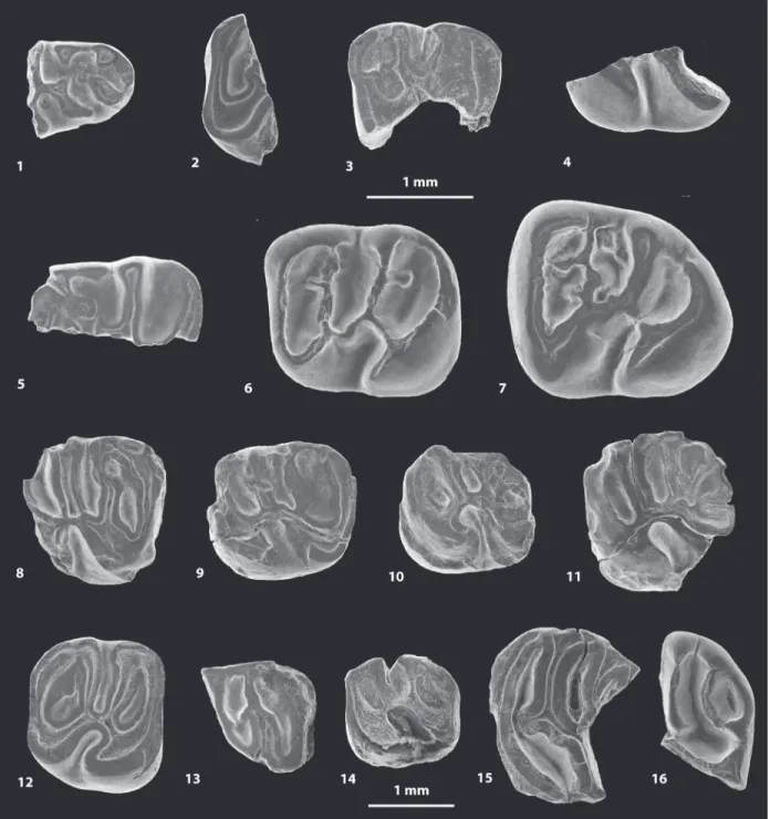
FIGURE 5. Scanning electron microscope images (in occlusal view) of fossil caviomorph teeth from CTA-47 (1-4), CTA-51 (5-12), CTA-73 (13-14) and CTA-66 (15-16). ?Canaanimys sp. (1-2),?Cachiyacuy kummeli (3), Caviomorpha indet. 1 (4), Cachiyacuy cf. contamanensis 1 (5-7), Caviomorpha indet 2 (8-10), Cachiyacuy cf. kummeli (11), Eoespina sp. (12), Caviomorpha indet 3 (13), Caviomorpha indet 4 (14) and Eobranisamys sp. (15-16). 1, fragmen-tary right dp4 (MUSM 2645); 2, fragmenfragmen-tary left lower molar (MUSM 2646); 3, fragmenfragmen-tary left dP4 (MUSM 2648); 4, fragmentary right lower molar (MUSM 2647); 5, fragmentary right dp4 (MUSM 2651, reversed); 6, left m1 (MUSM 2652); 7, left m3 (MUSM 2653); 8, fragmentary right lower molar (MUSM 2656); 9, fragmentary left dP4 (MUSM 2657); 10, fragmentary left upper molar (MUSM 2658); 11, fragmentary left M1 (MUSM 2654); 12, right M2 (MUSM 2655); 13, fragmentary right lower molar (MUSM 2659); 14, fragmentary left upper molar (MUSM 2660); 15, fragmen-tary left upper molar? (MUSM 2841); 16, fragmenfragmen-tary left upper molar (MUSM 2842). Top scale for 1-12, bottom scale for 13-16.
Locality. Contamana CTA-66, Loreto Department, Peru.
Formation and age. Pozo Formation, lower mem-ber, late middle Eocene (Antoine et al., 2016). Description. MUSM 2841 (Figure 5.15) could be a fragment of an upper molar, upon which it can be recognized a part of the anteroloph, the protoloph, the mure, the third transverse crest, and a mesial part of the anterior arm of the hypocone. The crests are very thick. The lingual protoloph is reduced to a short crestule, stemming from the labial branch of the protoloph and forwardly directed, but without connection with the protocone (pseudo-taeniodont pattern). The paracone displays short anterior and posterior arms connecting to the labial end of the anteroloph and the posterior arm of the mesostyle, respectively, thereby involving the labial closure of both the paraflexus and mesoflexus. The protoloph runs from the paracone to an oblique mure. The third transverse crest, labially linked to a strong mesostyle, is thin at the level of its lingual connec-tion with the anterior arm of the hypocone. Such a pattern suggests that the third transverse crest cor-responds to a long mesoloph, and possibly to a very short mesolophule lingually. The protoloph and third transverse crest are roughly parallel and slightly oblique.
MUSM 2842 (Figure 5.16) is a labiodistal frag-ment of a medium-sized upper molar, upon which it can be easily recognized labially, the mesostyle and metacone, and distolingually, the hypocone. The mesostyle is particularly strong, appearing as large as the metacone and hypocone. The meso-style is separate from the metacone by a narrow but deep notch, while the metacone is strongly connected to the posteroloph. The third transverse crest is strong, straight, and well-connected to the mesostyle, but it is distinct from the mesial end of the anterior arm of the hypocone. This pattern sug-gests that it is only formed by a mesoloph. The metaloph is well-defined, long, and well-curved. It runs lingually from the metacone, then turns back-wardly and connects to an accessory cusp of the posteroloph mediolingually (just below the middle line of the crown). The metaloph, mesoloph, and posteroloph remain well-separated. The hypocone displays a very long and oblique anterior arm. The posteroloph is strong and gently curved. The flexi are deep. The most posterior flexus (labial part of the posteroflexus) is entirely closed and forms a deep and oval posterofossette.
Comparisons. On MUSM 2842 (Figure 5.16), the presence of a strong and non-oblique third trans-verse crest, as well as a well-defined, long, and
curved backwardly metaloph, which is lingually connected to the posteroloph, in addition to the brachydonty, are characters found in three Paleo-gene Paleo-genera: Cachiyacuy, Eosallamys Frailey and Campbell, 2004, and Eobranisamys. In
Cachi-yacuy and Eosallamys, the development and
con-nections of the metaloph are highly variable. The metaloph can be long, reduced, or almost absent. It is totally absent on two upper molars of
Cachi-yacuy: on MUSM 2744 of C. contamanensis and
on MUSM 2785 of C. kummeli (Figure 4.12). When it is present in Cachiyacuy and Eosallamys, it can be connected either to the anterior arm of the hypocone, to the third transverse crest, or to the posteroloph. In Eobranisamys, the metaloph is present, well-defined, well-separated from its adja-cent mesial and distal transverse crests (third transverse crest and posteroloph, respectively), and it is always connected to the posteroloph. As for MUSM 2842, the mesostyle is always labially separate from the metacone in Eosallamys and
Eobranisamys, while in Cachiyacuy, these two
cusps can be linked. On MUSM 2842, the develop-ment and configuration of the metaloph generate a large, deep, and rounded posterofossette (labial part of the posteroflexus), a feature characteristic of Eobranisamys. In C. contamanensis and
Eosal-lamys, this posterofossette is smaller and
shal-lower. This fragmentary tooth from CTA-66 is therefore likely to be the distal part of an upper molar of an Eobranisamys-like taxon. The size of the specimens is closer to the species of
amys from CTA-27 (E. javierpradoi) and Eobranis-amys riverai than to Eobranisamys romeropittmanae. On MUSM 2842, as for E. romeropittmanae and E. riverai, the
posterofos-sette is larger than in E. javierpradoi. As for E.
javierpradoi, MUSM 2842 differs from E. romeropit-tmanae and E. riverai in showing sharper crests
and more salient cusps.
Superfamily OCTODONTOIDEA Waterhouse, 1839
Genus EOESPINA Frailey and Campbell, 2004 Type species. Eoespina woodi Frailey and Camp-bell, 2004
Species content. Only the type species.
Geographic and stratigraphic distribution. Con-tamana (CTA-51 locality), Pozo Formation, lower member (late middle Eocene), Loreto department, Peru; Santa Rosa (LACM 6289 locality), “Yahua-rango Formation” (?late Eocene/early Oligocene), Ucayali department, Peru.
Eoespina sp.
Figure 5.12, Appendix 3
2016 Cachiyacuy aff. kummeli Antoine et al., Supple-mentary data, p. 5.
2017 Cachiyacuy aff. kummeli Antoine et al., Supple-mentary data, p. 9.
Referred material. MUSM 2655, right M2 (Figure 5.12).
Locality. Contamana CTA-51, Loreto Department, Peru.
Formation and age. Pozo Formation, lower mem-ber, late middle Eocene (Antoine et al., 2016). Description. The M2 (MUSM 2655; Figure 5.12) is worn but all its occlusal structure is well-recogniz-able, with a subquadrate outline. The hypocone is slightly displaced labially and smaller than the pro-tocone, thereby indicating that this tetralophodont tooth is a M2. The transverse crests (anteroloph, protoloph, third transverse crest, and posteroloph) are subparallel, with a slightly oblique protoloph. Both the anteroloph and posteroloph are massive and strongly connected to the lingual and labial cusps. The tooth is not taeniodont. The mure is short and almost longitudinal. Labially, there is a large mesostyle, twinned with the metacone and strongly connected to the lingually thinning third transverse crest (therefore interpreted as a mesol-oph). A deep and narrow notch separates the mesostyle from the paracone, and thus the mesof-lexus remains open labially. The mesofmesof-lexus is par-ticularly narrow due to the close proximity of the mesoloph with the protoloph. Labially, the metacone is virtually indistinct and entirely incorpo-rated within the labial posteroloph. The mesial enamel edge of the posteroloph expends mesially, and forms an enamel platform-like surface. This structure might correspond to a relic of the metaloph. The paraflexus and the posteriormost flexus (fused meta- and posteroflexus) are close lingually. The hypoflexus is mesiodistally con-stricted but remains open.
Comparisons. This specimen has a size compara-ble to that of teeth of Cachiyacuy kummeli,
Canaanimys maquiensis, Eoespina woodi Frailey
and Campbell, 2004, and Eosachacui lavocati Frai-ley and Campbell, 2004. Except for Canaanimys, the concerned upper molars have a pattern close to that of MUSM 2655: non-taeniodont and with a metaloph reduced or absent. In Eosallamys, upper molars are non-taeniodont but their metaloph is still present and long. The subquadrate occlusal outline of MUSM 2655 and the strong connections of the anteroloph and posteroloph to the labial cusps, respectively, better match the conditions found in
Eoespina. Given the scarcity the available material,
we provisionally assign this tooth to Eoespina sp. Remarks. Following Frailey and Campbell (2004),
Eoespina woodi and Eosachacui lavocati, both
found at Santa Rosa, are two close taxa sharing many similarities. This is particularly shown in their brachydonty, non-taeniodonty, subquadrate upper molars with rounded corners, and pentalophodont/ tetralophodont upper molars with a very reduced or absent metaloph. Frailey and Campbell (2004, p. 88–91) described seven characters distinguishing both taxa. However, these characters are not found in all specimens referred to each taxon and can result from an intraspecific variation. A taxonomic revision of both taxa would hence be required, notably in considering their possible synonymy. According to Frailey and Campbell (2004, p. 88– 91):
1. Eoespina is slightly smaller than Eosachacui.
However, the size range of its dental variation matches that of Eosachacui;
2. Contrary to Eoespina, upper molars of
Eosachacui often display additional spurs or
cuspules in their metaflexus. However, this addition of enamel structures is only limited to three specimens (LACM 143292, 143387, and 143401; Frailey and Campbell, 2004, p. 128); 3. In Eosachacui, the deflections of the protoloph
are less severe than those observed in
Eoespina. These deflections of the protoloph
in Eosachacui probably correspond to the obliquity of the protoloph, which is more pro-nounced (strongly oblique) and tends to be in line with the mure. However, some upper molars of Eosachacui have a transverse pro-toloph (LACM 143387 and 143388; Frailey and Campbell, 2004, p. 128), while this crest can be slightly oblique in Eoespina (e.g., LACM 143286 and 149436; Frailey and Campbell, 2004, p. 124–125);
4. Contrary to Eosachacui, lower molars of
Eoespina display accessory cristulids and
cuspids in their anteroflexid. Nevertheless, one lower molar of Eosachacui shows these secondary structures (LACM 143325; Frailey and Campbell, 2004, p. 129). Interestingly, the presence or absence of these structures is also observed in Cachiyacuy contamanensis, but that remains very variable;
5. In lower molars of Eoespina, the second transverse cristid (named “metalophid” by Frailey and Campbell, 2004, p.91) is not uni-form either in height or thickness. Indeed, the
second transverse cristid appears as a combi-nation of two cristids (i.e., neomesolophid and posterior arm of the protoconid), more or less developed depending on the specimens. From our personal observation of the Santa Rosa specimens figured in Frailey and Camp-bell (2004, p. 124–130), this apparent com-posite second cristid in Eoespina is also found in lower molars of Eosachacui;
6. The second transverse cristid and hypolophid (named “protolophid” in Frailey and Campbell, 2004, p. 91) are mesiodistally closer in
Eosachacui than in Eoespina. However, in
both genera, these two cristids are variably spaced, which implies a variation of the shape and size of the flexids (anteroflexid and meso-flexid); and
7. Finally, in lower molars of Eosachacui, the hypolophid is transverse or slightly back-wardly directed, while it is transverse or slightly forwardly directed in Eoespina. How-ever, as noticed by Frailey and Campbell (2004, p. 91), this character is highly variable.
cf. Eoespina sp. Figure 4.17, Appendix 3
Referred material. MUSM 1913 (in Antoine et al., 2012, figure 2f’) and 2802 (Figure 4.17), left M2s; MUSM 1912, right M2 (in Antoine et al., 2012, fig-ure 2e’).
Locality. Contamana CTA-27, Loreto Department, Peru.
Formation and age. Pozo Formation, lower mem-ber, late middle Eocene (Antoine et al., 2012, 2016).
Description. Only three minute upper molars can be referred to cf. Eoespina sp. (MUSM 1912, 1913, and 2802; in Antoine et al., 2012, figure 2e’-f’; Fig-ure 4.17). They have a rounded crown outline in occlusal view. These teeth are primarily tetralopho-dont with strong and long third transverse crest, antero-, proto-, and posteroloph (no metaloph). The cusps are still well-defined. There is no para-style. Mesially, a strong anteroloph runs from the protocone to the mesiolabial aspect of the paracone. The teeth are not taeniodont and display a short and longitudinal mure, which is slightly situ-ated lingually to the medial axis of the crown. There is a strong but short anterior arm of the hypocone that connects the mure. The hypocone is more labial and reduced with respect to the proto-cone. The metacone is mesiodistally elongated and slightly more lingual than the paracone, and it is merged with a strong mesostyle. The paracone
and mesostyle are only separate by a narrow but deep notch, and the mesoflexus remains open labi-ally as a result. A strong and continuous third transverse crest runs from the mesial extremity of the anterior arm of the hypocone to the mesostyle. The protoloph and third transverse crest are nearly parallel. On the distolabial region of MUSM 1913 (in Antoine et al., 2012, figure 2f’), it may occur a vestigial metaloph, very low, short, backwardly directed, and practically subsumed within the pos-teroloph. On all teeth, the hypoflexus is narrow and somewhat constricted lingually.
Comparison. Teeth of this taxon are slightly smaller than those of Canaanimys maquiensis. These three diminutive upper molars from CTA-27 exhibit a tetralophodont/non-taeniodont pattern, with strong and long third transverse crest, antero-, proto-, and posteroloph (no metaloph or very reduced), as it can be observed in
“?Vallehermo-somys merlinae Vucetich et al., 2010,” Sallamys
Hoffstetter and Lavocat, 1970 or Eoespina/
Eosachacui. The general morphology of these
upper teeth from CTA-27, with notably a strong and long posterior arm of the paracone and a rounded crown outline, recall upper molars of E. woodi/E.
lavocati. However, these specimens from CTA-27
differ from E. woodi/E. lavocati in having slightly more transverse upper molars, usually character-ized by less inflated labial cusps. These upper molars have a longer and more mesially positioned third transverse crest contrary to the condition observed on the M3 of ?Vallehermosomys
mer-linae. Indeed, the latter displays a wider
mesof-lexus, and displays a third transverse crest that is clearly more displaced distally and close to the posteroloph. These upper molars from CTA-27 also differ from those of Sallamys in being lower-crowned, and in having a stronger third transverse crest, which always connects to the anterior arm of the hypocone.
CAVIOIDEA Fischer de Waldheim, 1817 or CHINCHILLOIDEA Bennett, 1833
Gen. et sp. indet. Figure 6.15-17, Appendix 3
2016 Eobranisamys sp. Antoine et al., Supplemen-tary data, p. 9.
2016 Cachiyacuy cf. contamanensis Antoine et al., Supplementary data, p. 9.
2016 Caviomorpha indet., sp. 1 Antoine et al., Sup-plementary data, p. 9.
2017 Eobranisamys sp. Antoine et al., Supplemen-tary data, p. 9.
2017 Cachiyacuy cf. contamanensis Antoine et al., Supplementary data, p. 9.
FIGURE 6. Scanning electron microscope images of fossil caviomorph teeth from CTA-29. Pozomys ucayaliensis gen. et sp. nov. (1-6), Cachiyacuy cf. contamanensis 2 (7-12), Caviomorpha indet. 5 (13), Caviomorpha indet. 6 (14) and Cavioidea or Chinchilloidea indet. (15-17). 1, right m1-2, occlusal view (MUSM 2822); 2, left p4, occlusal view (MUSM 2821, reversed); 3, fragmentary right M3, occlusal view (MUSM 2819); 4, right M2, occlusal view (MUSM 2833); 5, right m1-2, labial view (MUSM 2822); 6, right m1-2, lingual view (MUSM 2822); 7, left dp4, occlusal view (MUSM 2825); 8, right m1, occlusal view (MUSM 2827, reversed); 9, right dP4, occlusal view (MUSM 2828, reversed); 10, left M1, occlusal view (MUSM 2831); 11, right M2, occlusal view (MUSM 2563, reversed); 12, fragmen-tary left M2, occlusal view (MUSM 2832); 13, right dp4, occlusal view (MUSM 2838); 14, fragmenfragmen-tary right upper molar, occlusal view (MUSM 2839); 15, right m2, occlusal view (MUSM 2835); 16, right dp4, occlusal view (MUSM 2834); 17, fragmentary right upper molar, occlusal view (MUSM 2836, reversed).
2017 Caviomorpha indet. sp. 1, Antoine et al., Sup-plementary data, p. 9.
Referred material. MUSM 2834, right dp4 (Figure 6.16); MUSM 2835, right m2 (Figure 6.15); MUSM 2836–2837, fragmentary right upper molars (Fig-ure 6.17).
Locality. Contamana CTA-29, Loreto Department, Peru.
Formation and age. Pozo Formation, lower mem-ber, late middle Eocene (Antoine et al., 2016). Description. The dp4 (MUSM 2834; Figure 6.16) is relatively larger compared to other dp4s found at CTA-29. This tooth is pristine and brachydont, with slender transverse cristids, and small but well-defined and acute mesostylid, entoconid, and hypoconid. The metaconid and protoconid are much more crestiform. This deciduous lower pre-molar is pentalophodont. Mesially, the metalophulid I, well-curved, connects to the protoconid labially and ends and the base of the metaconid lingually. The metaconid, mesiodistally pinched, is faintly linked to the crestiform protoconid via its posterior arm, which forms a quasi-complete metalophulid II. There is a minute enamel cuspid on the anterof-lexid at the base of the distal wall of the metalophu-lid I. The metaconid does not develop a posterior arm. In the middle part of the tooth, the lingual mar-gin bears a well-defined and isolated mesostylid. The mesial ectolophid is almost transverse and appears in continuity with a mesolophid, which extends lingually to the mesostylid. Labially, the mesial and sub-transverse ectolophid displays an enamel swelling, which could be interpreted as a neomesoconid-like cuspid. The distal ectolophid is small and very low. As such, the hypoflexid and distal mesoflexid are virtually confluent. In this con-text, the third transverse cristid has no link with the surrounding cristids, and the hypoflexid-distal mesoflexid and mesial mesoflexid are transversely open. The hypoconid is the largest cuspid of the tooth, labially opposed to the entoconid. Both cus-pids are linked by a thin and sinuous hypolophid. The second transverse cristid, mesolophid, and hypolophid are parallel and slightly curved. The anterior arm of the hypoconid is faintly marked, lower and grooved, and it is weakly connected to the hypolophid (i.e., pseudo-taeniodont). Distolabi-ally, the posterolophid faintly links the hypoconid, distolingually ending its course far from the entoco-nid. Thus, the broad metaflexid remains open lin-gually.
The m2 (MUSM 2835; Figure 6.15) is cor-roded and damaged, with a dental structure still visible. This tetralophodont tooth is particularly
large with a trigonid as wide as the talonid. Two enamel wrinkles are directed backwardly to the metalophulid I, and one directed forwardly to the second transverse cristid. Due to wear, it is difficult to tell if this second cristid was connected or not to the mesostylid. This cristid could be a complete metalophulid II or a composite cristid made by a labial short posterior arm of the protoconid associ-ated with a short neomesolophid. The ectolophid is oblique and distally connected both to a short but strong anterior arm of the hypoconid (i.e., non-tae-niodont pattern) and to the transverse and strong hypolophid. The posterolophid is lingually separate from the entoconid. The mesoflexid and posterof-lexid are lingually open.
Both labial fragments (MUSM 2836 and 2837; Figure 6.17) document large upper molars. The labial cusps are well-defined: parastyle, paracone, mesostyle, and metacone. MUSM 2836 is tetralo-phodont (Figure 6.17): there is no trace of metaloph between the mesolophule and the pos-teroloph. The metacone is the largest cusp, at the labial end of the posteroloph. On MUSM 2836, all transverse flexi are labially open, whereas the metaflexus is labially closed on MUSM 2837 (more worn).
Comparisons. The morphology of MUSM 2834 (Figure 6.16) is reminiscent of that found in dp4s of
Eobranisamys from Santa Rosa (Peru, ?late
Eocene/early Oligocene; Frailey and Campbell, 2004) and Branisamys from Salla (Bolivia, late Oli-gocene; Hoffstetter and Lavocat, 1970; Lavocat, 1976), notably in lacking the connections between the second transverse cristid (metalophulid II) and the mesolophid (or with their associated cuspids). However, in Eobranisamys and Branisamys, there is a distal ectolophid between the mesolophid and the hypolophid, and a labial connection between the metalophulid I and the metalophulid II. MUSM 2834 is pentalophodont whereas the dp4s of
Eobranisamys and Branisamys are hexalophodont,
in showing the addition of a neolophid between the metalophulid I and the metalophulid II. MUSM 2835 (Figure 6.15) is similar in dental size to
Eobranis-amys, while Branisamys is significantly larger. That
m2 has a pattern close to that of lower molars of
Eobranisamys, which can have accessory cristids
on the metalophulid I and second transverse cris-tid. Although the size of the fragmentary upper molars (MUSM 2836 and 2837; Figure 6.17) is compatible with that of Eobranisamys, the former are tetralophodont while Eobranisamys and
Brani-samys have pentalophodont upper molars, with a
close affinities to Eobranisamys, and to a lesser extent to Branisamys. However, the particular char-acteristics of the dp4 and the tetralophodont pat-tern of upper molars do not allow for a formal generic assignment. Branisamys was initially attributed to the Dasyproctidae (i.e., Cavioidae) by Lavocat (1976), but this genus may be more closely related to the Chinchilloidea following Kra-marz et al. (2013) and Vucetich et al. (2015).
Eobranisamys was originally assigned to the
Cavi-oidea (Frailey and Campbell, 2004), but its alleged close relationship with Branisamys would question, in turn, the suprafamilial attribution of
Eobranis-amys. In the present state, we provisionally refer
the material of CTA-29 to as “Cavioidea or Chin-chilloidea indet.,” although a critical revision of
Eobranisamys and Branisamys would be
neces-sary in that purpose.
Superfamily indet. Genus POZOMYS gen. nov.
zoobank.org/903578CB-4B98-4430-AF8B-5C30839265D5 Type species. Pozomys ucayaliensis, sp. nov. Species content. Only the type species.
Etymology. From the Pozo Formation, which includes the CTA-29 locality, and mŷs, Greek for mouse.
Generic diagnosis. As for the type and only spe-cies.
Pozomys ucayaliensis sp. nov.
Figure 6.1-6, Appendix 3
zoobank.org/E38E9E2A-6303-4278-888F-0F8FC05C5010
2016 Canaanimys sp. Antoine et al., Supplementary data, p. 9.
2016 Cachiyacuy cf. kummeli Antoine et al., Supple-mentary data, p. 9.
2016 Chinchilloidea indet. Antoine et al., Supplemen-tary data, p. 9.
2017 Canaanimys sp. Antoine et al., Supplementary data, p. 9.
2017 Cachiyacuy cf. kummeli Antoine et al., Supple-mentary data, p. 9.
2017 Chinchilloidea indet. Antoine et al., Supplemen-tary data, p. 9.
Etymology. Refers to the Ucayali River, which is a major tributary of the Amazon River flowing near Contamana, Peru.
Holotype. MUSM 2833, right M2. Deposited in the Museo de Historia Natural de la Universidad Nacional Mayor de San Marcos, Lima, Peru. Referred material. In addition to the holotype (MUSM 2833) ̶ MUSM 2821, left p4 (Figure 6.2); MUSM 2820, fragmentary right p4; MUSM 2822, right mandibular fragment bearing m1 and m2
(Fig-ure 6.1, 6.5-6); MUSM 2833, right M2 (Fig(Fig-ure 6.4); MUSM 2819, fragmentary right M3 (Figure 6.3). Type Locality. Contamana CTA-29, Loreto Depart-ment, Peru.
Formation and age. Pozo Formation, lower mem-ber, late middle Eocene (Antoine et al., 2016). Diagnosis. Tiny rodent characterized by tetralo-phodont p4s and lower molars. Pozomys differs from Eoincamys in having no posterior arm of the protoconid (or very reduced one) on p4, and less oblique transverse cristids on the lower molars. Dif-fers from Cachiyacuy and Canaanimys in having a residual metaloph, which can be merged with the metacone-posteroloph complex on upper molars. Differs from Cachiyacuy, Eoespina/Eosachacui and Incamys in having a metalophulid I more or less disconnected to the protoconid on lower molars. Its teeth tend to be taeniodont, contrary to
Eoespina/Eosachacui and most teeth of Cachi-yacuy. Differs from Incamys in being
lower-crowned and in showing a similar thickness of the enamel layer on the mesial and distal flanks of the hypoflexus. Differs from Platypittamys and
Deseadomys in having a p4 with a hypolophid.
Dif-fers from Galileomys and Paulacoutomys in having a very thin lateral crest, which does not reaches the masseteric crest below the m1 on mandible. Description. The nearly complete p4 (MUSM 2821; Figure 6.2) is moderately eroded, and shows rounded corners. The shape of this premolar is characterized by a talonid, which is nearly twice wider than the trigonid. Despite of the wear, the four main cuspids (metaconid, entoconid, protoco-nid, and hypoconid) are well-recognizable, as they are well-defined. Although the tooth is slightly bro-ken mesially (it lacks the enamel), the metaconid and protoconid appear well-separated and con-nected mesially by a transverse and straight metalophulid I. The rounded protoconid seems to display a short posterior arm, which is in connec-tion and in line with a strongly oblique ectolophid. The hypolophid is also oblique and in line with the ectolophid. These two cristids form a distolingual-mesiolabial directed central and diagonal cristid, which links the entoconid and protoconid (its poste-rior arm?). Lingually, the mesostylid is faintly visible to indistinct and linked to a short and low posterior arm of the metaconid. The second transverse cris-tid is short, straight, and strikingly labiodistally directed. This second transverse cristid is lingually linked to the posterior arm of the metaconid-meso-stylid complex, and labially to the ectolophid, and as such it could represent a neomesolophid rather than a metalophulid II. Labiodistally, the hypoconid
is massive but crestiform (mesiodistally pinched), and bears a noticeably long labial outgrowth. Disto-labially, the hypoconid is entirely merged with the strong posterolophid. This latter cristid runs lin-gually and connects to a short, well-marked, and high posterior arm of the entoconid. A very short, low, and thin anterior arm of the hypoconid is faintly linked to the hypolophid (the tooth is almost tae-niodont). Therefore, the distal metafossettid is almost confluent with the hypoflexid, which exhibits a wide labial aperture. The fragment of p4 (MUSM 2820) is less eroded than the former one described above. This tooth fragment does not present any second transverse cristid. A thin and deep furrow separates the mesostylid from the metaconid.
MUSM 2822 is a mandibular fragment pre-serving m1 and m2 (Figure 6.1, 6.5-6). Labially, the broken masseteric crest is posteroventrally directed. It is widely prominent and reduced at its anterior tip, which probably ends below the premo-lar (p4). The part of the ascending ramus, which runs toward the coronoid process, begins below the m2. That mandibular fragment shows neither horizontal crest nor lateral crest nor notch for the insertion of the tendon of the zygomatico-mandibu-laris pars infraorbitalis. The m1 has a trigonid nar-rower than the talonid, whereas the trigonid is roughly as wide as the talonid on m2. Although the occlusal surface of the molars is worn, their cus-pids are still well-recognizable, notably on m2. Both teeth are tetralophodont. The metalophulid I is very thin in its labial part and it is clearly separate from the protoconid by a wide notch on m2. On both molars, the posterior arm of the metaconid is long and reaches a well-defined mesostylid. The latter is well-separated from the entoconid. The short second transverse cristid is straight and com-plete but constricted in its middle part, which sug-gests that it could result from the coalescence of two cristids (lingually, a neomesolophid, and labi-ally, a posterior arm of the protoconid). On m1, the pinching of the second cristid is labially located, which would indicate that the neomesolophid is dominant, whereas the two cristids are equal in length on m2. The hypolophid is strong and straight transversely. The second transverse cristid and hypolophid are parallel and mesiodistally close in position. Both isolate a labiolingually long and mesiodistally narrow furrow-like mesoflexid. The ectolophid, longitudinal, is short due to the advanced stage of wear. The anterior arm of the hypoconid, poorly developed (faintly visible and very low), separates the metaflexid from the hypof-lexid. Yet, given the weak development and low
elevation of the anterior arm of the hypoconid, we can describe this dental pattern as “pseudo-tae-niodont.” The hypoconid is crestiform (mesiodis-tally pinched), with a long and labially oriented outgrowth. This outgrowth and the distal flank of the protoconid isolate a narrow and nearly trans-verse hypoflexid (notably on m2). Lingually, on both teeth, the entoconid does not develop an anterior arm, and thus the furrow-like mesoflexid remains open lingually. The metaflexid is lingually open on m1 but closed on m2.
MUSM 2833 (Figure 6.4) is slightly wider than long. The hypocone is reduced compared to the protocone, and displaced labially like on some M3s and M2s. This specimen has interstitial facets on its mesial and distal margins, which allows us to identify it as a M2. The protocone, massive and slightly oblique, displays a short but thick posterior outgrowth. Connected to the protocone, the anter-oloph runs labially. It ends its course at the mesial base of the paracone, without connecting to it, thereby letting the paraflexus faintly open labially. The paracone is large and rounded, and much more differentiated than the metacone (nearly indistinct and subsumed within the posteroloph). From the paracone, the oblique labial protoloph runs distolingually and joins the mure-anterior arm of the hypocone complex, subparallel to the protol-oph. The lingual protoloph is lacking, and thus the hypoflexus connects the paraflexus, thereby illus-trating a full taeniodont pattern. Labially, at mid-dis-tance between the paracone and metacone-posteroloph, the mesostyle is strong and well-defined, but only connected to the metacone-pos-teroloph complex by a strong longitudinal crest (posterior arm of the mesostyle and/or anterior arm of the metacone). The central transverse crest between the mesostyle and the mesial extremity of the anterior arm of the hypocone is sinuous, indi-cating that this crest could be a composite crest, including a short mesolophular spur and a long mesoloph. Distolabially, the posteroloph shows a distinct mesiodistal enlargement, thus suggesting the presence of a residual metaloph, backwardly oriented and merged with the posteroloph. The metaflexus is the unique flexus entirely closed labi-ally.
The fragmentary MUSM 2819 (Figure 6.3) is subpentalophodont and non-taeniodont, with a rounded labial margin and a hypocone more labial than the protocone, which indicates that the speci-men is a M3. The protoloph is slightly oblique, close and parallel to the anteroloph (narrow paraf-lexus). Although broken labiomesially, these two
mesial crests seem not to be linked labially. The metacone is small, slightly more lingual than the paracone, and merged to the posteroloph. The mesostyle is almost as large as the paracone and isolated on the labial margin. Neither a posterior arm of the paracone nor an anterior arm of the metacone is developed, and the mesoflexus and metaflexus remain open labially. The hypocone displays a strong and sagittal anterior arm. It con-nects to the protoloph via a strong mure, although almost undifferentiated. The central transverse crest consists of two disjoint crests: lingually, a short mesolophule and labially, a long mesoloph stemming from the mesostyle. A short metaloph, stemming from the metacone, runs mesiolingually toward the labial extremity of the mesolophule, without being connected to it. Distolingually, the posteroloph is faintly connected to the hypocone, and a shallow notch separates both structures. Comparisons. The size of these specimens is comparable to that of Cachiyacuy kummeli,
Canaanimys maquiensis, Eoespina woodi/Eosach-acui lavocati, and Eoincamys ameghinoi Frailey
and Campbell, 2004. On the lateral view of the MUSM 2822 mandible (Figure 6.5), the general disposition and strong development of the masse-teric crest on the dentary is found in all Paleogene caviomorphs for which the mandible is known (e.g.,
Eobranisamys, Andemys Bertrand et al., 2012, Platypittamys Wood, 1949, Scotamys Loomis,
1914, Cephalomys Ameghino, 1897, Branisamys,
Incamys Hoffstetter and Lavocat, 1970, Sallamys, Migraveramus Patterson and Wood, 1982, Paula-coutomys Vucetich et al., 1993, Galileomys
Vuce-tich and Kramarz, 2003, Acarechimys Patterson, 1965 (in Patterson and Wood, 1982),
Leucokepha-los Vucetich et al., 2015, Loncolicu Vucetich et al.,
2015, and Llitun Vucetich et al., 2015). Contrary to MUSM 2822, Galileomys and Paulacoutomys develop a very thin lateral crest, stemming from the ascending ramus, and almost reaching the masse-teric crest below the m1. The lower molars of
Pozomys ucayaliensis nov. gen. et sp. display an
association of characters that can be found in
Canaanimys and/or in Eoincamys (Frailey and
Campbell, 2004). The molars of the MUSM 2822 mandible show a metalophulid I that tends to be disconnected to the protoconid, a configuration which can be observed in lower molars of
Eoin-camys and Canaanimys. As on the m2 of MUSM
2822, the m1s and m2s of Eoincamys pascuali clearly show an interrupted metalophulid I, which is separate from the protoconid by a large notch. In
Eoincamys ameghinoi and Canaanimys
maquien-sis, this feature is more variable: the metalophulid I
can show the same configuration than the m2 of MUSM 2822 and Eoincamys pascuali (on LACM 149435 [in Frailey and Campbell, 2004, p. 114] and MUSM 1893, 2788-2789 [in Antoine et al., 2012, figure 2x], respectively), but also it can be complete in linking the metaconid to the protoconid (on LACM 143435 [in Frailey and Campbell, 2004, p. 114] and MUSM 2787, 2790-2791, respectively). In
Eoincamys, the second transverse cristid is almost
limited to its lingual part (neomesolophid), which tends to be disconnected to a very short, spur-like posterior arm of the protoconid. In Canaanimys, the second transverse cristid is either complete or discontinuous (split into two or three parts). The molars of the MUSM 2822 mandible are character-ized by a pseudo-taeniodont pattern, like in some lower molars of Canaanimys (MUSM 1892 and 1893; in Antoine et al., 2012, figure 2w-x). This is also observed on a tooth of Eoincamys ameghinoi (LACM 149442; in Frailey and Campbell, 2004, p. 114). In contrast, Eoincamys pascuali has more taeniodont lower molars. Contrary to the m1-2 of P.
ucayaliensis and Canaanimys, the transverse
cris-tids of the lower molars are more oblique in
Eoin-camys: the hypolophid is slightly oblique and tends
to be aligned with an oblique ectolophid along with the posterior arm of the protoconid. The ectolophid is oblique in Eoincamys and Canaanimys, contrary to P. ucayaliensis. The p4 of P. ucayaliensis also resembles that of Eoincamys in showing a rounded crown outline and a reduced second cristid; yet, the posterior arm of the protoconid is more devel-oped in Eoincamys than in P. ucayaliensis (small or absent). A reduced second cristid is also found in
Draconomys Vucetich et al., 2015, Leucokephalos, Platypittamys, Deseadomys Wood and Patterson,
1959, and Incamys. As for Eoincamys, the p4 of P.
ucayaliensis is relatively shorter with respect to the
p4 of Draconomys, Leucokephalos, Platypittamys,
Deseadomys, and Incamys (Wood, 1949; Wood
and Patterson, 1959; Hoffstetter and Lavocat, 1970; Lavocat, 1976; Vucetich et al., 2010, 2015). Besides, the MUSM 2821 p4 (Figure 6.2) differs from Platypittamys and Deseadomys in the pres-ence of a hypolophid. Although being taeniodont, the M2 of P. ucayaliensis is very distinct from that of Eoincamys or Canaanimys. Indeed, there is a prominent outgrowth of the protocone on M1–2 in
C. maquiensis, which is less developed in P. ucay-aliensis. On MUSM 2833 (and MUSM 2819) of P. ucayaliensis, the mesoloph is still linked to the
mesolophular spur of the anterior arm of the hypo-cone, while the mesoloph tends to have no lingual