HAL Id: hal-01817697
https://hal.sorbonne-universite.fr/hal-01817697
Submitted on 18 Jun 2018
HAL is a multi-disciplinary open access archive for the deposit and dissemination of sci-entific research documents, whether they are pub-lished or not. The documents may come from teaching and research institutions in France or abroad, or from public or private research centers.
L’archive ouverte pluridisciplinaire HAL, est destinée au dépôt et à la diffusion de documents scientifiques de niveau recherche, publiés ou non, émanant des établissements d’enseignement et de recherche français ou étrangers, des laboratoires publics ou privés.
Critical Impact of Peptidoglycan Precursor Amidation
on the Activity of l,d -Transpeptidases from
Enterococcus faecium and Mycobacterium tuberculosis
Flora Ngadjeua, Emmanuelle Braud, Saidbakhrom Saidjalolov, Laura
Iannazzo, Dirk Schnappinger, Sabine Ehrt, Jean-Emannuel Hugonnet,
Dominique Mengin-Lecreulx, Delphine Patin, Mélanie Etheve-Quelquejeu, et
al.
To cite this version:
Flora Ngadjeua, Emmanuelle Braud, Saidbakhrom Saidjalolov, Laura Iannazzo, Dirk Schnappinger, et al.. Critical Impact of Peptidoglycan Precursor Amidation on the Activity of l,d -Transpeptidases from Enterococcus faecium and Mycobacterium tuberculosis. Chemistry - A European Journal, Wiley-VCH Verlag, 2018, 24 (22), pp.5743 - 5747. �10.1002/chem.201706082�. �hal-01817697�
Critical impact of peptidoglycan precursor amidation on the
activity of
L
,
D
-transpeptidases from Enterococcus faecium and
Mycobacterium tuberculosis
Flora Ngadjeua#,[a], Emmanuelle Braud#,[b], Saidbakhrom Saidjalolov[b], Laura Iannazzo[b], Dirk
Schnappinger[c], Sabine Ehrt[c], Jean-Emmanuel Hugonnet[a], Dominique Mengin-Lecreulx[d], Delphine
Patin[d], Mélanie Ethève-Quelquejeu*,[b], Matthieu Fonvielle,*,[a] and Michel Arthur*,[a]
Abstract: The bacterial cell wall peptidoglycan contains unusual L and D amino acids assembled in branched peptides. Insight into the biosynthesis of the polymer has been hampered by limited access to substrates and to suitable polymerization assays. Here we report the full synthesis of the peptide stem of peptidoglycan precursors from two pathogenic bacteria, Enterococcus faecium and Mycobacterium
tuberculosis, and the development of a sensitive post-derivatization
assay for their cross-linking by L,D-transpeptidases. Access to series of stem peptides showed that amidation of free carboxyl groups is essential for optimal enzyme activity, in particular the amidation of diaminopimelate (DAP) residues for the cross-linking activity of the
L,D-transpeptidase LdtMt2 from M. tuberculosis. Accordingly,
construction of a conditional mutant established the essentiality of AsnB indicating that this DAP amidotransferase is an attractive target for the development of anti-mycobacterial drugs.
Peptidoglycan is an essential and specific component of the bacterial cell wall.[1] The main role of this giant (cell-sized)
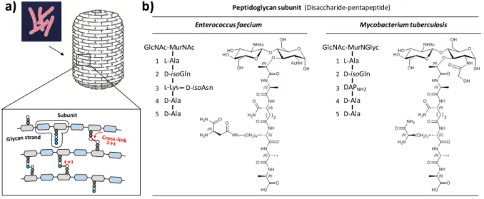
macromolecule is to protect bacterial cells against the osmotic pressure of the cytoplasm. The peptidoglycan subunit consists of a disaccharide substituted by a pentapeptide stem (figure 1), which is polymerized by glycosyltransferases for the elongation of the glycan chains (all glycosidic bonds are β-1,4) and by D,D -transpeptidases for cross-linking the glycan chains to each other.[2] The amide bond formed by the D,D-transpeptidases links
the carbonyl of D-Ala at the 4th position of an acyl donor stem to
the side-chain amino group at the 3rd position of an acyl acceptor
stem (4→3 cross-link). These enzymes are the targets of β-lactam antibiotics such as penicillin.
The structure of peptidoglycan is generally conserved in bacteria belonging to the same species, but highly diverse between species, including members of the same genus.[3] The
polymorphisms include the deacetylation, O-acetylation, and N-glycolylation of either or both GlcNAc and MurNAc. These modifications are mostly, if not exclusively, due to maturation of
subunits containing the canonic GlcNAc-MurNAc motif. The most frequent variations in the sequence of the pentapeptide stem
occur at the 3rd (e.g. L-Lys instead of diaminopimelic acid [DAP])
and at the 5th (e.g. D-Lac instead of D-Ala) positions (figure 1b).
Modifications of the pentapeptide stem involve the addition of a side-chain to the 3rd residue (e.g. D-isoAsn or Gly
5) and the
amidation of the carboxyl groups (e.g. the α-carboxyl of D-Glu and
D-isoAsp or the ε-carboxyl of DAP). A last source of polymorphism originates from the presence of 3→3 instead of 4→3 cross-links in mycobacteria (e.g. Mycobacterium tuberculosis) and in β-lactam-resistant mutants generated in vitro (e.g. Enterococcus
faecium) (figure 1a). The 3→3 cross-links are formed by
transpeptidases of the L,D specificity, which cleave the L-Lys3-D
-Ala4 or DAP3-D-Ala4 bond of an acyl donor containing a
tetrapeptide stem and form L-Lys3→L-Lys3 or DAP3→DAP3
cross-links.
Variability in the peptidoglycan structure has been known for decades based on biochemical analyses of the cell wall.[3a] The
corresponding enzymes have been described more recently, mostly because the complexity of their substrates has hampered their characterization.[4] Consequently, the biological significance
of structural variability is poorly understood. It may involve various selective advantages.[3b] Resistance to vancomycin is mediated
by replacement of D-Ala by D-Lac or D-Ser at the 5th position of
peptide stems since this prevents binding of the drug to the precursors. Specific links in mature peptidoglycan are cleaved by hydrolytic enzymes produced by eukaryote hosts, such as lysozyme, which cleaves the MurNAc-GlcNAc β-1,4 bond, or by competing bacteria, such as lysostaphin, which cleaves glycyl-glycine bonds in the D-Ala4→(Gly
5)-L-Lys3 cross-bridges of
Staphylococcus aureus. Variations in peptidoglycan structure are
therefore potential defense mechanisms against hydrolytic enzymes.
Diversification of the structure of peptidoglycan precursors associated with speciation is thought to lead to a parallel evolution of the substrate specificity of the transpeptidases.[5] Genetic
evidence in favor of this hypothesis is limited since impaired maturation of peptidoglycan precursors may have combined effects on numerous peptidoglycan biosynthetic steps in addition to transpeptidation. Scarce evidence has been provided by biochemical studies due to limited access to purified enzymes and substrates.[6] In this study, we have developed the chemical
synthesis of peptidoglycan precursor analogues and a post-derivatization assay to directly assess the impact of amidation of peptidoglycan precursors on the formation of cross-links by purified L,D-transpeptidases from E. faecium and M. tuberculosis. We show that defects in amidation strongly impair the efficacy of these enzymes indicating that the amidotransferases[7] are
attractive targets to develop alternatives to transpeptidase inhibition by β-lactam antibiotics in drug resistant bacteria.
[a] Dr. Flora Ngadjeua, Dr. Jean-Emmanuel Hugonnet, Dr. Matthieu Fonvielle*, Dr. Michel Arthur*, INSERM UMRS 1138, Sorbonne Universités, UPMC Univ Paris 06; Sorbonne Paris Cité, Université Paris Descartes, Université Paris Diderot; Centre de Recherche des Cordeliers, 75006 Paris, France. E-mail: michel.arthur@crc.jussieu.fr; matthieu.fonvielle@crc.jussieu.fr
[b] Dr. Emmanuelle Braud, Saidbakhrom Saidjalolov, Dr. Laura Iannazzo, Dr. Mélanie Ethève-Quelquejeu*, Laboratoire de Chimie et de Biochimie Pharmacologiques et Toxicologiques, Université Paris Descartes, UMR 8601, Paris, F-75005 France; CNRS UMR 8601, Paris, F-75006 France. E-mail: melanie.etheve-quelquejeu@parisdescartes.fr
[c]
Dr. Dirk Schnappinger, Dr. Sabine Ehrt, Department of Microbiology and Immunology, Weill Cornell Medical College, New York, NY 10021, USA. [d] Dr. Dominique Mengin-Lecreulx, Delphine Patin, Institute for Integrative Biology of the Cell (I2BC), CEA, CNRS, Univ Sud, Université Paris-Saclay, 91198, Gif-sur-Yvette cedex, France.
Figure 1. Peptidoglycan structure. a) Peptidoglycan is a giant mesh-like polymer that completely surrounds bacterial cells. b) Peptidoglycan is polymerized from
disaccharide-pentapeptide subunits, which are assembled in the cytoplasm. MurNAc, N-acetyl muramic acid; MurNGlyc, N-glycolyl muramic acid; GlcNAc, N-acetyl glucosamine; D-isoGln, D-iso-glutamine; D-isoAsn, D-iso-asparagine; DAPNH2, diaminopimelic acid amidated at the ε position.
Peptidoglycan precursors contain unusual amino acids (D
-isoGln, D-Ala, D-isoAsn, amidated DAP), a non-peptide bond
linking the γ-carboxyl of D-isoGln to the α-amino group of L-Lys or DAP, and a side-chain linked to the ε-amino group of L-Lys (figure 1b). Thus, solid-support peptide synthesis required access to non-commercial Fmoc-protected amino acids and the use of orthogonal protecting groups for the synthesis of branched peptides (Scheme 1). Synthesis of the Fmoc-protected DAP and amidated DAP has been performed using a cross-metathesis reaction as a key step (Scheme 1a).[8] Synthesis of the other
protected amino acids is described in the Supplementary Material. Orthogonal Fmoc and 1-(4,4-dimethyl-2,6-dioxocyclohexylidene)-3-methylbutyl (ivDde) protecting groups were used for sequential assembly of the peptide stem and of the side-chain residue branched at the 3rd position, respectively (Scheme 1b). Using this
approach 9 peptides mimicking peptidoglycan precursors have been synthesized and fully characterized.
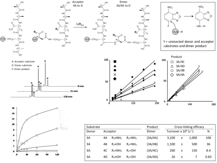
A post-functionalization assay was developed to quantify the substrates and the product of the enzymatic cross-linking reaction catalyzed by Ldtfm from E. faecium, the prototypic enzyme of the
L,D-transpeptidase family (figure 2a). We took advantage of the presence of a single primary amine on these molecules to specifically introduce a 4-fluoro-7-nitrobenzofurazan group by nucleophilic aromatic substitution. This post-functionalization reaction applied to the crude enzymatic reaction converted the unreacted donor and acceptor substrates as well as the L,D -transpeptidation product (dimer) into fluorescent peptides. The post-functionalized peptides were separated by rpHPLC and detected by fluorescence (figure 2b). Since the fluorescence yield may vary with the acetonitrile concentration required for elution and with the molecular environment of the fluorophore within the peptides, calibration curves were obtained for every substrate and every product (figure 2c). For this purpose, each substrate and each product were individually post-functionalized with 4-fluoro-7-nitrobenzofurazan and known amounts of each fluorescent peptide were analyzed by rpHPLC. Access to sensitive determination of the peptides in the 10- to 200-pmol range provided a versatile method to determine the rate of the cross-linking reaction catalyzed by Ldtfm.
Scheme 1. a) Synthetic scheme for the protected DAP and amidated DAP building blocks. i) 5% Grubb’s II catalyst, CH2Cl2, RT, 12 h; ii) 3% PtO2, H2, CH2Cl2/CH3OH/H2O (9/1/1), RT, 12 h; iii) 20% Grubb’s II catalyst, CH2Cl2, 70°C, 48 h; iv) PtO2, H2, CH3OH, 4 atm, RT, 18 h. b) Solid-phase-synthesis of the peptidoglycan precursor analogues using orthogonal Fmoc and ivDde protecting groups.
O O (S) (CH2)4-NH-ivDde H2N Wang-L-Lys(ivDde) i. Coupling ii. Deprotection HO O NHFmoc R n O O (S) (CH2)4-NH-ivDde HN O NH (S) O HN (R) O OtBu i. Coupling ii. Deprotection NHFmoc R1 + ivDde deprotection O i. Fmoc deprotection ii. Side chain deprotection iii. Resin cleavage R1 NHFmoc R1 OH O O O (S) (CH2)4-NH2 HN O NH (S) O HN (R) O OtBu R1 O O (S) (CH2)4-NH HN O NH (S) O HN (R) O OtBu R1 O NH2 R2 OH O (S) (CH2)4-NH HN O NH (S) O HN (R) O OH R2 R2= CO2H or CONH2 (S) BnO2C NHFmoc (R) NHBoc CO2tBu (R) NHBoc CONH2 BnO2C(S) NHFmoc (R)CO2tBu NHBoc BnO2C(S) NHFmoc (R)CONH2 NHBoc HO2C(S) NHFmoc (R)CO2tBu NHBoc HO2C(S) NHFmoc (R)CONH2 NHBoc i ii iii iv + R1= CO2tBu or CONH2 (R) (R) (R) (R) (R) (R) (R) b) a) strand 4→3 Subunit (Disaccharide-pentapeptide) Peptidoglycan subunit
Enterococcus faecium Mycobacterium tuberculosis
b)
a)
L-Ala GlcNAc-MurNAc D-isoGln L-Lys D-Ala D-Ala D-isoAsn 1 2 3 4 5 L-Ala D-isoGln DAPNH2 D-Ala D-Ala 1 2 3 4 5 GlcNAc-MurNGlycThe reaction catalyzed by Ldtfm involves two peptidoglycan
precursors that act as an acyl donor and as an acyl acceptor (figure 2a). For the donor, we used a linear tetrapeptide that cannot be used as an acyl acceptor since it does not harbor the
D-isoAsn residue branched to L-Lys. Conversely, the peptides used as acyl acceptors cannot be used as acyl donors since they do not harbor the essential C-terminal D-Ala residue (D-Ala4). To
specifically assess the impact of amidation in the acceptor substrate, we tested a tetrapeptide donor containing a D-isoGln residue (acyl donor 3A) and acceptors containing the four combinations of amidation of the D-isoGlu and D-isoAsp α-carboxyl groups (acyl acceptors 4A to 4D). The rate of formation of peptidoglycan dimers by Ldtfm was the highest for the fully
amidated acceptor, corresponding to the amidation status found in the peptidoglycan of the E. faecium host (figures 2d and 2e). The lack of amidation of D-isoGln had a moderate impact on the transpeptidase activity of Ldtfm (36% residual activity). The impact
of the lack of amidation was greater for the side-chain D-isoAsn residue (8.4% residual activity), whereas the combination of both modifications almost completely abolished the activity of Ldtfm
(0.83% residual activity). No transpeptidation product was
observed for substrates fully lacking amidation both in the acyl donor and acceptor substrates (data not shown). In conclusion, amidation of the D-isoGlu and D-isoAsp α-carboxyl groups of the acyl acceptor substrate was essential for optimal formation of peptidoglycan cross-links by Ldtfm in vitro.
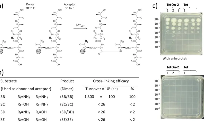
Our following objective was to evaluate the role of amidation of the α- and ε-carboxyl groups of D-isoGlu and DAP on the efficacy of formation of 3→3 cross-links by L,D-transpeptidases from M. tuberculosis. We based our analysis on LdtMt2 as a
representative of the five L,D-transpeptidase paralogues produced by this species. Substrates containing the four combinations of amidation of the linear tetrapeptide stem of M.
tuberculosis were synthesized and independently tested in the
quantitative cross-linking assay (figure 3). No cross-linked dimer was observed with substrates lacking amidation of DAP, D-isoGlu, or both. To assess the impact of the lack of amidation of peptidoglycan precursors on the growth of M. tuberculosis, we constructed mutants of strain H37Rv conditionally producing the DAP amidotransferase AsnB (Rv2201). This analysis showed that the amidation of the ε-carboxyl of DAP is required for growth of M.
tuberculosis H37Rv.
Figure 2. Post-functionalization assay for the cross-linking activity of peptidoglycan transpeptidases. a) Reaction catalyzed by the L,D-transpeptidase Ldtfm. Inset, post-functionalization reaction. b) Separation by rpHPLC of the post-functionalized substrates 3A and 4A and of the reaction product (dimer 3A/4A), which were detected by fluorescence (λex = 470 nm; λem = 530 nm). c) Calibration curves for quantitative determination of the substrates (left panel) and reaction products (right panel). d) Kinetics of dimer synthesis by Ldtfm. e) Impact of amidation of the acyl acceptor on the cross-linking efficacy of Ldtfm.
3A/4C 3A/4D 3A/4A 3A/4B Product
R = unreacted donor and acceptor substrates and dimer product
Substrate Product Cross-linking efficacy Donor Acceptor Dimer Turnover x 106 (s-1) %
3A 4A R1=NH2 R2=NH2 (3A/4A) 3,100 ± 1,000 100
3A 4B R1=OH R2=NH2 (3A/4B) 1,100 ± 500 36
3A 4C R1=NH2 R2=OH (3A/4C) 260 ± 150 8.4
3A 4D R1=OH R2=OH (3A/4D) 26 ± 7 0.83
Ldtfm Acceptor 4A to D 3A/4A to DDimer A: Acceptor substrate D: Donor substrate P: Dimer product
Figure 3. Impact of impaired peptidoglycan precursor amidation on the activity of M. tuberculosis L,D-transpeptidase LdtMt2. a) Transpeptidation reaction catalyzed by LdtMt2. b) In vitro cross-linking activity of LdtMt2. c) Impact of impaired amidation of DAP by AsnB (Rv2201) on the growth of M. tuberculosis H37Rv. Ten-fold dilutions of cultures of mutants Rv2201-TetON-2 and Rv2201-TetON-6 were spotted on the indicated media showing that depletion of AsnB in the absence of anhydrotetracycline prevents growth.
In conclusion, we have developed routes of synthesis of the peptide stems of peptidoglycan precursors that provide access to the full structural diversity found in bacteria, including the presence of DAP or L-Lys at the 3rd position, the presence or
absence of a side-chain, and the amidation of carboxyl groups. We have also developed a sensitive assay for the detection of the products of the cross-linking reaction. Based on these new tools, functional analysis was focused on the impact of amidation of carboxyl groups in the D-isoGlu and DAP residues of peptidoglycan precursors on the activity of L,D-transpeptidases. For the first time, we directly establish that the diversification of the structure of peptidoglycan precursors is associated with a parallel evolution of the substrate specificity of cross-linking enzymes. Amidation of carboxyl groups was essential for the in
vitro activity of L,D-transpeptidases from E. faecium and M.
tuberculosis. Amidation of DAP was also essential for growth of M. tuberculosis indicating that the amidotransferase AsnB is a
potential target for development of new drugs active on multi-drug resistant bacilli.
Acknowledgements
This work was supported by the project NAPCLI from the JPI AMR program to MA.
Keywords: Amidation • Amidotransferase • Mycobacterium
tuberculosis • Peptidoglycan • Transpeptidase
[1] A. J. Egan, R. M. Cleverley, K. Peters, R. J. Lewis, W. Vollmer, FEBS J
2017, 284, 851-867.
[2] E. Sauvage, M. Terrak, Antibiotics (Basel) 2016, 5, pii: E12.
[3] a) K. H. Schleifer, O. Kandler, Bacteriol Rev 1972, 36, 407-477; b) J. L. Mainardi, R. Villet, T. D. Bugg, C. Mayer, M. Arthur, FEMS Microbiol Rev
2008, 32, 386-408.
[4] a) S. Sarkar, E. A. Libby, S. E. Pidgeon, J. Dworkin, M. M. Pires, Angew
Chem Int Ed Engl 2016, 55, 8401-8404; b) D. Münch, T. Roemer, S. H.
Lee, M. Engeser, H. G. Sahl, T. Schneider, PLoS Pathog 2012, 8, e1002509.
[5] A. Arbeloa, J. E. Hugonnet, A. C. Sentilhes, N. Josseaume, L. Dubost, C. Monsempes, D. Blanot, J. P. Brouard, M. Arthur, J Biol Chem 2004, 279, 41546-41556.
[6] a) J. L. Mainardi, M. Fourgeaud, J. E. Hugonnet, L. Dubost, J. P. Brouard, J. Ouazzani, L. B. Rice, L. Gutmann, M. Arthur, J Biol Chem 2005, 280, 38146-38152; b) Y. Qiao, V. Srisuknimit, F. Rubino, K. Schaefer, N. Ruiz, S. Walker, D. Kahne, Nat Chem Biol 2017, 13, 793-798; c) L. Y. Huang, S. H. Huang, Y. C. Chang, W. C. Cheng, T. J. Cheng, C. H. Wong, Angew
Chem Int Ed Engl 2014, 53, 8060-8065.
[7] M. Levefaudes, D. Patin, C. de Sousa-d'Auria, M. Chami, D. Blanot, M. Hervé, M. Arthur, C. Houssin, D. Mengin-Lecreulx, J of Biol Chem 2015,
290, 13079-13094.
[8] a) A. R. Chowdhury, G.-J. Boons, Tetrahedron Lett 2005, 46, 1675-1678; b) D. B. Kastrinsky, P. Kumar, G. A. Marriner, C. E. Barry,
Synthesis-Stuttgart 2012, 44, 3043-3048.
a)
b)
c)
100 10-1 10-2 10-3 10-4 10-5 10-6 100 10-1 10-2 10-3 10-4 10-5 10-6 LdtMt2 Donor 3B to E Acceptor3B to E TetOn-2 1 2 3 Tet 1 TetOn-2 1 2 3 Tet 1 With anhydrotetraSubstrate Product Cross-linking efficacy
(Used as donor and acceptor) (Dimer) Turnover x 106 (s-1) %
3B R1=NH2 R2=NH2 (3B/3B) 1,300 ± 100 100
3C R1=OH R2=NH2 (3C/3C) < 26 < 2
3D R1=NH2 R2=OH (3D/3D) < 26 < 2
Layout 1:
COMMUNICATION
Synthetic routes to the peptide stem of peptidoglycan precursors and a sensitive fluorescent cross-linking assay were developed to assess the impact of structural variability on peptidoglycan polymerization. In the search for new targets for anti-mycobacterial drug development, this strategy was applied to the evaluation of diaminopimelate amidation in Mycobacterium tuberculosis, revealing the essential role of the AsnB amidotransferase for peptidoglycan transpeptidation both in vitro and in vivo.
Flora Ngadjeua#, Emmanuelle Braud#,
Saidbakhrom Saidjalolov, Laura Iannazzo, Dirk Schnappinger, Sabine Ehrt, Jean-Emmanuel Hugonnet, Dominique Mengin-Lecreulx, Delphine Patin, Mélanie Ethève-Quelquejeu*, Matthieu Fonvielle,* and Michel Arthur*
Page No. – Page No.
Critical impact of peptidoglycan precursor amidation on the activity of L,D -transpeptidases from Enterococcus
faecium and Mycobacterium tuberculosis Synthesis of peptidoglycan fragments Sensitive fluorescent cross-linking assay Impact of amidation
AsnB amidotransferase: new target in M . tuberculosis
1
Supporting Information
Critical impact of peptidoglycan precursor amidation on the activity of
L,
D-transpeptidases from Enterococcus faecium and Mycobacterium tuberculosis
Flora Ngadjeua, Emmanuelle Braud, Saidbakhrom Saidjalolov, Laura Iannazzo, Dirk
Schnappinger, Sabine Ehrt, Jean-Emmanuel Hugonnet, Dominique Mengin-Lecreulx, Delphine
Patin, Mélanie Ethève-Quelquejeu, Matthieu Fonvielle, and Michel Arthur
Table of contents
1- General information………p2
2- Organic synthesis………...p3
3- Solid-phase synthesis of linear peptides (3A to 3E) ……….……p10
4- Solid-phase synthesis of branched peptides (4A to 4D) ………p11
5- Mass spectrometry analyses of the peptides………p11
6. rpHPLC analysis of peptides………..p14
7- Protein purification……….………p15
8- Cross-linking assay………..……p15
9- Conditional production of the DAP amidase AsnB in M. tuberculosis……….p17
2 1- General information
All reagents were obtained from commercial suppliers and used without further purification. Solvents were dried using standard methods and distilled before use. TLC: recoated silica gel thin layer sheets 60F254 plates (Merck) were used for analytical thin layer chromatography. Flash column chromatography was performed on silica gel 60 (40-63 µm, Merck). Optical rotations were measured with a sodium lamp (589 nm) at 20°C on a Perkin Elmer polarimeter. NMR spectra were recorded at 300 K on Bruker AM250 or Bruker Advance II 500 spectrometers. Chemical shifts (δ) are expressed in ppm relative to the residual solvent resonance and coupling constants (J) are in Hertz (Hz). Multiplicities are reported as follows: singlet (s), doublet (d), triplet (t), multiplet (m), and broad resonance (br). Low resolution mass spectra (LRMS) were obtained on a LCQ Advantage mass spectrometer (ThermoElectron) and high resolution mass spectra (HRMS) were recorded on a TOF mass analyzer under electrospray ionisation.
3 2- Organic synthesis
2a- Synthesis of Fmoc-D-Glu-NHTrt (Compound 4)
Scheme S1. Synthesis of Fmoc-D-Glu-NHTrt (4).
(R)-Benzyl 5-amino-4-((tert-butoxycarbonyl)amino)-5-oxopentanoate (1)
Ethyl chloroformate (0.31 mL, 3.25 mmol) was added dropwise at -20 °C to a solution of Boc-D
-Glu(OBn)-OH (1.00 g, 2.96 mmol) and triethylamine (0.45 mL, 3.25 mmol) in anhydrous THF (15 mL). The solution was stirred for 30 minutes before addition of NH4OH (28%, 1 mL, 14.80 mmol) at 0 °C. The reaction mixture was stirred at 0 °C for 2 hours. The solvent was removed under reduced pressure and the residue dissolved in dichloromethane. The organic phase was washed with a saturated solution of NH4Cl, dried over MgSO4, filtered and concentrated to afford Boc-D-Glu(OBn)-NH2 1 as a white powder (0.96 g, 96%). 1H NMR (250 MHz, CDCl
3): δ 7.39 (br s, 5H, Ar-H), 6.23 (br s, 1H, NH), 5.43 (br s, 1H, NH), 5.28 (d, J = 7.2 Hz, 1H, NH), 5.16 (s, 2H, CH2), 4.26-4.19 (m, 1H, CH), 2.68-2.43 (m, 2H, CH2), 2.27-2.13 (m, 1H, CH2), 2.03-1.88 (m, 1H, CH2), 1.46 (s, 9H, CH3-Boc). 13C NMR (125 MHz, CDCl3): δ 175.7 (C=O), 172.8 (C=O), 156.3 (C=O), 136.1, 128.1, 127.7, 79.3 (C-Boc), 65.9 (CH2), 53.5 (CH), 30.0 (CH2), 27.3 (CH3), 27.2 (CH2). LRMS: Calcd for C17H24N2NaO5 [M+Na]+ 359.3; Found: 359.3.
(R)-Benzyl 4-((((9H-fluoren-9-yl)methoxy)carbonyl)amino)-5-amino-5-oxopentanoate (2)
A solution of Boc-D-Glu(OBn)-NH2 1 (0.94 g, 2.85 mmol) in HCl (4 N) /dioxane (21 mL) was stirred at room temperature for 2 hours. The reaction mixture was concentrated and the residue was dissolved in H2O/dioxane (80 mL, v/v). NaHCO3 (1.43 g, 17.10 mmol) and FmocCl (0.96 g, 3.70 mmol) were then added. After 2 hours stirring at room temperature, EtOAc was added to the mixture. The organic phase was washed with a saturated solution of NaHCO3, dried over MgSO4, filtered, and concentrated under reduced pressure. A silica gel chromatography (cyclohexane/EtOAc, 8:2) was performed on the residue to obtain the Fmoc-D-Glu(OBn)-NH2 2 as a white powder (0.86 g, 71%). [α]D- 2.2° (10 mg/mL, DMF). 1H NMR (250 MHz, DMSO): δ 7.89 (d, J = 7.2 Hz, 2H, Ar-H), 7.73 (d, 2H, Ar-H), 7.47-7.29 (m, 9H, Ar-H), 7.07 (s, 1H, NH), 5.10 (s, 2H, CH2), 4.30-4.21 (m, 3H, CH2-Fmoc and CH-Fmoc), 4.06-3.92 (s, 1H, CH), 2.39 (t,
4
(C=O), 155.9 (C=O), 143.8, 143.7, 140.7, 136.1, 128.4, 127.9, 127.8, 127.6, 127.0, 125.2, 120.0, 65.6 (CH2), 65.4 (CH2), 53.7 (CH), 46.7 (CH), 30.2 (CH2), 27.1 (CH2). LRMS: Calcd for C27H26N2NaO5 [M+Na]+ 481.4; Found: 481.3.
(R)-Benzyl 4-((((9H-fluoren-9-yl)methoxy)carbonyl)amino)-5-oxo-5-(tritylamino)pentanoate (3) To a mixture of Fmoc-D-Glu(OBn)-NH2 (2) (0.68 g, 1.60 mmol) and trityl alcohol (4.16 g, 16.00 mmol) in acetic acid (5.6 mL), sulfuric acid (51 µL, 0.96 mmol) and acetic anhydride (4.00 mmol) were added at 50 °C. The reaction mixture was stirred at 50 °C for 3.5 hours and diluted with EtOAc. The organic phase was washed with a saturated solution of NaHCO3, dried over MgSO4, filtered and concentrated under reduced pressure. Silica gel chromatography (cyclohexane/EtOAc, 7:3) afforded Fmoc-D
-Glu(OBn)-NHTrt3 as a white powder (0.82 g, 73%). [α]D+ 31.2° (10 mg/mL, MeOH). 1H NMR (250 MHz, CDCl3): δ 7.78 (d, 2H, J = 7.5 Hz, Ar-H), 7.60-7.57 (m, 2H, Ar-H), 7.46-7.28 (m, 24H, Ar-H), 5.64-5.62 (m, 1H, NH), 5.14 (br s, 2H, CH2), 4.40 (m, 3H, CH2-Fmoc and CH-Fmoc or CH), 4.22-4.19 (m, 1H, CH-Fmoc or CH), 2.61-1.87 (m, 4H, 2 CH2). 13C NMR (125 MHz, CDCl3): δ 172.8 (C=O), 171.6 (C=O), 157.2 (C=O), 144.2, 143.8, 143.2, 141.2, 141.1, 136.1, 128.4, 128.1, 127.8, 127.7, 127.3, 126.7, 126.5, 124.8, 124.7, 119.5, 70.1 (Cq), 66.6 (CH2), 66.0 (CH2), 54.6 (CH), 48.4 (CH), 29.8 (CH2), 26.2 (CH2). LRMS: Calcd for C46H40N2NaO5 [M+Na]+ 723.2; Found: 722.9.
(R)-4-((((9H-Fluoren-9-yl)methoxy)carbonyl)amino)-5-oxo-5-(tritylamino)pentanoic acid (4)
10 wt. % Pd/C (0.04 g, 0.38 mmol) was added to a solution of Fmoc-D-Glu(OBn)-NHTrt 3 (0.40 g, 0.57
mmol) in anhydrous THF (6 mL). The solution was hydrogenated overnight under atmospheric pressure. The catalyst was removed by filtration through celite and the filtrate was evaporated. Silica gel chromatography (dichloromethane/MeOH, 95:5) afforded the Fmoc-D-Glu-NHTrt 4 as a white
powder (0.29 g, 83%). [α]D + 42.6° (10 mg/mL, MeOH). 1H NMR (250 MHz, CDCl3): δ 7.78 (d, J = 5.2 Hz, 2H, Ar-H), 7.60-7.57 (m, 2H, Ar-H), 7.55-7.42 (m, 2H, Ar-H), 7.39-7.23 (m, 17H, Ar-H), 5.66 (d, J = 6.5 Hz, 1H, NH), 4.49-4.39 (m, 3H, CH2-Fmoc and CH), 4.22-4.16 (t, J = 6.8 Hz, 1H, CH-Fmoc), 2.53-2.07 (m, 3H, CH2), 1.95-1.83 (m, 1H, CH2). 13C NMR (125 MHz, CDCl3): δ 176.4 (C=O), 170.4 (C=O), 156.8 (C=O), 144.1, 143.7, 143.6, 141.3, 128.6, 127.9, 127.8, 127.1, 127.0, 125.1, 120.0, 70.6 (Cq), 67.5 (CH2), 54.2 (CH), 47.0 (CH), 29.7 (CH2), 27.7 (CH2). HRMS: Calcd for C39H34N2NaO5 [M+Na]+ 633.2360; Found: 633.2411.
5 2b- Synthesis of Boc-D-Asp-NH2 (compound 6)
Scheme S2. Synthesis of Boc-D-Asp-NH2 (6).
(R)-Benzyl 4-amino-3-((tert-butoxycarbonyl)amino)-4-oxobutanoate (5)
Ethyl chloroformate (0.15 mL, 1.70 mmol) was added dropwise at -20 °C to a solution of Boc-D
-Asp(OBn)-OH (0.50 g, 1.55 mmol) and triethylamine (0.23 mL, 1.70 mmol) in anhydrous THF (15 mL). The solution was stirred for 30 minutes. NH4OH (28%, 0.52 mL, 7.75 mmol) was added at 0 °C and the reaction mixture was stirred at 0 °C for 2 hours. The solvent was removed under reduced pressure and the residue was dissolved in dichloromethane. The organic phase was washed with water, dried over MgSO4, filtered and concentrated affording Boc-D-Asp(OBn)-NH2 5 as a white powder (0.46 g, 93%). 1H NMR (250 MHz, CDCl3): δ 7.38 (br s, 5H, Ar-H), 6.45 (br s, 1H, NH), 5.70 (br s, 1H, NH), 5.43 (br s, 1H, NH), 5.17 (s, 2H, CH2), 4.56 (br s, 1H, CH), 3.10 (dd, J = 17.0 Hz, J = 4.0 Hz, 1H, CH2), 2.74 (dd, 1H, CH2), 1.48 (s, 9H, CH3-Boc). The spectroscopic data were consistent with the literature data for the S enantiomer.[1]
(R)-4-Amino-3-((tert-butoxycarbonyl)amino)-4-oxobutanoic acid (6)
10 wt. % Pd/C (0.014 g, 0.13 mmol) was added to a solution of Boc-D-Asp(OBn)-NH2 5 (0.46 g, 1.43 mmol) in anhydrous THF (16 mL). The reaction mixture was hydrogenated overnight under atmospheric pressure. The solution was filtered through celite and evaporated under reduced pressure. Flash chromatography (Dichloromethane/MeOH, 9:1) afforded Boc-D-Asp-NH2 6as a white powder (0.28 g, 86%). [α]D + 29.4° (10 mg/mL, DMF). 1H NMR (250 MHz, DMSO): δ 12.29 (br s, 1H, CO2H), 7.18 (br s, 1H, NH), 7.03 (br s, 1H, NH), 6.93 (d, J = 8.2 Hz, 1H, NH), 4.25-4.16 (m, 1H, CH), 2.62 (dd, J = 16.2 Hz, J = 5.2 Hz, 1H, CH2), 2.44 (dd, 1H, CH2), 1.49 (s, 9H, CH3-Boc). 13C NMR (125 MHz, CDCl3): δ 173.0 (C=O), 171.9 (C=O), 155.1, (C=O), 78.1 (Cq), 50.8 (CH), 36.3 (CH2), 28.1 (CH3-Boc). LRMS: Calcd for C9H15N2O5 [2M-H]- 463.1; Found: 463.1.
6 2c- Synthesis of D-lactic acid derivative (compound 8)
Scheme S3. Synthesis of D-lactic acid derivate (8).
(R)-Benzyl 2-(tert-butoxy)propanoate (7)
A solution of D-lactic acid-benzyl ester (5.00 g, 27.74 mmol) in dry DCM (18 mL) was treated with di-t-butyl dicarbonate (15.14 g, 69.40 mmol) and magnesium perchlorate (0.63 g, 2.80 mmol), and the mixture was refluxed for 12 hours. The solution was poured into a saturated aqueous NaHCO3 solution and the mixture was extracted with EtOAc. The organic layer was washed with water, dried with brine and over MgSO4, filtered and concentrated under reduced pressure. The residue was purified by flash chromatography (silica gel, cycloexane/AcOEt 75/25) affording O-tBu-D-lactic acid benzyl ester 7 as a
colorless oil (4.00 g, 62%). [α]D + 35.2˚ (10 mg/mL, MeOH). 1H NMR (500 MHz, CDCl3): δ 7.34-7.29 (m, 5H, Ar-H), 5.14 (dd, J = 12.5 Hz, 2H, CH2), 4.14 (q, J = 7 Hz, 1H, CH), 1.33 (d, J = 7 Hz, 3H, CH3), 1.15 (s, 9H, CH3-tBu), 13C NMR (125 MHz, CDCl3): δ 174.8 (C=O), 135.8 (C-Ar), 128.5 (CH-Ar), 128.2 (CH-Ar), 128.2 (CH-Ar), 74.9 (Cq), 67.5 (CH), 66.4 (CH2), 27.7 (CH3-tBu), 20.5 (CH3). LRMS: Calcd for C14H20NaO3 [M+Na]+ 254.1; Found: 254.0.
(R)-2-(tert-Butoxy)propanoic acid (8)
A suspension of compound 7 (1.80 g, 7.62 mmol) in MeOH (75 mL) was treated with 10 wt. % Pd/C (0.26 g, 2.44 mmol) under 4 bar H2. The reaction mixture was stirred for 12 hours at room temperature. The residue was filtered through celite and concentrated under reduced pressure to afford D-lactic acid derivative 8 as a colorless oil (1.00 g, 90%). [α]D + 42.3˚ (10 mg/mL, EtOH). 1H NMR (500 MHz, CDCl3): δ 4.12 (q, J = 7 Hz, 1H, CH), 1.36 (d, J = 7 Hz, 3H, CH3), 1.20 (s, 9H, tBu). 13C NMR (125 MHz, CDCl3): δ 175.3 (C=O), 75.2 (Cq), 66.4 (CH), 26.8 (CH3-tBu), 19.4 (CH3). LRMS: Calcd for C7H15O3+ [M+H]+ 147.1; Found: 146.9.
7 2d- Synthesis of DAP derivative (compound 12)
Scheme S4. Synthesis of DAP derivative (12).
(2S,6R,E,Z)-1-Benzyl 7-tert-butyl 2-((((9H-fluoren-9-yl)methoxy)carbonyl)amino)-6-((tert-butoxycarbonyl)amino)hept-3-enedioate (11)[2]
A solution of vinyl-glycine 92 (0.42 g, 1.06 mmol) in dichloromethane (10 mL) was treated with allyl-glycine 10[3] (0.24 g, 0.88 mmol) and Grubb’s second generation catalyst (0.04 g, 0.04 mmol). The solution was stirred for 18 hours at room temperature. Dichloromethane was removed under reduced pressure. The obtained residue was purified by flash chromatography (Cyclohexane/EtOAc, 7:3) affording olefin 11 as a colorless oil (0.32 g, 55%). 1H NMR (500 MHz, CDCl3): δ 7.80 (d, J = 7.5 Hz, 2H, Ar-H), 7.64 (d, J = 5 Hz, 2H, Ar-H), 7.46-7.34 (m, 9H, Ar-H), 5.72-5.68 (m, 2H, Hallyl), 5.53 (d, J = 7.5 Hz, 1H, NH), 5.28-5.10 (m, 3H, CH2-Bn and NH), 4.99-4.94 (m, 1H, CH), 4.45-4.39 (m, 2H, CH2-Fmoc), 4.30-4.22 (m, 2H, 2 CH), 2.62-2.43 (m, 2H, CH2), 1.45 (s, 18H, CH3-tBu/Boc). HRMS: Calcd for C38H45N2O8 [M+H]+ 657.3170; Found: 657.3176.
(2S,6R)-2-((((9H-Fluoren-9-yl)methoxy)carbonyl)amino)-7-(tert-butoxy)-6-((tert-butoxycarbonyl)amino)-7-oxoheptanoic acid(12)[2]
The product 11 (0.32 g, 0.49 mmol) was dissolved in DCM/MeOH/H2O (9:1:1 v/v/v) (33 mL) and PtO2 (0.004 g, 0.02 mmol) was added under H2 atmosphere. The reaction mixture was allowed to stir for 18 hours at room temperature. The solution was filtered through celite and concentrated under reduced pressure. The obtained residue was purified by flash chromatography (dichloromethane/MeOH, 9:1) to afford protected diaminopimelic acid (DAP) 12 as a white powder (0.29 g, 90%). 1H NMR (500 MHz, MeOD): δ 7.75 (d, J = 7.8 Hz, 2H, Ar-H), 7.64 (d, J = 7.8 Hz, 2H, Ar-H), 7.39-7.26 (m, 2H, Ar-H), 7.00-6.79 (m, 2H, Ar-H), 4.36-4.31 (m, 2H, CH2-Fmoc or 2 CH), 4.21-4.07 (m, 2H, CH2-Fmoc or 2 CH), 3.97-3.93 (m, 1H, CH), 1.89-1.59 (m, 6H, CH2), 1.44 (s, 9H, CH3), 1.43 (s, 9H, CH3). 13C NMR (125 MHz, MeOD): δ 173.8 (C=O), 158.5 (C=O), 158.1 (C=O), 145.4, 145.2, 142.6, 128.7, 128.2, 126.2, 120.9, 82.5 (Cq), 80.4 (Cq), 67.8 (CH2-Fmoc), 55.9 (CH), 54.7 (CH), 49.7 (CH-Fmoc), 32.5 (CH2), 30.6 (CH2), 28.7 (CH3), 28.3 (CH3) 23.4 (CH2). HRMS: Calcd for C31H41N2O8 [M+H]+ 569.2857; Found: 569.2861.
8 2e- Synthesis of D-Allyl-Glycine (14)
Scheme S5. Synthesis of D-Allyl-Glycine (14).
(R)-2-((tert-Butoxycarbonyl)amino)pent-4-enoic acid (13)[4]
Toa solution of commercially available (2S)-2-aminopent-4-enoic acid (1.00 g, 8.70 mmol) in 1 M NaOH (20 mL) and dioxane (10 mL) at 0 ˚C was added di-tbutyl dicarbonate (2.28 g, 10.5 mmol). The reaction mixture was allowed to warm to room temperature and stirred for an additional 18 hours. The pH was checked and adjusted to basic when necessary. The reaction mixture was concentrated under reduced pressure and the aqueous phase washed with Et2O (2 x 10 mL). The aqueous phase was acidified to pH = 2 with 2 M H2SO4 and extracted with EtOAc (4 x 20 mL) while saturating the aqueous phase each time with NaCl. The combined organic layers were dried over MgSO4, filtered and concentrated under reduced pressure to afford the product 13 as a colorless oil (1.68 g, 90%). 1H NMR (250 MHz, CDCl3): δ 5.75-5.61 (d, J = 15 Hz, 1H, Hallyl), 5.14-5.03 (m, 2H, Hallyl), 4.47-4.40 (m, 1H, CH), 2.49-2.43 (m, 2H, CH2), 1.33 (s, 9H, CH3-Boc).
(R)-tert-Butyl (1-amino-1-oxopent-4-en-2-yl)carbamate (14)
To a solution of compound 13 (1.00 g, 4.67 mmol) in dry THF (40 mL), triethylamine (0.71 mL, 5.12 mmol) and ethyl chloroformate (0.43 mL, 5.6 mmol) were added at 0 ˚C. The reaction mixture was warmed to room temperature and stirred for 2 hours. The reaction mixture was then cooled to 0 ˚C and treated dropwise with a solution of NH4OH (28%, 1 mL, 14.80 mmol). The solution was stirred for extra 3 hours at room temperature. THF was removed under reduced pressure and the residue was dissolved in dichloromethane. The reaction mixture was washed with brine, dried over MgSO4, filtered and concentrated under reduced pressure. The obtained residue was purified by flash chromatography (Cyclohexane/EtOAc ,7:3) to afford the allyl-glycine14 as a white solid (0.90 g, 90%). [α]D + 7° (10 mg/mL, CHCl3). 1H NMR (500 MHz, CDCl3): δ 6.08 (br s, 1H, NH), 5.84-5.68 (m, 1H, Hallyl), 5.44 (br, 1H, NH), 5.19-5.14 (m, 2H, Hallyl), 4.16-4.13 (m, 1H, CH), 2.52-2.47 (m, 2H, CH2), 1.42 (s, 9H, CH3-Boc). 13C NMR (125 MHz, MeOD): δ 175.7 (C=O), 156.2 (C=O), 133.2 (C=CH2), 117.1 (CH2=CH), 79.0 (Cq), 54.0 (CH), 36.3 (CH2), 26.9 (CH3). HRMS: Calcd for C10H19N2O3 [M+H]+ 215.1390; Found: 215.1387.
9 2f- Synthesis of amidated DAP (16)
Scheme S6. Synthesis of amidated DAP (16).
(2S,6R,E,Z)-Benzyl 2-((((9H-fluoren-9-yl)methoxy)carbonyl)amino)-7-amino-6-((tert-butoxycarbonyl) amino)-7-oxohept-3-enoate (15)[5]
A solution of vinyl-glycine 9[2] (0.22 g, 0.53 mmol) in dichloroethane (10 mL) was treated with allyl-glycine 14 (0.27 g, 1.06 mmol) and Grubb’s Second Generation Catalyst (0.09 g, 0.16 mmol). The reaction mixture was stirred for 48 hours at 70 ˚C. The solution was cooled to room temperature, treated with DMSO (0.5 mL), and stirred for additional 8 hours. The solution was diluted in dichloromethane (100 mL), washed with water (2 x 20 mL), dried over MgSO4, filtered and concentrated under reduced pressure. The obtained residue was purified by flash chromatography (DCM/MeOH, 9:1) to afford a mixture (0.14 g) of 15 and unreacted 14 with a ratio of 1:2 (15:14) which was engaged in the next step. 1H NMR (500 MHz, CDCl
3): δ 7.81 (d, J = 7.5 Hz, 2H, Ar-H), 7.63 (d, J = 7.5 Hz, 2H, Ar-H), 7.46-7.38 (m, 9H, Ar-H), 6.1 (br, 1H, NH), 5.79 (br, 1H, NH), 5.69-5.68 (m, 2H, Hallyl), 5.30 (br, 1H, NH), 5.18 (s, 2H, CH2-Ar), 4.89 (m, 1H, CH-Fmoc or CH), 4.43-4.31 (m, 2H, CH2-Fmoc), 4.26-4.10 (m, 1H, CH-Fmoc or CH), 2.63-2.37 (m, 2H, CH2), 1.46 (s, 9H, CH3-Boc). LRMS: Calcd for C34H38N3O7 [M+H]+ 600.2; Found: 600.1.
(2S,6R)-2-((((9H-Fluoren-9-yl)methoxy)carbonyl)amino)-7-amino-6-((tert-butoxycarbonyl)amino)-7-oxoheptanoic acid (16)
The mixture 14:15 (0.22 g) was dissolved in MeOH (15 mL) and treated with PtO2 (0.10 g, 0.44 mmol) under 4 bar H2. The reaction mixture was stirred for 24 hours at room temperature. The mixture was filtrated through celite and concentrated under reduced pressure. The obtained residue was purified by flash chromatography (DCM/MeOH/H2O, 8:2:1) to afford amidated DAP 16 as a white powder (0.06 g, 30% over 2 steps). [α]D + 2˚ (5 mg/mL, MeOH). 1H NMR (500 MHz, MeOD): δ 7.76 (d, J = 7.5 Hz, 2H, Ar-H), 7.65-7.62 (m, 2H, Ar-H), 7.37-7.27 (m, 4H, Ar-H), 4.33-4.31 (m, 2H, CH2-Fmoc), 4.20-4.17 (m, 1H, CH-Fmoc), 4.05-3.99 (m, 2H, 2CH), 1.89-1.49 (m, 6H, CH2) 1.40 (s, 9H, CH3-Boc). 13C NMR (125 MHz, MeOD): δ 178.1 (C=O), 158.5 (C=O), 157.8 (C=O), 145.4 (Cq), 145.2 (Cq), 142.6 (Cq), 128.8 (CH-Fmoc), 128.2 (CH-Fmoc), 126.2 (CH-Fmoc), 120.9 (CH-Fmoc), 80.6 (Cq), 67.9 (CH2-Fmoc), 56.6 (CH), 55.8 (CH),
10
48.4 (CH-Fmoc), 33.2 (CH2), 33.1 (CH2), 28.7 (CH3-Boc), 23.30 (CH2). HMRS: Calcd for C27H32N3O7 [M - H]- 510.2246; Found: 510.2245.
3- Solid-phase synthesis of linear peptides (3A to 3E)
Peptides were manually assembled on a Fmoc-D-Ala-Wang resin (0.80 mmol.g-1, 75 mg). Coupling
reactions were performed in 2 mL of anhydrous DMF containing 3 or 1.5 molar equivalent excess of commercial and non-commercial Fmoc-amino acids, respectively, 3 equivalents of K-Oxyma, and 6 equivalents of DIC and DIPEA. Each coupling was performed twice for 5 hours and 12 hours. Fmoc was removed twice (5 minutes and 7 minutes) with a 20% solution of piperidine in DMF. Final deprotection of acid labile protecting groups and cleavage from the resin were carried out under gentle agitation at room temperature for 2 hours with 2 mL of a TFA solution containing DCM, TIPS and water (80:20:5:5 v/v/v/v). The solution was filtered and the resin was washed with 1 mL of TFA. The TFA solutions were pooled and evaporated under reduced pressure. The crude product was dissolved in a 10% acetic acid solution (5 mL) and extracted with chloroform (3 x 10 mL). The aqueous solution was lyophilized and the peptides were purified by rpHPLC on a 5 µm Nucleosil preparative C-18 column (22 x 250 mm) using a linear gradient (0 to 100% buffer B) applied between 10 minutes and 40 minutes (Buffer A: 0.1 % TFA in H2O; buffer B: 0.1 % TFA in CH3CN; 10 mL.min-1). Peptide-containing fractions were identified at 214 nm and analyzed by mass spectrometry (Supplementary figure S1). Peptides were lyophilized and dissolved in water at a final concentration of 10 mg.mL-1. Purity was assessed by rpHPLC (analytical C18 Nucleosil column, 3 µm, 4.6 x 250 mm) using a linear gradient (0 to 50 buffer B) applied between 10.5 and 40.5 min (Buffer A: 0.1 % TFA in H2O; buffer B: 0.1 % TFA in CH3CN; 1 mL.min-1) (Supplementary figure S2). Peptide concentration was determined in duplicate by acid hydrolysis and injection into a Hitachi L8800 amino acid analyzer equipped with a 2620 MSC-PS column.
HO (R) NH (S) HN (R) NH (S) HN O O NH2 O O (CH2)4 H2N O (R) OH O 3A HO (R) NH (S) HN (R) NH (S) HN O O NH2 O O (CH2)3 (R) O (R) OH O 3B H2N H2N O HO (R) NH (S) HN (R) NH (S) HN O O OH O O (CH2)3 (R) O (R) OH O 3C H2N H2N O HO (R) NH (S) HN (R) NH (S) HN O O NH2 O O (CH2)3 (R) O (R) OH O 3D H2N HO O HO (R) NH (S) HN (R) NH (S) HN O O OH O O (CH2)3 (R) O (R) OH O 3E H2N HO O
11 4- Solid-phase synthesis of branched peptides (4A to 4D)
HO (R) NH (S) HN (R) NH (S) HO O O NH2 O O (CH2)4 N H O (R) H2N O H2N O 4A HO (R) NH (S) HN (R) NH (S) HO O O OH O O (CH2)4 N H O (R) H2N O H2N O 4B HO (R) NH (S) HN (R) NH (S) HO O O NH2 O O (CH2)4 N H O (R) H2N O HO O 4C HO (R) NH (S) HN (R) NH (S) HO O O OH O O (CH2)4 N H O (R) H2N O HO O 4D
Branched peptides were synthesized via divergent Fmoc solid phase peptide synthesis using orthogonal protecting groups on the lysine residue. The starting material was a commercial Wang resin containing L-Lys with the α and ε NH2 groups protected with Fmoc and ivDde groups, respectively. Peptides were manually assembled on the resin (0.69 mmol.g-1, 75 mg), starting with the linear tripeptide to afford OtBu-D-Lac-L-Ala-D-iGlx-L-Lys(ivDde)-WANG resin, as described above. Then, the ε NH2 group of L-Lys was deprotected by hydrazinolysis of the ivDde protective group (5% of hydrazine mono-hydrate in DMF, 3 x 15 minutes). Fmoc-D-isoAsn or Fmoc-D-isoAsp was incorporated and the
final deprotection steps were performed as described above. 5- Mass spectrometry analyses of the peptide
Peptide Formula for [M+H]+ Calc. Found
3A C20H37N6O8+ 489.27 489.27 3B C21H38N7O9+ 532.27 532.20 3C C21H37N6O10+ 533.26 533.13 3D C21H37N6O10+ 533.26 533.20 3E C21H36N5O11+ 534.24 534.13 4A C21H38N7O9+ 532.27 532.27 4B C21H37N6O10+ 533.26 533.27 4C C21H37N6O10+ 533.26 533.27 4D C21H36N5O11+ 534.24 534.20
12 p p , , , T:+ p ESI Full ms [150,00-2000,00] 200 400 600 800 1000 1200 1400 1600 1800 2000 m/z 0 5 10 15 20 25 30 35 40 45 50 55 60 65 70 75 80 85 90 95 100 R el at iv e A bunda nc e 489,27 977,20 391,13 511,27 849,07 1048,13 1222,13 1465,20 284,33 1709,53 217,07 648,87740,93 1402,47 1636,93 1822,731912,67 p p , , ,
T:+ p ESI Full ms2 489,30@cid28,00 [150,00-2000,00]
180 200 220 240 260 280 300 320 340 360 380 400 420 440 460 480 500 m/z 0 5 10 15 20 25 30 35 40 45 50 55 60 65 70 75 80 85 90 95 100 R el at iv e A bunda nc e 383,13 472,20 400,07 489,13 195,13 240,00 346,20 218,00 454,07 257,00 271,93 328,93 355,07 365,07 417,20 222,27 311,07 444,33 500,93 201,00 177,40 284,07 T:+ p ESI Full ms [250,00-2000,00] 300 400 500 600 700 800 900 1000 1100 1200 1300 1400 1500 1600 1700 1800 1900 2000 m/z 0 5 10 15 20 25 30 35 40 45 50 55 60 65 70 75 80 85 90 95 100 R el at iv e A bundanc e 532,20 1063,20 660,27 554,27 1117,20 1967,60 1595,33 515,20 1191,20 1328,67 1861,73 404,27 935,87 327,27 682,40 846,07 1505,47 1722,60 p p _p _
T:+ p ESI Full ms2 532,30@cid26,00 [150,00-550,00]
260 280 300 320 340 360 380 400 420 440 460 480 500 520 m/z 0 5 10 15 20 25 30 35 40 45 50 55 60 65 70 75 80 85 90 95 100 R el at iv e A bundanc e 515,13 426,07 443,07 389,13 498,13 283,07 300,07 261,13 409,07 532,13 355,13 327,00 372,20 265,07 290,27 310,07 337,33 423,20 438,27 453,00 470,20 487,13 p y p g , , , , , T:+ p ESI Full ms [250,00-2000,00] 300 400 500 600 700 800 900 1000 1100 1200 1300 1400 1500 1600 1700 1800 1900 2000 m/z 0 5 10 15 20 25 30 35 40 45 50 55 60 65 70 75 80 85 90 95 100 R el at iv e A bundanc e 533,13 662,13 1065,13 376,27 503,07 604,27 789,07 893,93 362,00 704,20 1020,33 1127,201194,471306,60 1487,731587,601661,27 1827,871914,20 g p p g g
T:+ p ESI Full ms2 533,10@cid27,00 [150,00-550,00]
160 180 200 220 240 260 280 300 320 340 360 380 400 420 440 460 480 500 520 540 m/z 0 5 10 15 20 25 30 35 40 45 50 55 60 65 70 75 80 85 90 95 100 R el at iv e A bundanc e 301,07 444,00 390,07 172,07 515,00 261,07 426,07 283,07 244,07 372,07 497,07 533,00 155,07 199,07 227,00 265,00 327,13342,93 409,07 462,00479,80 T:+ p ESI Full ms [250,00-2000,00] 300 400 500 600 700 800 900 1000 1100 1200 1300 1400 1500 1600 1700 1800 1900 2000 m/z 0 5 10 15 20 25 30 35 40 45 50 55 60 65 70 75 80 85 90 95 100 R el at iv e A bunda nc e 533,20 1065,13 516,13555,20 1526,93 328,13 427,00 595,07 705,13 822,20887,60937,00 1087,20 1192,07 1332,13 1450,53 1597,13 1785,331897,73
T:+ p ESI Full ms2 533,30@cid25,00 [250,00-2000,00]
260 280 300 320 340 360 380 400 420 440 460 480 500 520 540 560 m/z 0 5 10 15 20 25 30 35 40 45 50 55 60 65 70 75 80 85 90 95 100 R el at iv e A bunda nc e 516,07 427,07 444,00 390,13 533,13 284,07 262,13 301,07 372,93 498,20 452,80 488,07 520,33 382,00398,67 328,13 355,93 471,13 271,87 289,00 409,13 HO (R) NH (S) HN (R) NH (S) HN O O NH2 O O (CH2)3 (R) O (R) OH O 3B H2N H2N O HO (R) NH (S) HN (R) NH (S) HN O O OH O O (CH2)3 (R) O (R) OH O 3C H2N H2N O HO (R) NH (S) HN (R) NH (S) HN O O NH2 O O (CH2)3 (R) O (R) OH O 3D H2N HO O HO (R) NH (S) HN (R) NH (S) HN O O NH2 O O (CH2)4 H2N O (R) OH O 3A
13 p p , , , T:+ p ESI Full ms [250,00-2000,00] 300 400 500 600 700 800 900 1000 1100 1200 1300 1400 1500 1600 1700 1800 1900 2000 m/z 0 5 10 15 20 25 30 35 40 45 50 55 60 65 70 75 80 85 90 95 100 R el at iv e A bundan ce 534,13 1067,13 1600,47 391,20445,20 895,00 1334,00 302,13 553,00 706,20 836,60 977,93 1131,071238,80 1420,33 1535,07 1779,801867,871956,93 p p , , ,
T:+ p ESI Full ms2 534,20@cid25,00 [250,00-2000,00]
260 280 300 320 340 360 380 400 420 440 460 480 500 520 540 m/z 0 5 10 15 20 25 30 35 40 45 50 55 60 65 70 75 80 85 90 95 100 R el at iv e A bundan ce 445,07 391,13 302,07 534,13 516,13 262,13 427,20 283,93 328,00 355,47 373,20 409,33 452,87 471,00 498,00 273,13 p p _ T:+ p ESI Full ms [150,00-2000,00] 200 400 600 800 1000 1200 1400 1600 1800 2000 m/z 0 5 10 15 20 25 30 35 40 45 50 55 60 65 70 75 80 85 90 95 100 R el at iv e A bundanc e 532,27 1063,27 944,53 1527,00 554,47 1101,27 471,27 418,20 660,40 1287,53 1776,67 219,13 397,80 718,40845,20 1003,20 1136,67 1484,73 1692,33 1960,47 p p _
T:+ p ESI Full ms2 532,30@cid26,00 [150,00-2000,00]
200 220 240 260 280 300 320 340 360 380 400 420 440 460 480 500 520 540 m/z 0 5 10 15 20 25 30 35 40 45 50 55 60 65 70 75 80 85 90 95 100 R el at iv e A bundanc e 487,13 418,13 515,07 498,13 355,07 389,27 470,13 532,27 372,27 261,07 327,27 452,20 426,13 216,07 244,27 272,20290,07310,00 332,00 407,53 T:+ p ESI Full ms [150,00-2000,00] 200 400 600 800 1000 1200 1400 1600 1800 2000 m/z 0 5 10 15 20 25 30 35 40 45 50 55 60 65 70 75 80 85 90 95 100 R el at iv e A bundanc e 533,27 1065,20 419,33 1987,87 555,27 472,00 391,13 1104,27 1597,13 1687,60 1917,33 217,07 697,00760,53 902,131005,07 1289,73 1425,87 1814,33
T:+ p ESI Full ms2 533,20@cid26,00 [150,00-2000,00]
160 180 200 220 240 260 280 300 320 340 360 380 400 420 440 460 480 500 520 540 m/z 0 5 10 15 20 25 30 35 40 45 50 55 60 65 70 75 80 85 90 95 100 R el at iv e A bundanc e 488,07 390,13 419,07 261,00 533,07 515,07 498,07 470,00 372,00 215,87227,27 310,47327,80 355,13 401,20 452,07 197,60 273,33 426,27 p p , , , , , T:+ p ESI Full ms [150,00-2000,00] 200 400 600 800 1000 1200 1400 1600 1800 2000 m/z 0 5 10 15 20 25 30 35 40 45 50 55 60 65 70 75 80 85 90 95 100 R el at iv e A bundanc e 533,27 1065,20 328,20 516,20555,20 661,33 849,93 1103,331234,331332,071402,87 1598,20 1800,531864,07 279,40 741,80 1003,13 1933,73 p p , , ,
T:+ p ESI Full ms2 533,30@cid26,00 [150,00-2000,00]
200 220 240 260 280 300 320 340 360 380 400 420 440 460 480 500 520 540 m/z 0 5 10 15 20 25 30 35 40 45 50 55 60 65 70 75 80 85 90 95 100 R el at iv e A bundanc e 516,13 373,13 390,13 498,13 355,27 262,13 418,20 487,20 533,20 470,00 328,13 244,07 275,07288,47310,13 401,20 433,87444,27 226,20 216,07 337,93 HO (R) NH (S) HN (R) NH (S) HO O O NH2 O O (CH2)4 N H O (R) H2N O H2N O 4A HO (R) NH (S) HN (R) NH (S) HO O O OH O O (CH2)4 N H O (R) H2N O H2N O 4B HO (R) NH (S) HN (R) NH (S) HO O O NH2 O O (CH2)4 N H O (R) H2N O HO O 4C HO (R) NH (S) HN (R) NH (S) HN O O OH O O (CH2)3 (R) O (R) OH O 3E H2N HO O
14
Supplementary figure S1. Mass spectrometry analysis of peptides. Left panels, mass spectrometry; right panels, tandem mass spectrometry.
6- rpHPLC analysis of peptides T:+ p ESI Full ms [150,00-2000,00] 200 400 600 800 1000 1200 1400 1600 1800 2000 m/z 0 5 10 15 20 25 30 35 40 45 50 55 60 65 70 75 80 85 90 95 100 R el at iv e A bundanc e 534,20 1067,20 1180,00 391,33 556,13 1105,20 1469,60 1600,40 328,20 516,27 1334,53 1692,67 1867,471937,93 216,47 656,93 738,33 894,271002,33 1205,33
T:+ p ESI Full ms2 534,30@cid26,00 [150,00-2000,00]
200 250 300 350 400 450 500 550 m/z 0 5 10 15 20 25 30 35 40 45 50 55 60 65 70 75 80 85 90 95 100 R el at iv e A bundan c e 391,13 262,13 516,13 373,07 419,13 488,20 534,27 498,20 355,07 244,20 470,13 198,13216,07 290,07 328,20 400,67 444,13 182,00 309,87 162,67 540,33 0 50 100 150 200 250 300 0 5 10 15 20 25 30 35 40 Abs or ba nc e / m AU Volume / mL 3A 0 100 200 300 400 500 600 700 0 5 10 15 20 25 30 35 40 Abs or ba nc e / m AU Volume / mL 3B 0 50 100 150 200 250 0 5 10 15 20 25 30 35 40 Abs or ba nc e / m AU Volume / mL 3C 0 50 100 150 200 250 300 0 5 10 15 20 25 30 35 40 Abs or ba nc e / m AU Volume / mL 3D 0 100 200 300 400 500 600 700 800 0 5 10 15 20 25 30 35 40 Abs or ba nc e / m AU Volume / mL 3E HO (R) NH (S) HN (R) NH (S) HO O O OH O O (CH2)4 N H O (R) H2N O HO O 4D
15
Supplementary figure S2. Analysis of peptides by rpHPLC. 7- Protein purification
The catalytic domain of Ldtfm (residues 341 to 466) and a soluble form of LdtMt2 (residues 55 to 408) were produced in Escherichia coli BL21(DE3) and purified by metal affinity and size-exclusion chromatography, as previously described.[6] Protein concentration was determined by the Bradford method (Bio-Rad protein assay).
8- Cross-linking assay
Formation of 3→3 cross-links was determined in 140 µl of phosphate buffer (15 mM, pH 7.0) containing Ldtfm (2.5 µM), the donor peptide 3A (30 µM), and one of four branched acceptor peptides (4A, 4B,
4C, or 4D; 30 µM). LdtMt2 (10 µM) was independently incubated with one of four DAP-containing tetrapeptides (3B, 3C, 3D, or 3E; 100 µM), which were each used both as a donor and as an acceptor by the L,D-transpeptidase. The reaction was allowed to proceed at 37°C and aliquots of 4 µl were
withdrawn at various times (0 to 120 min). For post-derivatization, each aliquot was incubated with the 4-fluoro-7-nitrobenzofurazan probe (12.5 mM; NBDF; SIGMA®F5883) for 8 min at 65°C in phosphate buffer (100 mM, pH 7.5) containing EDTA (1 mM) and β-mercaptoethanol (10 mM) in a total volume of 12.5 µl. The excess of unreacted NBDF was removed in a three-step extraction procedure comprising (i) acidification with citric acid (10% v/v) (total volume 162 µl), (ii) three extractions with 250 µl of isoamyl alcohol/chloroform (50/50 v/v), and (iii) five washings with 250 µl of chloroform. A portion of the aqueous phase containing the substrates and products (60 µl and 30 µl for the analyses of the reactions catalyzed by Ldtfm and LdtMt2, respectively) was mixed with an equal volume of the
0 50 100 150 200 250 300 350 400 450 500 0 5 10 15 20 25 30 35 40 Abs or ba nc e / m AU Volume / mL 4A 0 100 200 300 400 500 600 700 800 0 5 10 15 20 25 30 35 40 Abs or ba nc e / m AU Volume / mL 4B 0 100 200 300 400 500 600 0 5 10 15 20 25 30 35 40 Abs or ba nc e / m AU Volume / mL 4C 0 50 100 150 200 250 300 350 400 450 0 5 10 15 20 25 30 35 40 Abs or ba nc e / m AU Volume / mL 4D
16
rpHPLC buffer A (H2O, TFA 0.05%) and injected into a C18 rpHPLC column (Nucleosil ®100-3, EC 250/4.6; Macherey-Nagel) at a flow rate of 1 mL.min-1. For the substrates and products of Ldtfm, a three-step gradient with buffer B (50% ACN, 0.035% TFA) was applied between 10 and 15 min (0 to 40%), 15 and 20 min (40 to 60%), and 20 and 40 min (60 to 100%). For the substrates and products of LdtMt2, a three-step gradient with buffer B was applied between 10 and 12.5 min (0 to 20%), 12.5 and 32.5 min (20 to 60%), and 32.5 and 40 min (60 to 100%). Derivatized peptides were detected by fluorescence (λex = 470 nm; λem = 530 nm).
To determine the relative fluorescence intensity of the substrates and products, authentic peptides (6 to 240 pmol) were derivatized and independently analyzed by rpHPLC, as described above. For this approach, dimers were obtained by incubation of Ldtfm or LdtMt2 with their respective substrates andpurified by rpHPLC. The concentration of the peptides was determined twice by acid hydrolysis and injection on a Hitachi L8800 amino acid analyzer equipped with a 2620 MSC-PS column.
17
Supplementary Figure S3. a) Calibration curves for quantitative determination of the
substrates (left panel; 3B, square; 3C, triangle; 3D, circle; 3E, star) and the reaction products
(right panel; 3B/3B, diamond) of Ldt
Mt2from M. tuberculosis. b) Kinetics of dimer synthesis
by Ldt
Mt2. No dimer was detected with substrates 3C, 3D and 3E.
9- Conditional production of the DAP amidase AsnB in M. tuberculosis
To generate mutants that conditionally produce AsnB (Rv2201), asnB was tagged in situ with a DAS+4 tag as described.[7] The resulting strain was transformed with plasmids that express the DAS+4-recognition protein SspB under the control of a tetracycline-controlled promoter and integrate into the chromosomal Giles phage attachment site. We used two regulated SspB-encoding plasmids (TetON-2 and TetON-6) that mediate SspB production only in the absence of anhydrotetracycline (atc). In the resulting mutants, designated AsnB-TetOn-2 and AsnB-TetOn-6, SspB-mediated AsnB degradation occurs in the absence of atc.
10- References
[1] E. Y. Melikhova, R. D. C. Pullin, C. Winter, T. J. Donohoe, Angew. Chem. Int. Ed. 2016, 55, 9753-9757.
[2] a) A. R. Chowdhury, G.-J. Boons, Tet. Let. 2005, 46, 1675–1678; b) A. Roychowdhury, M.A. Wolfert, G.-J. Boons, Chembiochem 2005, 6(11), 2088-2097.
[3] Z. Jakopin, M. Gobec, J. Kodela, T. Hazdovac, I. Mlinaric-Rascan, M. Sollner Dolenc, Eur. J. Med. Chem. 2013, 69, 232-243.
[4] K. K. Kuhn, T. Ertl, S. Dukorn, M. Keller, G. Bernhardt, O. Reiser, A. Bushauer, J. Med. Chem. 2016, 59, 6045-6058.
[5] D.B. Kastrinsky, P. Kumar, G.A. Marriner, C.E. Barry, Synthesis 2012, 44, 3043-3048. [6] a) M. Cordillot, V. Dubée, S. Triboulet, L. Dubost, A. Marie, J. E. Hugonnet, M. Arthur, J. L. Mainardi, Antimicrob. Agents Chemother. 2013, 57, 5940-5945; b) L. Lecoq, V. Dubée, S. Triboulet, C. Bougault, J. E. Hugonnet, M. Arthur, J. P. Simorre, ACS Chem. Biol. 2013, 8, 1140-1146.
[7] D. Schnappinger, K. M. O'Brien, S. Ehrt, Methods Mol. Biol. 2015, 1285, 151-175.
0 20 40 60 80 100 0 20 40 60 80 100 R el at iv e p eak ar ea (% )
Peptide amount (pmol)
0 20 40 60 80 100 120 140 0 10 20 30 40 50 R el at iv e p eak ar ea (% )
Peptide amount (pmol)