HAL Id: cea-02946507
https://hal-cea.archives-ouvertes.fr/cea-02946507
Submitted on 23 Sep 2020
HAL is a multi-disciplinary open access
archive for the deposit and dissemination of
sci-entific research documents, whether they are
pub-lished or not. The documents may come from
teaching and research institutions in France or
abroad, or from public or private research centers.
L’archive ouverte pluridisciplinaire HAL, est
destinée au dépôt et à la diffusion de documents
scientifiques de niveau recherche, publiés ou non,
émanant des établissements d’enseignement et de
recherche français ou étrangers, des laboratoires
publics ou privés.
Studies on the cytochrome b 6 / f complex. I.
Characterization of the complex subunits in
Chlamydomonas reinhardtii
Claire Lemaire, Jacqueline Girard-Bascou, Francis-André Wollman, Pierre
Bennoun
To cite this version:
Claire Lemaire, Jacqueline Girard-Bascou, Francis-André Wollman, Pierre Bennoun.
Stud-ies on the cytochrome b 6 / f complex.
I. Characterization of the complex subunits in
Chlamydomonas reinhardtii. Biochimica biophysica acta (BBA) - Bioenergetics, Elsevier, 1986,
851 (2), pp.229-238. �10.1016/0005-2728(86)90130-1�. �cea-02946507�
Biochimica et Biophysica Acta 851 (1986) 229-238 229 Elsevier
BBA42094
Studies on the cytochrome b 6 / f complex. I. Characterization of the complex
subunits in
Chlamydomonas reinhardtii
Claire Lemaire, Jacqueline Girard-Bascou, Francis-Andr~ Wollman
and Pierre Bennoun
Service de Photosynthbse, Institut de Biologic Physico-Chiraique, 13 rue Pierre et Marie Curie, 75231 Paris Cedex 05 (France)
(Received February 10th, 1986)
Key words: Cytochrome b r / f complex; Peroxidase; Thylakoid membrane; Protein assembly; Chloroplast biogenesis We have analyzed the heme-associated peroxidase activity in thylakoid membranes from the. green algae
Chlamydomonas reinhardtii after electrophoresis in the presence of sodium dodecyl sulfate. Besides
cytochrome f and cytochrome b6, we observed peroxidase activity in two other bands, of 34 and 11 kDa, ofunknown origin. Characterization of the
b6/f
complex subunits was undertaken by means of a comparisonof the polypeptide deficiencies in several
b6/f
mutants with the polypeptide content of preparationsenriched in
b6/f
complexes. We conclude that theb6/f
complex consists of five subunits. Usingsite-specific translation inhibitors, we show that cytochrome f , cytochrome b 6 and subunit IV are of chloroplast origin, whereas the Rieske protein and probably subunit V are translated on cytoplasmic ribosomes. A model of assembly of the complex is proposed: a cytochrome moiety, comprising the subunits of chloroplast origin, is assembled in the thylakoid membranes prior to the insertion and assembly of the subunits encoded in the nuclear genome.
Introduction
In recent years, there have been several reports on the purification of
b6/f
complexes from higher plants [1-4] and cyanobacteria [5]. Cytochromes f and b 6, and the Rieske protein have been identi- fied among the putative subunits of the isolated complexes. The cytochromes were characterized by their redox differential absorption spectra and by heme staining upon SDS-polyacrylamide gel electrophoresis of the complex, whereas the Fe-S center-containing Rieske protein was identified by its characteristic EPR signal at g = 1.89. The useAbbreviations: Cyt, cytochrome; PS, Photosystem; HP, high potential; LP, low potential; a, antibody; TMBZ, 3,3',5,5'-te- tramethylbenzidine; DCMU, 3-(3,4-dichiorophenyl)-l,l-di- methylurea; PBS, phosphate-buffered saline; F0, initial yield of fluorescence; Fmax, maximal yield of fluorescence.
of antibodies against these subunits have proved useful for their identification in thylakoid mem- branes from different species and in subsequent preparations enriched in
br/f
complexes [6-8].There is a controversy about the number of additional subunits in the
br/f
complex. Besides a polypeptide in the 14-18 kDa region, quoted as subunit IV and supposedly related to cytochrome b 6 [9,10], polypeptides in the 35 kDa region and polypeptides of molecular weights smaller than 10 kDa were observed in some instances [2,3,5].Analysis of the pattern of polypeptide de- ficiencies in mutants lacking the
br/f
complex can be regarded as an alternative approach to the identification of the polypeptides constitutive of the complex. The absence of the main subunits of theb6/f
complex was reported in mutants fromMaize
[11] andLemna
[12]. Maroc and Gamier [13] described mutants fromChlarnydomonas rein-
hardtii lacking the two cytochromes, but they did not report on the possible absence of other poly- peptides.
In the present study, we attempted to char- acterize the subunits of the b6/f complex from C.
reinhardtii through a comparison of the poly-
peptide content of preparations, enriched in b6/f
complexes, with polypeptide deficiencies in several photosynthesis mutants altered in this complex. We have successfully used a similar approach for the identification of the subunits of protein com- plexes containing the P S I and PS II reaction centers [14,15]. The sites of synthesis of the vari- ous subunits were investigated by pulse-labelling experiments in the presence of site-specific trans- lation inhibitors. The comparative analysis of the pulse-labeled thylakoids from the mutant strains and the wild type gave some insight on the mode of assembly of the complex in the thylakoid mem- brane.
Materials and Methods
Mutant strains. In this study, we analysed
several mutants from C. reinhardtii, showing al- tered b6/f complexes, with respect to the wild type and, in some instances, to the F u D l l 2 mutant lacking in PS II reaction centers [16]. The mutant strains, blocked in the electron transfer chain at the level of the b6f complex, are listed below. - The nuclear mutant, ac21, and the chloroplast mutant, FuD2, were isolated and characterized by Levine and Smillie [17] and Bennoun et al. [18], respectively. Contrarily to the other mutants de- scribed in this paper, the FuD2 mutant still grows in phototrophic conditions although at reduced rates as compared to the wild type.
- The chloroplast mutants, FuD4, FuD6, FuD8, and the nuclear mutant, F18, were obtained after mutagenesis with 5-fluorodeoxyuridine for the former [18], and 5-fluorouracyl for the latter [14]. The enrichment step was performed according to Schmidt et al. [19] as modified by Bennoun and Delepelaire [20]. The b6/f mutants were screened by their high stationary level of fluorescence as described by Bennoun and Levine [21]. They dis- play fluorescence characteristics similar to that of the FuD2 mutant previously described by Ben- noun et al. [18]: same F 0 as in the wild type and a continuous fluorescence rise with no decay phase.
Their Fma × level is identical to that observed in the presence of D C M U in wild-type cells.
The genetic analysis of the mutants was per- formed according to Levine and Ebersold [22]. We analysed an average of 12 tetrads per cross and checked the nuclear segregation of the mating type. For chloroplast mutations, the transmission was almost exclusively from the mating type + parental strain, while a 2 : 2 segregation was ob- served for all nuclear mutations.
Methods. All the strains were grown at 25 o C in
Tris-acetate-phosphate medium at a light intensity of 300 Ix. The cells were harvested in late ex- ponential phase ( 4 - 1 0 6 cells/ml), except for pulse-labelling experiments ( 2 . 1 0 6 cells/ml).
Pulse labelling was carried out according to Delepelaire [23]. Purified thylakoid membranes were prepared according to Chua and Bennoun [24]. Preparations enriched in br/f complex were obtained by a procedure similar to that of Pick and Racker [25], using thylakoid membranes from the wild type or FuD50 mutant lacking the chloro- plast ATPase [26], solubilized in the absence of ( N H a ) z S O 4 by a mixture of 1% octylglucoside and 0.5% cholate. After fractionation of the super- natant by (NH4)2SO 4 precipitation, and sucrose gradient centrifugation, the upper reddish brown band was collected and analysed for heme content after SDS-polyacrylamide gel electrophoresis.
Electrophoresis were run in the Laemmli sys- tem [27] using 7.5-15% acrylamide gradients (re- ferred to as a 'conventional gel system' in the present paper) or 12-18% acrylamide gradients in the presence of 8 M urea [28]. Heme staining was performed according to Thomas et al. [29]. Poly- peptides were stained by Coomassie blue or de- tected by autoradiography of the dried gels using Agfa-Gevaert industrial P films. Molecular weights of the polypeptides were estimated using a low molecular-weight calibration kit from Pharmacia.
Electrophoretic transfers were carried out according to Towbin et al. [30]. Protein saturation of the nitrocellulose sheets was performed using either 0.2% tween and 0.25% gelatin in phosphate- buffered saline or 5% low-fat milk in phosphate- buffered saline. Binding of the a-Rieske and a-b 6,
generously provided by G. Hauska, was detected using radioiodinated protein A according to Burnette [31].
R e s u l t s
In order to characterize the heine deficiencies in the
br/f
mutants we first compared the heme content of the thylakoid membranes fromC. rein-
hardtii
and spinach by TMBZ staining of urea gels (Fig. 1A). Both types of thylakoid displayed the same two bands of apparent molecular weight, 41 000 and 23 000, in this gel system. In conven- tional SDS-polyacrylamide gel electrophoresis they had an apparent molecular weight of 38 000 and 25 000 (not shown). On the other hand we noted the presence of two stained bands inC. rein-
hardtii,
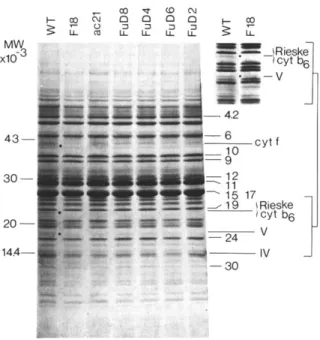
quoted h I and h E o n Fig. 1A, of apparent molecular weights, 34 kDa and 11 kDa, respec-04 tO 03 ,.Q o ~ M W ;10 "3) , y t f - - (41) h 1 - - (34) ; y t b 6 - (23) : h 2 " ~ (11 ) (A) (B)
Fig. 1. H e m e staining of polypeptides from purified thylakoids a n d from a b6/f complex preparation, using urea SDS poly- acrylamide gel eleetrophoresis (A) comparison of a b6/f com- plex preparation with thylakoid m e m b r a n e s from the wild type of C. reinhardtii and from spinach. Cyt f and Cyt b 6 are observed in the three samples. Note the presence of two h e m e stainable bands, h I a n d h 2, in thylakoid m e m b r a n e s from C.
reinhardtii. (B) comparison between thylakoids from different strains of C. reinhardtii. Cyt f a n d Cyt b 6 are still visible in the ac21 mutant, b u t are absent in the F u D 4 , F u D 8 a n d F18 mutants. Traces of Cyt f are detected in the F u D 6 mutant. T h e F u D 2 m u t a n t contains Cyt f and a modified Cyt b 6. Note the presence of h 1 a n d h 2 in all strains, including the F u D l l 2 m u t a n t lacking PS II reaction centers. WT, wild type.
231
tively. In spinach thylakoids, h 2 was never ob- served whereas a faint staining in the position of h 1 could be detected in some instances. The stained bands of 38 and 25 kDa (41 and 23 kdA, respec- tively, in urea gels) have been previously attri- buted in spinach to Cyt f and Cyt b 6, respectively [1]. Accordingly, purified
b6/f
complexes from C.reinhardtii
contained these two bands (Fig. 1A), whereas most of theb6/f
mutants showed no heme staining in these electrophoretic positions (Fig. 1B). In contrast, h 1 and h 2 were absent in theb6/f
complex preparations but they remained in the variousb6/f
mutants that we have char- acterized. These bands were still visible in mutants lacking in PS II reaction centers ( F u D l l 2 on Fig. 1B). Therefore they do not correspond to Cyt b-559HV which is structurally associated with PS II reaction centers [32] and is lacking in PS II mutants (Wollman, F.-A., unpublished results).Three types of
b6/f
mutant could be dis- tinguished on the basis of their heme content: - mutants devoid of Cyt f and Cyt b 6, like FuD4, FuD8 and F18, or showing only traces of Cyt f , like FuD6;- mutants displaying a heme-staining pattern sim- ilar to that in the wild type, like ac21;
- mutants showing a modification in the electro- phoretic position of one of the two cytochromes. This was the case of the FuD2 mutant which contains a somewhat modified Cyt b 6.
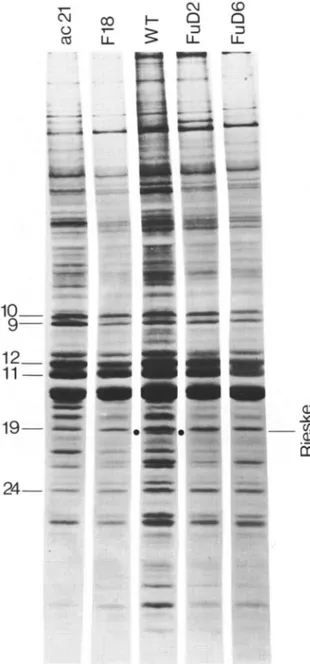
We first analysed the group of mutants devoid of both cytochromes. After Coomassie blue stain- ing of urea gels (Fig. 2), we noted the absence of the 41 kDa polypeptide, corresponding to Cyt f , in the four mutants thought to lack the
b6/f
complex on the basis of their heme-staining pat- tern. This group of mutants showed two other deficiencies in the 23 and 19.5 kDa positions (see inset at the right-hand corner of Fig. 2). The former deficiency corresponds to the position where Cyt b 6 was detected by the heme-staining procedure used in Fig. 1. No other modifications were observed among the population of poly- peptides of still lower molecular weight, which is best resolved with this gel system.
We then performed immunoblotting experi- ments in order to check the absence of the Rieske protein in these mutants. This is shown on Figs. 3 and 4, for the F18 mutant taken as typical of the
MW'
xlO-
4 3I
cO ~ 0 0 0 0 W- oo t - X• 5. ~
~---w. E ¢,DOi!:!ii iiii!i!i i~i iii!~!ii!ili~i!i
!~ i~ill iiiiiiii:i
!?;~iiii:i!~[!
ii2iii i~ iii!::ii:~! iiiii!iill i!ii:iiiii~il i}!iii!ii!!! i~i!i!ii: i;1111!il-
MW
x 10 -3
23--
~- X 0 (D m ~ N 8 ,,Fig. 2. Polypeptide patterns of thylakoid membranes from wild type (WT) and b6/f mutant strains from C. reinhardtii, using urea-SDS polyacrylamide gel electrophoresis after Coomassie blue staining. Polypeptides are numbered according to Ben- noun et al. [15]. Besides the absence of Cyt f in several mutants, two polypeptide deficiencies are observed in the 23 and 19 kDa regions. Comparison of these regions in the F18 and wild type thylakoid membranes is shown the insert at the right upper corner.
first g r o u p of m u t a n t s in the above classification. I n spinach as well as in the wild-type of C. rein-
hardtii the a - R i e s k e labeled a single b a n d of 23
k D a in the urea gel system. N o labeling was observed in the F18 m u t a n t (Fig. 3A). W h e n similar experiments were p e r f o r m e d with a a - C y t b 6, a 23 k D a b a n d was also labeled in spinach a n d in the wild type of C. reinhardtii. This label was absent in the F18 m u t a n t (Fig. 3B). T h a t these observations revealed a c o m i g r a t i o n of the Rieske protein and Cyt b 6 in this gel system, and n o t a cross-reaction o f the antibodies, is illustrated on Fig. 4: in c o n v e n t i o n a l gel systems (i.e., in the absence of urea) the two proteins migrated at different positions giving rise to a label in the 25 k D a region with the a - C y t b 6 a n d in the 19 k D a region with the a-Rieske. T h e absence of radio- labeled b a n d s in these positions in the F18 thylakoids c o n f i r m e d the absence of the two anti- gens in this type o f mutants.
- Rieske
c~-b
(A)
(B) 6
Fig. 3. Immunoblots on a b6/f complex preparation and on thylakoid membranes from wild type (WT) and b6/f mutant strains after urea-SDS polyacrylamide gel eleetrophoresis. Using a-Rieske protein (A) and a-Cyt b 6 (B) coupled to radioiodinated protein A. Note that 23 kDa bands are labeled with the two antibodies in spinach and C. reinhardtii wild-type thylakoids as well as in the b6/f complex preparation. No such labeling is observed in the F18 mutant. The ac21 mutant shows no 23 kDa labeling with the a-Rieske only.
I n agreement with their heme staining pattern, the ac21 a n d F u D 2 m u t a n t s c o n t a i n e d Cyt f , c o r r e s p o n d i n g to the 41 k D a polypeptide in the urea gel shown on Fig. 2. However, the 23 k D a b a n d was lacking in the ac21 m u t a n t a n d was present in the F u D 2 mutant. I m m u n o b l o t t i n g ex- periments (Figs. 3 and 4) showed that the Rieske protein was lacking in the ac21 m u t a n t and was present in the F u D 2 mutant, whereas C y t b 6 was present in b o t h strains with a modified electro- phoretic position in the case of the F u D 2 mutant. These observations indicated that the 23 k D a band, revealed b y C o o m a s s i e blue staining of urea gels, c o r r e s p o n d e d to the Rieske protein only. There- fore, it should be emphasized that C y t b 6 is not stained efficiently e n o u g h to be detected a m o n g the polypeptides o f the thylakoid m e m b r a n e .
2 5 - - 1 9 - -
0 4 O . ~ 1 O .
I.I- I.I- ~ IJ- IJ-
J
- b 6 ~- Rieske
( A ) ( B )
Fig. 4. Same as Fig. 3 after SDS polyacrylamide gel electro- phoresis. Distinct bands are labeled by the a-Rieske protein (19 kDa) and the a-cytb6 (25 kDa). Note the absence of labeling with a-Rieske protein in the ac21 mutant and the absence labeling by the a-Rieske protein and a-Cyt b 6 in the F18 mutant. WT, wild type.
whole
b6/f
complex, thylakoids from the ac21 mutant were lacking in a 19.5 kDa polypeptide in the urea gel system (Fig. 2). This polypeptide was still present in the FuD2 thylakoids, although showing a modified electrophoretic mobility in some experiments.In order to understand if the
br/f
complex consisted of only the four polypeptides deficient in theb6/f
mutants, we attempted to purify the complex fromC. reinhardtii.
Preparations enrichedin
b6/f
complexes showed abr/f
ratio of 1.76(using millimolar extinction coefficients, ~543-553, of 21 for Cyt f [33] and c563_570 of 14 for Cyt b 6 [34]). Similar experiments performed on intacts thylakoids yielded a
b6/f
ratio of 1.94. Therefore,our
br/f
complex preparations showed a slightdeficiency ( = 10%) in Cyt b 6. The polypeptide content of this preparation is shown on Fig. 5. In conventional SDS-polyacrylamide gel electro- phoresis (Fig. 5A), Cyt f appeared as a main band
233 cyt f - - cyt b 6 - Rieske I v ~i~ii~i~iii~iiii ~ MW_ 3 xlO ... ~i!!!/! - - ~ ~ - - c y t f ,:~ Rleske cyt b 6 - - ~ V • _ _ ~ , ~ , .... -- I V H H
Fig. 5. Comparison of the polypeptide patterns in preparations enriched in br/f complexes and in thylakoid membranes from the wild-type strain. (A) Heated samples after SDS poly- acrylamide gel electrophoresis. (B) Heated samples after urea- SDS-polyacrylamide gel electrophoresis. The asterisk indicates contaminant polypeptides of high molecular weight, the amount of which varied from one preparation to another.
in the 38 kDa region. Among the other poly- peptides present in this preparation were a 25 kDa polypeptide, comigrating with the heme staining due to Cyt b 6 and recognized by a-Cyt b 6 (Fig. 4A), a 19 kDa polypeptide and a doublet of about 16 kDa. The 19 kDa band was labeled by the a-Rieske (Fig. 4B). A rather different pattern was observed in urea gels (Fig. 5B): besides the 41 kDa band, attributed by heme staining to Cyt f (Fig. 1), we observed only three bands, of ap- parent molecular weights of 23000, 19500 and 16 000. As discussed above, Cyt b 6 and the Rieske protein migrated in the position of the 23 kDa band (see Figs. 1 and 3). Other polypeptides of high molecular weight, thought to be contami- nants of the
b6/f
complex, since their amounts varied from one preparation to another, are clearly visible in the two gel systems of Fig. 5A and B.Thus the isolated
br/f
complex contained the four polypeptides lacking in theb6/f
mutants. However, a fifth polypeptide, of 16 kDa in urea gels, was also visible in these preparations (see Fig. 5B). It could be either a contaminant or aconstitutive subunit of the complex. In the latter case, the absence of modifications in the 16 k D a region of the polypeptide patterns of the mutants should be attributed to a comigration of several polypeptides, masking the absence of the b6/f
subunit.
In an attempt to distinguish between comigrat- ing polypeptides in this region, on the basis of possible differences in their origin of synthesis, we performed pulse labeling experiments in the pres- ence of site-specific translation inhibitors on whole cells of the wild type and b6/f mutants of C.
reinhardtii. These experiments were also designed
to determine the site of synthesis of the four subunits deficient in most of the mutants, and could be used in the identification of subunit IV, known as a chloroplast-encoded subunit from the work of Alt et al. [7]. To this end, we have isolated thylakoid m e m b r a n e s from cells pulse-labeled for 45 min with t4C-acetate in the presence of cyclo- heximide (Fig. 6), which inhibits cytoplasmic translation, or chloramphenicol (Fig. 7.), which inhibits chloroplast translation. It is worth men- tioning that these experiments give access to the insertion of individual subunits of the complex in the thylakoid m e m b r a n e s of the mutants, even though they would not assemble or accumulate to the same extent as in the wild-type m e m b r a n e s [35,16].
Three subunits of the b6/f complex - namely Cyt f , Cyt b 6 and subunit IV - have been re- ported as chloroplast translates in higher plants [7]. The chloroplast translates in the thylakoids of
C. reinhardtii are shown on Fig. 6. The use of
conventional gels (left of Fig. 6) allowed us to observe the insertion of Cyt b6, during the time of the pulse, in all the strains we have analysed. Only did the F u D 2 m u t a n t show insertion of an a b n o r m a l Cyt b 6, with an apparent molecular weight about 1 k D a larger than usual. Cyt f was lacking from the thylakoid m e m b r a n e s of the F u D 4 mutant, but it was inserted in the thylakoids f r o m the other strains in which the complex does not accumulate (see for instance the F18 or F u D 6 m u t a n t s in Fig. 6). The urea gel system on the fight of Fig. 6 shows the absence of insertion of a 16 k D a polypeptide in the F u D 6 thylakoids. In- sertion of this 16 k D a polypeptide occurred in the other mutant strains (not shown). Thus, the 16
04 ~" ¢D
u_ u_ u_ 2
A
[3
Fig. 6. Autoradiograms of chloroplast translates, inserted in the thylakoid membranes from wild-type (WT) and b6/f
mutant strains, viewed after SDS polyacrylamide gel electro- phoresis (A) and urea-SDS polyacrylamide gel electrophoresis (B). Cells were pulse-labeled for 45 min with [14C]acetate, in the presence of cycloheximide. Note the modified position of Cyt b 6 in the FuD2 mutant and the absence of Cyt f and of subunit IV in the FuD4 and FuD6 mutants, respectively. k D a polypeptide that we observed in the poly- peptide pattern of the b6/f complex (Fig. 5) is p r o b a b l y of chloroplast origin. Therefore, it should correspond to subunit IV in the b6/f complex of
C. reinhardtii.
The polypeptides, translated on cytoplasmic ribosomes and inserted in the thylakoid m e m - branes during the time of our pulse labeling, are shown on the urea gel of Fig. 7. There was no detectable insertion of the 23 k D a polypeptide, corresponding to the Rieske protein, in the ac21 nuclear mutant which contains Cyt f and Cyt b 6. On the contrary the 23 k D a b a n d was visible in
235 T ' - C'4 ¢D CO I--- v-- ... ... iiiii~iiiiii~iiiiii!i!
1
1 2 , m1 1 m
1 9 - - • !ii!~iiiiiiiii3!iiiiii!ill ¸ !!ii~i!iiT!~ii~ii~!!iii!!iii! Q ¢q ¢D O D LL II v" _ _ 09 Ct"Fig. 7. Autoradiograms of cytoplasmic translates, inserted in
the thylakoid membranes from the wild-type (WT) and b6/f
mutant strains, viewed after urea-SDS polyacrylamide gel elec- trophoresis. N o t e the absence of the Rieske protein in all m u t a n t s but F u D 2 .
the pulse-labeled thylakoids from the FuD2 mutant, as expected from the immunoblotting ex- periments and from its polypeptide pattern after Coomassie blue staining. More striking is the ob- servation that the Rieske protein was not inserted
in the thylakoid membranes from any of the other mutants (see for instance F18 and FuD6 in Fig.
7).
Although a 19.5 kDa polypeptide was clearly missing in the mutant thylakoids after Coomassie blue staining (Fig. 2), we observed no deficiencies in labeled polypeptides in this region in urea gels whether the pulse labeling experiments were per- formed in the presence of chloramphenicol or in the presence of cycloheximide (see Figs. 6 and 7). It was therefore impossible to determine, in these experiments, the site of synthesis of the fifth sub- unit of the b6/f complex from C. reinhardtii. How- ever, indirect evidence for its nuclear origin arises from the absence of labeled polypeptides in the 19.5 kDa region in the thylakoids from the wild type when pulse labeled with cycloheximide (Fig.
6).
D i s c u s s i o n
Polypeptides showing peroxidase activity in the thylakoid membrane of. reinhardtii
As previously reported by Maroc and G a m i e r [13], Cyt f and Cyt b 6 from C. reinhardtii were easily identified among thylakoid polypeptides after TMBZ staining of polyacrylamide gels. We showed that thylakoid membranes of C. rein-
hardtii contained two other stained bands, quoted
h 1 and h 2 in this paper, distinct from Cyt f and Cyt b 6, since they remained in the b6/f mutants. Although the procedure might detect peroxidase activities due to prosthetic groups other than the hemes, we looked for a possible correspondance between h 1 and h 2 and Cyt b-559, Hp or LP, and Cyt b-560, recently detected by Bendall and Sanguansermsri (personal communication). We can rule out the first possibility, since we observed h I and h 2 in thylakoid membranes from a mutant lacking in PS II reaction centers although PS II mutants are devoid of Cyt b-559Hv. These PS II mutants would also lack Cyt b-560, but would still contain significant amounts of Cyt b-559Le (Bendall and Sanguansermsri, personal communi- cation). Therefore, among the cytochromes al- ready identified in the thylakoid membranes from
C. reinhardtii, Cyt b-559Lp is the more likely to
correspond to either hi and h z. Such correlations clearly require further investigations. Still another
possibility is the participation of h 1 and h 2 in the chlororespiratory pathway described by Bennoun
[36] in C. reinhardtii. The activity of the putative
oxidase, responsible for the reoxidation of the plastoquinone pool in this pathway, was reported to be independent from the presence or absence of
b6/f
complexes [37]. h I and h 2 might then be related to the oxidase itself or to a cytochrome complex, specific of the chlororespiratory path- way.Characterization of the subunits of the b 6 / f complex
We have identified five subunits in the b6/f
complex from the green algae C. reinhardtii. The
complex comprised Cyt f and Cyt b 6, which are chloroplast translates, the Rieske protein, trans- lated on cytoplasmic ribosomes, and two smaller
subunits. That Cyt br/f complexes from C. rein-
hardtii contain three non-heme subunits in ad-
dition to Cyt f and Cyt b 6 has also been observed by Bendall and Sanguansermsri (personal com- munication). The electrophoretic properties of the two cytochromes and of the Rieske protein showed
minor differences in Spinach and C. reinhardtii.
These observations arose from the comparison of the positions of the heme-stained bands and from immunoblotting experiments performed on the thylakoids from the two organisms. The apparent molecular weights of these subunits varied from one gel system to another. Therefore we consider that the differences in their molecular weights as
reported by different groups (reviewed in Ref. 38) may not be significant. In conventional SDS-poly- acrylamide gel electrophoresis, Cyt f was found in the 33-38 k D a region, Cyt b 6 was in the 19-25 k D a region and the Rieske protein was about 19-20 kDa. However, we observed marked changes in this electrophoretic pattern when 12-18% acrylamide gradients in the presence of 8 M urea were used. Cyt f migrated with a higher apparent molecular weight (41 kDa), whereas Cyt b, and the Rieske protein comigrated with an apparent molecular weight of 23 kDa. This is worth men-
tioning, since two other subunits of the b6/f com-
plex, with apparent molecular weights, 19.5 and 16 kDa, were resolved in this type of gel and could be misquoted as the Rieske protein and subunit IV.
It has been previously reported that subunit IV from spinach has an apparent molecular of about 17 kDa (2) and is encoded by the chloroplast genome [7]. Out of the two small subunits that we
identified in the b6/f complex from C. reinhardtii,
only the 16 kDa, resolved in urea gels, was shown unambiguously to be translated on chloroplast ribosomes. Therefore, its correspondence with subunit IV from spinach is very likely.
The role of the 19.5 kDa subunit, quoted as subunit V in this paper, remains to be elucidated. Owing to the absence of a labeled band, in this region of molecular weights, in the pattern of thylakoid polypeptides translated on chloroplast ribosomes, we believe that subunit V is translated
TABLE I
POLYPEPTIDES OF CHLOROPLAST OR CYTOPLASMIC ORIGIN, ACCUMULATED (A) OR INSERTED (B) IN THE THYLAKOID MEMBRANES
+, polypeptides present; - , polypeptides below detection; ND, not determined; Chip, chloroplast; Cytp, cytoplasmic.
A B
Cyt f Cyt b 6 IV Rieske V Cyt f Cyt b 6 IV Rieske V
(Chlp) (Chlp) ( C h l p ) ( C y t p ) ( C y t p ? ) ( C h i p ) ( C h i p ) ( C h i p ) ( C y t p ) (Cytp?) Wild type + + ND + + + + + + ND FuD2 + + a ND + + + + a + + ND ac21 + + N D - - + + + - N D FuD4 - - N D - - - + + - ND F u D 8 - - N D - - + + + - N D F18 - - N D - - + + + - ND FuD6 + / _ b _ ND -- -- + / - - + - -- ND
a Polypeptide present in a modified position. b Polypeptide in a markedly reduced amount.
237 in the cytoplasm. Preliminary experiments, using
preparations enriched in
b6/f
complexes after pulse labelling the cells of C. reinhardtii with inhibitors of chloroplast translation, favor this hypothesis. It should be noted that plastocyanin is also of low molecular weight, 10 kDa [39], and is translated in the cytoplasm [40]. However, thylakoids from the ac208 mutant fromC. rein-
hardtii,
lacking in plastocyanin [41], still showed a 19.5 kDa polypeptide upon urea-SDS-poly- acrylamide gel electrophoresis (unpublished ob- servation). It is then unlikely that subunit V corre- sponded to some plastocyanin retained in the iso- lated complex. That such subunits of low molecu- lar weight may exist inb6/f
complexes from higher plants has been previously mentioned [2,5].The assembly of the b6/f complex
The comparison of the polypeptide content in the various mutants gave some insight on the mode of assembly of the
b6/f
complexes in the thylakoid membranes ofC. reinhardtii.
We can hypothesize at least three steps in the assembly of a multiple subunit membrane protein: the inser- tion of the subunits in the membrane, their accu- mulation and subsequent assembly in a functional membrane protein complex. Our data, relevant to these processes in the case of theb6/f
complex, are gathered on Table I. The majority of the mutants that we analysed in the present paper, lack the five subunits of the complex. However, one mutant, the ac21, was deficient only in the Rieske protein and in subunit V. These observa- tions tend to define two parts in the complex: a cytochrome moiety, comprising Cyt f, Cyt b 6 and subunit IV, which can accumulate and assemble in the membrane in the absence of the other part of the complex, consisting of the Rieske protein and subunit V. This latter part would not accumulate in the thylakoid membrane in the absence of the cytochrome moiety.We observed that the subunits of the cyto- chrome moiety were inserted independently in thylakoids isolated from pulse-labeled cells of C.
reinhardtii:
for instance, Cyt b 6 and subunit IV were inserted in the absence of Cyt f in the FuD4 mutant, whereas the two cytochromes were in- serted in the absence of subunit IV in the FuD6 mutant. However the inserted subunits, in the twomutants, did not accumulate to the same extent as in the wild-type thylakoids. Only did Cyt f show a slight accumulation in the absence of subunit IV in the FuD6 mutant. These observations support a concerted accumulation of the subunits by a mechanism probably involving the assembly of the newly inserted subunits in the thylakoid mem- branes. The other part of the complex, exemplified by the case of Rieske protein, could not be de- tected, by pulse labeling, in the membranes of the mutants showing no accumulation of the cyto- chrome moiety. This indicates that the insertion of the Rieske subunit was dependent on the preced- ing accumulation of the cytochrome moiety and further substantiates the existence of an assembly between to parts of the
b6/f
complex.Thus we propose the following mode of assem- bly of
b6/f
complexes in the thylakoid mem- branes: as a first step, the subunits encoded in the chloroplast genome - Cyt b 6 and subunit IV which show sequence homologies with Cyt b from mitochrondria [9], and Cyt f - are inserted inde- pendently, then assembled in a cytochrome moiety which allows the subsequent insertion and con- comitant assembly of the subunits encoded in the nuclear genome (the Rieske protein and subunit V). It is worth mentioning that a similar situation probably prevails in the assembly of the subunits of the PS II complex: the chloroplast encoded subunits were functionally assembled in a PS II mutant fromC. reinhardtii
devoid of most of the nuclear-encoded subunits which participate in the formation of the O2-evolving site [15]. The reverse situation was never observed and could be easily interpreted by the fact that the O2-evolving site subunits are peripheral membrane proteins [35]. In this respect, the case of thebr/f
complex is somewhat different, since the Rieske protein has been described as a transmembrane protein [42,43].Acknowledgements
We thank D. Bendall for fruitful exchanges of experimental observations. P. and A. Joliot for stimulating discussions and G. Hauska for his encouragement at the beginning of this work and for the use of his antibodies. C. Lemaire is a recipient of te Agence Fran~aise pour la MaStrise de l'l~nergie. This work was supported by the
C N R S ( A T P g 6 n & i q u e d e s a l g u e s , c o n t r a c t n o . 2 2 2 1 ) .
References
1 Hurt, E. and Hauska, G. (1981) Eur. J. Biochem. 117, 591-599
2 Hurt, E. and Hauska, G. (1982) J. Bioenerg. Biomembranes 14, 405-424
3 Phillips, A.L. and Gray, J.C. (1983) Eur. J. Biochem. 137, 553
4 Clark, R.D. and Hind, G. (1983) J. Biol. Chem. 258, 10348-10354
5 Krinner, M., Hauska, G., Hurt, E. and Lockau, W. (1982) Biochim. Biophys. Acta 681, 110-117
6 Hurt, E., Hauska, G. and Malkin, R. (1981) FEBS Lett. 134, 1-5
7 Alt, J., Westhoff, P., Sears, B.B., Nelson, N., Hurt, E., Hauska, G. and Herrmann, G. (1983) EMBO J. 2, 979-986 8 Bullerjahn, G.S., Riethman, H.C. and Sherman, L.A. (1985)
Biochim. Biophys. Acta 810, 148-157
9 Widger, W.R., Cramer, W.A. Herrmann, R.G. and Trebst, A. (1984) Proc. Natl. Acad. Sci. USA 81,674-678 10 Heinemeyer, W., Alt. J. and Herrmann, R.G. (1984) Curr.
Genetics 8 543-549
1l Metz, J.G., Milles, D. and Rutherford, A.W. (1983) Plant Physiol. 73, 452-459
12 Lam, E. and Malkin, R. (1985) Biochim. Biophys. Acta 810, 106-109
13 Maroc, J. and Garnier, J. (1981) Biochim. Biophys. Acta 637, 473-480
14 Girard, J., Chua, N.H. Bennoun, P., Schmidt, G. and Delosme, M. (1980) Curr. Genetics 2, 215-221
15 Bennoun, P., Diner, B., Wollman, F.-A. Schmidt, G.W. and Chua, N.H. (1981) in Photosynthesis, Vol. III (Akoyunoglou, G., ed.), pp. 839-849, Balaban Interna- tional Science Services, Philadelphia, PA
16 Bennoun, P., Spierer-Herz, M., Erickson, J., Girard-Bascou, J., Pierre, Y. Delosme, M. and Rochaix, J.D. (1986) Plant Mol. biol., in the press
17 Levine, R.P. and Smillie, R.M. (1962) Proc. Natl. Acad. Sci. USA 48, 417-421
18 Bennoun, P., Masson, A., Piccioni, R. and Chua, N.H. (1978) in Chloroplast Development (Akoyunoglou, G. and Argyroudi-Akoyunoglou, J.H., eds.), pp. 721-726, Else- vier/North-Holland Biomedical Press, Amsterdam
19 Schmidt, G.W., Matlin, K.S. and Chua, N.H. (1977) Proc. Natl. Acad. Sci. USA 74, 610-614
20 Bennoun, P. and Delepelaire, P. (1982) in Chloroplast Molecular Biology (M., Hallick, R.B. and Chua, N.-H., eds.) pp. 25-38, Elsevier Biomedical Press, Amsterdam 21 Bennoun, P. and Levine, R.P. (1967) Plant Physiol. 42,
1284-1287
22 Levine, R.P. and Ebersold, W.T. (1960) Annu. Rev. Micro- biol. 14, 197-214
23 Delepelaire, P. (1983) Photochem. and Photobiophys. 6, 279-291
24 Chua, N.-H. and Bennoun, P. (1975) Proc. Natl. Acad. Sci. USA 72, 2175-2179
25 Pick, U. and Racker, E. (1979) J. Biol. Chem. 254, 2793-2799
26 Woessner, J.P., Masson, A., Harris, E.H., Bennoun, P., Gilham, N.W. and Boynton, J.E. (1984) Plant Mol. Biol. 3, 177-190
27 Laemmli, U.K. (1970) Nature (Lond.) 227, 680-685 28 Piccioni, R.G., Bennoun P. and Chua, N.H. (1981) Eur. J.
Biochem. 117, 93-102
29 Thomas, P.E., Ryan, D. and Levin, W. (1976) Anal. Bioch. 75, 168-176
30 Towbin, H., Staehelin, T. and Gordon, J. (1979) Proc. Natl. Acad. Sci. USA 76, 4350-4354
31 Burnette, W.N. (1981) Anal. Biochem. 112, 195-203 32 Satoh, K. (1985) Photochem. and Photobiol. 42, 845-853 33 Nelson, N. and Neumann, J. (1972) J. Biol. Chem. 247,
1817-1824
34 Stuart A.L. and Wasserman, A.R. (1973) Biochim. Biophys. Acta 314, 284-297
35 Delepelaire, P. (1984) EMBO J. 3, 701-706
36 Bennoun, P. (1982) Proc. Natl. Acad. Sci. USA 79, 4352-4356
37 Bennoun, P. (1983) FEBS Lett. 156, 363-365
38 Hauska, G., Hurt, E., Gabellini, N. and Lockau, W. (1983) Biochim. Biophys. Acta 726, 97-133
39 Bohner, H., Bohme, H. and Boger, P. (1981) Febs Lett. 131, 386-388
40 Haslett, B.G. and Cammack, R. (1974) Biochem. J. 144, 567-573
41 Gorman, D.S. and Levine, R.P. (1965) Proc. Natl. Acad. Sci. USA 54, 1665-1669
42 Mansfield, R.W. and Anderson, J.M. (1985) Biochim. Bio- phys. Acta 809, 435-444
43 Ortiz, W. and Malkin, R. (1985) Biochim. Biophys. Acta 808, 164-170