HAL Id: hal-02379510
https://hal.archives-ouvertes.fr/hal-02379510
Submitted on 2 Dec 2019HAL is a multi-disciplinary open access archive for the deposit and dissemination of sci-entific research documents, whether they are pub-lished or not. The documents may come from teaching and research institutions in France or abroad, or from public or private research centers.
L’archive ouverte pluridisciplinaire HAL, est destinée au dépôt et à la diffusion de documents scientifiques de niveau recherche, publiés ou non, émanant des établissements d’enseignement et de recherche français ou étrangers, des laboratoires publics ou privés.
Self-Assembly
Sara Ayala, Pierre Genevaux, Christelle Hureau, Peter Faller
To cite this version:
Sara Ayala, Pierre Genevaux, Christelle Hureau, Peter Faller. (Bio)chemical Strategies To Modulate Amyloid-β Self-Assembly. ACS Chemical Neuroscience, American Chemical Society (ACS), 2019, 10 (8), pp.3366-3374. �10.1021/acschemneuro.9b00239�. �hal-02379510�
Review: (Bio)Chemical strategies to modulate amyloid- self-assembly
Sara Ayala1,2,Ŧ, Pierre Genevaux2, Christelle Hureau1, Peter Faller,1,3
1 LCC, CNRS & University of Toulouse (UPS, INPT), 205 route de Narbonne, 31077 Toulouse, France 2 Laboratoire de Microbiologie et de Génétique Moléculaires, Centre de Biologie Intégrative (CBI),
Université de Toulouse, CNRS, UPS, Toulouse, France.
3 Institut de Chimie, UMR 7177, CNRS-Université de Strasbourg, 4 rue Blaise Pascal, 67000, Strasbourg,
France.
Ŧ Current address:Leibniz-Research Institute for Molecular Pharmacology (FMP), Robert-Roessle-Straße
10, 13125, Berlin, Germany.
Abstract: Amyloid plaques are one of the two hallmarks of Alzheimer’s disease (AD). They consist mainly of fibrils made of self-assembled amyloid- (A) peptides. A is produced in healthy brains from proteolytic cleavage of the amyloid precursor protein. A aggregates, in particular smaller, soluble aggregates, are toxic to cells. Hence modulating the self-assembly of A became of very active field of research, with the aim to reduce the amount of the toxic aggregates of A or to block their toxic action. A great variety of molecules, chemical and biological, are able to modify the aggregation of A. Here we give an overview of the different mechanistic ways to modulate A aggregation and on which step in the self-assembly molecules can interfere. We discuss the aggregation modulators according to different important parameters, including the type of interaction (weak interaction, coordination or covalent bonds), the importance of kinetics and thermodynamics, the size of the modulating molecules, and binding specificity.
Abbreviations:
AD: Alzheimer’s disease A: amyloid beta
APP: amyloid precursor protein PSEN1: presenilin 1
PSEN2: presenilin 2 polyP: polyphosphates HSP: heat shock proteins CLU: clusterin
TOC:
Keywords:
Amyloids: the biophysical definition is based on the structure, i.e. aggregates of self-assembled
proteins/peptides that form cross-beta structures.
Aggregates: off pathways in protein biogenesis in which misfolded proteins accumulate. In the
amyloid context, aggregates often mean ordered assemblies of several units of the same protein of any structure into protein fibers.
Inert-labile: describes the kinetic stability, in contrast to the thermodynamic stability, which is
strong-weak. An inert bond can exist for a long time, and hence be biologically relevant, despite it is not thermodynamically stable.
A oligomers: A aggregates that are soluble and relatively small (roughly from dimer to ~one-
two dozen of monomers)
Polyphenols: Organic compounds (native or synthetic) that contain several, covalently linked
phenols
Polyphosphate: cyclic or linear polymer of phosphates linked by ester bonds. In nature linear
Molecular chaperones: key players of the cellular proteostasis network, they assist protein
folding and/or protein assembly into higher order structures without being part of the final structures. Molecular chaperones can also disassemble already formed aggregates in order to either reactivate aggregated proteins or target them to degradation machineries.
Chemical chaperones: Small chemical molecules that have similar activity like the molecular
chaperons.
Polyoxometalate: discrete molecular anionic structures composed of early transition metals (e.g.,
1) Introduction
Alzheimer’s disease (AD) is the most common form of dementia, accounting for 50-70% of cases. The number of AD cases is expected to reach 75 million by 2030, and 130 million by 2050 1. A major common point between familial AD (related to specific mutations) and sporadic AD (no mutation background) is the accumulation of a 40-42 amino acids peptide, amyloid- (A) in form of amyloid plaques. The higher aggregation prone A1-42 form is enriched compared to the most abundant but lower aggregation prone A1-40 isoform. In familial cases, mutations on the Amyloid Precursor Protein (APP), the Presenilin 1 (PSEN1) or the Presenilin 2 (PSEN2) genes result in an overall increased production of A. Sporadic cases are rather characterized by a reduced clearance of A due to a decrease in A-degrading enzymes 2.
A peptide production through APP sequential cleavage by - and γ-secretases occurs already during brain embryogenesis and seems to be required for normal brain development. A has been reported to enhance neurons survival in vitro, neural progenitor cells differentiation, neuron proliferation and synapse regulation 3,4. How a physiologically necessary peptide becomes a major actor in the development of AD has been proposed to occur through a cascade of events known as the “amyloid hypothesis” proposed in 1992 5. A accumulation promotes conformational changes
in the peptide, which lead to its gradual non-covalent polymerization into a heterogeneous array of oligomeric species that eventually evolves towards amyloid fibrils. Experimental data points to A oligomers species as the most toxic ones, exerting their deleterious effect through a variety of mechanisms 6, including direct interaction with membranes forming pore-like structures and disrupting their proper permeability, and binding to cellular receptors with deleterious consequences 7,8.
A aggregation is thus key in AD neurodegeneration and it has stimulated multiple studies with the objective of elucidating the exact mechanism by which aggregation occurs. One of the observations is that A aggregation is a modifiable process. Environmental factors, such as peptide concentration, temperature, pH, ion strength, added solvents, agitation, etc., have been studied and are known to affect A fibrillization 9. A great variety of molecules, chemical and biological, are able to modify as well the aggregation of A. Here, we shortly review the bases of A aggregation and structure, and focus then on the different strategies and classes of chemical and biological modulators of the A aggregation. We do also not treat the strategies to disaggregate oligomers or amyloid fibrils, despite it is general importance and refer to the literature 10. The purpose is not to be exhaustive in terms of mentioning all tested compounds, but to show the different classes and principles that can be exploited.
2) Aggregation mechanism and structures
Amyloid formation occurs through a complex mechanism that involves a heterogeneous array of species. One can describe amyloid formation process as a function of time with a sigmoidal curve characterized by a lag phase followed by rapid growth and plateau phases. During lag phase, polypeptides suffer a spontaneous transition from soluble to non-soluble β-cross species, which ultimately assemble leading to the formation of amyloid fibrils. Amyloids can be formed with intrinsically disordered peptides/proteins, such as Aβ, or with natively folded proteins. In latter case the fold has to be destabilized first. Once a peptide has adopted the characteristic cross-β structure of amyloids, it will serve as a template for the upcoming monomers that will add-up to the growing fibril as they become equally cross-β structured. The initial cross-β structural shift is known as primary nucleation, while the addition of new monomers to the growing fibril is named elongation. Primary nucleation directly affects the lag phase duration that will depend on the time taken by the cross-β structures to reach a detectable concentration of aggregates. Once primary nucleation has occurred, fragmentation and secondary nucleation processes can take over and dominate amyloid formation. Fragmentation depends on the probability that fibrils break increasing the number of termini in which new cross-β monomers polymerize to elongate fibrils. Intact fibrils can, on their side, serve as catalytic surfaces that promote the formation of secondary nuclei of aggregation. Quantitative analysis of amyloid growth has revealed that primary nuclei form within the first milliseconds of the reaction, allowing elongation and secondary processes to coexist, at different rates, through all the amyloid formation phases 11,12. Alteration of the microscopic processes rates result in a modification of the amyloid formation curve. For example, a variation in the primary nucleation leads to changes in the duration of the lag phase. On their side, changes in the rates of elongation, fragmentation and secondary nucleation profoundly affect the lag phase duration as well as the growth phase slope.
Aβ1-42 fibril growth, in particular, is governed by secondary nucleation. Moreover, Aβ1-42 toxic oligomeric species seem to originate from monomers mainly through secondary nucleation 13. Thus, modulation of specific microscopic steps of the amyloid formation is a promising area of study that could lead to future therapeutics.
2.2) Importance of biomembranes
Biomembranes play a very crucial role in the biology of Aβ. First Aβ is cleaved of a
transmembrane protein called amyloid-precursor protein, in which the Aβ sequence is partially in the transmembrane region 2,3. Thus, cleaved Aβ has a hydrophobic part with affinity to
membranes. Thus membranes can on one hand serve as a template to foster Aβ aggregation and on the other hand interaction of Aβ-aggregates with membranes has been reported as a
mechanism of Aβ toxicity, including disruption, pore formation or destabilization. Therefore, studying the different modulators outlined below in the context of biomembranes is crucial and changing the lipid constitution of the membranes is also a very potent way to influence Aβ aggregation and toxicity. (for recent review see 14,15).
2.3) Structural aspects of amyloid-beta: monomer, oligomer, fibrils
Hugh attempts have been done to shed light on the molecular structure of the different aggregation states of A. The best characterized structures are A fibrils, with a few complete models available from mainly solid-state NMR studies and cryo-TEM 17. Oligomers, due to their transient
and heterogenic nature, have been more difficult to describe. Several studies have been published but their structures remain elusive.
A fibrils contain cross- motifs in their structure. This means that A monomers contain -strand segments that assemble into -sheets, with -strands running perpendicular to the fibril axis, and hydrogen bonds between -strands running parallel to it. Cross- motifs can arise both from parallel and antiparallel -sheets. A fibrils comprise parallel in-register -sheets. Nevertheless, antiparallel -sheets can be observed in A intermediate aggregation species 18. One preliminary model for A1-40 fibrils consists in two cross- units each one formed by monomers with a disordered N-terminus followed by two strand segments that form two parallel in-register -sheets and that are separated by a 180° bent stabilized by a salt bridge 19. Conditions under which
fibrils are prepared can significantly impact their structure. Indeed, while mild agitation conditions correlate with fibrils that contain two cross- units with two-fold symmetry (striated ribbon morphology), fibrils prepared under quiescent conditions contain three cross- units with a three-fold symmetry (twisted morphology). Later proposed models, as well as a S-shaped A1-42 fibril, and A1-40 Iowa and Osaka mutations fibrils models are available 12,20.
The structure of A oligomers is even less clear. Due to the difficulties encountered to obtain homogeneous and stable samples of oligomers, a great variety of models are described in the literature. The majority of the available observations suggest that oligomeric species lack the characteristic in-register parallel -sheets of fibrils 18. However, some groups have reported oligomers with cross- structure 21,22.
3) Modulation of A aggregation by molecules
Conceptually one can distinguish modulations by molecules on different levels:
i) the type of interaction, i.e. which type of bonding is involved. We consider here the three main types of interaction: weak bonds (including H-bonds, hydrophobic interactions, Pi-stacking, Van-der Waals etc.), coordination and covalent bonds. In general, the strength of the bonds decreases from covalent to coordination to weak bonds.
ii) the structure targeted by the molecule, i.e. aggregation species to which the molecule binds iii) the action that is triggered upon binding of the molecule: inhibition or promotion, slow-down or acceleration, new pathway, etc.
Therefore, based on the hypothesis that certain oligomeric species are the most toxic, there are several strategies to avoid their formation:
ii) acceleration of fibril formation and stabilization of fibrils (this lowers the concentration of oligomeric species)
iii) triggering other pathways that lead to non-toxic species, like favoring non-toxic amorphous aggregates
iv) binding to the toxic oligomers and inhibiting their toxic action, for instance inhibiting binding of oligomers to their pathological target (like membrane, receptor, metal ions, etc.).
This is summarized in Figure 1.
Figure 1 Schematic view of the various ways (bio)-molecular or synthetic chaperones can act on
the aggregation process are shown in red. This simplified scheme shows only three type of aggregates, oligomers (most toxic), fibrils (like found in the amyloid plaques; not or less toxic) and other non-toxic aggregated species (off-pathway species, e.g. amorphous aggregates). Various modulations to reduce the toxicity of Aoligomers are: push the equilibrium away from the toxic oligomers, either to monomers (A), to fibrils (B) or to non-toxic aggregates (E), block the interaction of oligomers with the biological partners that triggers the toxicity (D; e.g. a membrane receptor); orange color), inhibit secondary nucleation to form oligomers (C).
4) Modulation by weak interactions
4.1) Small organic molecules
A lot of small organic molecules have been studied concerning their modulation of aggregation. The advantages of such molecules are that they are interesting as drug, because they are cheap, can often be tuned to pass the blood brain barrier passively. These compounds contain very often (poly)-aromatic systems and are often planar. A reason for that might be possibility of intercalation with beta-sheet structures and the hydrophobic interaction with the central hydrophobic patch of Aβaround the Phe-Phe at position .02/91A problem is to target the monomeric Aβ, with the idea to inhibit the formation of toxic aggregates (Figure 1, path A). The affinity of a small organic molecule with an intrinsically disorder peptide Aβ is often relatively low (µM) and not very specific. This can be explained by the limited weak interaction a small molecule is able to make, and by the entropic penalty of a disordered peptide to form this interaction. However, interaction with structured oligomers and fibrils can have higher affinities. Several types of mechanisms have been described. Such compounds can:
i) bind to fibrils and reduce fragmentation (Figure 1, path C), like proposed for the flat compound BAF31 (Binder of Amyloid Fibers 31), which was identified upon a structure-based screening. BAF31 does not reduce A fiber formation but it reduces A cytotoxicity by increasing fiber stability and shifting the equilibrium of A from oligomers to fibers 23.
ii) accelerate the formation of fibrils (Figure 1, path B) by reducing the lifetime of toxic oligomers. The compound O4 (orcein-related polyphenol) is such an example 24
iii) bind to toxic oligomers and change aggregation into non-toxic species (Figure 1, path E). An example is epigallocatechin gallate (EGCG) 24,25.
iv) inhibit interaction with biological target (Figure 1, path D). An example is ALI6 (Amyloid-LilrB2 Inhibitor 6), identified upon a structure-based screening. ALI6 inhibits Aβ interaction with the neuronal cell surface receptor LilrB2 reducing Aβ cytotoxicity 26.
4.2) Peptide derivatives
Peptides and derivatives have often been used with the aim to inhibit aggregation of Aβ (Figure 1, path A). Among different applied strategies, the uses of peptides (and peptoids) as β-sheet breakers was often reported. The basic idea behind this approach is that for aggregation, Aβ has to recognize itself. Under focus were mainly the central part around Phe19-Phe20, thought to be the first contact between two Aβ molecules. Thus, investigations started with small peptides of the LVFF region (Aβ17-20), supposed to bind to the parent similar sequence in Aβ1-40/42 and inhibit the self-recognition of Aβ and hence its aggregation 27. Then several derivatives of pure
peptides have been developed in order to increase the activity, like cyclization, methylation of the amide bond, introduction of non-native amino acids (see e.g. 28–30). These studies also showed the efficiency of the peptide derivatives to inhibit amyloid fibrils formation. However, at least for some, it is clear that they do not stabilize the monomeric form and/or inhibit nucleation or
elongation as aimed by the design, but that they rather promote formation on fibrillar, non-toxic A aggregates (Figure 1, path E) 28.
4.3) Chemical chaperones
4.3.1) Chemical and pharmacological chaperones
These are small molecules with activities similar to that of molecular chaperones, assisting proper protein folding and refolding misfolded proteins. According to Cortez and Sim 31, one should
distinct between pharmacological and chemical chaperones. Pharmacological chaperones have a specific binding site with proteins in order to stabilize the native fold or induce refolding. In contrast, chemical chaperones have often a non-specific mode of action. Thus, they have often only an effect at higher concentrations, which can be limiting for therapeutic approaches. On the other hand, certain of these chemical chaperones can be natively present at very high concentrations (like sugars or aminoacids).
Two main classes of chemical chaperones are osmolytes and hydrophobic compounds. Examples for osmolytes are sugars (e.g. trehalose, mannitol), inositols, aminoacid (proline), etc. 32. Hydrophobic compounds are often detergents or fatty acids. Their proposed mode of action involves the binding to exposed hydrophobic patches and hence inhibition of aggregation (Figure 1, path A). There also some larger molecules studied, like cyclodextrins or polyphosphates 33,34.
4.3.2. Polyphosphates as modulator of A aggregation
Polyphosphates (polyP) are chains of inorganic phosphates linked by phosphoanhydride bonds, which biological role is just starting to be unveiled. In eukaryotes, polyP are involved in blood clotting, apoptosis, mTOR activation and neuronal signaling 35–38, but recent work unveiled a new role for polyP as a chaperone 39. PolyP were shown to inhibit the aggregation of various chaperone substrate proteins, which remained in a state that was compatible for refolding, suggesting that polyP act in a non-specific and promiscuous manner. Since polyP synthesis does not require transcription nor translation, they may constitute a backup system during stresses hindering chaperone overexpression and their ATP-dependent function. In the context of protein-misfolding diseases, it has been discovered that polyP are able to accelerate amyloid fibril formation by a series of proteins including ⍺-synuclein, Tau and A1-40/42. PolyP accelerate protein fibrillation (Figure 1, path B) by serving as a scaffold that recruits a maximum number of monomers to the cross- structure and stabilize them to prevent secondary nucleation processes (Figure 1, path C). Through this mechanism, polyP reduce the time window of cell exposition to A oligomers 33.
In general, chemical chaperones are attractive because they have multiple modes of action and they are often tolerated at high concentrations. And in the case of small organic molecules they can have good blood–brain barrier penetration property (particular the hydrophobic type). Disadvantage is
that they are more unspecific and can have a variety of side effect and that often high concentrations are needed.
4.4. Proteins
4.4.1 Modulation of A aggregation by molecular chaperones
Molecular chaperones interact, stabilize and help nonnative proteins to reach their native conformation 40. They assist de novo protein folding, disaggregation and refolding of stress-denatured proteins, oligomeric assembly and disassembly of protein complexes, protein targeting and translocation through biological membranes, and assistance to proteolytic degradation 41. Most chaperones belong to the heat shock proteins (HSP) families that are generally classified on the basis of their sequence homology as observed for HSP100, HSP90, HSP70, HSP60, HSP40, or small HSP family members 42,43. Other many important bona fide chaperones including prefoldin, clusterin, haptoglobin, α2-macroglobulin, SecB, Trigger Factor or caseins do not belong to HSP families 44–47. Among these, several chaperones have proven ability to modulate A aggregation
(see Table I).
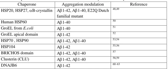
Table 1: Chaperones studied towards the modulation of A self-assembly
Chaperone Aggregation modulation Reference HSP20, HSP27, B-crystallin A1-42, A1-40, E22Q Dutch
familial mutant
48,49
Human HSP60 A1-40 50
GroEL from E.coli A1-40 51 GroEL apical domain A1-42 52 HSP70 , HSP90 A1-42, A1-40 53,54
HSP104 A1-42 55,56
BRICHOS domain A1-42, A1-40 57 Clusterin (CLU) A1-42, A1-40 58,59
DNAJB6 A1-42 60–63
Kinetic studies have shown that molecular chaperones can target specific steps of the protein aggregation process. This is of particular importance in the case of A aggregation because its aggregation is dominated by the generation of highly toxic oligomers through secondary nucleation processes. Human BRICHOS domain has a high affinity for A1-42 fibrils to which it remains bound abolishing their breakage as well as the catalysis of new nuclei (Figure 1, path C)
64. Clusterin (CLU) shields the hydrophobic region of A1-40 and A1-42 oligomers (Figure 1,
processing and degradation . Finally, DNAJB6 (HSP40 human member) delays Aβ1-42 aggregation by interacting with aggregated but not with monomeric forms of the peptide (Figure 1, path A) 60. Interestingly, the same protein inhibits Huntingtin aggregation by interacting with
monomeric species 62. This illustrates how chaperones do not have a unique mechanism of handling amyloid fibrils, and that discreet steps of aggregation affected by one chaperone may depend on the intrinsic characteristics of the amyloid-forming protein itself.
As natural existing modulators of Aβ fibril formation, molecular chaperones have become one of the therapeutic targets of AD, as well as other protein-misfolding diseases. Current approaches include the identification of small chemical compounds able to upregulate chaperones expression or to enhance their activity, and the direct administration of recombinant chaperones via intranasal administration 65. One downside of these strategies arises from the chaperones broad substrate specificity 66. Up-regulating or increasing the pool of one chaperone is likely to affect the disease-associated protein but also other proteins that naturally interact with that one chaperone. Therefore, in order to therapeutically use chaperones, it may be important to first engineer chaperones that are specifically directed towards disease-related proteins, including Aβ67.
4.4.2 Modulation of Aβ aggregation by antibodies and alike:
Antibody-antigen binding is mediated by hydrogen bonds, van der Waals forces and ionic
interactions. Common Kd values range from 5 x 10⁻⁴-10-11 M 68. Specific binding and promotion of degradation are the two characteristics that have brought attention to immunotherapy in the neurodegeneration field. Passive immunotherapy consists in stimulating the organism to produce its own antibodies against a specific antigen. Active immunotherapy is the direct administration of pre-produced antibodies 69. Both, passive and active immunotherapy against Aβ have been
extensively developed with the aim of arresting Alzheimer’s mental deterioration. Many antibodies that bind Aβ have been engineered in the last decades (for detailed reviews refer to
70,71.
Antibodies can have different impact on Aβ: i) they can direct the immune system towards Aβ aggregates (preferentially oligomers) which then can be cleared by the microglia. ii) they can bind to oligomers and inhibit their interaction with the binding-partner responsible for the toxicity, e.g. membranes of receptors, and iii) they can modulate Aβ aggregation, by stabilizing the monomeric form of Aβ. To which extent the different mechanisms are involved in the in vivo experiments is often not clear.
Antibodies and antibody fragments have been designed to inhibit Aβ aggregation, via for instance a β-sheet breaker mechanism72. They have the advantage to be very specific towards their target, and can even distinguish between different aggregation states of Aβ, such as oligomers and fibrils
71. Moreover, their affinity can be very strong allowing the binding to nano-picoM concentrations
of Aβ. A potential drawback to the use of antibodies is their large size; although the use of antibody fragments or of camel single domain antibodies might be very promising 73. In addition, aptamers, oligonucleotide ligands with high affinity and specificity, have also been developed toward Aβ
Antibodies having the ability to i) enter the brain where Aβ plaques are formed, ii) induce microglial phagocytosis of Aβ, and iii) reduce Aβ burden, have been successfully tested in mice Yet, data from human trials were less promising 71.
5) Modulation by coordination bonds
Coordination bonds often have a strength between covalent and weak bonds. In biology, they mostly occur between positive charged metal ions and electron donors like nitrogen, oxygen or sulfur. Coordination bonds could be strong enough to occur even at low concentrations (nM, pM) and can be reversible (with labile metal ions) or not (with inert metal ions). Hence, the strength of the interaction (thermodynamic) is not the only factor that counts, but also the kinetics. Certain metal ions (like Cu, Zn, Fe, Mn, etc) are generally kinetically labile, meaning they will rapidly bind to the highest affinity ligand. In contrast certain metal ions (like Pt(II), Ru(II), Ir(III)) are kinetically inert, meaning that the reactions are very slow, and these metals can bind to a target for a very long time, although it is not the thermodynamically most favorable. The canonical example is the binding of cis-Pt to its therapeutic target DNA. Only a few percent bind to DNA, the other complexes bind to other targets 75.
5.1. Labile metals, mainly Zn, and Cu
5.1.1 Labile metal ions in solution: The effects of metal ions on A aggregation has been widely
studies. 76,77. Mostly Cu(II) and Zn(II) has been investigated, due to the fact that these two metal ions are bound to A in amyloid plaques 78. The main binding sites for Cu(I), Cu(II) and Zn(II) to
the soluble monomeric A are well known 79,80. The binding sites in aggregated A are less well defined, but seems to be generally similar. In contrast, the effects of the main studied metal ions Zn(II) and Cu(II) on the aggregation are not consensual. The only consensus is that these metal ions affect the aggregation behavior, but how, in terms of kinetic and structures is not clear. There are several parameters playing an important role, like concentrations, metal-peptide ratios, pH, A pretreatment, and batch to batch differences 76,77,81.
Potential important effects of metal ion binding are i) the change in the structure, ii) change in the overall charge, and iii) the possibility to bind to two peptides simultaneously, i.e. bringing two peptides close together. It is clear that binding of Cu(I), Cu(II) and Zn(II) change the structure(s) of the A. As A is an intrinsically disordered protein, it stays very disordered when metals are bound, but the structural propensities and the dynamics change significantly. And such changes are metal and oxidation specific, i.e. the induced changes are different for the different metal ions 82.
In the case of A, binding of metal ions can change the overall charge, but one has also to consider that metal ions often displace H+, which are released. In general, Cu(I/II) and Zn(II) increase slightly the net-charge at pH 7.4 and can render it more aggregation-prone 83.
The importance of bridging metal ions in the case of Cu(I/II) and Zn(II) is not very clear. Stable bridging metal ions in oligomers and fibrils have been suggested, but there is no strong evidence for them. However it is clear that metal-ion bridging occurs transiently and for Cu(II), the transiently formed A–Cu(II)-A have been proposed to be less aggregation prone 84,85. In contrast, it has been shown that substoichiometric Zn compared to Aβhave a strong impact on aggregation kinetics 86–88.
5.1.2 polyoxometalates: labile metal ions in solid state/assembly:
Metal substituted polyoxometalates (POMs) are among the few inorganic complexes based on a labile central ion probed for their ability to modulate A aggregation via interaction with the His residues. In a seminal and key study, Dawson-type POMs were able to reduce A aggregation with a dependence on the central ion (POM-NI > POM-Co > unsubstituted POM) via a mechanism where oligomers formation is disfavored and monomers stabilized (Figure 1, path A) 89.
5.2. Inert metal complexes:
Complexes made of d-block metal ions of the second and third rows have been used to impact the A aggregation process 90–92. The rationale behind this approach is the formation of kinetically stable L-M-A ternary species, where the ligand L is generally made of bulky aromatic ancillary scaffold to target the hydrophobic sequence involved in aggregation and where M is mainly Pt(II), Ru(II) and to a lesser extent Pt(IV), Ru(III), Ir(III), Rh(III). Such species would then modify the aggregation propensity of the apo-A and hamper binding of Cu and Zn ions in their natural/native sites 93,94 thus further modifying the aggregation properties in presence of biological ions. It was shown that the interaction to the central metal ions occurs via the His residues of the peptide. Due to the intrinsic toxicity of Pt(II) and Ru(II) species, pro-drug approach was also developed using less-toxic Pt(IV) or Ru(III) central ions that could be further reduced biologically 95,96. The main modes of action of such inert-metal complexes are interaction with monomers and further preclusion of native A aggregation (Figure 1, path A).
6) Modulation via covalent bonds
Covalent bonds are normally the strongest bond and are mostly not reversible. Thus, derivatization of A by a covalent bond can be a very strong and persisting way to modulate aggregation. As in the case for slow exchanging metals, a main problem is the selectivity, i.e. how to target A only. Moreover, a modulation of all A by a covalent bond needs as much of the modulator as A, thus this is not catalytic. However, it is likely that partially derivatization of A is enough to modulate aggregation.
Polyphenols are well known to modulate A aggregation (see above) (recent review: 97). The exact mechanism is not completely understood, and often only weak interactions are considered. However, polyphenols could also form covalent bonds. In the case of EGCG, it was proposed that oxidized EGCG molecules could react with free amines of A via formation of Schiff
bases 98. Similarly, Taxifolin, a catechol type flavonoid, was able to suppress A1-42 aggregation via reaction of its oxidized form (o-quinone) with the Lys in A 99. Moreover, further analysis suggested that Taxifolin specifically targets the elongation phase, rather than the nucleation phase. Thus, it is possible that also other polyphenols than EGCG and Taxifolin modulate A aggregation via weak and covalent bonds.
Recently Kino et al. 100 conceived a covalent A aggregation modifier. They used a cyclic peptide (cyclo-KLVFF), known to bind to A by weak interaction and inhibiting A aggregation. They decorated the cyclo-KLVFF with a diazirine. UV irradiation resulted in a covalent bond formation with Tyr 10. Further experiments showed that this photoinduced derivatization reduced amyloid formation and attenuated cell toxicity.
7) Conclusions
A aggregation is an auto-catalyzed self-assembly process with a lot of different pathways and species of various morphology that can be form. Thus, it is inherently a process very sensitive to conditions and effectors. Hence, it is not astonishing that a lot of different conditions and chemical or biological molecules influence this process. We reviewed the different classes of effectors, by focusing more on the type of interactions, and their pros and cons. An ideal effector would be cheap, penetrates very well to the brain, is specific for A, not toxic and gives no side effects and is highly efficient so that only low amount is needed.
When going through all the different type of effectors on A aggregation, it seems that no strategy assembles all these advantages. Thus, may be a trade-off has to be found. Another strategy might be to use a combination of different effectors, to combine the positive parameters.
Acknowledgments:
S.A. was supported by the grant CHAPROZINC from « APR de l’Université Fédérale Toulouse Midi-Pyrénées 2014 (COMUE) ». P.F. acknowledges the Frontier Research in Chemistry Foundation (Strasbourg), Installation grant (PF). C.H. acknowledges the European Research Council (ERC aLzINK, StG-638712) for its participation to this research activity at the frontier with the aLzINK project.
References
(1) Winblad, B.; Amouyel, P.; Andrieu, S.; Ballard, C.; Brayne, C.; Brodaty, H.; Cedazo-Minguez, A.; Dubois, B.; Edvardsson, D.; Feldman, H.; et al. Defeating Alzheimer’s Disease and Other Dementias: A Priority for European Science and Society. Lancet Neurol. 2016, 15 (5), 455–532. https://doi.org/10.1016/S1474-4422(16)00062-4.
(2) Selkoe, D. J. Alzheimer’s Disease : Genes , Proteins , and Therapy. Physiol. Rev. 2001, 81 (2), 741– 767. https://doi.org/10.1016/0092-8674(88)90462-x.
(3) Chasseigneaux, S.; Allinquant, B. Functions of Aβ, sAPPα and sAPPβ: Similarities and Differences. J.
Neurochem. 2012, 120 (SUPPL. 1), 99–108. https://doi.org/10.1111/j.1471-4159.2011.07584.x.
(4) Pearson, H.; Peers, C. Physiological Roles for Amyloid β Peptides. J. Physiol. 2006, 575 (Pt 1), 5–10. https://doi.org/10.1113/jphysiol.2006.111203.
(5) Hardy, J.; Higgins, G. Alzheimer’ S Disease : The Amyloid Cascade Hypothesis. Science 1992, 256, 184–185. https://doi.org/10.1126/science.1566067.
(6) Kirkitadze, M. D.; Bitan, G.; Teplow, D. B. Paradigm Shifts in Alzheimer’s Disease and Other Neurodegenerative Disorders: The Emerging Role of Oligomeric Assemblies. J. Neurosci. Res. 2002, 69 (5), 567–577. https://doi.org/10.1002/jnr.10328.
(7) Sakono, M.; Zako, T. Amyloid Oligomers: Formation and Toxicity of Aβ Oligomers. FEBS J. 2010,
277 (6), 1348–1358. https://doi.org/10.1111/j.1742-4658.2010.07568.x.
(8) Sengupta, U.; Nilson, A. N.; Kayed, R. The Role of Amyloid-β Oligomers in Toxicity, Propagation, and Immunotherapy. EBioMedicine 2016, 6, 42–49. https://doi.org/10.1016/j.ebiom.2016.03.035. (9) Tiiman, A.; Krishtal, J.; Palumaa, P.; Tõugu, V. In Vitro Fibrillization of Alzheimer’s Amyloid-β
Peptide (1-42). AIP Adv. 2015, 5 (9), 092401. https://doi.org/10.1063/1.4921071.
(10) Citron, M. Alzheimer’s Disease: Strategies for Disease Modification. Nat. Rev. Drug Discov. 2010, 9 (5), 387–398. https://doi.org/10.1038/nrd2896.
(11) Arosio, P.; Knowles, T. P. J.; Linse, S. On the Lag Phase in Amyloid Fibril Formation. Phys. Chem.
Chem. Phys. 2015, 17 (12), 7606–7618. https://doi.org/10.1039/C4CP05563B.
(12) Knowles, T. P. J.; Vendruscolo, M.; Dobson, C. M. The Amyloid State and Its Association with Protein Misfolding Diseases. Nat. Rev. Mol. Cell Biol. 2014, 15 (6), 384–396.
https://doi.org/10.1038/nrm3810.
(13) Cohen, S. I. A.; Linse, S.; Luheshi, L. M.; Hellstrand, E.; White, D. A.; Rajah, L.; Otzen, D. E.;
Vendruscolo, M.; Dobson, C. M.; Knowles, T. P. J. Proliferation of Amyloid-β42 Aggregates Occurs through a Secondary Nucleation Mechanism. Proc. Natl. Acad. Sci. U. S. A. 2013, 110 (24), 9758– 9763. https://doi.org/10.1073/pnas.1218402110.
(14) A. Kotler, S.; Walsh, P.; R. Brender, J.; Ramamoorthy, A. Differences between Amyloid-β
Aggregation in Solution and on the Membrane: Insights into Elucidation of the Mechanistic Details of Alzheimer’s Disease. Chem. Soc. Rev. 2014, 43 (19), 6692–6700.
https://doi.org/10.1039/C3CS60431D.
(15) Sciacca, M. F. M.; Tempra, C.; Scollo, F.; Milardi, D.; La Rosa, C. Amyloid Growth and Membrane Damage: Current Themes and Emerging Perspectives from Theory and Experiments on Aβ and hIAPP. Biochim. Biophys. Acta BBA - Biomembr. 2018, 1860 (9), 1625–1638.
https://doi.org/10.1016/j.bbamem.2018.02.022.
(16) Nasica-Labouze, J.; Nguyen, P. H.; Sterpone, F.; Berthoumieu, O.; Buchete, N.-V.; Coté, S.; De Simone, A.; Doig, A. J.; Faller, P.; Garcia, A.; et al. Amyloid β Protein and Alzheimer’s Disease: When Computer Simulations Complement Experimental Studies. Chem. Rev. 2015, 115 (9), 3518– 3563. https://doi.org/10.1021/cr500638n.
(17) Gremer, L.; Schölzel, D.; Schenk, C.; Reinartz, E.; Labahn, J.; Ravelli, R. B. G.; Tusche, M.; Lopez-iglesias, C.; Hoyer, W.; Heise, H.; et al. Fibril Structure of Amyloid-ß ( 1-42 ) by Cryo-Electron Microscopy. 2018, 358 (6359), 116–119. https://doi.org/10.1126/science.aao2825.Fibril. (18) Tycko, R. Molecular Structure of Aggregated Amyloid-β: Insights from Solid-State Nuclear
Magnetic Resonance. Cold Spring Harb. Perspect. Med. 2016, 6 (8), 1–22. https://doi.org/10.1101/cshperspect.a024083.
(19) Petkova, A. T.; Ishii, Y.; Balbach, J. J.; Antzutkin, O. N.; Leapman, R. D.; Delaglio, F.; Tycko, R. A Structural Model for Alzheimer’s β-Amyloid Fibrils Based on Experimental Constraints from Solid State NMR. Proc. Natl. Acad. Sci. U. S. A. 2002, 99 (26), 16742–16747.
https://doi.org/10.1073/pnas.262663499.
(20) Meier, B. H.; Riek, R.; Böckmann, A. Emerging Structural Understanding of Amyloid Fibrils by Solid-State NMR. Trends Biochem. Sci. 2017, 42 (10), 777–787.
https://doi.org/10.1016/j.tibs.2017.08.001.
(21) Stroud, J. C.; Liu, C.; Teng, P. K.; Eisenberg, D. Toxic Fibrillar Oligomers of Amyloid-β Have Cross-β Structure. Proc. Natl. Acad. Sci. 2012, 109 (20), 7717–7722.
https://doi.org/10.1073/pnas.1203193109.
(22) Chimon, S.; Shaibat, M. A.; Jones, C. R.; Calero, D. C.; Aizezi, B.; Ishii, Y. Evidence of Fibril-like β-Sheet Structures in a Neurotoxic Amyloid Intermediate of Alzheimer’s β-Amyloid. Nat. Struct. Mol.
Biol. 2007, 14 (12), 1157–1164. https://doi.org/10.1038/nsmb1345.
(23) Jiang, L.; Liu, C.; Leibly, D.; Landau, M.; Zhao, M.; Hughes, M. P.; Eisenberg, D. S. Structure-Based Discovery of Fiber-Binding Compounds That Reduce the Cytotoxicity of Amyloid Beta. eLife 2013,
2, e00857. https://doi.org/10.7554/eLife.00857.
(24) Bieschke, J.; Russ, J.; Friedrich, R. P.; Ehrnhoefer, D. E.; Wobst, H.; Neugebauer, K.; Wanker, E. E. EGCG Remodels Mature α-Synuclein and Amyloid-β Fibrils and Reduces Cellular Toxicity. Proc.
Natl. Acad. Sci. 2010, 107 (17), 7710–7715. https://doi.org/10.1073/pnas.0910723107.
(25) Andrich, K.; Bieschke, J. Natural Compounds as Therapeutic Agents for Amyloidogenic Diseases; 2015; Vol. 863. https://doi.org/10.1007/978-3-319-18365-7.
(26) Cao, Q.; Shin, W. S.; Chan, H.; Vuong, C. K.; Dubois, B.; Li, B.; Murray, K. A.; Sawaya, M. R.; Feigon, J.; Black, D. L.; et al. Inhibiting Amyloid-ß Cytotoxicity through Its Interaction with the Cell Surface Receptor LilrB2 by Structure-Based Design. Nat. Chem. 2018, 10, 1213–1221.
https://doi.org/10.1038/s41557-018-0147-z.
(27) Soto, C.; Sigurdsson, E. M.; Morelli, L.; Kumar, R. A.; Castaño, E. M.; Frangione, B. β-Sheet Breaker Peptides Inhibit Fibrillogenesis in a Rat Brain Model of Amyloidosis: Implications for Alzheimer’s Therapy. Nat. Med. 1998, 4 (7), 822. https://doi.org/10.1038/nm0798-822.
(28) Amijee, H.; Bate, C.; Williams, A.; Virdee, J.; Jeggo, R.; Spanswick, D.; Scopes, D. I. C.; Treherne, J. M.; Mazzitelli, S.; Chawner, R.; et al. The N-Methylated Peptide SEN304 Powerfully Inhibits Aβ(1-42) Toxicity by Perturbing Oligomer Formation. Biochemistry (Mosc.) 2012, 51 (Aβ(1-42), 8338–8352. https://doi.org/10.1021/bi300415v.
(29) Arai, T.; Araya, T.; Sasaki, D.; Taniguchi, A.; Sato, T.; Sohma, Y.; Kanai, M. Rational Design and Identification of a Non-Peptidic Aggregation Inhibitor of Amyloid-β Based on a Pharmacophore Motif Obtained from Cyclo[-Lys-Leu-Val-Phe-Phe-]. Angew. Chem. Int. Ed Engl. 2014, 53 (31), 8236–8239. https://doi.org/10.1002/anie.201405109.
(30) Stellato, F.; Fusco, Z.; Chiaraluce, R.; Consalvi, V.; Dinarelli, S.; Placidi, E.; Petrosino, M.; Rossi, G. C.; Minicozzi, V.; Morante, S. The Effect of β-Sheet Breaker Peptides on Metal Associated Amyloid-β Peptide Aggregation Process. Biophys. Chem. 2017, 229, 110–114.
https://doi.org/10.1016/j.bpc.2017.05.005.
(31) Cortez, L.; Sim, V. The Therapeutic Potential of Chemical Chaperones in Protein Folding Diseases.
(32) Rabbani, G.; Choi, I. Roles of Osmolytes in Protein Folding and Aggregation in Cells and Their Biotechnological Applications. Int. J. Biol. Macromol. 2018, 109, 483–491.
https://doi.org/10.1016/j.ijbiomac.2017.12.100.
(33) Cremers, C. M.; Knoefler, D.; Gates, S.; Martin, N.; Dahl, J. U.; Lempart, J.; Xie, L.; Chapman, M. R.; Galvan, V.; Southworth, D. R.; et al. Polyphosphate: A Conserved Modifier of Amyloidogenic Processes. Mol. Cell 2016, 63 (5), 768–780. https://doi.org/10.1016/j.molcel.2016.07.016. (34) Re, F.; Airoldi, C.; Zona, C.; Masserini, M.; Ferla, B. L.; Quattrocchi, N.; Nicotra, F. Beta Amyloid
Aggregation Inhibitors: Small Molecules as Candidate Drugs for Therapy of Alzheimer’s Disease. 2010. https://doi.org/https://doi.org/10.2174/092986710791959729.
(35) Hassanian, S. M.; Dinarvand, P.; Smith, S. A.; Rezaie, A. R. Inorganic Polyphosphate Elicits Proinflammatory Responses through Activation of mTOR Complexes 1 and 2 in Vascular Endothelial Cells. J Thromb Haemost 2015, 13 (5), 860–871.
https://doi.org/10.1177/0963721414541462.Self-Control.
(36) Holmström, K. M.; Marina, N.; Baev, A. Y.; Wood, N. W.; Gourine, A. V; Abramov, A. Y. Signalling Properties of Inorganic Polyphosphate in the Mammalian Brain. Nat. Commun. 2013, 4, 1362. https://doi.org/10.1038/ncomms2364.
(37) Hernandez-Ruiz, L.; Gonzalez-Garcia, I.; Castro, C.; Brieva, J. a.; Ruiz, F. a. Inorganic Polyphosphate and Specific Induction of Apoptosis in Human Plasma Cells Laura. Haematologica 2006, 91, 1180– 1186.
(38) Smith, S. A.; Choi, S. H.; Davis-harrison, R.; Huyck, J.; Boettcher, J.; Rienstra, C. M.; Morrissey, J. H. Polyphosphate Exerts Differential Effects on Blood Clotting , Depending on Polymer Size. Blood 2010, 116 (20), 4353–4359. https://doi.org/10.1182/blood-2010-01-266791.
(39) Gray, M.; Wholey, W. Y.; Wagner, N.; Cremers, C.; Mueller-Schickert, A.; Hock, N.; Krieger, A.; Smith, E.; Bender, R.; Bardwell, J. a.; et al. Polyphosphate Is a Primordial Chaperone. Mol. Cell 2014, 53 (5), 689–699. https://doi.org/10.1016/j.molcel.2014.01.012.
(40) Ellis, R. J. Assembly Chaperones: A Perspective. Philos. Trans. R. Soc. Lond. B. Biol. Sci. 2013, 368 (1617), 20110398. https://doi.org/10.1098/rstb.2011.0398.
(41) Hartl, F. U.; Hayer-Hartl, M. Converging Concepts of Protein Folding in Vitro and in Vivo. Nat.
Struct. Mol. Biol. 2009, 16 (6), 574–581. https://doi.org/10.1038/nsmb.1591.
(42) Kampinga, H. H.; Hageman, J.; Vos, M. J.; Kubota, H.; Tanguay, R. M.; Bruford, E. a; Cheetham, M. E.; Chen, B.; Hightower, L. E. Guidelines for the Nomenclature of the Human Heat Shock Proteins.
Cell Stress Chaperones 2009, 14 (1), 105–111. https://doi.org/10.1007/s12192-008-0068-7.
(43) Lindquist, S.; Craig, E. A. The Heat -Shock Proteins. Annu. Rev. Genet. 1988, 22, 631–677. https://doi.org/10.1146/annurev.ge.22.120188.003215.
(44) Hoffmann, A.; Bukau, B.; Kramer, G. Structure and Function of the Molecular Chaperone Trigger Factor. Biochim. Biophys. Acta BBA - Mol. Cell Res. 2010, 1803 (6), 650–661.
https://doi.org/10.1016/j.bbamcr.2010.01.017.
(45) Sala, A.; Bordes, P.; Genevaux, P. Multitasking SecB Chaperones in Bacteria. Front. Microbiol. 2014, 5, 1–12. https://doi.org/10.3389/fmicb.2014.00666.
(46) Vainberg, I. E.; Lewis, S. A.; Rommelaere, H.; Ampe, C.; Vandekerckhove, J.; Klein, H. L.; Cowan, N. J. Prefoldin, a Chaperone That Delivers Unfolded Proteins to Cytosolic Chaperonin. Cell 1998, 93 (5), 863–873. https://doi.org/10.1016/S0092-8674(00)81446-4.
(47) Wyatt, A. Extracellular Chaperones and Proteostasis. Aust. Biochem. 2016, 47 (2), 14–17. https://doi.org/10.1146/annurev-biochem-072711-163904.
(48) Wilhelmus, M. M. M.; Boelens, W. C.; Otte-Höller, I.; Kamps, B.; de Waal, R. M. W.; Verbeek, M. M. Small Heat Shock Proteins Inhibit Amyloid-β Protein Aggregation and Cerebrovascular Amyloid-β Protein Toxicity. Brain Res. 2006, 1089 (1), 67–78.
(49) Raman, B.; Ban, T.; Sakai, M.; Pasta, S. Y.; Ramakrishna, T.; Naiki, H.; Goto, Y.; Rao, C. M. αB-Crystallin, a Small Heat-Shock Protein, Prevents the Amyloid Fibril Growth of an Amyloid β-Peptide and β2-Microglobulin. Biochem. J. 2005, 392 (Pt 3), 573–581.
https://doi.org/10.1042/BJ20050339.
(50) Mangione, M. R.; Vilasi, S.; Marino, C.; Librizzi, F.; Canale, C.; Spigolon, D.; Bucchieri, F.; Fucarino, A.; Passantino, R.; Cappello, F.; et al. Hsp60, Amateur Chaperone in Amyloid-Beta Fibrillogenesis.
Biochim. Biophys. Acta - Gen. Subj. 2016, 1860 (11), 2474–2483.
https://doi.org/10.1016/j.bbagen.2016.07.019.
(51) Yagi-Utsumi, M.; Kunihara, T.; Nakamura, T.; Uekusa, Y.; Makabe, K.; Kuwajima, K.; Kato, K. NMR Characterization of the Interaction of GroEL with Amyloid β as a Model Ligand. FEBS Lett. 2013,
587 (11), 1605–1609. https://doi.org/10.1016/j.febslet.2013.04.007.
(52) Ojha, B.; Fukui, N.; Hongo, K.; Mizobata, T.; Kawata, Y. Suppression of Amyloid Fibrils Using the GroEL Apical Domain. Sci. Rep. 2016, 6 (August), 31041. https://doi.org/10.1038/srep31041. (53) Evans, C. G.; Wisén, S.; Gestwicki, J. E. Heat Shock Proteins 70 and 90 Inhibit Early Stages of
Amyloid Beta-(1-42) Aggregation in Vitro. J. Biol. Chem. 2006, 281 (44), 33182–33191. https://doi.org/10.1074/jbc.M606192200.
(54) Yoshiike, Y.; Minai, R.; Matsuo, Y.; Chen, Y.-R.; Kimura, T.; Takashima, A. Amyloid Oligomer Conformation in a Group of Natively Folded Proteins. PLOS ONE 2008, 3 (9), e3235. https://doi.org/10.1371/journal.pone.0003235.
(55) Arimon, M.; Grimminger, V.; Sanz, F.; Lashuel, H. A. Hsp104 Targets Multiple Intermediates on the Amyloid Pathway and Suppresses the Seeding Capacity of Aβ Fibrils and Protofibrils. J. Mol. Biol. 2008, 384 (5), 1157–1173. https://doi.org/10.1016/j.jmb.2008.09.063.
(56) E. DeSantis, M.; H. Leung, E.; Sweeny, E. A.; Jackrel, M. E.; Cushman-Nick, M.; Neuhaus-Follini, A.; Vashist, S.; Sochor, M. A.; Knight, M. N.; Shorter, J. Operational Plasticity Enables Hsp104 to Disaggregate Diverse Amyloid and Non-Amyloid Clients. Cell 2012, 151 (4), 778–793. https://doi.org/10.1038/nature09421.Oxidative.
(57) Wilander, H.; Presto, J.; Askarieh, G.; Biverstal, H.; Frohm, B.; Knight, S. D.; Johansson, J.; Linse, S. BRICHOS Domains Efficiently Delay Fibrillation of Amyloid β-Peptide. J. Biol. Chem. 2012, 287, 31608–31617. https://doi.org/10.1074/jbc.M112.393157.
(58) Beeg, M.; Stravalaci, M.; Romeo, M.; Carra, A. D.; Cagnotto, A.; Rossi, A.; Diomede, L.; Salmona, M.; Gobbi, M. Clusterin Binds to Aβ1-42 Oligomers with High Affinity and Interferes with Peptide Aggregation by Inhibiting Primary and Secondary Nucleation. J. Biol. Chem. 2016, 291, 6958–6966. https://doi.org/10.1074/jbc.M115.689539.
(59) Cascella, R.; Conti, S.; Tatini, F.; Evangelisti, E.; Scartabelli, T.; Casamenti, F.; Wilson, M. R.; Chiti, F.; Cecchi, C. Extracellular Chaperones Prevent Aβ42-Induced Toxicity in Rat Brains. Biochim.
Biophys. Acta BBA - Mol. Basis Dis. 2013, 1832 (8), 1217–1226.
https://doi.org/10.1016/j.bbadis.2013.04.012.
(60) Månsson, C.; Kakkar, V.; Monsellier, E.; Sourigues, Y.; Härmark, J.; Kampinga, H. H.; Melki, R.; Emanuelsson, C. DNAJB6 Is a Peptide-Binding Chaperone Which Can Suppress Amyloid Fibrillation of Polyglutamine Peptides at Substoichiometric Molar Ratios. Cell Stress Chaperones 2014, 19 (2), 227–239. https://doi.org/10.1007/s12192-013-0448-5.
(61) Hussein, R. M.; Hashem, R. M.; Rashed, L. A. Evaluation of the Amyloid Beta-GFP Fusion Protein as a Model of Amyloid Beta Peptides-Mediated Aggregation: A Study of DNAJB6 Chaperone. Front.
Mol. Neurosci. 2015, 8 (40). https://doi.org/10.3389/fnmol.2015.00040.
(62) Kakkar, V.; Månsson, C.; de Mattos, E. P.; Bergink, S.; van der Zwaag, M.; van Waarde, M. A. W. H.; Kloosterhuis, N. J.; Melki, R.; van Cruchten, R. T. P.; Al-Karadaghi, S.; et al. The S/T-Rich Motif in the DNAJB6 Chaperone Delays Polyglutamine Aggregation and the Onset of Disease in a Mouse Model. Mol. Cell 2016, 62 (2), 272–283. https://doi.org/10.1016/j.molcel.2016.03.017.
(63) Månsson, C.; van Cruchten, R. T. P.; Weininger, U.; Yang, X.; Cukalevski, R.; Arosio, P.; Dobson, C. M.; Knowles, T.; Akke, M.; Linse, S.; et al. Conserved S/T Residues of the Human Chaperone DNAJB6 Are Required for Effective Inhibition of Aβ42 Amyloid Fibril Formation. Biochemistry
(Mosc.) 2018, 57 (32), 4891–4902. https://doi.org/10.1021/acs.biochem.8b00353.
(64) Cohen, S. I. A.; Arosio, P.; Presto, J.; Kurudenkandy, F. R.; Biverstål, H.; Dolfe, L.; Dunning, C.; Yang, X.; Frohm, B.; Vendruscolo, M.; et al. A Molecular Chaperone Breaks the Catalytic Cycle That Generates Toxic Aβ Oligomers. Nat. Struct. Mol. Biol. 2015, 22 (3), 207–213.
https://doi.org/10.1038/nsmb.2971.
(65) Bobkova, N. V.; Garbuz, D. G.; Nesterova, I.; Medvinskaya, N.; Samokhin, A.; Alexandrova, I.; Yashin, V.; Karpov, V.; Kukharsky, M. S.; Ninkina, N. N.; et al. Therapeutic Effect of Exogenous Hsp70 in Mouse Models of Alzheimer’s Disease. J. Alzheimers Dis. 2014, 38 (2), 425–435. https://doi.org/10.3233/JAD-130779.
(66) Mack, K. L.; Shorter, J. Engineering and Evolution of Molecular Chaperones and Protein Disaggregases with Enhanced Activity. Front. Mol. Biosci. 2016, 3 (March), 8.
https://doi.org/10.3389/fmolb.2016.00008.
(67) Sala, A. J.; Bordes, P.; Ayala, S.; Slama, N.; Tranier, S.; Coddeville, M.; Cirinesi, A.-M.; Castanié-Cornet, M.-P.; Mourey, L.; Genevaux, P. Directed Evolution of SecB Chaperones toward Toxin-Antitoxin Systems. Proc. Natl. Acad. Sci. 2017, 114 (47), 12584–12589.
https://doi.org/10.1073/pnas.1710456114.
(68) Alberts, B.; Johnson, A.; Lewis, J.; Raff, M.; Roberts, K.; Walter, P. B Cells and Antibodies. In
Molecular biology of the cell; 2002.
(69) Baxter, D. Active and Passive Immunity, Vaccine Types, Excipients and Licensing. Occup. Med. 2007, 57 (8), 552–556. https://doi.org/10.1093/occmed/kqm110.
(70) Montoliu-Gaya, L.; Villegas, S. Aβ-Immunotherapeutic Strategies: A Wide Range of Approaches for Alzheimer’s Disease Treatment. Expert Rev. Mol. Med. 2016, 18, 1–15.
https://doi.org/10.1017/erm.2016.11.
(71) van Dyck, C. H. Anti-Amyloid-β Monoclonal Antibodies for Alzheimer’s Disease: Pitfalls and Promise. Biol. Psychiatry 2018, 83 (4), 311–319. https://doi.org/10.1016/j.biopsych.2017.08.010. (72) Giorgetti, S.; Greco, C.; Tortora, P.; Aprile, F. A. Targeting Amyloid Aggregation: An Overview of
Strategies and Mechanisms. Int. J. Mol. Sci. 2018, 19 (9), 2677. https://doi.org/10.3390/ijms19092677.
(73) Lafaye, P.; Achour, I.; England, P.; Duyckaerts, C.; Rougeon, F. Single-Domain Antibodies Recognize Selectively Small Oligomeric Forms of Amyloid Β, Prevent Aβ-Induced Neurotoxicity and Inhibit Fibril Formation. Mol. Immunol. 2009, 46 (4), 695–704.
https://doi.org/10.1016/j.molimm.2008.09.008.
(74) Rahimi, F. Aptamers Selected for Recognizing Amyloid β-Protein—A Case for Cautious Optimism.
Int. J. Mol. Sci. 2018, 19 (3), 668. https://doi.org/10.3390/ijms19030668.
(75) Reedijk, J. Fast and Slow versus Strong and Weak Metal-DNA Binding: Consequences for Anti-Cancer Activity. Metallomics 2012, 4 (7), 628–632. https://doi.org/10.1039/c2mt20032e.
(76) Faller, P.; Hureau, C.; Berthoumieu, O. Role of Metal Ions in the Self-Assembly of the Alzheimer’s Amyloid-β Peptide. Inorg. Chem. 2013, 52 (21), 12193–12206. https://doi.org/10.1021/ic4003059. (77) Atrián-Blasco, E.; Conte-Daban, A.; Hureau, C. Mutual Interference of Cu and Zn Ions in
Alzheimer’s Disease: Perspectives at the Molecular Level. Dalton Trans. 2017, 46 (38), 12750– 12759. https://doi.org/10.1039/C7DT01344B.
(78) Lovell, M. A.; Robertson, J. D.; Teesdale, W. J.; Campbell, J. L.; Markesbery, W. R. Copper, Iron and Zinc in Alzheimer’s Disease Senile Plaques. J. Neurol. Sci. 1998, 158, 47–52.
(79) Atrián-Blasco, E.; Gonzalez, P.; Santoro, A.; Alies, B.; Faller, P.; Hureau, C. Cu and Zn Coordination to Amyloid Peptides: From Fascinating Chemistry to Debated Pathological Relevance. Coord.
Chem. Rev. 2018, 371, 38–55. https://doi.org/10.1016/j.ccr.2018.04.007.
(80) Aliès, B.; Borghesani, V.; Noël, S.; Sayen, S.; Guillon, E.; Testemale, D.; Faller, P.; Hureau, C. Mutations of Histidine 13 to Arginine and Arginine 5 to Glycine Are Responsible for Different Coordination Sites of Zinc(II) to Human and Murine Peptides. Chem. – Eur. J. 2018, 24 (53), 14233–14241. https://doi.org/10.1002/chem.201802759.
(81) Rana, M.; Sharma, A. K. Cu and Zn Interactions with Aβ Peptides: Consequence of Coordination on Aggregation and Formation of Neurotoxic Soluble Aβ Oligomers. Metallomics 2019, 11, 64–84. https://doi.org/10.1039/C8MT00203G.
(82) Faller, P.; Hureau, C.; La Penna, G. Metal Ions and Intrinsically Disordered Proteins and Peptides: From Cu/Zn Amyloid-β to General Principles. Acc. Chem. Res. 2014, 47 (8), 2252–2259.
https://doi.org/10.1021/ar400293h.
(83) Viles, J. H. Metal Ions and Amyloid Fiber Formation in Neurodegenerative Diseases. Copper, Zinc and Iron in Alzheimer’s, Parkinson’s and Prion Diseases. Coord. Chem. Rev. 2012, 256 (19–20), 2271–2284. https://doi.org/10.1016/j.ccr.2012.05.003.
(84) Faller, P. Copper and Zinc Binding to Amyloid-β: Coordination, Dynamics, Aggregation, Reactivity and Metal-Ion Transfer. ChemBioChem 2009, 10 (18), 2837–2845.
https://doi.org/10.1002/cbic.200900321.
(85) Pedersen, J. T.; Østergaard, J.; Rozlosnik, N.; Gammelgaard, B.; Heegaard, N. H. H. Cu(II) Mediates Kinetically Distinct, Non-Amyloidogenic Aggregation of Amyloid-β Peptides. J. Biol. Chem. 2011,
286 (30), 26952–26963. https://doi.org/10.1074/jbc.M111.220863.
(86) Abelein, A.; Gräslund, A.; Danielsson, J. Zinc as Chaperone-Mimicking Agent for Retardation of Amyloid β Peptide Fibril Formation. Proc. Natl. Acad. Sci. U. S. A. 2015, 112 (17), 5407–5412. https://doi.org/10.1073/pnas.1421961112.
(87) Alies, B.; Solari, P.-L.; Hureau, C.; Faller, P. Dynamics of ZnII Binding as a Key Feature in the Formation of Amyloid Fibrils by Aβ11-28. Inorg. Chem. 2012, 51 (1), 701–708.
https://doi.org/10.1021/ic202247m.
(88) Matheou, C. J.; Younan, N. D.; Viles, J. H. The Rapid Exchange of Zinc2+ Enables Trace Levels to Profoundly Influence Amyloid-β Misfolding and Dominates Assembly Outcomes in Cu2+/Zn2+ Mixtures. J. Mol. Biol. 2016, 428 (14), 2832–2846. https://doi.org/10.1016/j.jmb.2016.05.017. (89) Gao, N.; Sun, H.; Dong, K.; Ren, J.; Duan, T.; Xu, C.; Qu, X. Transition-Metal-Substituted
Polyoxometalate Derivatives as Functional Anti-Amyloid Agents for Alzheimer’s Disease. Nat.
Commun. 2014, 5, 3422. https://doi.org/10.1038/ncomms4422.
(90) Hureau, C.; Faller, P. Platinoid Complexes to Target Monomeric Disordered Peptides: A Forthcoming Solution against Amyloid Diseases? Dalton Trans. 2014, 43 (11), 4233–4237. https://doi.org/10.1039/C3DT52954A.
(91) Valensin, D.; Gabbiani, C.; Messori, L. Metal Compounds as Inhibitors of β-Amyloid Aggregation. Perspectives for an Innovative Metallotherapeutics on Alzheimer’s Disease. Coord. Chem. Rev. 2012, 256 (19), 2357–2366. https://doi.org/10.1016/j.ccr.2012.04.010.
(92) Suh, J.-M.; Kim, G.; Kang, J.; Lim, M. H. Strategies Employing Transition Metal Complexes To Modulate Amyloid-ß Aggregation. Inorg. Chem. 2019, 58, 8–17.
https://doi.org/10.1021/acs.inorgchem.8b02813.
(93) Barnham, K. J.; Kenche, V. B.; Ciccotosto, G. D.; Smith, D. P.; Tew, D. J.; Liu, X.; Perez, K.; Cranston, G. A.; Johanssen, T. J.; Volitakis, I.; et al. Platinum-Based Inhibitors of Amyloid-β as Therapeutic Agents for Alzheimer’s Disease. Proc. Natl. Acad. Sci. 2008, 105 (19), 6813–6818.
(94) Collin, F.; Sasaki, I.; Eury, H.; Faller, P.; Hureau, C. Pt(II) Compounds Interplay with Cu(II) and Zn(II) Coordination to the Amyloid-β Peptide Has Metal Specific Consequences on Deleterious Processes Associated to Alzheimer’s Disease. Chem. Commun. 2013, 49 (21), 2130–2132.
https://doi.org/10.1039/C3CC38537J.
(95) Kenche, V. B.; Hung, L. W.; Perez, K.; Volitakes, I.; Ciccotosto, G.; Kwok, J.; Critch, N.; Sherratt, N.; Cortes, M.; Lal, V.; et al. Development of a Platinum Complex as an Anti-Amyloid Agent for the Therapy of Alzheimer’s Disease. Angew. Chem. 2013, 125 (12), 3458–3462.
https://doi.org/10.1002/ange.201209885.
(96) Messori, L.; Camarri, M.; Ferraro, T.; Gabbiani, C.; Franceschini, D. Promising in Vitro Anti-Alzheimer Properties for a Ruthenium(III) Complex. ACS Med. Chem. Lett. 2013, 4 (3), 329–332. https://doi.org/10.1021/ml3003567.
(97) Velander, P.; Wu, L.; Henderson, F.; Zhang, S.; Bevan, D. R.; Xu, B. Natural Product-Based Amyloid Inhibitors. Biochem. Pharmacol. 2017, 139, 40–55. https://doi.org/10.1016/j.bcp.2017.04.004. (98) Palhano, F. L.; Lee, J.; Grimster, N. P.; Kelly, J. W. Toward the Molecular Mechanism(s) by Which
EGCG Treatment Remodels Mature Amyloid Fibrils. J. Am. Chem. Soc. 2013, 135 (20), 7503–7510. https://doi.org/10.1021/ja3115696.
(99) Sato, M.; Murakami, K.; Uno, M.; Nakagawa, Y.; Katayama, S.; Akagi, K.; Masuda, Y.; Takegoshi, K.; Irie, K. Site-Specific Inhibitory Mechanism for Amyloid β42 Aggregation by Catechol-Type
Flavonoids Targeting the Lys Residues. J. Biol. Chem. 2013, 288 (32), 23212–23224. https://doi.org/10.1074/jbc.M113.464222.
(100) Kino, R.; Araya, T.; Arai, T.; Sohma, Y.; Kanai, M. Covalent Modifier-Type Aggregation Inhibitor of Amyloid-β Based on a Cyclo-KLVFF Motif. Bioorg. Med. Chem. Lett. 2015, 25 (15), 2972–2975. https://doi.org/10.1016/j.bmcl.2015.05.027.