Publisher’s version / Version de l'éditeur:
Diseases of Aquatic Organisms, 86, pp. 253-262, 2009-11-09
READ THESE TERMS AND CONDITIONS CAREFULLY BEFORE USING THIS WEBSITE. https://nrc-publications.canada.ca/eng/copyright
Vous avez des questions? Nous pouvons vous aider. Pour communiquer directement avec un auteur, consultez la première page de la revue dans laquelle son article a été publié afin de trouver ses coordonnées. Si vous n’arrivez pas à les repérer, communiquez avec nous à PublicationsArchive-ArchivesPublications@nrc-cnrc.gc.ca.
Questions? Contact the NRC Publications Archive team at
PublicationsArchive-ArchivesPublications@nrc-cnrc.gc.ca. If you wish to email the authors directly, please see the first page of the publication for their contact information.
Archives des publications du CNRC
This publication could be one of several versions: author’s original, accepted manuscript or the publisher’s version. / La version de cette publication peut être l’une des suivantes : la version prépublication de l’auteur, la version acceptée du manuscrit ou la version de l’éditeur.
For the publisher’s version, please access the DOI link below./ Pour consulter la version de l’éditeur, utilisez le lien DOI ci-dessous.
https://doi.org/10.3354/dao02121
Access and use of this website and the material on it are subject to the Terms and Conditions set forth at
Changes induced by two strains of Vibrio splendidus in haemocyte
subpopulations of Mya arenaria, detected by flow cytometry with
LysoTracker
Mateo, Dante R.; Spurmanis, Aleks; Siah, Ahmed; Araya, Mebrahtu T.;
Kulka, Mariana; Berthe, Franck C. J.; Johnson, Gerry R.; Greenwood,
Spencer J.
https://publications-cnrc.canada.ca/fra/droits
L’accès à ce site Web et l’utilisation de son contenu sont assujettis aux conditions présentées dans le site LISEZ CES CONDITIONS ATTENTIVEMENT AVANT D’UTILISER CE SITE WEB.
NRC Publications Record / Notice d'Archives des publications de CNRC:
https://nrc-publications.canada.ca/eng/view/object/?id=7e8da9f4-8a48-4c4d-b876-896a8aaaa765
https://publications-cnrc.canada.ca/fra/voir/objet/?id=7e8da9f4-8a48-4c4d-b876-896a8aaaa765
DISEASES OF AQUATIC ORGANISMS Dis Aquat Org
Vol. 86: 253–262, 2009
doi: 10.3354/dao02121 Published November 9
INTRODUCTION
Haemocytic ontogeny and typing are undefined aspects of haemocytic characterisation in bivalve mol-luscs. In the absence of well-defined immunopheno-typic markers comparable to those immunopheno-typically employed
to study analogous human peripheral blood leuco-cytes, the characterisation of haemocytic subpopula-tions in bivalves relies upon morphological features including cell size, complexity or granularity, or func-tional characteristics including phagocytosis and oxidative burst (Huffman & Tripp 1982, Cajaraville &
© Inter-Research 2009 · www.int-res.com *Email: dmateo@upei.ca
Changes induced by two strains of Vibrio splendidus
in haemocyte subpopulations of Mya arenaria,
detected by flow cytometry with LysoTracker
Dante R. Mateo
1,*, Aleks Spurmanis
2, Ahmed Siah
1, Mebrahtu T. Araya
1,
Mariana Kulka
2, Franck C. J. Berthe
1, 3, Gerry R. Johnson
1, Spencer J. Greenwood
1, 4 1Department of Pathology and Microbiology, Atlantic Veterinary College, University of Prince Edward Island,550 University Avenue, Charlottetown, Prince Edward Island C1A 4P3, Canada
2Institute for Nutrisciences and Health, National Research Council (NRC), Regis and Joan Duffy Research Centre, University of Prince Edward Island, 550 University Avenue, Charlottetown, Prince Edward Island C1A 4P3, Canada
3Animal Health and Welfare Unit, European Food Safety Authority (EFSA), Largo N, Palli 5IA, 43100 Parma, Italy 4AVC Lobster Science Centre, Atlantic Veterinary College, University of Prince Edward Island, 550 University Avenue,
Charlottetown, Prince Edward Island C1A 4P3, Canada
ABSTRACT: Flow-cytometric characterisation of bivalve haemocytes is usually performed by light-scatter profiles based on size and complexity of the cells. Additional means of characterisation such as specific fluorescent dyes are not commonly used to discriminate cell subpopulations in challenged and unchallenged haemocytes. In the present study, we characterise the changes in haemocyte sub-populations of soft-shell clam Mya arenaria induced by in vivo challenge with 2 strains of Vibrio
splendidus by using a fluorescent probe. Responses were measured 24 h after infection with either a
local wild strain (7SHRW) or a modification (LGP32-GFP) of a strain associated with oyster mortalities in France (LGP32). Changes in haemocyte subpopulations were analysed using flow cytometry based on 2-parameter scatter profiles and lysosomal content reflected by LysoTracker staining. Forward and side-scatter profiles revealed 2 haemocyte subpopulations: hyalinocytes and granulocytes. Gran-ulocytes exhibited significantly higher levels of lysosomal staining (p < 0.01). Following infection with LGP32-GFP, both subpopulations merged into a single continuous group and their lysosomal content significantly decreased (p < 0.05). Independent modifications after infection were observed in the proportions of subpopulations established by their lysosomal content. While the subpopulation of hyalinocytes had lower levels of lysosomal content after infection, especially with LGP32-GFP (p < 0.001), the subpopulation of granulocytes had similar levels of lysosomes after infection with 7SHRW and significantly decreased levels after infection with LGP32-GFP (p = 0.001). Our data suggest spe-cific modulation of bivalve responses against pathogenic bacteria that would include degranulation. KEY WORDS: Mya arenaria · Vibrio splendidus · Haemocyte subpopulations · Flow cytometry · LysoTracker · Lysosome
Pal 1995, López et al. 1997, Pipe et al. 1997, Cima et al. 2000, Chang et al. 2005, Zhang et al. 2006, Aladaileh et al. 2007, García-García et al. 2008). Interspecies variability and methodological differences in specimen collection and processing have been considered the principal reasons for the current lack of consensus on the actual number of distinct haemocytic sub-populations that can be discerned morphologically (see reviews in Cheng 1981, 1984, Auffret 1988, Hine 1999). There is general agreement, however, that bivalve haemocytes can readily be classified into at least 2 morphologically distinct groups, namely the granulocytes, which tend to be larger and contain cytoplasmic granules, and hyalinocytes (or agranulo-cytes), which tend to be smaller and contain fewer or no granules.
The cytoplasmic granules that characterise granulo-cytes are mostly lysosomes, membrane-bound orga-nelles containing hydrolytic enzymes at low pH (Luzio et al. 2000). In bivalves, haemocyte lysosomes are known to be involved in the intracellular degradation of digestible material and the release of hydrolytic enzymes during phagocytosis in response to infection (reviewed in Cheng 1983). Since their membranes are susceptible to being destabilised by different stressors, this feature has been frequently used as a biomarker to monitor pollution and animal health (reviewed in Moore et al. 2004, 2006).
Since the introduction of flow cytometry to the study of bivalve haemocytes, this tool has gained gradual acceptance due to its practical use and avoidance of subjectivity compared with traditional methods of cell characterisation (Fisher & Ford 1988, Ashton-Alcox et al. 2000). The application of light-scatter profiling has been used to differentiate bivalve haemocyte subpop-ulations (Ashton-Alcox & Ford 1998, Allam et al. 2002, García-García et al. 2008) and to monitor changes in several immune indicators after bacterial challenges (Allam et al. 2001, 2006, Choquet et al. 2003, Lambert et al. 2003, Allam & Ford 2006, Labreuche et al. 2006). In addition, haemocyte characterisation using fluores-cent dyes with affinity to cellular organelles and mon-oclonal antibodies for specific cellular types conju-gated with fluorescent dyes has been used in combination with light-scatter profiles (Renault et al. 2001, Tu et al. 2007).
In soft-shell clams, granulocytes have been success-fully discriminated from agranulocytes based on both light-microscopic (Huffman & Tripp 1982) and flow-cytometric analyses (Brousseau et al. 1999, Fournier et al. 2001, 2002). Recently, using flow-cytometric analy-sis, we reported changes in cell numbers and adhesion of Mya arenaria haemocytes infected with 2 strains of
Vibrio splendidus (Mateo et al. 2009). A shift in the
dis-tribution of Manila clam Ruditapes philippinarum
granulocytes after in vitro challenge with V. tapetis has been reported using 2-parameter scatter profiles (Allam & Ford 2006). Whether bacterial challenge induces changes in the distribution of haemocyte sub-populations of M. arenaria is still unknown.
Given its acidic tropism, the commercially available probe LysoTracker has been used to detect lysosomes in studies of human cells (Haller et al. 1996, Via et al. 1998, Blander & Medzhitov 2004). Here, we report the changes in the distribution of haemocyte subpopula-tions of Mya arenaria induced by in vivo challenges with 2 strains of Vibrio splendidus using LysoTracker Red. By using this method we supplement light-scatter characterisation of subpopulations of soft-shell clam haemocytes with profiles of cellular lysosomal content.
MATERIALS AND METHODS
Clams.Wild soft-shell clams Mya arenaria (approxi-mately 4 to 5 cm in length and 17 g in weight) shipped from the Centre Maricole des Iles-de-la-Madeleine (CEMIM; Gulf of St. Lawrence, Canada) were used, as they are exposed to minimal levels of pollution. Clams were held in 300 l tanks with recirculating synthetic seawater (Instant Ocean®, Aquarium Systems). Throughout the acclimation period clams were fed Spat Formula (Innovative Aquaculture Products) every other day, and water temperature was kept at 16°C and salinity at 30.
Bacteria. Vibrio splendidus LGP32 is a strain
associ-ated with mortalities in juvenile oysters Crassostrea
gigas in France (Gay et al. 2004a,b), and we used a
modified strain (LGP32-GFP) which has a green fluo-rescence protein (GFP) gene insertion that confers flu-orescence through UV light. V. splendidus 7SHRW (GenBank accession no. FJ610758) is a wild strain iso-lated from sediments from Hillsborough River, Prince Edward Island (Gulf of St. Lawrence, Canada) (Mateo 2006). The identification of this strain as V. splendidus was based on the combination of conventional bio-chemical tests, BIOLOG automated identification and determination of its 16S rDNA sequence, which pos-sesses 98% similarity to the 16S rDNA on chromosome 1 of LGP32 (GenBank accession number FM954972).
Bacterial exponential growth was achieved over-night in trypticase soy broth (TSB; BD-Bacto™) sup-plemented with 2% NaCl at 16°C. Bacteria were sus-pended in filtered (0.22 µm) sterile seawater (FSSW) after 2 steps of centrifugation (5000 × g, 16°C, 10 min) and rinsing. Bacterial concentration was adjusted to approximately 3.8 × 10⁸ bacteria ml–1 in FSSW
according to 1 optical density (OD)600 nm = 4 × 10⁸
bacteria ml–1 as estimated by flow-cytometry cell
Mateo et al.: Vibrio splendidus changes Mya arenaria haemocytes
Clam pre-screening and inoculation. In order to avoid using samples from unhealthy or stressed clams, haemolymph was pre-screened through microscopic observation immediately before experiments as previ-ously described (Mateo et al. 2009). Briefly, a drop of haemolymph from each clam was placed on a slide for over 15 min to allow healthy haemocytes to adhere and stretch onto the glass surface. An Axio Imager A1 (Carl Zeiss) light-fluorescent microscope with phase contrast (×400) was used to detect bacteria and assess the per-centage of rounded haemocytes estimated by calculat-ing the average measurement from 5 different fields. A threshold of > 5% and/or obvious presence of bacteria were used to exclude unfit animals.
In a total of 21 clams, the posterior adductor muscle was injected with 200 µl of either FSSW (control) or 1 of the 2 bacterial suspensions, containing approximately 4.5 × 10⁶bacteria g–1of clam. To assure that the
bacte-rial suspension was retained, clams were kept out of the water for 1 h after injection before transferring them to containers with non-circulating artificial sea-water at 16°C.
Twenty-four hours after infection, haemolymph was withdrawn from the posterior adductor muscle of each
clam with a 3 ml syringe fitted with a 25-gauge needle containing 200 µl of anti-aggregate Alsever’s solution (Sigma). From each clam an aliquot of 400 µl of haemolymph was collected and screened through an 80 µm mesh to avoid large particles, and immediately placed on ice to prevent formation of haemocyte aggregates.
Morphological profiling.For the analysis of haemo-cyte subpopulations, 400 µl haemolymph samples were treated with LysoTracker™ Red (Invitrogen-Mol-ecular Probes), a fluorescent probe that stains lyso-somes (Fig. 1). A working solution of LysoTracker was prepared by diluting the stock solution 1:1000 in phos-phate-buffered saline (PBS) supplemented with 2% NaCl, and added to 400 µl haemolymph samples at a ratio of 1:20. Samples were incubated on ice and in darkness for 2 h, to allow adequate staining prior to analysis in a FACSaria cell sorter (BD Biosciences). PE-Texas Red channel (600 to 620 nm) was used for Lyso-Tracker detection. Excitation was performed with a blue laser (488 nm) and the detector PMT voltage was adjusted so that unstained cells appeared in the first decade (i.e. relative fluorescence < 300). The distribu-tion of haemocytes was characterised according to
255
Fig. 1. Mya arenaria haemocytes (×1000). Granulocyte under (a) normal light and (b) fluorescent light showing many lysosomes stained by Lyso-Tracker. Hyalinocyte under (c) normal light and (d) fluorescent light showing a few lysosomes stained by Lyso-Tracker. (e) Rounded cell after infec-tion showing lysosomal content after
their size and complexity (dependent on the presence of granules and organelles) using 2-parameter scatter profiling: side scatter versus forward scatter. Arbitrary gating was drawn around distinct subpopulations of cells that were readily discernible in healthy speci-mens. The intensity of fluorescence emitted by Lyso-Tracker was simultaneously measured in a separate fluorescence channel from each gate both before and after infection.
Haemocytes from control and LGP32-GFP-infected clams belonging to each of the subpopulations estab-lished by combining the light-scatter and the Lyso-Tracker-staining profiles were physically sorted and immediately fixed with 6% formalin (prepared with FSSW) for observation by light-fluorescent microscopy under a rhodamine filter. Images were obtained using an Axio Cam imaging system.
Statistical analysis. Statistical analysis was per-formed using MINITAB 15.1.0.0 statistical software. The differences in fluorescence intensity among haemocyte subpopulations were tested using 1-way general linear ANOVA followed by Bonferroni pair-wise comparison to log10-transformed data. The
differ-ences in the proportions of haemocytic subpopulations were also tested using ANOVA and Bonferroni pair-wise comparison of the arcsine of the square-root-transformed data. When a normal distribution was not achieved, the Kruskal-Wallis nonparametric model was applied followed by the Mann-Whitney test for pairwise comparisons. A statistical significance level of 0.05 was used for differences in all tests.
RESULTS
Forward versus side-scatter profiles revealed that healthy Mya arenaria haemocytes were distributed into 2 discernible subpopulations: 1 group (69.1 ± 23.1% SE of the haemocytes) composed of larger and more complex cells, considered to be granulocytes, and another group (30.9 ± 3.1% of the haemocytes) composed of smaller and less complex cells, consid-ered to be hyalinocytes or agranulocytes (Figs. 2 & 3a). After infection with 7SHRW, the latter subpopulation in the scatter profile became less discernible (Fig. 3b), while with LGP32-GFP infection, both subpopulations appeared to coalesce into a single continuous group (Fig. 3c). When the arbitrary gates established for healthy clam haemocyte subpopulations were main-tained, the proportions of these 2 subpopulations did not change significantly after infection with either strain (p > 0.05, Figs. 2 & 3).
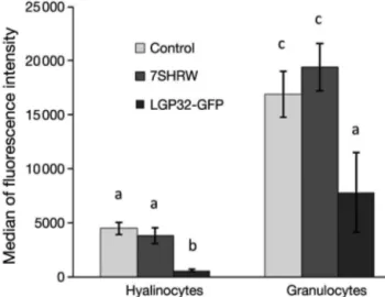
Comparison of the median of fluorescent intensity values of LysoTracker staining showed that in control clams, granulocytes exhibited significantly more
lyso-somal staining than did hyalinocytes (p < 0.01) (Fig. 4). A similar tendency was found after infection with 7SHRW (p < 0.001) and LGP32-GFP (p < 0.001). How-ever, the median fluorescence intensity revealed that the lysosomal staining in both haemocyte subpopula-tions in LGP32-GFP-infected clams was significantly lower than those from control clams (p < 0.01 for granulocytes, p < 0.0001 for hyalinocytes) and from 7SHRW-infected clams (p < 0.001 for granulocytes, p = 0.0001 for hyalinocytes) (Fig. 4).
When each haemocyte subpopulation was further subdivided according to lysosomal content (herein defined as the relative amount of LysoTracker fluores-cence observed) into ‘low’ and ‘high’ (Fig. 5), the pro-portions among groups significantly changed after infection, for both hyalinocytes (p < 0.001) and granu-locytes (p = 0.001) (Fig. 6). Among hyalinocytes, the proportion of cells with high lysosomal content signifi-cantly decreased from 85.3 ± 2.2% in the control clams to 69.4 ± 4.7% in the 7SHRW-infected clams and to 21.1 ± 5.4% in the LGP32-GFP-infected clams (Fig. 6a). Among granulocytes, the proportion of cells with high lysosomal content varied little between the control and the 7SHRW-infected groups, each having > 80% of cells with high lysosomal content (87.8 ± 0.4% and 86.6 ± 1.8%, respectively) and <15% of cells with low lysosomal content (12.2 ± 0.4% and 13.4 ± 1.8%, respectively). These proportions, however, were somewhat inversed in clams infected with LGP32-GFP, with 60.2 ± 10.6% of cells having low and 39.8 ± 10.6% of cells having high lysosomal content (Fig. 6b).
Microscopic observation of the sorted cells belong-ing to each of the 4 subpopulations established by the 2-scatter and LysoTracker staining profiles
re-Fig. 2. Mya arenaria haemocytes. Proportions (± SE) of hyali-nocytes and granulocytes from clams 24 h after injection with filtered sterile seawater (FSSW; control), Vibrio splendidus 7SHRW or V. splendidus LGP32-GFP (n = 21). Differences
Mateo et al.: Vibrio splendidus changes Mya arenaria haemocytes
vealed cells of different sizes and cytoplasm content (Fig. 7). Hyalinocytes had a diameter of approxi-mately 5 to 7 µm (Fig. 7a,b,e,f) whereas in granulo-cytes it was around 9 to 13 µm (Fig. 7c,d,g,h). Haemocytes sorted from the ‘high’ LysoTracker staining subpopulation (Fig. 7b,d,f,h) appeared to have a higher granularity compared with those with ‘low’ staining (Fig. 7a,c,e,g). To some extent, the cytoplasm content of the control haemocytes appeared to be homogeneously distributed, while in LGP32-GFP-infected cells it appeared to be localised towards one side of the cell and the nucleus was often indistinguishable.
DISCUSSION
Flow cytometry is a useful technique for the charac-terisation of haemocyte subpopulations in bivalves. Analyses using these methods are mostly based on 2-side scatter profiles that delineate subpopulations according to cell size and complexity. Additional fluo-rescent dyes can be used to reveal complementary information about cellular components for a more com-plete characterisation. In the present study, the use of LysoTracker, as an indicator of lysosomal content, in addition to light-scatter profiles, revealed different haemocyte subpopulations in Mya arenaria. These subpopulations were shown to undergo changes in proportions that suggest interesting cellular processes induced by 2 strains of Vibrio splendidus, a local wild
257
Fig. 3. Mya arenaria haemocyte distribution profile. Forward-scatter (FSC) and side-Forward-scatter (SSC) plot profiles of granulo-cytes and hyalinogranulo-cytes from clams 24 h after injection with (a) filtered sterile seawater (FSSW), (b) Vibrio splendidus
7SHRW or (c) V. splendidus LGP32-GFP
Fig. 4. Mya arenaria haemocytes. Median (± SE) fluorescence intensity of LysoTracker retention in granulocytes and hya-linocytes from clams 24 h after injection with filtered sterile seawater (FSSW; control), Vibrio splendidus 7SHRW or V.
splendidus LGP32-GFP (n = 21). Letters show statistical
equivalence (same letters) or difference at p < 0.05 (different letters) between pairs of haemocyte subpopulations and
strain and a strain associated with oyster mortalities in France.
Through the flow-cytometry scatter profiles (forward and side-scatter) we observed 2 discernible subpopu-lations of haemocytes in Mya arenaria: a discrete sub-population of smaller and less complex cells and another, more dispersed, subpopulation of larger and more complex cells. We think that the former subpop-ulation was composed of hyalinocytes and/or agranu-locytes (with no or few cytoplasmic granules) and the latter, granulocytes. This latter subpopulation might include what others have classified as small and large granulocytes (reviewed in Cheng 1981). Previously, using flow-cytometric toxicological studies of M.
are-naria haemocytes, 2 discernible subpopulations using
2-scatter profiles were also noticed (Brousseau et al. 1999, Fournier et al. 2001, 2002).
Prior reports involving flow-cytometric light-scatter profiling revealed that haemocytes from a number of bivalve species can typically be classified into 2, 3 or 4 subpopulations. Haemocytes from clams Ruditapes
philippinarum, R. decussatus and Mercenaria merce-naria have been classified into 2 subpopulations:
hyalinocytes and granulocytes, located in the lower and higher channels of both light-scatter axes, respec-tively (Allam et al. 2001, 2002, 2006, Allam & Ford 2006). In the scallop Chlamys farreri, 2 haemocyte types have been identified: granulocytes and hyalino-cytes (Xing et al. 2002). The hard clam Meretrix lusoria has been reported to have 3 subpopulations: hya-linocytes and small and large granulocytes (Tu et al. 2007). In mussels Mytilus galloprovincialis, 3 subpopu-lations (hyalinocytes and small and large granulocytes) (Parisi et al. 2008) and 4 subpopulations (large
granu-Fig. 5. Mya arenaria haemocytes with LysoTracker. Median fluorescence intensity of LysoTracker (LT) retention (arbitrarily es-tablished as ‘high’ and ‘low’) in (a) hyalinocytes and (b) granulocytes from clams 24 h after injection with filtered sterile seawater
Mateo et al.: Vibrio splendidus changes Mya arenaria haemocytes
locytes, large semigranulocytes, smaller granulocytes and small agranulocytes or hyalinocytes) (García-Gar-cía et al. 2008) have been considered. Oysters
Cras-sostrea virginica and C. gigas were shown to possess 3
haemocyte subpopulations, although they were regarded slightly differently by different researchers (Ashton-Alcox & Ford 1998, Allam et al. 2002, Lambert et al. 2003, Goedken & De Guise 2004). In the Sydney rock oyster Saccostrea glomerata, up to 4 subpopula-tions were distinguished, although hyalinocytes and granulocytes were the 2 most abundant phenotypes (Aladaileh et al. 2007).
Following infection with Vibrio splendidus 7SHRW, the proportion of hyalinocytes slightly decreased (Fig. 2) and became less discernible in the scatter pro-file (Fig. 3b), while with the strain LGP32-GFP, changes were more striking, as both subpopulations merged into a single continuous group (Fig. 3c). Dras-tic changes in haemocyte distribution according to size and granularity have been previously noticed by Allam
& Ford (2006). They observed a clear shift of granular cells towards the agranular cell population, resulting in unimodal distribution of haemocytes from clams
Rudi-tapes philippinarum and Mercenaria mercenaria after in vitro exposure to V. tapetis and V. splendidus.
We supplemented our light-scatter profiling data with LysoTracker staining in order to provide a mea-sure of lysosome content. LysoTracker is a fluorescent probe that accumulates in acidic compartments (Fre-undt et al. 2007) and has been used previously in stud-ies of human-cell lysosomes (Haller et al. 1996, Via et al. 1998, Blander & Medzhitov 2004). This approach, applied for the first time to bivalve haemocytes in the present study, revealed independent responses in haemocyte subpopulations (Figs. 4, 5 & 6). LysoTracker staining showed that granulocytes from the control clams contained significantly more lysosomes than the hyalinocytes. This is in agreement with microscopic observations, and indirectly with enzyme studies, which have shown that granulocytes have abundant
259
lysosomes (Pipe 1990, Cajaraville & Pal 1995, Cima et al. 2000, Matozzo et al. 2007).
When challenged bacterially, hyalinocytes had lower levels of lysosomal content, especially after infection with LGP32-GFP (Figs. 4, 5a & 6a), whereas granulocytes had similar levels of lysosomes after infection with 7SHRW and significantly decreased levels after infection with LGP32-GFP (Figs. 4, 5b & 6b). Lysosomes are cellular organelles that contain hydrolytic enzymes (Luzio et al. 2000), including lysozymes, that are involved in intracellular degrada-tion and host defence (Olsen et al. 2003). Many lyso-somes have secretory functions (Holt et al. 2006). Similar to mammalian macrophages, in several bi-valves including Mya arenaria, lysosomes release their content upon infection during degranulation of actively phagocytosing cells (Cheng & Rodrick 1974, Rodrick 1979; reviewed in Cheng 1983, Chu 1988). Lysosomal degranulation may be one possible mecha-nism accounting for the decrease of lysosomal con-tent, expressed as loss of fluorescent intensity (Figs. 4 & 5) and change in proportions (Fig. 6) in haemocytes from Vibrio splendidus LGP32-GFP-infected clams.
Fig. 6. Mya arenaria haemocyte subpopulations. Proportion (± SE) that are (a) hyalinocytes (H) or (b) granulocytes (G) ac-cording to LysoTracker (LT; ‘high’ or ‘low’) staining 24 h after injection of clams with filtered sterile seawater (FSSW; con-trol), Vibrio splendidus 7SHRW or V. splendidus LGP32-GFP (n = 21). Letters show statistical equivalence (same letters) or
difference at p < 0.05 (different letters) among treatments
Fig. 7. Mya arenaria haemocytes. Phase-contrast images of immediately fixed haemocytes from each subpopulation. Control group: hyalinocytes with (a) ‘low’ and (b) ‘high’ LysoTracker staining; granulocytes with (c) ‘low’ and (d) ‘high’ LysoTracker staining. Vibrio splendidus LGP32-GFP-infected group: hyalinocytes with (e) ‘low’ and (f) ‘high’ LysoTracker staining;
Mateo et al.: Vibrio splendidus changes Mya arenaria haemocytes
The decreased responsiveness to V. splendidus 7SHRW insult could, by extension, be attributable to a decreased capacity to activate degranulation in
Mya arenaria haemocytes.
Considering that 7SHRW is a wild endemic strain isolated from sediments from an area relatively close to the source area of our clams, where there is no history of bacterial infections, this strain may be recognised by the haemocyte’s pattern-recognition receptors as a non-threat, non-self particle. On the other hand, LGP32 is a non-native strain that has been associated with mortalities of juvenile Pacific oysters Crassostrea
gigas in Europe (Gay et al. 2004a,b). Moreover, it has
been found that this strain possesses the pathogenic factor vsm, a metalloprotease recently demonstrated to be the most important toxicity factor in the extracellu-lar products of LGP32 (Binesse et al. 2008). It is likely that this pathogenic factor is associated with the signif-icant degranulation of Mya arenaria haemocytes in the present study. The specific response of M. arenaria haemocytes to the strain LGP32-GFP is in accordance with our previous findings that this strain induces sig-nificant changes in haemocyte structure, number and adhesion on this host species, whereas changes induced by 7SHRW are minor or nonexistent (Mateo et al. 2009).
The decrease in lysosomal content in hyalinocytes (Figs. 4 & 5b) or the increase of hyalinocytes with low levels of lysosomal content (Figs. 5a & 6a) might not only be due to degranulation but possibly due to the increased presence of precursor haemocytes that have not yet developed cytoplasmic granules. They might be released prematurely to fight the infection. A simi-lar phenomenon, known as ‘left shift’, occurs in mam-malian leucocytes upon inflammation and septic shock (Opdenakker 2001). Undifferentiated and small stem cells have been described as blast-like cells or haemoblasts, a subtype of agranular cells with a high nucleus:cytoplasm ratio and which lack organelles (Hine 1999, Cima et al. 2000, Chang et al. 2005, Aladaileh et al. 2007, Matozzo et al. 2008). Further research is, however, required to confirm the nature of the abundant smaller and less complex cells we found after Vibrio splendidus LGP32-GFP challenge.
In conclusion, we found that responses of haemo-cytic subpopulations are not only specific to the pathogen strain, but are modulated independently of each other and possibly through independent cellular mechanisms. These changes in haemocytic subpopula-tions were monitored by using LysoTracker as an indi-cator of the lysosomal content, and more striking responses were found after infection with Vibrio
splen-didus LGP32-GFP. Functional studies are needed to
confirm the activation of degranulation and the sus-pected release of immature haemocytes.
Acknowledgements. We acknowledge the kind support of
Dr. F. Le Roux (Harvard Medical School, formerly at Institut Pasteur) for providing the bacterial strain LGP32-GFP and of L. Chevarie (Centre Maricole des Iles-de-la-Madeleine, CEMIM) for providing the clams. This work was funded by the Industrial Research Assistance Program (IRAP), the Nat-ural Sciences and Engineering Research Council of Canada (NSERC) and Technology PEI. D.R.M. is supported by a PhD scholarship from AVC.
LITERATURE CITED
Aladaileh S, Nair SV, Birch D, Raftos DA (2007) Sydney rock oyster (Saccostrea glomerata) haemocytes: morphology and function. J Invertebr Pathol 96:48–63
Allam B, Ford SE (2006) Effects of the pathogenic Vibrio
tapetis on defence factors of susceptible and
non-suscepti-ble bivalve species: I. Haemocyte changes following in
vitro challenge. Fish Shellfish Immunol 20:374–383
Allam B, Ashton-Alcox KA, Ford SE (2001) Haemocyte para-meters associated with resistance to brown ring disease in
Ruditapes spp. clams. Dev Comp Immunol 25:365–375
Allam B, Ashton-Alcox KA, Ford SE (2002) Flow cytometric comparison of haemocytes from three species of bivalve molluscs. Fish Shellfish Immunol 13:141–158
Allam B, Paillard C, Auffret M, Ford S (2006) Effects of the pathogenic Vibrio tapetis on defence factors of the suscep-tible and non-suscepsuscep-tible bivalve species: II. Cellular and biochemical changes following in vivo challenge. Fish Shellfish Immunol 20:384–397
Ashton-Alcox KA, Ford SE (1998) Variability in molluscan hemocytes: a flow cytometric study. Tissue Cell 30:195–204 Ashton-Alcox KA, Allam B, Ford SE (2000) Application of flow
cytometry to bivalve pathology. In: Fingerman M, Nagab-hushanam R (eds) Recent advances in marine biotechnol-ogy. Science Publishers, Enfield, NH, p 85–124
Auffret M (1988) Bivalve hemocyte morphology. Am Fish Soc Spec Publ 18:169–177
Binesse J, Delsert C, Saulnier D, Champomier-Vergès MC and others (2008) Metalloprotease vsm is the major deter-minant of toxicity for extracellular products of Vibrio
splendidus. Appl Environ Microbiol 74:7108–7117
Blander JM, Medzhitov R (2004) Regulation of phagosome maturation by signals from Toll-like receptors. Science 304:1014–1018
Brousseau P, Pellerin J, Morin Y, Cyr D, Blakley B, Boermans H, Fournier M (1999) Flow cytometry as a tool to monitor the disturbance of phagocytosis in the clam Mya arenaria hemocytes following in vitro exposure to heavy metals. Toxicology 142:145–156
Cajaraville MP, Pal SG (1995) Morphofunctional study of the haemocytes of the bivalve mollusc Mytilus
galloprovin-cialis with emphasis on the endolysosomal compartment.
Cell Struct Funct 20:355–367
Chang SJ, Tseng SM, Chou HY (2005) Morphological charac-terization via light and electron microscopy of the haemo-cytes of two cultured bivalves: a comparison study between the hard clam (Meretrix lusoria) and Pacific oys-ter (Crassostrea gigas). Zool Stud 44:144–153
Cheng TC (1981) Bivalves. In: Ratcliffe NA, Rowley AF (eds) Invertebrate blood cells, Vol I. Academic Press, London, p 233–300
Cheng TC (1983) The role of lysosomes in molluscan inflam-mation. Am Zool 23:119–144
Cheng TC (1984) A classification of molluscan haemocytes based on functional evidence. In: Bulla LA, Cheng TC 261
➤
➤
➤
➤
➤ ➤➤
➤
➤
➤
➤
➤
➤
➤
➤
➤
➤
(eds) Comparative pathobiology, Vol 6. Plenum Press, New York, p 111–146
Cheng TC, Rodrick GE (1974) Identification and characteriza-tion of lysozyme from hemolymph of the soft-shelled clam,
Mya arenaria. Biol Bull (Woods Hole) 147:311–320
Choquet G, Soudant P, Lambert C, Nicolas JL, Paillard C (2003) Reduction of adhesion properties of Ruditapes
philippinarum haemocytes exposed to Vibrio tapetis. Dis
Aquat Org 57:109–116
Chu FLE (1988) Humoral defense factors in marine bivalves. Am Fish Soc Spec Publ 18:178–188
Cima F, Matozzo V, Marin MG, Ballarin L (2000) Haemocytes of the clam Tapes philippinarum (Adams & Reeve, 1850): morphofunctional characterisation. Fish Shellfish Immu-nol 10:677–693
Fisher WS, Ford SE (1988) Flow cytometry: a tool for cell research in bivalve pathology. Am Fish Soc Spec Publ 18: 286–291
Fournier M, Pellerin J, Clermont Y, Morin Y, Brousseau P (2001) Effects of in vivo exposure of Mya arenaria to organic and inorganic mercury on phagocytic activity of hemocytes. Toxicology 161:201–211
Fournier M, Pellerin J, Lebeuf M, Brousseau P, Morin Y, Cyr D (2002) Effects of exposure of Mya arenaria and
Mac-tromeris polynyma to contaminated marine sediments on
phagocytic activity of haemocytes. Aquat Toxicol 59:83–92 Freundt EC, Czapiga M, Lenardo MJ (2007) Photoconversion
of Lysotracker Red to a green fluorescent molecule. Cell Res 17:956–958
García-García E, Prado-Álaverz M, Novoa B, Figueras A, Ros-ales C (2008) Immune responses of mussel hemocyte sub-populations are differentially regulated by enzymes of the PI 3-K, PKC, and ERK kinase families. Dev Comp Immunol 32:637–653
Gay M, Berthe F, Le Roux F (2004a) Screening of Vibrio iso-lates to develop an experimental infection model in the Pacific oyster Crassostrea gigas. Dis Aquat Org 59:49–56 Gay M, Renault T, Pons AM, Le Roux F (2004b) Two Vibrio
splendidus related strains collaborate to kill Crassostrea gigas: taxonomy and host alterations. Dis Aquat Org 62:
65–74
Goedken M, De Guise S (2004) Flow cytometry as a tool to quantify oyster defence mechanisms. Fish Shellfish Immunol 16:539–552
Haller T, Dietl P, Deetjen P, Völkl H (1996) The lysosomal compartment as intracellular calcium store in MDCK cells: a possible involvement in InsP3-mediated Ca2+ release.
Cell Calcium 19:157–165
Hine PM (1999) The inter-relationship of bivalve haemocytes. Fish Shellfish Immunol 9:367–385
Holt OJ, Gallo F, Griffiths GM (2006) Regulating secretory lysosomes. J Biochem 140:7–12
Huffman JE, Tripp MR (1982) Cell types and hydrolytic enzymes of soft shell clam (Mya arenaria) haemocytes. J Invertebr Pathol 40:68–74
Labreuche Y, Lambert C, Soudant P, Boulo V, Huvet A, Nico-las JL (2006) Cellular and molecular hemocyte response of the Pacific oyster, Crassostrea gigas, following bacterial infection with Vibrio aestuarianus strain 01/32. Microbes Infect 8:2715–2724
Lambert C, Soudant P, Choquet G, Paillard C (2003) Mea-surement of Crassostrea gigas hemocyte oxidative metab-olism by flow cytometry and the inhibiting capacity of pathogenic vibrios. Fish Shellfish Immunol 15:225–240 López C, Carballal MJ, Azevedo C, Villalba A (1997)
Morpho-logical characterization of the hemocytes of the clam,
Ruditapes decussates (Mollusca: Bivalvia). J Invertebr
Pathol 69:51–57
Luzio JP, Pryor PR, Bright NA (2000) Lysosome-endosome fusion and lysosomes biogenesis. J Cell Sci 113:1515–1524 Mateo DR (2006) Assessment of antimicrobial resistance in marine environments of Prince Edward Island using a selected bacterial group. MSc dissertation, Atlantic Vet-erinary College, University of Prince Edward Island, Char-lottetown, PEI
Mateo DR, Siah A, Araya MT, Berthe FCJ, Johnson GR, Greenwood SJ (2009) Differential in vivo response of soft-shell clam hemocytes against two strains of Vibrio
splen-didus: changes in cell structure, numbers and adherence.
J Invertebr Pathol 102:50–56
Matozzo V, Rova G, Marin MG (2007) Haemocytes of the cockle Ceratoderma glaucum: morphological characteri-sation and involvement in immune responses. Fish Shell-fish Immunol 23:732–746
Matozzo V, Marin MG, Cima F, Ballarin L (2008) First evi-dence of cell division in circulating haemocytes from the Manila clam Tapes philippinarum. Cell Biol Int 32: 865–868
Moore MN, Depledge MH, Readman JW, Leonard DRP (2004) An integrated biomarker-based strategy for ecotoxicologi-cal evaluation of risk in environmental management. Mutat Res 552:247–268
Moore MN, Allen JI, McVeigh A (2006) Environmental prog-nostics: an integrated model supporting lysosomal stress responses as predictive biomarkers of animal health sta-tus. Mar Environ Res 61:278–304
Olsen ØM, Nilsen IW, Sletten K, Myrnes B (2003) Multiple invertebrate lysozymes in blue mussel (Mytilus edulis). Comp Biochem Physiol B 136:107–115
Opdenakker G (2001) New insights in the regulation of leuko-cytosis and the role played by leukocytes in septic shock. Verh K Acad Geneeskd Belg 63:531–538
Parisi MG, Li H, Jouvet LBP, Dyrinda EA, Parrinello N, Cam-marata M, Roch P (2008) Differential involvement of mus-sel hemocyte sub-populations in the clearance of bacteria. Fish Shellfish Immunol 25:834–840
Pipe RK (1990) Hydrolytic enzymes associated with the gran-ular haemocytes of the marine mussel Mytilus edulis. His-tochem J 22:595–603
Pipe RK, Farley SR, Coles JA (1997) The separation and char-acterisation of haemocytes from the mussel Mytilus edulis. Cell Tissue Res 289:537–545
Renault T, Xue QG, Chilmonczyk S (2001) Flow cytometric analysis of European flat oyster, Ostrea edulis, haemo-cytes using a monoclonal antibody specific for granulo-cytes. Fish Shellfish Immunol 11:269–274
Rodrick GE (1979) Selected enzyme activities in Mya arenaria hemolymph. Comp Biochem Physiol B 62:313–316 Tu CY, Hung SW, Tsou LT, Chang YC, Wang WS (2007)
Simultaneous flow cytometry assessment for cellular types and phagocytic abilities of the haemocytes of the hard clam, Meretrix lusoria. Fish Shellfish Immunol 23: 16–23
Via LE, Fratti RA, McFalone M, Pagán-Ramos E, Deretic D, Deretic V (1998) Effects of cytokines on mycobacterial phagosome maturation. J Cell Sci 111:897–905
Xing J, Zhan WB, Zhou L (2002) Endoenzymes associated with haemocyte types in the scallop (Chlamys farreri). Fish Shellfish Immunol 13:271–278
Zhang W, Wu X, Wang M (2006) Morphological, structural, and functional characterization of the haemocytes of the scallop, Argopecten irradians. Aquaculture 251:19–32
Editorial responsibility: Mike Hine, Fouras, France
Submitted: April 30, 2009; Accepted: July 29, 2009 Proofs received from author(s): October 19, 2009