Basolateral Amygdala Circuits for Differentiating
Positive and Negative Associations
By
Praneeth Namburi
B.Eng. in Electrical and Electronic Engineering
Nanyang Technological University, 2008
Submitted to the Department of Brain and Cognitive Sciences in partial fulfillment of the requirements for the degree of
DOCTOR OF PHILOSOPHY IN NEUROSCIENCE at the
MASSACHUSETTS INSTITUTE OF TECHNOLOGY June 2016
C Massachusetts Institute of Technology 2016. All rights reserved.
Signature of Author:
Certified by:
Signature redacted
Department of Brain and Cognitive Sciences
April 1 2th 2016
S
ignature redacted~
Kay M. Tye stant Professor of Neuroscience Thesis Supervisor
Accepted
b4Signature
redacted
Matthew A. Wilson
S an Fairchild Professor of Neuroscience and Picower Scholar Director of Graduate Education for Brain and Cognitive Sciences
1 MASSACHUSETTS INSTITUTE OF TECHNOLOGY
JUL 2
0
2015
LIBRARIES
K
MITLibraries
77 Massachusetts Avenue Cambridge, MA 02139 hftp://Iibraries.mit-edu/ask
DISCLAIMER NOTICE
Due to the condition of the original material, there are unavoidable
flaws in this reproduction. We have made every effort possible to
provide you with the best copy available.
Thank you.
The images contained in this document are of the
best quality available.
Basolateral Amygdala Circuits for Differentiating Positive
and Negative Associations
By
Praneeth Namburi
Submitted to the Department of Brain and Cognitive Sciences on April 12th, 2016 in partial
fulfillment of the requirements for the degree of DOCTOR OF PHILOSOPHY IN NEUROSCIENCE
Abstract
The ability to differentiate between rewarding and threatening stimuli, and engaging the appropriate behavioral response is critical for survival. The Basolateral Amygdala (BLA) is an almond shaped structure in the brain where rewarding and fearful associations are encoded by different populations of neurons. However, identifying features of these populations have remained an enigma. My thesis work shows that populations of BLA neurons that differ in their
long range anatomical connectivity play opposing roles in the acquisition of positive and negative associations, and dissects a mechanism by which these associations are encoded in the
BLA. We show that BLA neurons projecting to the nucleus accumbens (NAc) and BLA neurons projecting to the centromedial nucleus of the amygdala (CeM) undergo opposing changes at their input synapses after rewarding and fearful associations. We then establish the in vivo ramifications of these opposing changes in synaptic strength in response to rewarding and fearful associations
by assaying neural activity from BLA neurons and identifying the NAc and CeM projectors.
Finally, in order to compare and contrast the role of BLA neural populations in encoding positive and negative associations, we propose a model that parametrizes neural responses to positive and negative cues from large scale electrophysiological recordings. My thesis work identifies
functional roles of specific circuit components based on their long range anatomical connectivity, identifies differentially expressed receptors within these circuit components and
provides a mechanistic explanation, on synaptic, cellular, circuit and molecular levels, for how
positive and negative associations can be formed within, and diverge from the BLA.
Thesis Supervisor: Kay M. Tye
Table of Contents
A b str a c t...3
D e d ic a tio n ... 9
A ck n ow led gm en ts ... 1 1 C h ap ter 1 - In trod uction ... 13
1 .1 V a le n c e ... 14
1.2 Reward and aversion are widely represented in the brain ... 16
1.3 Studying valence at the level of neural populations ... 18
1.4 Advances in targeting specific neural populations ... 20
1.5 Representation of reward and aversion in the BLA ... 23
1.6 Learning-induced plasticity in the BLA ... 27
1.7 Studying mechanisms of valence coding in the BLA ... 29
Chapter 2 - A circuit mechanism for differentiating positive and negative associations ... 31
2 .1 S u m m ary ... 3 2 2 .2 B ack g rou n d ... 3 3 2 .3 R e su lts ... 3 5 2.3.1 NAc and CeM projecting BLA neurons undergo opposing synaptic changes following fear and reward conditioning... 35
2.3.2 Photostimulation of NAc or CeM projecting BLA neurons causes positive or negative reinforcement, respectively ... 40
2 .4 D iscu ssio n ... 5 0
2.4.1 Specificity of inputs undergoing experience-dependent changes ... 50
2.4.2 Food restriction-induced synaptic changes... 51 2.4.3 The BLA on valence and arousal - differential roles for the LA and the BA .... 52 2.4.4 Potential diversity among NAc-projecting BLA neurons...54 2.4.5 A circuit-based approach to identify novel drug targets ... 55 2 .5 C on clu sion ... 5 6 2.6 M ethods and M aterials... 57 2.7 Supplem ental Figures... 73
Chapter 3 -Divergent routing of positive and negative information from the amygdala .... 99 3 .1 S u m m ary ... 10 0 3 .2 B ack grou n d ... 10 1 3 .3 R e su lts ... 10 3
3.3.1 Optogenetic-mediated photoidentification of BLA projection neurons during
retriev a l ... 10 3 3.3.2 Half of BLA neurons respond to sucrose and/or quinine cues ... 107 3.3.3 BLA projectors send collaterals to multiple downstream targets ... 111
3.3.4 ChR2-expressing neurons have shorter photoresponse latencies compared to non-expressing neighbors ... 113 3.3.5 BLA projector populations differentially respond to sucrose and quinine
c u e s ... 1 1 5
3.3.6 BLA neurons projecting to NAc and CeA preferentially code for opposite
v a len ce ... 12 5
3 .4 D iscu ssio n ... 13 1 3.4.1 Divergent circuits for encoding positive and negative valence ... 131
3.4.3 Design Advantages ... 133
3.4.4 "Both" cells and collaterals: hypotheses...135
3.4.5 Valence: operational definitions ... 136
3.4.6 Innate versus learned valence: responses to the US...138
3.5 Conclusion ... 140
3.6 M ethods and M aterials...141
3.7 Supplem ental Figures...154
Chapter 4 - A m odel for quantifying valence representation...173
4.1 Sum m ary ... 174
4.2 A N eurobiological Definition of Valence...175
4.3 A M odel for Investigating Valence Representation...177
4.4 Cross-study and between-population comparison ... 185
4 .5 C o n clu sio n ... 18 7 Chapter 5 - The Conclusion...189
5.1 The BLA as a selector...192
5.2 Selection through opposing gain modulation between functional components...195
5.3 Flexibility of Valence Representation in the BLA ... 197
Dedicated to my mother, Swarupa Kumari.
Acknowledgments
As one of my best friends Zenna Tavares said, "It is all about the process", I am extremely grateful to everyone who have made this journey better than anything I had dreamed about.
Kay, I have you to thank most of all for this amazing journey. Reflecting upon the past 5 years, a significant portion of the good and vivid memories I have are due to the scientific and social environment you have created. Thank you for the opportunity to be part of your creation, thank you for constantly caring, identifying my developmental needs, always being there to guide me towards being a better scientist, and pausing to celebrate. You are an amazing mentor, and drawing inspiration from you, one day I hope to create an environment around me that is supportive, creative and inclusive - a scientific family striving for each others' success.
Anna, thank you for being you. You are an amazing person, and I am so grateful for the opportunity to cross paths with you. Working with you has made me a better collaborator, scientist and friend. Thank you for being there for me when I needed support the most - I could not have
made it this far without you. I owe you a peanut butter banana smoothie.
Dr. Michale Fee, I have had two long one-on-one conversations with you - the first one made me really want to come to MIT for grad school, and the second one has helped me clarify what I would like to do after graduate school. Thank you for inspiring me with the way you think and thank you for being the chair of my thesis committee.
Dr. Mark Bear, thank you being a member of my thesis committee. I am grateful for our conversations and your constant support. Your support helped me secure funding for the last two years of my PhD.
Dr. Naoshige Uchida, thank you for agreeing to be the outside reviewer on my thesis committee. My interaction with you has helped me clarify which skills I would like to strengthen in the next leg of my neuroscientific journey.
Edward, thank you for our friendship, and sharing your graduate school journey with me.
I am so grateful that we got to watch each other grow in graduate school, and for having a friend
who knows exactly what I'm going through at each phase of graduate school. Thank you for your support.
Gwen, thank you for working with me, and thank you for your friendship and support. It has been such a pleasure learning from you. Thank you for being the new leader of Team
Craig, one day I would like to start my own lab, and when I do, I hope to find someone like you. Thank you for making everything possible. Chris, working with you over the past year has been amazing. I have learned a lot, and thanks to you, working on my new phase of experiments has been such a great experience. Thank you Ada, Stephen, Gillian, Romy, Caitlin, Evelien, Alienor, Tony, and the rest of the Tye Lab for being such wonderful and supportive colleagues.
Zenna, Valerie, Jack, Melissa, Galen, Max and Julian, thank you for being such good roommates and friends, and for the little things that I will end up remembering more than any figure or paper down the line. Thank you guys, and Swati, Shaiyan, Emily and Steph, for your
friendship and all the adventures - Ski trips, bike rides, A4 pizza nights, smash bro nights,
Colombia, etc, etc, etc.
I enjoy learning, and MIT is the best place to learn anything as long there is the desire. I
am extremely grateful for my friends in the MIT ballroom dance team. I started dancing in graduate school, and within a short period of time, dancing has become very important to me, thanks to you.
Armin, Fil, Allena, Soli, Amanda, Amanda, Melissa and Maria -thanks to you I have been able to
immerse myself in something outside the lab.
I would also like to thank the people who helped me on my way towards graduate school
- my mentors in Singapore Dr. Cindy Goh, Dr. Vitali Zagorodnov, Dr. Cuntai Guan, Dr. Michael
Chee and Dr. George Augustine, and my friends from the Chee and Augustine labs. Without my experiences with you, graduate school would not have been the same.
Chapter 1
Introduction
1.1 Valence
For more than a century following William James' original thesis on emotion (James,
1884), psychologists have attempted to determine whether the diverse range of human affect can
be understood using few independent factors. On the basis of self-reported emotional states, early theorists charted emotions in two or more dimensions (Nowlis and Nowlis, 1956; Russell, 1980; Schlosberg, 1954). Popular among these models is the two-dimensional circumplex model of emotion (Russell, 1980), wherein emotions arise from the interaction between two neuropsychological systems-one representing the degree of pleasantness, ranging from aversive to appetitive (valence), and the other representing alertness (arousal) (Posner et al., 2005). Identifying and understanding the neurobiological substrates underlying these features of emotion is an active area of neuroscience research.
The idea that anatomically localized regions in the brain drive emotion and emotional behaviors was initially suggested by the finding that lesions to the temporal lobe and amygdala cause affective deficits (Klfiver and Bucy, 1939). Following this early work, animal models for studying affect have been instrumental in advancing our understanding of the neurobiological basis of emotion. Although the subjective aspect of emotions cannot be directly tested in animal models, the behavioral and physiological responses elicited by emotionally relevant stimuli can be objectively assessed.
Arousal is commonly studied in relation to consciousness, sleep, attention, sex, and emotion. Emotional arousal is an important aspect of emotion that is known to enhance emotional memory, either positive or negative. For a detailed review of the neural representation of arousal,
refer to (Adolphs et al., 1999; Harris and Aston-Jones, 2006; Lang et al., 1998; McGaugh, 2000,
2004; McIntyre and Roozendaal, 2007).
Monitoring neural activity evoked by emotionally salient stimuli in model organisms, such as non-human primates and rodents, has proved to be an invaluable method to investigate the neurobiological basis of valence. A stimulus that is inherently appetitive or pleasant is said to carry positive valence, whereas a stimulus that is inherently aversive is said to carry negative valence. These stimuli are sufficient to evoke appetitive or aversive responses, and are therefore designated positive or negative unconditioned stimuli (US), respectively. When a previously neutral stimulus (known as a conditioned stimulus or CS), such as a tone, odor, or image, predicts a positive or negative US, it acquires valence. Pavlovian conditioning (Pavlov, 1927; Rescorla, 1988), in which the CS and US are repeatedly paired, is a common behavioral paradigm for teaching an animal a
CS-US association. After the acquisition of a successful CS-US pairing, a positive CS is sufficient
to evoke appetitive behaviors such as approach toward a food dispenser, and a negative CS is sufficient to evoke fear- or avoidance-related behaviors such as freezing in response to a CS predicting a foot shock.
In several regions of the brain, neural responses to the CS change after the acquisition of
CS-US pairing. Moreover, these changes are different depending on whether the CS is positive or
negative. A neuron recorded during a behavioral paradigm such as Pavlovian conditioning or retrieval is said to represent valence if its output is differentially modulated by the positive and negative natures of the stimuli, independent of all other features.
1.2 Reward and aversion are widely represented in the brain
Certain anatomically localized populations of neurons show differential responses to reward or aversion-associated cues. These regions have been considered to represent valence, and include the BLA (Fuster and Uyeda, 1971; Paton et al., 2006; Shabel and Janak, 2009), nucleus accumbens (NAc) (Roitman et al., 2005), ventral tegmental area (VTA) (Bromberg-Martin et al., 2010; Cohen et al., 2012; Matsumoto and Hikosaka, 2009), orbitofrontal cortex (Schoenbaum et
al., 1999), lateral hypothalamus (LH) (Fukuda et al., 1990; Li et al., 2013; Nakamura et al., 1987;
Ono et al., 1986; Yamamoto et al., 1989), dorsal raphe nucleus (DRN) (Cohen et al., 2015), locus coeruleus (Bouret and Richmond, 2015; Hofmeister and Sterpenich, 2015; McCall et al., 2015), subthalamic nucleus (Sieger et al., 2015), and hippocampus (Fuster and Uyeda, 1971). Within many of these regions, there is marked heterogeneity among neural populations, such that neurons representing positive valence reside side-by-side with those representing negative valence.
Techniques that reveal two populations of neurons in the same brain-one active in response to a rewarding stimulus and the other active in response to an aversive stimulus-will be immensely useful for gaining a comprehensive understanding of brain regions containing topographically intermingled populations of neurons representing valence. These emergent strategies are already proving their utility; a recent study (Xiu et al., 2014) used a technique known as tyramide-amplified-immunohistochemistry-fluorescence in situ hybridization (TAI-FISH) to simultaneously label populations of neurons that respond to a rewarding stimulus such as cocaine, and an aversive stimulus, such as foot shock. TAI-FISH exploits the difference between the time courses of c-fos mRNA and protein expression to simultaneously label two populations of neurons, each active in response to a different stimulus. The authors found several brain regions containing intermingled populations of neurons activated by morphine and foot shock, including the ventral
division of the lateral septum, dorsomedial shell of the NAc, and the fusiform nucleus of the bed nucleus of stria terminalis.
Therefore, valence is represented across several brain regions in distributed networks, and, more importantly, all of the brain regions discussed above represent both positive and negative valences. Some brain regions, such as the basolateral amygdala (BLA), contain a greater proportion of neurons signaling valence (-40%) compared with others, such as the hippocampus (~25%) (Fuster and Uyeda, 1971). Valence-encoding neurons represent only a subset of neurons within each of these brain regions. Even among the valence-representing subset in a given region, there are some neurons signaling positive valence and some neurons signaling negative valence. Hence, we can posit that several brain regions contain either largely distinct or overlapping populations of neurons representing positive and negative valences, and dissecting neural mechanisms underlying valence at the level of neural populations (a group of neurons that share a
1.3 Studying valence at the level of neural populations
A population of neurons, in the most general sense, is simply a set of neurons that share a
unique feature. Neurons belonging to such a population can be identified across all members of a given species. A few examples of populations of neurons are (1) neurons in the NAc that are excited by a rewarding stimulus, (2) neurons in the BLA that send axons to the medial prefrontal cortex, (3) neurons in the VTA that express dopamine, (4) neurons in the ventral hippocampus that receive inputs from BLA neurons and release glutamate from their axons, and (5) neurons in the auditory thalamus excited by a stimulus of negative valence that project to the BLA.
A projection-target defined neural population refers to neurons whose axons synapse in a defined anatomical target. Perhaps populations of neurons defined based on the projection-target criterion have a more uniform functional role in representing valence. For example, perhaps BLA neurons projecting to region A are excited in response to stimuli representing positive valence, and neurons projecting to region B are excited in response to stimuli representing negative valence. In this thesis, we will deeply explore the role of projection-target defined BLA neural populations in representing valence. A second common criterion used to define a neural population is genetic, where the neural population consists of neurons that express a particular gene. Although explored to a lesser extent in this thesis, genetically defined populations of neurons also appear to have unique functional roles in valence coding.
Recent attempts to establish the identity of positive and negative valence-encoding populations can be broadly classified into forward and reverse approaches, similar to the definitions of forward and reverse genetics. In a forward approach, properties or features (e.g. genetic or projection-target) of functionally defined populations of neurons are examined, whereas
in the reverse approach the functional role of either anatomically- or genetically-defined populations of neurons is examined. An example of a forward approach in the basolateral amygdala (BLA) is activity-dependent labeling of BLA neurons activated by a positive and/or a negative stimulus (Gore et al., 2015; Han et al., 2009; Nonaka et al., 2014; Redondo et al., 2014), followed by the study of the properties of each subpopulation to understand their necessity, sufficiency, experience-dependent plasticity, and molecular identity. An example of a reverse approach in the BLA is examining the functional role of projection-target-defined BLA neurons (Senn et al., 2014), or genetically defined BLA interneurons (Wolff et al., 2014).
In the past few years, there has been an explosion in the number of tools available for dissecting neural populations. These tools have enabled us to tag and manipulate neural populations with a common genetic marker, or a common projection target, and can be used to understand the relationship between functionally-defined, genetically-defined and projection-target-defined neural populations. Next, we will review the tools that are used to target specific populations of neurons, and then review BLA neural populations and known relationships between them, which have been elucidated using these tools.
1.4 Advances in targeting specific neural populations
Recent advances in using viral vectors to target and express genes in specific neural populations have facilitated in asserting their role, necessity, and sufficiency in valence-learning. Here we summarize some of the modern tools available for targeting projection-target-defined and genetically defined neural populations. We will also summarize some of the tools available to selectively express genes in populations of neurons active during a specific time window, which we hope will evolve to target even more specific functional populations of neurons.
Retrograde viruses have been immensely useful for projection specific targeting-including the herpes simplex virus (HSV) (Breakefield and DeLuca, 1991; Glorioso et al., 1994; Kristensson et al., 1982; Lima et al., 2009), canine adenovirus (CAV) (Kremer et al., 2000; Soudais et al., 2001), and rabies virus (RV) (Mazarakis et al., 2001; Wickersham et al., 2007). A dual virus recombination approach can be used to drive gene expression selectively in a projection-target-defined population of neurons. In this approach, a cre-dependent construct introduced nonspecifically into a brain region is unlocked in specific cells with a retrograde virus carrying a construct to express Cre-recombinase (Hnasko et al., 2006; Lima et al., 2009; Nieh et al., 2015; Senn et al., 2014). Genetically defined populations can be targeted either using mouse lines expressing cre/flp recombinase in specific cell populations (Gong et al., 2007; Taniguchi et al.,
2011) or viral delivery of constructs expressing genes under the control of a promoter that is active
only in certain populations of neurons. There are also tools available to drive gene expression in populations complementary to those expressing Cre (Cre-out) (Cai et al., 2014; McDevitt et al., 2014), or, more generally, in populations specified by multiple cell-type features, such as DA neurons in the VTA that do not project to mPFC (Fenno et al., 2014). Targeting populations of
neurons that project to a specific subpopulation in a downstream region can be achieved using monosynaptic tracing technology employing replication-incompetent RV (Callaway and Luo, 2015; Ogawa et al., 2014; Pollak Dorocic et al., 2014; Watabe-Uchida et al., 2012; Wickersham et al., 2010).
Current activity-dependent tagging techniques involve gene expression under the control of an immediate early gene promoter, such as cFOS and/or Arc (Denny et al., 2014; Eguchi and Yamaguchi, 2009; Garner et al., 2012; Guenthner et al., 2013; Liu et al., 2012; Reijmers and Mayford, 2009; Reijmers et al., 2007). These are limited to labeling neurons whose activity is above a certain threshold within a time window that is in the order of hours. The time window for tagging is dictated either by a pharmacological agent (Guenthner et al., 2013), life time of protein degradation (Eguchi and Yamaguchi, 2009), or more recently, by light (Fosque et al., 2015). CaMPARI detects the coincidence between calcium levels in a cell (neural activity) and the presence of light (time window) (Fosque et al., 2015).
In addition, the advent of optical tools that confer spatiotemporal specificity of signaling will provide an additional layer of resolution. Recent efforts to utilize opto-XR receptors (modified G-protein-coupled receptors, GPCRs) to mimic endogenous neurotransmission through peptide and monoamine receptors (Airan et al., 2009; Gunaydin et al., 2014; Siuda et al., 2015) will further extend the possible selective targets for intervention in therapeutic realms. These receptors couple endogenous receptor-signaling domains to class A rhodopsin GPCRs, and upon photostimulation allow for rapid time-locked engagement of excitatory or inhibitory signaling in vivo in closed-loop
et al., 2014) for in vivo manipulations are now possible, and provide the ability to directly target
native pathways with unprecedented precision and will prove useful in studies of the mechanisms of plasticity within defined neuronal populations.
These advances in precise spatiotemporal control of specific neural populations are beginning to shed light on the necessity and sufficiency of neural populations in valence representation. Valence representation in the BLA is being dissected at the level of functional, genetic and projection-target defined neural populations, and much effort is being devoted to isolate a common property within neurons signaling one valence vs neurons signaling the opposite valence. In the next section, we will review functional, genetic and projection target defined BLA neural populations and examine the inter-relationships between them and in this thesis, we will establish the role of projection target defined populations of BLA neurons in valence representation.
1.5 Representation of reward and aversion in the BLA
The BLA complex, consisting of the lateral (LA) and the basal (BA) subdivisions, contains topographically intermingled populations of neurons related to a variety of behaviors (Zhang et al., 2013) including fear conditioning, reward conditioning, anxiety, feeding, and social interaction (Janak and Tye, 2015). Here we will focus on the functional populations of BLA neurons related to fear and reward behaviors. Advances in activity-dependent regulation of transgene expression is fueling dissection of such functionally defined BLA neural populations (Denny et al., 2014; Gore et al., 2015; Liu et al., 2012; Redondo et al., 2014; Reijmers et al., 2007). Optogenetic advances have allowed us to dissect BLA neurons based on criteria other than a functionally defined population, including projection target (Felix-Ortiz and Tye, 2014; Felix-Ortiz et al., 2013,
2015; Senn et al., 2014; Stuber et al., 2011; Tye et al., 2011) and genetic markers (Wolff et al.,
2014). The field is currently employing a substantial amount of effort toward understanding the relationship between populations of BLA neurons defined by the following three criteria:
functional, anatomical (projection target), and genetic identity.
The idea that multimodal information converges onto single neurons in the BLA was established at the dawn of single-unit recordings in the amygdala (Machne and Segundo, 1956), and increased firing of BLA neurons to behaviorally relevant stimuli was observed shortly thereafter (Sawa and Dalgado, 1963). Almost a decade later, 37% of neurons in the amygdala were reported to be selectively responsive to motivationally significant stimuli-a higher proportion compared with neurons in the hippocampus and piriform cortex (Fuster and Uyeda, 1971). Some
this reversal, or valence-tracking property, was present in about half of the BLA neurons
selectively responsive to affective stimuli (Schoenbaum et al., 1999). Furthermore, valence-tracking BLA neurons reversed their response properties before behavioral reversal in the animal (Belova et al., 2007; Schoenbaum et al., 1999). Taken together, we should expect about a fifth of all randomly sampled BLA neurons to track valence. Consistent with single-unit recording data (Zhang et al., 2013), the complementary approach of activity-dependent labeling of BLA neurons using nicotine or a conspecific of the opposite sex as a positive US and foot shock as a negative
US in the same animal reveal two largely non-overlapping, but topographically intermingled,
populations in the BLA (Gore et al., 2015; Redondo et al., 2014).
Populations of BLA neurons can also be classified into nonoverlapping sets based on the primary neurotransmitter they carry-glutamate or GABA (Sah et al., 2003). There are multiple partially overlapping subpopulations among the GABAergic population that can be distinguished based on their immunoreactivity to various proteins, such as parvalbumin (PV), somatostatin
(SOM), and calbindin and calretnin (Capogna, 2014; Kemppainen and Pitkanen, 2000; McDonald
and Mascagni, 2002).
Although the functional role of PV and SOM interneurons in the BLA during fear conditioning has been identified recently (Wolff et al., 2014), the functional role of BLA interneurons in reward learning has not yet been explored. PV cells primarily contact the soma of principal neurons (Muller et al., 2006) and inhibit SOM neurons. SOM neurons primarily contact the distal dendrites of principal neurons (Muller et al., 2007). PV neurons are active during an auditory CS, and inhibit SOM neurons, thereby disinhibiting distal principal neuron dendrites. Both PV and SOM neurons are inhibited by the US, thereby disinhibiting the principal neuron
(Wolff et al., 2014). The authors also find a population of PV neurons that is inhibited by the CS.
Perhaps distinct subpopulations of PV interneurons might disinhibit BLA principal neurons in the BLA differentially responsive to fear or reward cues (Janak and Tye, 2015).
BLA projection neurons have diverse targets in the brain, notably including the NAc (McDonald, 1991 a), the lateral division of the central nucleus of the amygdala, medial division of the central nucleus of the amygdala (Pitkanen et al., 1997), ventral hippocampus (Pikkarainen et
al., 1999), and the pre-limbic (PL) and infralimbic (IL) subdivisions of the mPFC (McDonald,
1991b). Although the exact extent of overlap between various projection-target-defined
subpopulations of neurons remains to be elucidated, retrograde tracing using two tracers suggests that some of the projection-target-defined populations of BLA neurons are largely nonoverlapping, such as ventral hippocampus (vHPC) vs medial prefrontal cortex (mPFC)-projecting BLA neurons (Senn et al., 2014), whereas other projection-target-defined BLA neurons are largely overlapping, especially striatal and prefrontal cortex-projecting BLA neurons (McDonald, 1991 b; Shinonaga et
al., 1994).
The broad relationship between projection-target- and neurotransmitter-defined populations of BLA neurons is relatively straight forward. About 70-90% of the projection neurons in the BLA are glutamatergic (McDonald and Augustine, 1993; Millhouse and DeOlmos,
1983; Washburn and Moises, 1992), with the exception of some SOM+ GABAergic neurons that
project to the basal forebrain (McDonald et al., 2012), entorhinal cortex (McDonald and Zaric,
It is also important to consider the interplay among different subpopulations of BLA neurons. Different populations of GABAergic interneurons target different cell compartments of glutamatergic BLA principal neurons (Capogna, 2014). Since projection-target-defined BLA neurons are being shown to have opposing functional roles (Senn et al., 2014), there may be interpopulation inhibition within the BLA (Janak and Tye, 2015). The first evidence for functional opposition among projection-target-defined BLA populations came from the anxiolytic properties of BLA projections to the lateral subdivision of the CeA (Tye et al., 2011) and the anxiogenic properties of BLA projections to the ventral hippocampus (Felix-Ortiz et al., 2013). In the context of conditioned associations, there is also evidence for projection-target-defined functional opposition as seen by an inversely correlated pattern of FOS expression between PL- and IL-projecting BLA neurons (Senn et al., 2014). Further work is required to establish an understanding of the interplay between BLA neurons that process positive and negative valences.
Understanding the mechanisms by which different BLA neural populations come to represent cues imbued with positive or negative valence requires an understanding of synaptic changes onto BLA neurons. In the next section, we will review experience dependent synaptic changes in the BLA, and in Chapter 2, we will show that experience dependent plasticity in the BLA is a function of the neural population sampled.
1.6 Learning-Induced Plasticity in the BLA
Acquisition of an association between a CS and either an aversive or appetitive outcome leads to an increase in AMPAR/NDMAR ratio (a proxy for synaptic strength) of internal capsule inputs to BLA neurons (Clem and Huganir, 2010; Rumpel et al., 2005; Tye et al., 2008).
Inputs onto BLA neurons undergo plastic changes upon fear and reward learning (McKernan and Shinnick-Gallagher, 1997; Rogan et al., 1997; Tye et al., 2008). This long-term plasticity is mediated via AMPA receptor trafficking to the postsynaptic membrane (Clem and Huganir, 2010; Rumpel et al., 2005; Tye et al., 2008). Emerging evidence suggests that these plastic changes can vary by cell type and projection-target-defined populations of BLA neurons.
Another study (Nonaka et al., 2014) expressed a fluorophore dVenus under the control of an Arc promoter (Eguchi and Yamaguchi, 2009) during a fear-conditioning paradigm, thus labeling the functional population of neurons involved in fear conditioning. The authors contrasted input synaptic transmission between dVenus-expressing and -non-expressing LA neurons after a cued fear-conditioning paradigm (Nonaka et al., 2014). The authors demonstrated an increase in synaptic transmission selectively onto dVenus-positive cells (ie, labeled by fear conditioning). This increase was at least in part due to an increase in probability of release from the presynaptic terminals of the cortical inputs. It is interesting to note that the authors did not see a difference in synaptic transmission from internal capsule inputs onto LA neurons with (dVenus-expressing) and without (dVenus-negative) activity-dependent labeling. Taken together with studies showing increased post-synaptic transmission from internal capsule inputs onto nonspecific LA neurons
changes in cortical pathway synaptic transmission signal changes specific to one aversive experience. A reward-conditioning analog of this study has yet to be conducted.
In summary, both fear and reward conditioning increase synaptic transmission onto BLA neurons (Clem and Huganir, 2010; Tye et al., 2008). How can such seemingly opposite experiences cause similar changes in synaptic transmission? Studying synaptic transmission in specific BLA neural populations can perhaps shed light on this question. In chapter 2, we will study changes in synaptic transmission onto projection-target defined BLA neural populations, induced by fear and reward conditioning.
1.7 Studying mechanisms of valence coding in the BLA
In summary, the BLA is an ideal candidate to study the mechanisms of valence coding because (1) BLA neurons respond to cues predictive of positive and negative valence, (2) information about the conditioning stimulus and unconditioned stimulus converges onto the BLA,
(3) learning modulates plasticity onto BLA neurons, and (4) BLA neurons send axons to diverse
structures in the brain, capable of regulating a diverse set of behaviors.
In this thesis, we will examine the role of projection-target defined BLA populations in valence coding at circuit, cellular, synaptic and molecular levels. In the next chapter, we will show that experience dependent synaptic changes onto BLA neurons is a function of the projection-target-defined BLA neural population. In chapter 3, we will examine the in vivo ramifications of these synaptic changes by combining electrophysiology with optogenetic identification of projection-target defined BLA neurons. In chapter 4, we will develop metrics to synthesize electrophysiological data from neural populations in the context of valence, and place BLA neural populations within the larger context of valence coding in the brain.
Chapter 2
A circuit mechanism for differentiating positive and negative
2.1 Summary
The ability to differentiate stimuli predicting positive or negative outcomes is critical for survival, and perturbations of emotional processing underlie many psychiatric disease states. Synaptic plasticity in the basolateral amygdala complex (BLA) mediates the acquisition of associative memories, both positive (Tye et al., 2008, 2010) and negative (Han et al., 2009; Maren,
2005; McKernan and Shinnick-Gallagher, 1997; Rogan et al., 1997; Rumpel et al., 2005).
Different populations of BLA neurons may encode fearful or rewarding associations (Paton et al.,
2006; Redondo et al., 2014; Shabel and Janak, 2009), but the identifying features of these
populations and the synaptic mechanisms of differentiating positive and negative emotional valence have remained unknown. Here we show that BLA neurons projecting to the nucleus accumbens (NAc projectors) or the centromedial amygdala (CeM projectors) undergo opposing synaptic changes following fear or reward conditioning. We find that photostimulation of NAc projectors supports positive reinforcement while photostimulation of CeM projectors mediates negative reinforcement. Photoinhibition of CeM projectors impairs fear conditioning and enhances reward conditioning. We characterize these functionally distinct neuronal populations
by comparing their electrophysiological, morphological and genetic features. Overall, we provide
a mechanistic explanation for the representation of positive and negative associations within the amygdala.
2.2 Background
The BLA, including lateral and basal nuclei of the amygdala (Sah et al., 2003), receives sensory information from multiple modalities (Fontanini et al., 2009; Pitkanen, 2000; Romanski et al., 1993), and encodes motivationally significant stimuli (Cardinal et al., 2002; Davis, 1992; Uwano et al., 1995). Partially non-overlapping populations of BLA neurons encode cues associated with appetitive or aversive outcomes (Paton et al., 2006; Shabel and Janak, 2009). The acquisition of the association between a neutral stimulus and an aversive outcome such as a foot shock has been shown to induce long term potentiation (LTP) of synapses onto lateral amygdala neurons (McKernan and Shinnick-Gallagher, 1997; Rogan et al., 1997), mediated by postsynaptic increases in a-amino-3-hydroxy-5-methyl-4-isoxazolepropionic acid receptor (AMPAR)-mediated currents (Clem and Huganir, 2010; Rumpel et al., 2005) in a N-methyl-D-aspartate receptor (NMDAR)-dependent manner (Fanselow et al., 1999; Miserendino et al., 1990). Similarly, increases in glutamatergic synaptic strength of inputs providing sensory information to BLA neurons are necessary for the formation of a stimulus-reward association (Tye et al., 2008). Yet the similarity in neural encoding and synaptic changes induced by learning a positive or negative association and the contrasting nature of the ensuing outputs (reward-seeking or fear-related behaviors) presents an ostensible paradox: How is it possible that potentiation of synapses onto neurons in the BLA can underlie learned associations that led to such different behavioral responses?
(Ambroggi et al., 2008; Cardinal et al., 2002; Stuber et al., 2011), while BLA projections to the
CeM have been linked to the expression of conditioned fear (Ciocchi et al., 2010; Haubensak et
al,, 2010; Jimenez and Maren, 2009). However, the unique synaptic changes onto projection-identified BLA neurons have never been explored.
2.3 Results
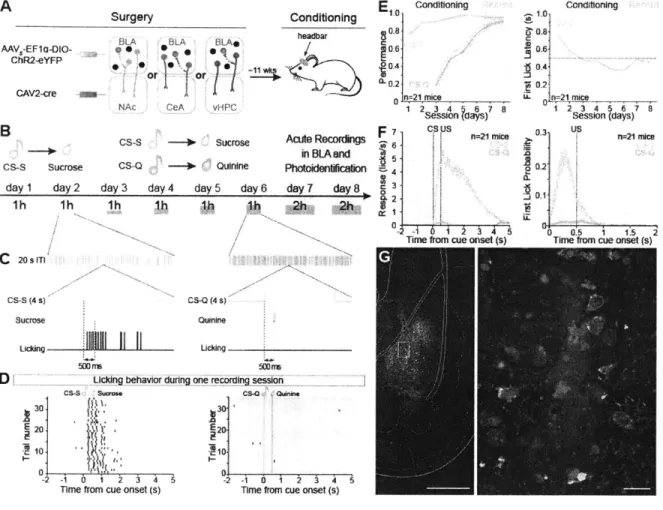
2.3.1 NAc and CeM projecting BLA neurons undergo opposing synaptic changes following fear and reward conditioning
To test the hypothesis that BLA neurons projecting to different downstream targets
undergo distinct synaptic changes, we selected the NAc and CeM as candidate target regions and examined the synaptic changes onto either NAc-projecting BLA neurons (NAc projectors) or CeM-projecting BLA neurons (CeM projectors) following fear conditioning or reward conditioning (Figure 1). To identify the projection target of BLA neurons, we injected retrogradely-traveling fluorescent beads (retrobeads) into either the NAc or CeM to label BLA neurons sending axon terminals to these regions (Figure la; Figure S1). After retrobead migration upstream to BLA cell bodies, we trained mice in fear or reward conditioning paradigms wherein a tone was paired with either a foot shock or sucrose delivery. Mice in reward conditioning groups were food restricted 1 day before the conditioning session to increase motivation to seek sucrose (Figure Si). AMPAR/NMDAR ratio, a proxy for glutamatergic synaptic strength, increases after either fear or reward conditioning in the BLA (Clem and Huganir, 2010; Rumpel et al., 2005; Tye et al., 2008, 2010). We used matched experimental parameters across groups in an acute slice preparation stimulating axons arriving via the internal capsule and performing whole-cell patch-clamp recordings in retrobead-identified NAc projectors and CeM projectors, which we observed to be topographically intermingled (Figure Ib; Figure S2).
a Retrobeads BLA NAc CeM conditioning OR * d 1-C)
p
n=1 1 Unpaired Paired 0-** C n=1a Naive FRK
C1 n=1 1 n=1 Unpaired LeamerL 1
K%-1
ELA neurons projectinq to CeM (CeM proiectors,
****
1C Unpaired Paired 0-n=13 V=10 Naive FR UnpairedK
Lo-L
0 inputs Baseline C 1-.2 0- n=11 Naive 25 pAL 50 maK
e 1-Ez
i Naive 50 ms 00
0 Leamer inputsA
Ce APr. inputs Cem IPrFigure 1. Opposite changes in AMPAR/NMDAR following fear or reward conditioning in BLA neurons projecting to NAc or CeM
a, After injecting retrobeads into NAc or CeM, animals underwent either fear or reward conditioning.
b, Confocal image of retrobead-labelled BLA neurons, with schematic of stimulation and
recording sites (left); region in white square is enlarged (right). DAPI is shown in blue.
c-f, One-way ANOVAs were performed on AMPAR/NMDAR ratios collected after conditioning. Open circles reflect individual data points, number of neurons are shown in each bar and representative traces for each group are below the bar. Results show mean and s.e.m. FR, food restricted.
c, AMPAR/NMDAR ratio was related to training condition (F2,33 = 5.844, P = 0.0070) and
significantly lower in the paired fear group relative to the unpaired fear group (t31 = 2.21, *P <
0.05).
d, AMPAR/NMDAR ratio was related to training condition (F2,3 1 = 6.53, P = 0.0046) and reward
learners showed a greater AMPAR/NMDAR ratio than mice in the unpaired reward group (t29 = 3.20, **P < 0.01).
e, In CeM projectors, AMPAR/NMDAR ratio was related to fear conditioning (F2,29 = 8.72, P =
f, In CeM projectors, AMPAR/NMDAR ratio was altered by reward learning (F2,32 = 3.63, P =
0.039), and was lower in learners relative to the unpaired group (t30 = 2.57, *P < 0.05).
g, Proposed model, arrow thickness represents relative synaptic strength. CeM pr., CeM
projectors; NAc pr., NAc projectors.
We found that in NAc projectors, fear conditioning decreased AMPAR/NMDAR ratio relative to controls exposed to the same number of tones and shocks, but where the tones and shocks were unpaired (Figure ic). Conversely, following the acquisition of the association between a tone and sucrose delivery, NAc projectors showed an increase in AMPAR/NMDAR ratio relative to unpaired controls that were also food restricted and received the same number of tones and volume of sucrose (Figure Id). Importantly, we also included naive and food restricted naYve groups (Figurel c-f) as food restriction itself could alter AMPAR/NMDAR ratio (Figure
S3).
In contrast, CeM projectors from the paired group showed an increase in AMPAR/NMDAR ratio following fear conditioning, relative to unpaired controls (Figure le). Following reward conditioning, CeM projectors from mice that learned the tone-sucrose association showed a decrease in AMPAR/NMDAR ratio relative to unpaired controls (Figure 1f). In addition to AMPAR/NMDAR ratios, we also examined paired-pulse ratio, and did not detect any differences between groups (Figure S3), suggesting a postsynaptic mechanism of plasticity.
These results support a model wherein NAc and CeM projectors undergo opposing changes in synaptic strength following fear and reward learning, such that relative synaptic strengths onto CeM projectors increase following fear conditioning and decrease following reward learning.
Conversely, relative synaptic strengths onto NAc projectors decrease following fear conditioning and increase following reward learning (Figure Ig). However, these findings raised several new questions. First, is there a causal relationship between the activity of BLA neurons projecting to the NAc and reward-related behaviors, and between the activity of CeM projectors and fearful or aversive behaviors? Second, what defining features of NAc and CeM projectors might endow them with their opposing functions?
2.3.2 Photostimulation of NAc or CeM projecting BLA neurons causes positive or negative reinforcement, respectively
To test whether there was a causal relationship between populations of projection-identified BLA neurons on behavior, we first used a retrogradely-infectious rabies viral (RV) vector (Wickersham et al., 2007) to express channelrhodopsin-2 (ChR2) fused to a fluorescent reporter (Venus), or a control virus carrying Venus alone (RV-Venus) in BLA neurons projecting to either the NAc or the CeM (Figure 2a).
RV-ChR2-Venus or RV-Venus OR NAc CeM 120- NAc Ch2Acbvo, $ Venus Active 0 CeM ChR2 Act 0 C60-0 E z 0-ICSS
C
-(D 5 0-- -0 z 0 60 120 Time (min)e
o E laser ON laser OFFCeM projectors 70- 50- 30-RTPA
m
7O
-
-7
00 0 ChR2 Venus ChR2 NAc pr. CeM pr.o
0 0 9 Q Q ChR2 Venus ChR2 NAc pr. CeM pr.Figure 2. Within the BLA, photostimulation of NAc or CeM projectors causes positive or negative reinforcement, respectively.
a, After rabies virus injection into NAc or CeM, animals were tested using intracranial
selfstimulation (ICSS) and real-time place avoidance (RTPA) assays.
b, Representative traces of nose poke responses during ICSS.
c, The relative number of active nose pokes was related to the experimental group (one-way
ANOVA, F2,18 = 10.50, P = 0.00 12), and was significantly increased by photostimuation of NAc
projectors (NAc pr.) in comparison to controls (t16=4.00, **P<0.01). CeM pr., CeM projectors.
d, Representative locomotor trace from an animal receiving CeM projector photostimulation
during RTPA.
e, The percentage of time spent in the photostimulation-paired zone was related to the experimental group (one-way ANOVA, F2,45=4.38, P=0.019) and was significantly decreased
by photostimulation of CeM projectors in comparison to controls (t43 = 2.25, *P < 0.05). Results show mean and s.e.m.
Following verification of functional ChR2 expression and retrograde transport from either the NAc or CeM back to the BLA (Figure S7), we tested animals receiving injections of RV-ChR2-Venus or RV-RV-ChR2-Venus into either the NAc or CeM and implantation of an optical fiber over the BLA
upon photostimulation of BLA cell bodies projecting to NAc (Figure 2b,c).. Given that we could not elicit robust nose poke responses for CeM projector photostimulation, we next tested CeM projectors in a real-time place aversion assay (RTPA), where an animal freely-explored two chambers, one in which the mouse received photostimulation of CeM projectors. Photostimulation of CeM projectors caused robust avoidance of the light-paired side (Figure 2d,e). Consistent with our data and previous studies, we demonstrated a causal relationship between NAc projectors and positive reinforcement and CeM projectors and negative reinforcement (avoidance).
2.3.3 Inhibition of CeM projecting BLA neurons impairs acquisition of fear associations, and enhances acquisition of reward associations.
We went on to probe the necessity of NAc or CeM projectors in mediating reward or fear conditioning. The acquisition of fear (Miserendino et al., 1990) and reward (Tye et al., 2008) associations are mediated by an NMDAR-dependent LTP mechanism thought to require simultaneous glutamate release and postsynaptic depolarization. Thus, we tested whether projection-specific hyperpolarization during the US presentation could impair learning in a valence-specific manner.
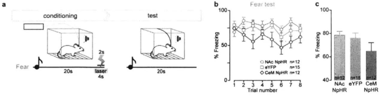
To this end, we bilaterally infused an adeno-associated viral vector (AAV5) carrying halorhodopsin (NpHR) fused to an enhanced yellow fluorescent protein (eYFP), or a no-opsin control (eYFP), in a cre-dependent manner (double-floxed inverted open reading frame; DIO) into the BLA (Figure S5). We then bilaterally infused a retrogradely-traveling canine adenovirus
(CAV) carrying cre-recombinase into either the NAc or CeM (Figure 3a). We illuminated the
BLA with yellow light only during the unconditioned stimulus (US), i.e. during shock or sucrose consumption. Indeed, photoinhibition of CeM projectors during the US impaired conditioned freezing (Figure 3b, Figure S6) and enhanced conditioned reward-seeking (Figure 3c).
Dual virus recombination
AAV5-EF1a-DlO-NpHR-eYFP AAV5-EF1a-DI0-NpHR-eYFP
OR A NAc 1001 C **T NAc NpHR, n=12 eYFP, n=16 <,CeM NpHR, n=13 2 3 4 5 6 7 8 Trial number 8-A) 6-V 0 cL4-0 0 P2- 0-conditioning 20s laser 4s OR faser T5s 6 NAc NpHR, n=9 eYFP. n=14 CeM NpHR, n!9
T T
TA
1
10 20 30 Time from CS (s)Figure 3. Photoinhibition of CeM projectors impairs fear learning and enhances reward learning.
a, Halorhodopsin (NpHR) was expressed bilaterally either in NAc- or CeM-projecting BLA
neurons using a dual-virus recombination strategy. Mice underwent fear or reward conditioning and yellow light was delivered to the BLA during each unconditioned stimulus.
b, Time course of percentage freezing and average freezing in trials 6-8 (inset). Average
freezing was related to experimental condition (one-way ANOVA, F2,40= 6.68, P = 0.0033) and
a
b
00 701 S40-100 C N 0,50 - 0-0jr
1was significantly reduced by photoinhibition of CeM projectors, relative to controls (t38 = 3.46,
**P < 0.01; see inset).
c, Time course of normalized number of port entries relative to cue presentation during reward conditioning and average number of normalized port entries (< 8 s latency, inset). z-score of port entries was related to the experimental condition (one-way ANOVA, F2,31 = 9.23, P =
0.0008) and was significantly increased by photoinhibition of CeM projectors, relative to
2.3.4 Distinguishing features of NAc and CeM projecting BLA neurons
Next, because there was no topographical separation between NAc and CeM projectors (Figure Ib), which are both glutamatergic (Pard et al., 1995; Sah et al., 2003; Stuber et al., 2011; Tye et al., 2011), we searched for distinguishing characteristics of these functionally-distinct neuronal populations. As the BLA is known to have some heterogeneity in electrophysiological and morphological characteristics (Sah et al., 2003; Washburn and Moises, 1992), we compared these features between NAc and CeM projectors. While we did not observe differences in action potential half-width (Figure 4a,b), threshold to spike (Figure 4c,d), nor intrinsic excitability (Figure 4e,f), we did observe a significant difference in action potential accommodation (Figure
4g and Figure S7). To investigate the morphological features of these functionally-distinct populations, we reconstructed projection-identified BLA neurons. We observed greater distal dendritic branching in CeM projectors (Figure 4h,i), though NAc projectors and CeM projectors contained both pyramidal and stellate cells (Figure 4i inset; Figure S8).
Finally, we compared the transcriptomes of BLA neurons projecting to the NAc or CeM (Figure 4j; Figure S9). Following retrobead injections into the NAc or CeM, we dissociated labeled neurons and performed RNA-Seq (Figure 4j). RNA-Seq revealed relatively few candidate genes expressed differentially between NAc and CeM projectors, consistent with the idea that these two populations are closely related (Figure 4k; Figure S9). However, these differentially expressed candidate genes may underpin the mechanisms both that contribute to the wiring of these distinct populations through development as well as rapid gain modulation of synaptic transmission during valence-specific learning.
a
2 ms pr NAc pr. CeM pr.lit
200 nsh
100 pm i . NAc pr. Retrobeads E 0 1.] t:0 a. 0 -tA c r. CeM pr. f 15-C -NAc pr. n=1 1 >CeM pr. n=11 C1E-30-
1-40--501 2DO ms g 0 5 -0 3 -0 0 150 30Injected current (pA)
A
CeM pr dissociation R; J U C ~20I
C C., $210 C8
0 0-Q NA 1 r. CeM pr. NAc pr. n=11 GeM pr. n=11 0.5 Time (s) Pyramidal Stellate 3 5 10 15 #ofcefis 250 500Distance from soma (pm)
NA-Seq sequence alignment
BLA 1 U OR NArc CM franscrplome comparison twansure Gabrd Penk Va Snx25 Prex1 V T P4ha3 Dusp1'S Cd Ntsr - Membrane Secreted c25a Cytoplasm
j
k
Pcdha2 S3c17a6 Sena3d Cbtn4 Figf Arhgef6 IFigure 4. Electrophysiological, morphological and transcriptional profiles of NAc and CeM projectors.
a, Population average of action potential traces. CeM pr., CeM projectors; NAc pr., NAc
projectors.
b, No detectable difference in action potential half-width (unpaired t-test, t20 = 1.82, P = 0.085).
Open circles represent individual data points.
c, Representative trace from action potential threshold detection protocol.
d, No detectable difference in action potential threshold between NAc and CeM projectors (unpaired t-test, t20 = 1.05, P50.3 1).
e-g, Representative trace (e) from current injection protocol to determine firing rate responses
(f) and action potential probability (g) over time, which was different between NAc and CeM
projectors (interaction, two-way ANOVA F9,180 = 2.32, P = 0.017) in the first 100 ms of current
injection (t200 = 4.55, ***P < 0.001).
h, Representative reconstructions showing dendritic branching pattern in the coronal plane. Cells were classified into pyramidal or stellate based on the presence of an apical tuft (right).
i, Sholl analysis of neuron reconstructions.
k, Candidate genes identified as differentially expressed between NAc and CeM projectors at a 0.01 quantile fold-change threshold, corresponding to a false discovery rate (FDR) of 26.2%
(see also Figure S9) across two independent repetitions of RNA-seq (n=8 mice for NAc pr., n=9 mice for CeM pr. total). Results show mean and s.e.m.
2.4 Discussion
2.4.1 Specificity of inputs undergoing experience-dependent changes
In this study, we show that internal capsule inputs to NAc and CeM projecting BLA neurons undergo opposing changes after fear and reward conditioning. The internal capsule is an axon fiber bundle conveying information from multiple subcortical structures that respond to multiple modalities. Axons in the internal capsule relay auditory and somatosensory information from the medial division of medial geniculate nucleus of the thalamus (MGM) and the posterior intralaminar nucleus of the thalamus (PIN), gustation-related somatosensory information through the ventral posterior medial (VPM) nucleus of the thalamus and the parabrachial (PB) nucleus, information about pain-related somatosensory information from the paraventricular nucleus of the thalamus (PVT), and dopaminergic input from the sub-parafasicular nucleus of the thalamus
(SPF), and inputs from the paratenial nucleus of the thalamus, which acts as a relay between the
BLA and the anterior cingulate cortex (ACC) (Ottersen and Ben-Ari, 1979).
According to the Hebbian model (Hebb, 2002) of fear conditioning in the LA (Johansen et al., 2011; Pare, 2002; Sah et al., 2008), synapses carrying information about the unconditioned stimulus (US) induce strong depolarization in the LA neurons, and co-active synapses onto the same neurons carrying information about the CS are strengthened by Hebbian mechanisms. There is indirect evidence to support this model, in terms of sensory convergence (Bordi and LeDoux,
1992; Fontanini et al., 2009; LeDoux et al., 1990; Romanski et al., 1993), synaptic strengthening
(Clem and Huganir, 2010; McKernan and Shinnick-Gallagher, 1997; Rogan et al., 1997; Rumpel et al., 2005; Tye et al., 2008) and sufficiency of LA neuron depolarization to act as a US (Johansen et al., 2010). Although it is widely suspected in the field that synapses conveying CS information to the BLA are strengthened, a direct test of this hypothesis is yet to be conducted.
2.4.2 Food restriction-induced synaptic changes
In this study, positive and negative associations were not the only experiences that caused changes in synaptic transmission onto BLA neurons. Food restriction was also observed to change population-specific changes synaptic transmission.
In order to motivate mice to seek rewards, a common practice is to restrict their food or water intake prior to an associative learning paradigm. In our experience, placing mice in a state of food deprivation clearly increases their performance measured in terms of the number of rewards retrieved and the latency to retrieve rewards in response to reward-predictive cues.
Food restriction has widespread effects in the central nervous system (Mattson et al., 2003). Food restriction is known to influence neurotransmitter systems (Diao et al., 1997; Kohsaka et al.,
1980; Verhagen et al., 2009), gene expression profiles (Lee et al., 2000), and synaptic plasticity in
several brain regions, including the visual cortex (Spolidoro et al., 2011) and hippocampus (Eckles-Smith et al., 2000; Talani et al., 2016). According to our data, food restriction induced opposing changes in AMPAR/NMDAR ratios in NAc and CeM projecting neural populations (Figure S3e,
j)
in the BLA. Food restriction decreased AMPAR/NMDAR ratios from the internal capsule onto NAc projectors and increased AMPAR/NMDAR ratios onto CeM projectors. Perhaps these changes are reflective of a state of reward-seeking wherein inputs onto NAc projecting neurons are more readily potentiated relative to a non-food restricted state. Even though both food restriction and fear/reward conditioning modify internal capsule inputs to the BLA, it is not yet clear if these inputs arise from the same structures.2.4.3 The BLA on valence and arousal - differential roles for the LA and the BA
Previous studies have shown that fear and reward conditioning strengthen syanpses onto BLA neurons (Clem and Huganir, 2010; Tye et al., 2008). How can associations with such different outcomes cause similar changes in synaptic transmission? One possibility is that synapses onto different populations of BLA neurons are strengthened after fear and reward conditioning. Even though we observe that synaptic transmission onto one population is strengthened (among NAc and CeM projecting BLA neurons), we also observe that synaptic transmission onto the other population is weakened. Therefore, among a random sampling of neurons, we are unlikely to see a net change in synaptic transmission after fear conditioning or reward conditioning.
The answer to observing increased synaptic transmission after fear and reward conditioning (Clem and Huganir, 2010; Tye et al., 2008) perhaps lies in the anatomical organization of the BLA, and the population of neurons sampled in these studies. Neurons in these two studies were sampled from the lateral sub-division of the BLA, whereas most of the long range projecting neurons are in the basal sub-division of the BLA. We reconcile the data from (Clem and Huganir, 2010; Tye et al., 2008) with our data by positing that the lateral and basal sub-divisions of the BLA have differential roles in arousal and valence, where LA recognizes relevant stimuli of either valence, and the BA then encodes valence through its various projector populations by gating activity to the appropriate downstream structure.
Although in this thesis we don't differentiate between the lateral and basal divisions of the BLA, these two nuclei differ in many ways. Relevant to this thesis are (1) LA receives a denser input from the internal capsule compared to the BA, and (2) most of the NAc and CeM projecting BLA neurons are in the BA. In our ex vivo experiments, we targeted a region of transition between