Publisher’s version / Version de l'éditeur:
Plant Cell, Tissue and Organ Culture, 116, 1, pp. 89-96, 2013-09-29
READ THESE TERMS AND CONDITIONS CAREFULLY BEFORE USING THIS WEBSITE. https://nrc-publications.canada.ca/eng/copyright
Vous avez des questions? Nous pouvons vous aider. Pour communiquer directement avec un auteur, consultez la première page de la revue dans laquelle son article a été publié afin de trouver ses coordonnées. Si vous n’arrivez Questions? Contact the NRC Publications Archive team at
PublicationsArchive-ArchivesPublications@nrc-cnrc.gc.ca. If you wish to email the authors directly, please see the first page of the publication for their contact information.
NRC Publications Archive
Archives des publications du CNRC
This publication could be one of several versions: author’s original, accepted manuscript or the publisher’s version. / La version de cette publication peut être l’une des suivantes : la version prépublication de l’auteur, la version acceptée du manuscrit ou la version de l’éditeur.
For the publisher’s version, please access the DOI link below./ Pour consulter la version de l’éditeur, utilisez le lien DOI ci-dessous.
https://doi.org/10.1007/s11240-013-0385-0
Access and use of this website and the material on it are subject to the Terms and Conditions set forth at
Isolated microspore culture of oat (Avena sativa L.) for the production
of doubled haploids : effect of pre-culture and post-culture conditions
Ferrie, A. M. R.; Irmen, K. I.; Beattie, A. D.; Rossnagel, B. G.
https://publications-cnrc.canada.ca/fra/droits
L’accès à ce site Web et l’utilisation de son contenu sont assujettis aux conditions présentées dans le site LISEZ CES CONDITIONS ATTENTIVEMENT AVANT D’UTILISER CE SITE WEB.
NRC Publications Record / Notice d'Archives des publications de CNRC:
https://nrc-publications.canada.ca/eng/view/object/?id=5a9526df-e02a-4e92-903a-a59bfa9ee24a https://publications-cnrc.canada.ca/fra/voir/objet/?id=5a9526df-e02a-4e92-903a-a59bfa9ee24a
Isolated microspore culture of oat (Avena sativa L.) for the production of doubled haploids: effect of pre-culture and
1
post-culture conditions
2
3
A.M.R. Ferrie1, K.I. Irmen1, A.D. Beattie2, B.G. Rossnagel2
4
5
1National Research Council
6
110 Gymnasium Place7
Saskatoon, SK8
S7N 0W99
10
2Crop Development Centre
11
University of Saskatchewan12
Saskatoon, SK13
S7N 5A814
15
16
17
Abstract
1
The production of doubled haploid (DH) plants from microspores is an important technique used in plant breeding
2
programs and basic research. Although doubled haploidy efficiencies in wheat and barley are sufficient for breeding
3
purposes, oat (Avena sativa L.) is considered recalcitrant. The objective of this project was to develop a protocol for
4
the production of microspore-derived embryos of oat and further develop these embryos into fertile doubled haploid
5
plants. A number of experiments were conducted evaluating the factors influencing microspore embryogenesis, i.e.,
6
donor plant conditions, pretreatments, media composition, and culture conditions. The initial studies yielded little
7
response, and it was not until high microspore densities (106 microspores/ml and greater) were used that
8
embryogenesis was achieved. Depending on the treatment, yields of over 5000 embryos /106microspores were
9
obtained for breeding line 2000QiON43. The doubled haploidy protocol includes: a 0.3 M mannitol pretreatment of
10
the tillers for 7 days, culture in W14 basal medium with a pH of 6.5 – 7.5, a microspore density of 106
11
microspores/mL, and continuous incubation at 28°C incubation. The resulting embryos observed after 28 days
12
were plated onto solidified W14 medium with 0.8 or 1.0 g/L activated charcoal. A colchicine treatment of 0.2%
13
colchicine for 4 h resulted in conversion of 80% of the plants from haploid to doubled haploid. This protocol was
14
successful for the production of oat microspore-derived embryos and doubled haploid green plants with minimal
15
albinism. Doubled haploid seed was produced and planted for evaluation in a field nursery.
16
17
18
Keywords: Avena sativa, doubled haploid, microspore culture, oat
19
20
Introduction
1
Doubled haploid methodology to generate homozygous, true-breeding plants is used routinely in many plant
2
breeding programs. This technology has benefits for practical application (e.g. varietal development, mutagenesis,
3
and transformation) and basic research (e.g. genomics, biochemical, and physiological studies) (Dunwell 2010,
4
Ferrie and Möllers 2011). Doubled haploids are commonly produced using one of four methods: culture of anthers or
5
microspores (androgenesis), culture of unfertilized ovules (gynogenesis), interspecific or intergeneric crosses followed
6
by chromosome elimination, or pollination with irradiated pollen.
7
8
Oat (Avena sativa L.) is considered one of the more recalcitrant cereal crops with respect to doubled haploidy. Wide
9
hybridization with maize pollen (Rines 2003; Rines and Daheen 1990; Sidhu et al. 2006) generates doubled haploids
10
as has anther culture (De Cesaro et al. 2009; Kiviharju et al. 2000; Kiviharju et al. 2005; Ponitka and
Slusarkiewicz-11
Jarzina 2009), but methods are inefficient. Haploid embryo production using wide crosses has an efficiency of 0.8 –
12
6.7% (Sidhu et al. 2006) and for anther culture up to 30 green plants/100 anthers has been reported (Kiviharju et al.
13
2005). Haploid and doubled haploid plants have been regenerated from isolated microspore culture, but again at
14
very low frequency; two green plants and 15 albino plants were regenerated (Sidhu and Davis 2009).
15
16
Developing an efficient doubled haploidy protocol involves evaluating factors which influence induction of
17
embryogenesis and the regeneration of those embryos to plants (Ferrie and Caswell 2011). Prior to the culture of
18
microspores, the conditions in which the donor plants are grown, pretreatments given to floral buds, and the
19
developmental stage of the microspores selected for culture affect embryo production. The microspore isolation
20
method, the culture media, as well as the post-isolation conditions also influence the embryogenic response. This
21
paper describes several parameters affecting the production of microspore-derived embryos from isolated oat
22
microspores and the subsequent regeneration of green, doubled haploid plants.
23
24
Materials and methods
1
Germplasm
2
Genotype 2000QiON43 (LA9326E86) was selected for protocol development based on preliminary experiments and
3
was provided by Stephen Harrison, LSU AgCenter, Louisiana, USA. 2000QiON43 is a non-released breeding line
4
from the LSU AgCenter oat breeding program.
5
6
Donor plant conditions
7
Plants were grown in controlled environment chambers (20°C day/15°C night, 16 h photoperiod, 360 µmol m-2 s-1
8
light intensity). Seeds were planted in a soil-less mix (Sunshine #3/LG3-Sun Gro Horticulture) with approximately
9
1 g of slow release fertilizer (Nutricote 100 14-14-14; 14% nitrogen, 14% phosphorous, and 14% potassium) in a 15
10
cm pot. Plants were watered as required with a 0.88 g/L nutrient solution (20-20-20; N-P-K).
11
12
Original microspore culture protocol
13
Tillers were harvested when the panicle was 2.5 cm from the secondary leaf and placed in water for 7 - 10 days at
14
4°C. Panicles were then removed and spikelets containing microspores at the late uninucleate to early binucleate
15
stage were placed into steel baskets for sterilization. The steel baskets were placed into a sterile beaker filled with
16
50% bleach (6.25% sodium hypochlorite) with a drop of Tween 20 surfactant and placed on a shaker for 10 minutes.
17
The steel baskets containing the spikelets were then rinsed four times in sterile water for 5 minutes each. After
18
sterilization, the panicles were put into a large blender cup (250 ml) containing 125 ml of 0.3 M mannitol (pH 5.8)
19
and blended for 5 seconds on low speed and 7 seconds on high speed until homogenized. The homogenate was
20
filtered through a sterile 90 μm Nitex screen into 50 mL centrifuge tubes and centrifuged at 489 g for 3 minutes.
21
The supernatant was removed, the pellet was resuspended in 5 mL of 0.3 M mannitol, and the new suspension
22
centrifuged as above. This was repeated 3 times. Prior to the last wash step, the microspore density was determined
23
using a hemacytometer. The microspores were suspended in liquid culture medium (W14 basal medium unless
24
otherwise specified; Ouyang et al. 1989) adjusting cell density as required in each experiment, plated in Petri dishes
25
(100 mm x 15 mm), wrapped in Parafilm®, and incubated in the dark at the temperature outlined for each
26
experiment. Embryos were counted after 4 weeks. Experiments were repeated three to 4 times, with a minimum of
27
three replicate plates per treatment for each experiment.
1
Factors evaluated to enhance microspore embryogenesis in oat:
2
Microspore density
3
Five microspore densities (5 x 10⁴, 1 x 10⁵, 5 x 10⁵, 1 x 10⁶, 2 x 10⁶microspores/mL) were compared and evaluated
4
for embryogenic response. Tillers were pre-treated in water for 7-10 days at 4°C and microspores were cultured in
5
W14 basal medium and incubated continuously at 28°C. Microspores were plated in Petri dishes (35 x 10 mm) with
6
1 mL of culture medium.7
8
Tiller pretreatment9
Tillers were harvested as previously described and treated in water or 0.3 M mannitol for 7 days at 4°C.
10
Microspores were isolated as described and cultured in W14 basal medium at densities of 1 x 10⁶, 2 x 10⁶or 4 x 10⁶
11
microspores/mL and incubated continuously at 28°C.
12
13
Culture temperature
14
Experiment 1: Tillers were pre-treated in 0.3 M mannitol for 7 days at 4°C. Microspores were isolated as described
15
and incubated at 22°C, 24°C, or 28°C continuously in W14 basal medium at a density of 1 x 10⁶microspores/mL.
16
17
Experiment 2: Tillers were pre-treated and microspores isolated as in experiment 1. Microspores were incubated
18
continuously at 22°C or 28°C, or 28°C for 1, 3, or 5 days then transferred to 22°C.
19
20
Donor plant conditions
21
Plants were grown in controlled environment chambers at temperatures of 20°C day/ 15°C night, or 10°C day/5°C
22
night, harvested as described above, and used immediately or grown at 20°C day/ 15°C night, harvested and given a
23
mannitol pretreatment (4°C for 7 days). Microspores were cultured in W14 basal medium at a density of 106
24
microspores/mL and incubated continuously at 28°C.
25
26
27
28
Media components
1
Five basal media were evaluated: W14 (Ouyang et al. 1989), NLN (Lichter 1982), modified FHG (Kasha et al.
2
2001), KFWC (Sidhu and Davies 2009), and TM (Tupý et al. 1991). Microspores were cultured at densities of 1 x
3
106or 2 x 10⁶microspores/mL. A mannitol pretreatment was used for the tillers.
4
5
pH
6
Donor plants (grown at 20°C day/15°C night) and microspores were treated as described previously (mannitol
7
pretreatment). Microspores were cultured in W14 basal medium with a pH range of 4.5 – 7.5, microspore density of
8
1 x 10⁶/mL, and an incubation temperature of 28°C.
9
10
Embryo and plantlet regeneration
11
Embryos (approximately 1-2 mm) were transferred onto solid W14 regeneration medium (Kiviharju 2009), solid B5
12
(Gamborg et al. 1968) or solid MS (Murashige and Skoog, 1962) basal media all with 2% maltose and 0.3%
13
phytagel and placed at 22°C continuously with a 16 h photoperiod (75 µmol m-2 s-1). After 3 - 4 weeks, plantlets
14
with green shoots were transferred to larger culture jars (Magenta™ GA7) with the same regeneration medium. To
15
enhance root development and regeneration, activated charcoal (0.6, 0.8, or 1.0 g/L) was added to the W14
16
regeneration medium. Root development (i.e. root length, number of roots) was observed after 2 weeks. Once a
17
well developed root system was established, plantlets were removed from the culture jars and the ploidy level of
18
each microspore-derived plantlet was determined using flow cytometry.
19
20
Ploidy determination
21
A 1 cm2piece of young leaf was chopped with a razor blade in 400 μL ice cold nuclei extraction buffer (Cystain UV
22
Precise, Partec) for 1 minute or until the leaf material was finely chopped. The suspension was filtered through a 30
23
μm screen to remove excess plant material leaving only plant cell nuclei for analysis. The extraction suspension was
24
incubated for 5 minutes on ice, then 1.6 mL of nuclei staining buffer (Cystain UV Precise, Partec) was added to the
25
suspension and the samples were incubated on ice for 1 minute. The Partec Cell Counter Analyzer (CCA) was used
26
to determine the ploidy of the stained nuclei.
27
28
Chromosome doubling
1
The microspore-derived plantlets determined to be haploid were treated with colchicine for chromosome doubling.
2
The roots and crown were soaked in a 0.1% or 0.2% colchicine solution for 1.5, 3, 4, or 5 h. After this time the
3
plantlets were rinsed for 1 h in water, planted in 15 cm pots with commercial growing mix (Sunshine Mix #3/LG3),
4
and covered with clear plastic cups to maintain a high relative humidity. The plastic cups were removed slowly
5
once the plantlets were established. Doubled-haploid plants were grown in an controlled environment growth room
6
(20°C day/15°C night, 16 h photoperiod, 360 µmol m-2 s-1) and bagged individually to ensure self-fertilization.
7
Resulting seed was planted in hill plots in a field nursery at the Crop Development Centre, University of
8
Saskatchewan, Saskatoon, Canada in 2012.
9
10
Results and discussion
11
Many factors influence the embryogenic response of microspores, and these factors can be modified so as to
12
generate microspore-derived embryos and subsequently doubled haploid plants. In our oat program, preliminary
13
studies evaluated a number of genotypes, media compositions, pretreatments, and culture conditions (data not
14
shown). From these experiments, genotype 2000QiON43 was selected for further experimentation. This was the
15
same germplasm as Sidhu and Davis (2009) found most responsive.
16
17
Microspore density
18
From preliminary experiments evaluating a range of microspore densities (25,000 – 100,000 microspores/ml), cell
19
division was observed in less than 1% of the microspores with no continued embryo development (data not shown).
20
In the same preliminary experiments, higher densities (200,000 – 400,000 microspores/mL), more cell divisions and
21
multi-cellular structures were observed, but no true-embryos (i.e. possessing both the scutellum and germ)
22
developed. Very high densities (1 x 10⁶, 2 x 10⁶microspores/mL) were then evaluated and found to be beneficial to
23
embryogenesis (Table 1). With a density of 1 x 10⁶microspores/mL, a significantly higher number of embryos (or
24
embryo-like structures, ELS) (1596/106msp) was produced than any other density tested; this density also resulted
25
in the highest number of large (1 – 2 mm in size with the scutellum and germ) embryos. There was little response at
26
lower densities (5 x 10⁴and 1 x 10⁵). Embryo development was not synchronous with each culture plate; although
27
there were many ELS only a few were large enough (1 – 2 mm) for conversion to plantlets after 3 – 4 weeks (Table
1). Smaller ELS remaining in the plate did develop further, although not all became large enough for culture and
1
plantlet development. Microspore density is an important factor in culture, as it affects both embryo quality and
2
quantity. Optimal microspore density varies depending on the species. Whereas a low microspore density (1 x 10⁴
3
microspores/ml) was beneficial for Camelina sativa (Ferrie and Bethune, 2011), a high microspore density (5 x 10⁵)
4
has been effective with barley (Kasha et al. 2003).
5
6
Tiller pre-treatments
7
Microspore-derived embryos and ELS were produced from both pre-treatments of tillers (water or mannitol). With
8
both pre-treatments, a microspore density of 1 x 10⁶microspores (msp)/mL produced significantly more embryos
9
and ELS than were produced at any other density. These results correspond with those from our previous
10
microspore density studies. At each of the densities evaluated, there was no significant difference between the
11
mannitol and water pre-treatment in terms of the number of ELS developed. However, mannitol pretreated tillers
12
produced more 1-2 mm embryos than tillers treated with water (Table 2). A total of 48 green plantlets were
13
produced from the mannitol pre-treatment whereas only 25 were produced from the water pre-treatment. There
14
were very few albino plantlets produced. This 7 day pre-treatment is different from that of Sidhu and Davis (2009)
15
who found a longer cold pre-treatment ( 6 – 9 weeks) beneficial in producing multicellular structures.
16
17
Culture conditions
18
In vitro embryogenesis can be influenced by the post isolation conditions of the microspores. Much research has
19
focused on appropriate culture temperature and its duration, which varies among species. Culture temperature
20
usually ranges from 24 – 27°C. For some species (e.g. Brassica), an elevated temperature (30 – 35°C) for 12 – 72
21
hours is required for embryo induction (Baillie et al. 1992; Ferrie 2003). A preliminary study in oat evaluating a
22
range of temperatures determined that a heat shock of 32°C or 35°C was not beneficial as the oat microspores
23
exhibited swelling but no further development. In contrast, microspores cultured at 28°C did produce multi-cellular
24
structures. A further study evaluated three temperatures (22°C, 24°C, 28°C continuously). Embryos and ELS were
25
produced from all temperature regimes, with significantly more ELS (1387 embryos/106msp) being produced from
26
the 28°C treatment (Table 3). No plantlets were regenerated.
27
28
Further experiments evaluated different heat shock periods (i.e. 1, 3, 5 days at 28°C followed by 22°C, continuous
1
28°C, or continuous 22°C). The longer the heat shock the more embryogenic the cultures. Microspore cultures
2
incubated at 28°C for 5 days or continuously at 28°C were the most embryogenic (Table 4) with 2411 and 3061
3
embryos (ELS)/106msp being produced, respectively.
4
5
Donor plant conditions
6
The environmental conditions in which donor plants are grown can influence embryogenic response. Healthy,
7
vigorous donor plants are essential for successful microspore culture. Studies have shown that embryo/ELS yield
8
can be affected by temperature, photoperiod, light intensity, and where the plant is grown (i.e. greenhouse, field, or
9
growth chamber). For many species, growth of the donor plants at a low temperature (10°C day/5°C night) is
10
beneficial (Baillie et al. 1992; Keller et al. 1987; Takahata et al. 1991). An experiment was designed to determine if
11
the cold pretreatment of the tillers in mannitol (4°C for 7 days) could be replaced by growing the donor plants under
12
cooler conditions (10/5°C). There was no significant difference in embryos or ELS/106 microspores between
13
mannitol/cold pre-treatment and plants grown at 10/5°C; however, the mannitol pre-treatment produced more 1-2
14
mm embryos than the other two donor plant growing regimes (Table 5).
15
16
Basal media
17
The composition of the media plays a major role in embryogenesis. There are numerous studies evaluating the
18
different components of media and their role in microspore culture. Initially, W14, a wheat and oat anther culture
19
medium (Kiviharju 2009; Ouyang et al. 1989) was used. Utilizing two high microspore culture densities and a
20
mannitol pre-treatment, five basal media were evaluated: W14, NLN [a medium used for microspore
21
embryogenesis of Brassica species (Lichter 1982)], modified FHG [a barley anther culture medium (Kasha et al.
22
2001)], KFWC [used in oat microspore embryogenesis (Sidhu and Davies 2009)], and TM [a tobacco maturation
23
medium (Tupý et al. 1991)]. Embryo-like structures were produced from all basal media and at both densities
24
(Table 6). Although W14 medium at a density of 106microspores/mL produced the most embryos/ELS (2067
25
embryos per 106msp), at a density was 2 x 10⁶microspores/mL, KFWC medium produced more embryos/ELS than
26
W14 at the same density.
27
28
pH
1
Sidhu and Davies (2009) reported induction of oat microspores when a high pH was applied (pH 8). In our
2
experiments, medium pH and number of embryos/ELS were correlated (Figures 1, 2A; Table 7); however, at pH 6.5,
3
7, and 7.5 approximately 10% of the larger embryos developed callus (data not shown). The higher pH levels (7 and
4
7.5) are significantly better than the lower pH levels (4.5, 5, 5.8) for embryo/ELS production and also produced
5
more large embryos than the lower pH levels.
6
7
Embryo culture regeneration
8
An important step in developing a doubled haploidy protocol is the conversion of embryos to plants (Figure 2 B, C).
9
Observations from previous experiments indicated a low regeneration rate, which has also been reported for other
10
cereals (Pauk et al. 2003, Pulli and Guo, 2003). Three basal media (W14, B5, MS) were compared and resulted in
11
regeneration rates ranging from 10 – 21% (Table 8). The majority of the embryos/ELS did not develop or produced
12
callus. The W14 medium produced 88% green plants along with 12% albinos. Use of the other basal media
13
resulted in a higher frequency of albino plants.
14
15
Plantlet culture
16
With the addition of activated charcoal to the W14 medium, 0.6, 0.8, or 1.0 g/L, the embryos produced roots that
17
were 9.7, 17.1, and 17.1 cm long, respectively, after two weeks. There was no root growth on W14 basal medium
18
without activated charcoal. Overall, the total number of microspore derived green plantlets (Figure 2E) produced
19
from genotype 2000QiON43 was 648 (91.7%) and the number of albino plantlets produced was 59 (8.3% albino).
20
The low frequency of albino plants regenerated is beneficial as albinism can be a problem in many cereal crops
21
(Torp and Andersen, 2009).
22
23
Ploidy determination
24
The ploidy, as described in the material and methods section, of 104 green plantlets was determined prior to
25
chromosome doubling. Only 6% of plants had spontaneously doubled, while the majority were haploid (data not
26
shown). Haploid plantlets (98) were treated with colchicine. Doubled haploid and non-haploid (haploid and
27
mixaploid) were observed (Table 9). Plant survival rates for 0.1 % and 0.2 % colchicine were 97.9% and 93.6%,
respectively. The 4 hour treatment with 0.2% colchicine produced the highest conversion of haploid to doubled
1
haploid plants (80%) and was concluded to be the best chromosome doubling treatment.
2
3
Field and greenhouse evaluation
4
Seed from 115 plants was evaluated in the greenhouse and in hill plots in the field (Figure 2F). Despite a wet
5
growing season in 2012, the DH lines preformed well when compared to the control (parental) hill plots. Within the
6
DH rows, plants were uniform as expected with DH lines. The plants tended to be shorter than the control, which
7
has been observed in other DH lines grown in the field (Ferrie et al. 2011). This may also have been due to the
8
background of the original material.
9
10
In conclusion, an improved doubled haploidy protocol that will yield sufficient doubled haploid plants for a breeding
11
program was developed for oat as illustrated in Figure 2. Embryo/ELS yields of over 5000 embryos/106
12
microspores were obtained from genotype 2000QiON43. Plantlet regeneration from these embryos needs further
13
improvement but those plantlets that did regenerate could be converted to doubled haploid plants with a colchicine
14
treatment at a high rate of efficiency (80%). The protocol described here is different from previous published
15
protocols as a short pre-treatment is used (7 days) and non-conditioned media is utilized both of which saves time
16
and resources. Further research is required to expand the protocol to other oat germplasm.
17
18
19
Acknowledgements
20
The authors acknowledge and thank the Saskatchewan Ministry of Agriculture and the Agriculture and
21
Development Fund in supporting and providing funding for the project, Shelley Duncan and the field staff at the
22
Crop Development Center (Saskatoon, SK) for the field evaluation, and K.L. Caswell and S.M.H. Slater for
23
evaluation of the manuscript.
24
25
1
References
2
Baillie AMR, Epp DJ, Hutcheson D, Keller WA (1992) In vitro culture of isolated microspores and regeneration of
3
plants in Brassica campestris. Plant Cell Rep 11:234-237
4
5
De Cesaro T, Baggio MI, Zanetti A, Suzin M, Augustin L, Brammer SP, Lorczeski EJ, Milach SCK (2009)
6
Haplodiploid androgenetic breeding in oat: genotypic variation in anther size and microspore development stage. Sci
7
Agri (Piracicaba, Braz ) 66:118-122
8
9
Dunwell JM (2010) Haploids in flowering plants: origins and exploitation. Plant Biotechnol J 8:377-424
10
11
Ferrie AMR (2003) Microspore culture of Brassica species. In: Maluszynski M, Kasha KJ, Forster BP, Szarejko I
12
(eds) Doubled haploid production in crop plants. A manual. Kluwer Academic Publishers, p 205-216
13
14
Ferrie AMR, Bethune TD (2011) A microspore embryogenesis protocol for Camelina sativa, a multi-use crop. Plant
15
Cell Tissue Organ Cult 106:495-501.
16
17
Ferrie AMR, Bethune TD, Arganosa GC, Waterer D (2011) Field evaluation of doubled haploid plants in the
18
Apiaceae: Dill (Anethum graveolens L.), caraway (Carum carvi L.), and fennel (Foeniculum vulgare Mill.). Plant
19
Cell Tissue Organ Cult 104:407-413
20
21
Ferrie AMR, Caswell KL (2011) Isolated microspore culture techniques and recent progress for haploid and doubled
22
haploid plant production. Plant Cell Tissue Organ Cult 104:301-309
23
24
Ferrie AMR, Mӧllers C (2011) Haploids and doubled haploids in Brassica spp. for genetic and genomic research.
25
Plant Cell Tissue Organ Cult 104:375-386
26
27
Gamborg OL, Miller RA, Ojima K (1968) Nutrient requirements of suspension cultures of soybean root cells. Exp
28
Cell Res 50:151-158
29
30
Kasha KJ, Simion E, Oro R, Shim YS (2003) Barley isolated microspore culture protocol. In: Maluszynski M,
31
Kasha KJ, Forster BP, Szarejko I (eds) Doubled Haploid Production in Crop Plants: A Manual. Kluwer Academic
32
Publishers, pp 43-47
33
34
Kasha KJ, Simion E, Oro R, Yao QA, Hu TC, Carlson AR (2001) An improved in vitro technique for isolated
35
microspore culture of barley. Euphytica 120: 379-385
36
37
Keller WA, Arnison PG, Cardy BK (1987) Haploid from gametophytic cells: recent developments and future
38
prospects. In: Green CE, Somers DA, Hackett WP, Biesboer DD (eds) Plant Tissue and Cell Culture, New York:
39
Allan R. Liss, pp 233-241
40
41
Kiviharju EM (2009) Anther culture derived doubled haploids in oat. In: Touraev A, Forster BP, Jain, SM (eds.)
42
Advances in Haploid Production in Higher Plants, Springer, pp 171-178
43
44
Kiviharju E, Moisander S, Laurila J (2005) Improved green plant regeneration rates from oat anther culture and the
45
agronomic performance of some DH lines. Plant Cell Tissue Organ Cult 81:1-9
46
47
Kiviharju E, Puolimatka M, Saastamoinen M (2000) Extension of anther culture to several genotypes of cultivated
48
oats. Plant Cell Rep 19:674-679
49
50
Lichter R (1982) Induction of haploid plants from isolated pollen of Brassica napus. Z Pflanzenphysiol 105:427434
51
52
Murashige T, Skoog F (1962) A revised medium for rapid growth and bioassays with tobacco tissue cultures.
1
Physiol Plant 15:473-497
2
3
Ouyang JW, Jia SE, Zhang C, Chen X, Feng G (1989) A new synthetic medium (W14) for wheat anther culture.
4
Annu Rep Inst Genet Acad Sin 1987-1988. Beijing, pp91-92
5
6
Pauk J, Mihály R, Puolimatka M (2003) Protocol for wheat (Triticum aestivum L.) anther culture. In: Maluszynski
7
M, Kasha KJ, Forster BP, Szarejko I (eds) Doubled Haploid Production in Crop Plants: A Manual. Kluwer
8
Academic Publishers, pp 59-64
9
10
Ponitka A, Slusarkiewicz-Jarzina A (2009) Regeneration of oat androgenic plants in relation to induction media and
11
culture conditions of embryo-like structures. Acta Soc Bot Pol 78:209-213
12
13
Pulli S, Guo Y-D (2003) Anther culture and isolated microspore culture in timothy. In: Maluszynski M, Kasha KJ,
14
Forster BP, Szarejko I (eds) Doubled Haploid Production in Crop Plants: A Manual. Kluwer Academic Publishers,
15
pp 173-177
16
17
Rines HW (2003) Oat haploids from wide hybridization. In: Maluszynski M, Kasha KJ, Forster BP, Szarejko I (eds)
18
Doubled haploid production in crop plants. Kluwer, Dordrecht, pp 155-159
19
20
Rines HW, Daheen LS (1990) Haploid oat plants produced by application of maize pollen to emasculated oat florets.
21
Crop Sci 30:1073-1078
22
23
Sidhu P, Davies PA (2009) Regeneration of fertile green plants from oat isolated microspore culture. Plant Cell Rep
24
28:571-577
25
26
Sidhu PK, Howes NK, Aung T, Zwer PK, Davies PA (2006) Factors affecting oat haploid production following oat
27
x maize hybridization. Plant Breed 125:243-247
28
29
Takahata Y, Brown DCW and Keller WA (1991) Effect of donor plant age and inflorescence age on microspore
30
culture of Brassica napus L. Euphytica 58:51-55
31
32
Torp AM, Andersen SB (2009) Albinism in microspore culture. In: Touraev A, Forster BP, Jain, SM (eds.)
33
Advances in Haploid Production in Higher Plants, Springer, pp 155-160
34
35
Tupý J, Řihová L, Žárský V (1991) Production of fertile tobacco pollen from microspores in suspension culture and
36
its storage for in situ pollination. Sexual Plant Reprod 4:284-287
37
38
39
40
41
42
43
44
1
Figure 1: Effect of medium pH (4.5 – 7.5) on microspore-derived embryogenesis of oat genotype 2000QiON43.
2
3
4
5
6
Figure 2: Doubled haploidy process in oat genotype 2000QiON43.
7
A. Microspore-derived embryos, B. Microspore-derived embryo on solid medium, C. Germinating embryo, D.
8
Plantlet, bar = 1 cm, E. Haploid/doubled haploid plants in greenhouse, F. Doubled haploid (DH) plants in the field
9
(Summer 2012). Bars for A, B, C = 1 mm.
10
11
12
13
14
15
16
17
A
B
F
E
DH
D
C
4.5 5.0 5.8 6.5 7.0 7.5
1
Table 1: Influence of microspore (msp) density on microspore embryogenesis of oat genotype 2000QiON43.
2
3
Microspore density msp/mL
Mean number of embryos/106msp Large embryos (1 – 2 mm)
Total number (per 106msp)
2 x 10⁶ 802 b 21 (2.6)
1 x 10⁶ 1596 a 110 (6.5)
5 x 10⁵ 608 bc 9 (0.8)
1 x 10⁵ 220 bc 7(0.2)
5 x 10⁴ 32 c 0 (0)
Means within a column followed by different letters in a column are significantly different at P = 0.05 level as
4
determined by Duncan’s multiple range test. Results are based on 3 experiments with 3 replicate plates per
5
treatment per experiment.
6
7
8
9
Table 2: Influence of mannitol or water pretreatment on microspore embryogenesis of oat genotype 2000QiON43.
10
11
Microspore density (msp/mL)
Mean number of
embryos/106msp Large embryos ( 1 – 2 mm)
Total number (per 106msp)
Number of green plantlets Number of albino plantlets Water pre-treatment 1 x 10⁶ 2285 a 123 (2.9) 10 0 2 x 10⁶ 1558 b 114 (8.8) 9 1 4 x 10⁶ 1057 c 77 (12.8) 6 0 Mannitol pre-treatment 1 x 10⁶ 2155 a 184 (4.5) 16 2 2 x 10⁶ 1544 b 40 (3.6) 0 0 4 x 10⁶ 1188 c 177 (29.5) 32 2
Means within a column and per pre-treatment followed by different letters in a column are significantly different at
12
P = 0.05 level as determined by Duncan’s multiple range test. Results are based on 3 experiments with 3 replicate
13
plates per treatment per experiment.
14
15
16
17
Table 3: Comparison of incubation temperatures on microspore embryogenesis of oat genotype 2000QiON43.
1
2
Temperature (°C) Mean number of embryos/ 106msp Large embryos (1 – 2 mm)
Total number (per 106msp)
22 297 b 0
24 659 b 0
28 1387 a 33 (2.8)
Means within a column followed by different letters in a column are significantly different at P = 0.05 level as
3
determined by Duncan’s multiple range test. Results are based on 3 experiments with 3 replicate plates per
4
treatment per experiment.
5
6
7
8
Table 4: Effect of temperature (28°C) and duration of heat shock on microspore embryogenesis of oat genotype
9
2000QiON43.
10
11
Temp (°C) and duration (days) Mean number of embryos/106msp
22 – continuously 739 c
28 – 1 day 757 c
28 – 3 day 2135 b
28 – 5 day 2411 ab
28 - continuously 3061 a
Means within a column followed by different letters in a column are significantly different at P = 0.05 level as
12
determined by Duncan’s multiple range test. Results are based on 4 experiments with 3 replicate plates per
13
treatment per experiment.
14
15
16
17
18
Table 5: Influence of donor plant conditions on microspore culture and embryo regeneration of oat genotype
1
2000QiON43.2
3
Donor plant conditions/pre-treatmentMean number of embryos/ 106 msp Large embryos (1 – 2 mm)
Total number (embryos /106msp)
20/15°C Mannitol pre-treatment 2061 a 42 (3.0) 10/5°C No pre-treatment 1526 ab 5 (0.4) 20/15°C No pre-treatment 1157 b 26 (1.6)
Means within a column followed by different letters in a column are significantly different at P = 0.05 level as
4
determined by Duncan’s multiple range test. Results are based on 3 experiments with 3 replicate plates per
5
treatment per experiment.
6
7
8
9
Table 6: Effect of basal medium on microspore embryogenesis of oat genotype 2000QiON43.
10
11
Embryo production (mean number embryos /106 msp)
Media composition Original density 1 x 10⁶ msp/ml Original density 2 x 10⁶ msp/ml
W14 2067 a 730 b
KFWC 1401 b 1232 a
FHG 767 c 540 b
TM 290 d 161 c
NLN 155 d 446 bc
Means within a column followed by different letters in a column are significantly different at P = 0.05 level as
12
determined by Duncan’s multiple range test. Results are based on 3 experiments with 3 replicate plates per
13
treatment per experiment.
14
15
16
17
18
Table 7: Influence of basal medium pH on oat microspores genotype 2000QiON43.
1
2
pH Mean number of embryos/1 x 106msp Large embryos
Total number (per 106msp)
4.5 2458 c 272 (22.7) 5 2811 bc 321 (26.8) 5.8 2977 bc 544 (45.3) 6.5 3959 ab 1551 (129.3) 7 5045 a 2029 (169.1) 7.5 5158 a 2081 (173.4)
Means within a column followed by different letters in a column are significantly different at P = 0.05 level as
3
determined by Duncan’s multiple range test. Results are based on 4 experiments with 3 replicate plates per
4
treatment per experiment.
5
6
7
8
Table 8: Effect of basal medium on embryo conversion to plants in oat genotype 2000QiON43.
9
10
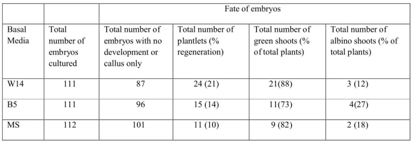
Fate of embryos Basal Media Total number of embryos cultured Total number of embryos with no development or callus only Total number of plantlets (% regeneration) Total number of green shoots (% of total plants) Total number of albino shoots (% of total plants) W14 111 87 24 (21) 21(88) 3 (12) B5 111 96 15 (14) 11(73) 4(27) MS 112 101 11 (10) 9 (82) 2 (18)11
Results are based on 4 experiments.
12
13
14
15
Table 9: Influence of colchicine concentration and duration on ploidy of oat plantlets derived from microspores.
1
2
Colchicine concentration and duration (h)
Haploid plantlets (%) Double haploid plantlets (%) 0.1% Colchicine 1.5 h 46.2 53.8 3 h 66.7 33.3 4 h 50.0 50.0 5 h 54.5 45.5 0.2% Colchicine 1.5 h 53.8 46.2 3 h 41.7 58.3 4 h 20.0 80.0 5 h 45.5 54.5
3
Results are based on 1 experiment with a total of 101 microspore-derived plants.