Aging of the Shoulder Joint of Guinea Pigs.
Electron Microscopic and Quantitative
Histochemical Aspects
1
Ruth Silberberg, MD, Mary Hasler, BS, and Peggy A. Lesker, BS
2HPHE tendency of aging joints to develop
•*- osteoarthrosis varies with the site of the articulation; these differences originally ob-served in human individuals (Heine, 1926), were also noted in mice of different strains (Sokoloff, 1956). Since the lesions of osteo-arthrosis are closely related to aging processes (Silberberg, & Silberberg, 1941; Bennet, Waine, & Bauer, 1942) the latter might also vary from one joint to another. In order to explore this possibility, we examined ultrastructural and enzymatic changes in the shoulder joints of aging guinea pigs in conformity with similar investigations of the joints of the lower extremi-ties of these animals (Silberberg, Stamp, Lesker, & Hasler, 1970, Silberberg & Lesker, 1971, Sil-berberg, Lesker, & Hasler, 1972).
MATERIAL AND METHODS
Cartilage from the head of the humerus and from the socket of the shoulder joint was ob-tained from 88 male or female quinea pigs 2 weeks, 12 weeks, 1 year, 2l/2 years, and 5 %
years of age and used previously for the investi-gation of joints of the lower extremity. From each age group, one joint was retained for histological examination; from each of the re-maining humeral heads one slice of cartilage was prepared for electron microscopy; the re-maining cartilage was collected individually from each animal, immediately frozen in liquid nitrogen, lyophilized, and stored for enzyme as-says. In some instances, cartilage from the soc-ket of the shoulder and head of the humerus were handled separately.
1
The investigation was supported by grant AM 04213 of the Na-tional Institutes of Health, Public Health Service, Bethesda, Md.
2 From the Depts. of Pathology and Orthopedic Surgery,
Wash-ington University School of Medicine, St. Louis, Mo. 63110, and the Institute for Pathologic Anatomy of the University of Zurich.
18
The techniques used for electron microscopy, for quantitative histochemical enzyme assays, and for the determination of DNA were the same as those used in our earlier investigations. (Silberberg et al., 1970, Silberberg & Lesker, 1971). Owing to the scarcity of animals in the oldest age group and to the small amount of cartilage available in old animals, not all enzymes could be examined in each animal. However, each value shown on Tables 2-6 represents the mean of at least eight assays, two of each of 4 animals in each age group. The values for DNA (Table 1) are means of all values obtained from each individual sam-ple following comsam-pletion of the enzyme assays. The following enzymes were investigated:
Glycolytic.—Hexokinase (HK),
phosphoglu-comutase (PGM), phosphorylase (total, PI), glucose-6-phosphate dehydrogenase (G6PDH), phosphofructokinase (PFK), aldolase (Aid), a-glycerophosphate dehydrogenase (aGPDH), pyruvate kinase (PK), lactate dehydrogenase (LDH),—up to 2y2 years in males, up to 5%
years in females.
Lysosomal.—Cathepsin D (cathepsin B and
C not being demonstrable), sulfatase, /?-galac-tosidase, /?-glucuronidase, up to 5% years in males and females.
Enzyme activity was calculated on the basis of both dry weight of tissue and of DNA con-tents of the individual samples on which en-zyme determinations were made.
RESULTS
I. Electron microscopy of the cartilage of the head of the humerus
2-week-old animals.—The most superficial
cup-derlying chondrocytes. The matrix about the superficial cells was of uniform density: elec-tron opaque ground substance contained a net-work of collagen fibrils, 15 to 50 nm in diam-eter, with fibrils of 25 to 30 nm predominating. Orientation was mostly circumferential, but some fibrils were also arranged at various angles with the surface (Fig. 2a). Moreover, small tufts of delicate filaments were superimposed on the surface of the cartilage. Scattered among the fibrils were small vesicles about 50 nm in diameter and osmiophilic globules of about the same size.
With increasing distance from the surface, chondrocytes were more commonly paired than in the surface region, and some cells were in the process of division. Compared to superficial chondrocytes, cytoplasm and nuclei were en-larged, cytoplasmic footlets were elongated, and organelles were more crowded. The endoplas-mic reticulum formed stacks of up to eight lamellae, and the cisterns were strikingly di-lated. The Golgi complex had large vacuoles filled with electron opaque granules; centrioles, cytoplasmic pools, mitochondria and lysosome-like bodies were increased in number. Lipid inclusions appeared here and there. Cells of the lowest layer had the typical appearance of hypertrophic chondrocytes with large quantities of glycogen replacing part of the cytoplasm and often aggregating around lipid inclusions. The matrix of the midzone contained a network of fibrils, disposed in all directions and measuring 40 to 50 nm or occasionally more in diameter
(Fig. 2b).
12-week-old animals.—Chondrocytes of both
superfiical and midzone were well organized with predominantly smooth nuclei, abundant, often dilated endoplasmic reticulum, an increas-ingly prominent Golgi apparatus, few but well defined mitochondria, and a few lysosome-like bodies. Postdivisional cells were common. Cell size and organellar development increased with increasing distance from the articular surface, and lipid inclusions and dense bodies were
A few microscars were noted (Fig. 3a).
1-year-old animals.—Many cells were
degen-erating or dying, and there were large cell free areas. Advancing calcification had caused con-siderable narrowing of the articular covering, calcium deposits being found as close as 10 (x from the surface. Chondrocytes varied in their degree of development from small cells with smooth nuclei and poorly supplied with or-ganelles to large polygonal forms with long cytoplasmic footlets and nuclei with irregular contour (Fig. 4 ) . In these cells organelles were crowded: numerous densely approximated la-mellae of endoplasmic reticulum, with uni-formly wide or dilated cisterns often overshad-owed the Golgi complex; the usual array of mitochondria with well defined cristae, dense and lysosome-like bodies, cytoplasmic pools were present; glycogen was less abundant whereas lipid inclusions were more prominent than in the younger animals, especially in the deeper layers of the tissue. There was a tend-ency of the chondrocytes to become walled off from the remainder of the matrix by a band of closely packed collagen fibrils (Fig. 4 ) . This band separated the plasmalemma from the zone of calcification, which encroached upon cells as high as the second layer. Increasing fibrillar-ity was present throughout the cartilage, ow-ing to closer packow-ing as well as to increased thickness of the collagen fibrils. In some places the very surface of the joint was composed of closely packed circumferentially oriented fibrils 50 or more nm in diameter, superimposed merely by small aggregates of delicate fila-ments. Microscars interrupted the otherwise uniform fibrillar pattern (Fig. 3b). The thick-ness of individual fibrils varied with largest diameters measuring about 100 nm.
2Y2-year-old animals.—Many superficial and
midzonal chondrocytes were less crowded with organelles than the mature cells seen at 1 year of age. Nuclei were usually oval and smooth; the endoplasmic reticulum was slightly vesicu-lated, a finding which in association with the
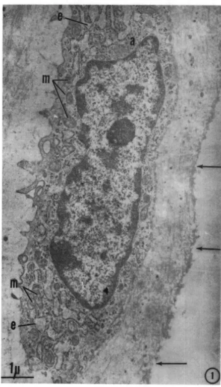
All electron micrographs are from the cartilage of the humeral heads of guinea pigs.
Fig. 1. 2-week-old male. Superficial chondrocyte. Dilated endoplasmic reticulum (e), delicate Golgi apparatus (a); several mitochondria (m); undulating articular surface (arrows). Approx. 15,000 X.
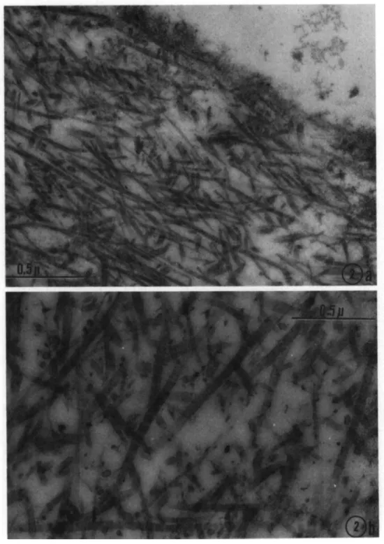
Fig. 2a. 2-week-old female. Superficial fibrils with distinct periodicity, some, but not all, in circumferential orientation. Tufts of delicate filaments protruding over the surface. Approx. 58,000 X.
Fig. 2b. Same animal as in Fig. 2a. Deep midzonal fibrils in haphazard orientation. Abundant interfibrillary ground substance. Fibrils thicker than those shown in Fig. 2a. Approx. 58,000 X.
Fig. 3a. 12-week-old female. Part of a midzonal chondrocyte in left upper corner (Z). Situated in a cell bay close to a cytoplasmic footlet a thick collagen fibril (C), which joins a cluster of similar fibrils to form a micro-ground substance. Approx. 58,000 X.
Fig. 3b. 1-year-old female. Densely packed collagen fibrils from the deep midzone with little interfibrillary ground substances. Approx. 58,000 X.
Fig. 4. 1-year-old male. Fully developed midzonal chondrocyte. Abundant endoplasmic reticulum (e), large Golgi apparatus (a), mitochondria (m), dense bodies (d), lipid inclusions (L), small amount of glycogen (s). Fibrillar matrix forms a somewhat wavy band (H) around part of the cell periphery, separating the latter from the calcified cartilage (K). Approx. 16,000 X.
smooth nucleus gave the cells a youthful ap-pearance. Ribosomes, attached to the mem-branes of the ER were irregularly spaced; the Golgi complex consisted of narrow tubules and a few vacuoles; mitochondria were small but had well defined cristae. Characteristically, from this age on, microtubules 15-30 nm in diameter and microfilaments, 7 nm in width became increasingly conspicuous: they were disposed in bundles and partly or wholly encircled the nucleus. Lipid inclusions were less prominent than before (Fig. 5).
In the matrix, fibrils formed a dense network about the periphery of the cells as well as at some distance. Clusters of thick fibrils were also present in the bays formed by the plasmalemma (Fig. 6).
53/4-year-old animals.—There were no basic
changes from the findings in the previous age group. The majority of cells appeared com-paratively inactive with stacks of short loops of endoplasmic reticulum, which sometimes widened into small sacs, an inconspicuous Golgi apparatus, few mitochondria and a variety of multivesicular bodies, dense and lysosome-like bodies in small numbers. Microfilaments and microtubules were prominent (Fig. 7), ex-tending in bandlike fashion from the peri-nuclear region toward the plasmalemma. Some cells had the typical appearance of fully de-veloped chondrocytes.
In cell-free areas the matrix showed a fairly uniform distribution of fibrils in scanty ground substance. Pericellularly, the fibrillar bands
had become denser with calcium deposits en-croaching upon them from several directions. The joint surface was undulating, with focal deposits of tiny filaments. The most superficial fibrils, arranged circumferentially, measured about 20 nm in diameter (Fig. 8a); in the deep layers, there was no preferential orientation, and fibrils measured up to 160 nm in thickness (Fig. 8b).
II. Histochemical assays of cartilage of the shoulder joint
DNA (Table 1).—In both males and females
a rise in the level during the first months of life was followed by a steep drop during the next 9 mo. Only insignificant changes occurred thereafter; however, in females, there was a slight tendency of a renewed rise in the last age group.
Glycolytic enzymes
Males.—Between the ages of 2 weeks and 2]/2
years, enzyme activity as calculated on the basis of dry weight declined for most enzymes (HK, PI., G6PDH, PFK, Aid., a-GPDH, LDH) or remained steady with minor rises and falls for some (PGM, PK) (Table 2). As calcu-lated per gm DNA (Table 3), enzyme activity in general changed little during the early months of life; from there on, it increased up to 2!/2 years of age, the degree of the change ranging from about 6 times the previous level for G6PDH to the 16-fold increase in the activ-ity of aldolase.
Table 1. Age Changes in DNAa of Articular Cartilage of the Shoulder of Guinea Pigs. 2 Weeks Males 4.4±0.37 Females 3.02±0.29 12 Weeks 3.37±0.28 4.45±0.53 "Calculated as gm/kg dry weight of tissue.
1 Year 0.89±0.05 1.49±0.21
Table 2. Age Changes in Enzyme Activity* of Articular Cartilage
Hexokinase (mM) Phosphoglucomutase (M) Phosphorylase (total) (mM) Glucose-6-P'dehydrogenase (mM) Phosphofructokinase (mM) Aldolase (mM) <x-glycerol-P dehydrogenase (mM) Pyruvic Kinase (M) Lactic dehydrogenase (M) Sulfatase (MM) /3-galactosidase (mM) Cathepsin D (Hb) (gm) 0-glucuronidase (mM) 2 Weeks 197±27 3.56±0.80 66.1±1.3 558±20 130.0±24.0 115±16 29.4±4.3 2.89±0.98 12.10zfc0.80 1.30±0.20 2.93±0.35 4.55±0.50 5.55±0.43 12 Weeks 139±35 2.78±1.20 48.9±4.6 507±138 46.7±4.4 150±8 45.5±7.1 2.31db0.79 6.48±0.81 1.76±0.30 3.05±0.53 3.97±0.50 3.11±0.44 1\i Years 1.69±0.17 1.57±0.21 5% Years 1.56±0.11 1.40±0.20
of the Upper Extremity of Male Guinea Pigs.
1 Year 118±30 3.58±0.60 41.6±2.7 330±56 41.4±1.4 116±7 46.2±2.0 3.80±0.67 3.26±0.77 0.96±0.16 1.73±0.19 2.05±0.42 2.65±0.35 VA Years 104±20 3.49±0.10 22.2±3.4 188±18 47.2±1.2 96±3 22.5±3.4 2.26±0.64 3.23±0.78 1.43±0.33 2.10±0.26 2.93±0.39 3.18±0.40 5% Years 1.28±0.29 1.56±0.16 1.74±0.22 2.13±0.41 a
Table 3. Age Changes in-Enzyme Activity*1 of Articular Cartilage of the Upper Extremity of Male Guinea Pigs. Hexokinase (mM) Phosphoglucomutase (M) Phosphorylase (total) (mM) Glucose-6-P dehydrogenase (mM) Phosphofructokinase (mM) Aldolase (mM) a-glycerol-P dehydrogenase (mM) Pyruvic Kinase (M) Lactic dehydrogenase (M) Sulfatase (/xM) /3-galactosidase (mM) Cathpsin D (gm) /3-glucuronidase (mM) 2 Weeks 35=1=1 0.64±0.03 11.9±0.8 100=1=11 23.3±0.7 21±1 5.3±0.9 0.52d=0.02 2.17±0.77 0.28±0.02 0.75=1=0.11 0.83±0.06 1.12±0.08 "Calculated as moles (M), millimoles (mM), micromoles (/nM),
12 Weeks 23=1=1 0.47=1=0.04 8.2±0.8 85=1=5 7.8=1=0.5 25dbl 7.6=1=0.9 0.39d=0.01 1.08±0.81 0.45±0.09 O.84±O.O8 1.12±0.10 0.96±0.10 1 Year 158d=21 4.80±0.63 55.8d=2.1 442=1=59 55.5=1=2.4 155±20 61.9=t2.5 5.09=1=0.71 4 . 3 7 ± 0 . 8 8 1.17±0.12 1.87±0.16 2.58±.0.50 2 . 9 5 ± 0 . 3 2 2K Years 353=blO 1.18=1=0.16 75.3=1=4.2 637±32 160.0=1=6.0 326=tll 76.2=1=4.3 7.66±0.52 10.90±1.00 0.89±0.15 1.42±0.17 1.78±0.16 1.68±0.12 or gram (gm/gm DNA/hour. Note.—=b Indicates standard error.
h% Years
0.78db0.05 0.96±0.05 1.18=1=0.20 1.43±0.17
Table 4. Age Changes in Enzyme Activity11 of Articular Cartilage of the Upper Extremity of Female Guinea Pigs.
Hexokinase (mM) Phosphoglucomutase (M) Phosphorylase (total) (mM) Glucose-6-P dehydrogenase (mM) Phosphofructokinase (mM) Aldolase (mM) a-glycerol-P dehydrogenase (mM) Pyruvic Kinase (M) Lactic dehydrogenase (M) Sulfatase (/xm) /3-galactosidase (mM) Cathepsin D (Hb) (gm) /3-glucuronidase (mM) 2 Weeks 243=1=22 3 . 1 1 ± 0 . 3 7 6 2 . 4 ± 4 . 4 775±71 159.0=1=11.0 186=1=13 77.6±3.6 4.01±0.50 12.80=1=1.40 1.69=1=0.19 3.17±0.24 1.89=b0.39 6.38±0.50 "Calculated as moles (M), millimoles (mM), micromoles (/xM),
12 Weeks 152=1=40 1.74±0.33 5 8 . 2 ± 6 . 8 396=t34 64.4=1=3.2 214±9 6 6 . 7 ± 2 . 8 1.87=1=0.50 5.81=1=0.51 1.95=1=0.30 2.48=1=0.30 4.47±0.48 3.30=t0.45 1 Year 144=1=7 2.60±0.12 23.6=1=1.5 447=h46 42.8=1=1.6 9 2 ± 4 31.0±7.8 3 . 8 1 ± 0 . 0 8 6.54=1=0.44 0.78=1=0.12 1.56±0.36 2.47=1=0.66 3.13=1=0.49 or gram (gm)/kilogram dry weight/hour.
2}/2 Years 87=bl 0.33=1=0.02 21.3=1=1.8 108±5 35.1±2.2 132±9 19.4d=2.9 2.08±0.08 2.55±0.04 1.45±0.17 1.30=1=0.20 3.67±0.39 2.73±0.45 b% Years 192=1=23 3 8 6 ± 2 5 54.8=t4.9 274±16 3.38±0.39 5.16=1=0.16 0.89±0.19 0.66±0.10 2.75=1=0.52 1.05=1=0.18 Note.—± Indicates standard error.
Table 5. Age Changes in Enzyme Activitya of Articular Cartilage of the Upper Extremity of Female Guinea Pigs.
Hexokinase (mM) Phosphoglucomutase (M) Phosphorylase (total) (mM) Glucose-6-P dehydrogenase (mM) Phosphofructokinase (mM) Aldolase (mM) a - g l y c e r o l - P dehydrogenase (mM) Pyruvic Kinase (M) Lactic dehydrogenase (M) Sulfatase (iiM) /3-galactosidase (mM) Cathepsin D (gm) /3-glucuronidase (mM) 2 Wce'cs 58=1=1 0.74±0.04 14.9=1=0.9 185±7 37.9=1=1.2 44=1=8 18.5=1=0.7 0.96±0.05 3.05±0.20 0.70=fc0.02 1.21±0.04 1.60=1=0.08 2.30±0.12 "Calculated as moles (M),r
millimoles (mM), micromoles (/uM),
12 Weeks 21=1=1 0.31=1=0.01 8.2=1=0.8 560=1=8 9.0±0.5 21=tl 9.5=1=0.7 0.26=t0.01 8.16±0.72 0.47=h0.03 0.81±0.06 1.07=1=0.11 0.85±0.10 1 Year 326=1=11 7.00=1=0.85 ' 63.3±1.6 1070d=82 96.3±1.3 207±12 70.1=k3.6 9.92±0.57 9.19±0.20 1.10d=0.16 1.37+0.13 3.68=1=0.80 3.01=1=0.31 or gram (gm)/gm DNA/hour. Note.—±
2]/2 Years 118±6 0.48±0.03 28.9=1=0.9 164=1=9 47.7=1=1.1 180=1=5 26.4±1.2 2.83=1=0.72 3.47±0.14 l . l l ± 0 . 1 1 0.93±0.10 2.84=1=0.20 2.00=1=0.23 h%/i Years 155=1=3 3 3 1 ± 2 1 42.6=fcl.O 221±10 2.73=1=0.71 4.16±0.21 0.59±0.08 0.65±0.03 1.22±0.26 O.76=tO.O4 Indicates standard error.
Fig. 5. 2'/2-year-old male. Midzonal chondrocyte. Smooth nucleus; the cytoplasm sparsely populated with or-ganelles: narrow endoplasmic reticulum (e), delicate Golgi apparatus (a), mitochondria (m), lipid inclusion (L), and microtubules and microfilaments (T). Fibrillar band in pericellular matrix (H). Approx. 14,000 X.
Fig. 6. 2'/2-year-old male. Two midzonal chondrocytes with large nucleus and narrow cytoplasm. From cell bays clusters of thick collagen fibrils (F) extend into the matrix. Approx. 14,000 X.
%m&mzmii
Fig. 7. 5%-year-old male. Midzonal chondrocyte. Large, smooth nucleus, few organelles, except for a promi-nent band of microtubules (T) and microfilaments circling the nucleus. Approx. 11,000 X.
Fig. 8a. 5%-year-old male. Superficial fibrils more densely packed than in the young animal (Fig. 2a). Ap-prox. 58,000 X.
Fig. 8b. Same animal as in Fig. 8a. Deep midzonal fibrils are much thicker than those near the joint surface. Approx. 58,000 X.
Lysosomal Enzymes
Males.—As calculated on the basis of dry
weight, the activity of cathepsin D declined from the age of 2 weeks to 1 year with minor deviations in either direction later in life. (Ta-ble 2 ) . Sulfatase changed little during the en-tire life-span. The activity of /?-glucuronidase closely followed that of cathepsin D, /?-galac-tosidase activity varying even less from early life through old age.
As calculated per gm DNA, all four enzymes assayed peaked at 1 year of life (Table 3 ) ; terminal levels of activity equalled or slightly exceeded those observed during growth.
Females.—As calculated on the basis of dry
weight, the activities of cathepsin D and of sulfatase alternatingly rose and fell during life; the only common feature was the peak of activ-ity at 12 weeks of age. Actual differences how-ever, were slight, the highest and lowest levels of the respective activities not differing more than 21/2-fold (Table 4 ) . By contrast, the
ac-tivities of /?-glucuronidase and /?-galactosidase declined gradually from the youngest to the oldest age group tested.
As calculated per gm DNA, the activities of the four enzymes assayed followed a similar pattern: dropping at 12 weeks, rising sharply at 1 year, and receding gradually thereafter into old age (Table 5).
COMPARISON BETWEEN AGING CHANGES IN U P -PER AND LOWER EXTREMITY
Electron microscopy
During growth, chondrocytes and matrix of the humeral and femoral heads resembled each other closely, and from 1 year of age on dead or dying cells were numerous in both sites. From the age of 2l/2 years on, there were
con-spicuous differences in the appearance of cells and fibrils. Whereas in the femoral head, the majority of preserved chondrocytes was crowded with organelles, there were, in the humeral head, comparatively few active appearing cells; instead, in most chondrocytes the nuclei ap-peared youthful and the cytoplasmic organelles, especially the ER and the Golgi complex were poorly developed, while lipid inclusions were few and small. Microscars with bundles of thick collagen fibrils were present in both ex-tremities, but otherwise, the collagen fibrils of the humeral cartilage were narrower than those seen at corresponding sites of the femoral head.
Histochemistry
As calculated per dry weight (Tables 2, 4 ) , the cartilage of the shoulder joint was on the whole less active enzymatically than the carti-lage of the lower extremity. This was true for 9 of 12 enzymes assayed in males and in 8 of 10 glycolytic enzymes assayed in females. In the remaining assays, activities were either found to be equal in both extremities, or there was a temporary, usually minor increase in the upper over the lower leg.
The age curves of the individual enzymes cal-culated per gram DNA are shown in Figure 9 (males) and 10 (females). Solid lines repre-sent the values obtained for the shoulder joints and shown in detail in Tables 3 and 5; broken lines represent the corresponding figures for the joints of the lower extremities, published previ-ously in detail (Silberberg et al., 1970; Silber-berg & Lesker, 1971; SilberSilber-berg, Lesker, & Has-ler, in press). The four curves for each enzyme in males or females and upper or lower extrem-ity respectively are drawn on the same scale; the scales for different enzymes are not always identical, since activities are variously expressed in moles, millomoles, micromoles, or grams.
In both, males and females, the curves for DNA of the two extremities were similar.
In males, assays for glycolytic enzymes yielded, up to 2l/2 years of age, curves that were
either closely approximated to each other or that indicated higher activity in joints of the lower as compared to those of the upper ex-tremity. However, at one point each of the curves for PGM and PI, values for the upper extremity surpassed those obtained for the lower. Curves for lysosomal enzymes were conspic-uously flatter in the upper compared to the lower extremity. The difference was particu-larly striking for cathepsin D and sulfatase, while it was less marked with regard to /?-glu-curonidase and /?-galactosidase.
In females, likewise, comparable values were obtained for enzyme activity up to 2l/2 years
of age, the curves either being closely apposed or following a similar pattern of rises and falls. A noticeable difference, however, was present in enzyme activities in the oldest age group: the rise seen in the lower extremity in the ac-tivities of HK, PFK, Aid, PK, and LDH was less striking or failed to develop in the cartilage of the shoulder. Upper and lower extremities differed similarly in regard to the lysosomal en-zymes cathepsin D and sulfatase, whereas the
aGPDH
LDH CathD
Sulf
bGluc
bGal
Fig. 9. Curves comparing enzyme activity in the articular cartilage of aging male guinea pigs. Solid lines: up-per, broken lines: lower extremity.
assays for /?-glucuronidase and /?-galactosidase yielded comparable curves for both extremities.
In a few instances, the absolute differences between single points on the curves for the two extremities respectively were especially wide, the upper rising above the lower; this was true for PGM, G6PDH, to some extent for sulfatase, and particularly for cathepsin D. A possible reason for this was sought in the fact that in the early assays no distinction was made be-tween cartilage of the socket and of the humeral head, respectively. In subsequent assays the activities of a few lysosomal enzymes were de-termined separately for these two sites. The
results are shown in Table 6. From this table it appears that after one year of age, enzyme activity tended to be higher in the socket than in the humeral head. Moreover, whenever curves for upper and lower extremities deviated considerably from each other as in the case of cathepsin D and sulfatase, enzyme activity of the socket was strikingly in excess of that of the humeral head. It was thus concluded that unusually high values for the upper extremity were caused by the presence in the sample of a comparatively large quantity of cartilage from the socket of the joint.
aGPDH PK Cath D LDH Suit bGal bGluc
Fig. 10. Curves comparing enzyme activity in the articular cartilage of aging female guinea pigs. Solid lines: upper, broken lines: lower extremity.
DISCUSSION
Age-linked changes in ultrastructure, DNA content and in enzyme activity of the cartilage of the shoulder joint of guinea pigs of both sexes were determined and compared with the corresponding changes in the articular cartilage of the lower extremity of the same animals. Morphologically, chondrocytes of lower and upper extremity resembled one another during the first year of life. During the second part of the life-span, chondrocytes of the humeral head were comparatively underdeveloped and the collagen fibrils of the matrix were more delicate than in the femoral head. The late ultrastruc-ture thus was compatible with relatively de-creased chondrocyte function in the shoulder
and in concordance with the histochemical data. Regardless of conditions in the lower extrem-ity, the youthful appearance of many chondro-cytes in the humeral head of old animals was striking. Apparently cell renewal took place in the replacement of dead cells, keeping the number of cells more or less constant. The presence of large numbers of microfilaments and microtubules in these chondrocytes is diffi-cult to explain: These structures are believed to be related to movement of intracellular ma-terial (Ledbetter & Porter, 1963; Slautterback, 1963) or to contractility of cells or cell parts (Allison, Davies, & Petris, 1971; Ishikawa, Bischoff, & Holtzer, 1969; Ledbetter & Porter, 1963). Neither of these functions is likely to
Head Socket 9 Sulfatase (/xM) Head Socket /3-galactosidase (mM) Head Socket Cathepsin D (gm) Head Socket 0-glucuronidase (mM) Head Socket 1.12±0.16 l.ll±0.09 0.70±0.06 0.71±0.06 1.09±0.06 1.33±0.07 1.29±0.06 1.91±0.13 2.04±0.19 2.56±0.22 0.76±0.14 1.15±0.11 0.49±0.07 0.44±0.05 0.90±0.10 0.72±0.10 0.93±0.19 1.21±0.10 0.88±0.19 0.82±0.17 2.58±0.52 3.31±0.30 0.82±0.08 1.38±0.22 1.08±0.09 1.64±0.20 1.26±0.22 6.08±0.99 3.21±0.43 2.80±0.38 1.59±0.13 1.77±0.29 0.90±0.12 1.32±0.23 0.53±0.08 1.33±0.08 1.82±0.22 3.86±0.32 1.16±0.12 2.84±0.22 0.89±0.12 1.96±0.22 0.46±0.06 0.72±0.13 0.61±0.04 0.68±0.05 0.66±0.12 1.78±0.14 0.82±0.09 0.70±0.06 "Calculated as millimoles (mM,), micromoles QuM), or gram (gm)/gram DNA/hour. Note.—± Indicates standard error.
be significantly enhanced in the compara-tively inactive chondrocytes, in which micro-tubules and filaments are most prominent. Possibly, these structures may become un-masked or aggregate under conditions in which other organelles regress, and the usual trans-port mechanisms fail. Likewise of interest is the intense focal fibrillogenesis about some of the chondrocytes, which contrasted with the comparative delicacy of the fibrils elsewhere in the matrix. Judged by the appearance of endo-plasmic reticulum, protein synthesis by chon-drocytes was not increased, and the question arises of whether the cells gave off stored colla-gen precursors or substances intensifying focal accretion of collagen present in the matrix.
As a tissue cartilage of the shoulder was less active enzymatically than that of the lower extremity. This reduction may have been due either to a relative decrease in the number of cells, to a relative decrease in the activity of the individual chondrocytes of the shoulder, or to both. Since, during the second half of the life-span, values for DNA were close for both ex-tremities, differences in enzyme activity should primarily be due to variations in the activity of individual chondrocytes in the respective lo-cations. The observed differences in enzyme activity according to site varied in degree from enzyme to enzyme. This may be related to the nature of the samples tested. Articular
chon-drocytes vary in structure and presumably in function according to their location with ref-erence to the joint surface; the slices of tissue shaved off with a razor blade certainly were heterogeneous as to the cell population. More uniform samples than those presently examined might be obtainable by microdissection. The relatively low level of the activity of lysosomal enzymes agrees with the small numbers of lysosomes found in the chondrocytes. Both findings raise the question as to how much chondrocyte lysosomes contribute to the age-linked degradation of the matrix.
The cause or causes of the variations in en-zymatic activity at different articular sites are unknown. Intensified joint motion and the re-sulting improved nutrition of the cartilage would create a favorable environment for chon-drocytes; after a period of relatively high activ-ity, however, they age and die more rapidly than those kept under nutritionally marginal conditions (Silberberg & Silberberg, 1964). The fact that some degradative enzymes were shown to be more active in the fixed socket than in the movable head of the humeral would be con-sistent with this suggestion and with increased activity of some lysosomal enzymes observed following prolonged compression of the articular cartilage (Thompson & Clerk, 1970). Regard-less of these points, however, the present mor-phologic and enzymatic findings indicate a
comparatively slow rate of aging and are thus compatible with the lesser tendency of the shoulder joint to develop arthrosis as compared to the articulations of the lower extremity, espe-cially the knee joint.
SUMMARY
Cartilage of the shoulder joint of guinea pigs of both sexes and 2 weeks to 5% years of age was examined electron microscopically and as to the activity of nine glycolytic and four ly-sosomal enzymes. The findings were compared with those obtained previously in joints of the lower extremity. Ultrastructurally, the articu-lar cartilage of the upper resembled that of the lower extremity during the first year of life; during the second half of the life-span, articular chondrocytes of the upper extremity were com-paratively underdeveloped and collagen fibrils of the matrix were thinner than in the lower extremity. The activities of the glycolytic en-zymes were of comparable intensity in both lo-cations during the first part of the life-span; at later ages, most activities were lower in the shoulder than in the cartilage of knee and hip. The activities of lysosomal enzymes was either similar in both locations or decreased in the upper compared to the lower extremity. The findings are consistent with the relatively low tendency of the shoulder joint to develop osteo-arthrosis.
REFERENCES
Allison, A. C , Davies, P., & Petris, S. X. Role of con-tractile microfilaments in macrophage movement and
endocytosis. Nature, 1971, 232, 153-155.
Bennett, G. A., Waine, H., & Bauer, W. Changes in
the kneejoints at various ages with particular refer-ence to the nature and development of degenerative joint disease. New York: Commonwealth Fund,
Ox-ford Univ. Press, 1942.
Heine, J. Ueber die Arthritis deformans. Virchows
Archiv fur pathologishe Anatomie und Physiologie,
1926, 260, 521-663.
Ishikawa, H., Bischoff, R., & Holtzer, H. Formation of arrowhead complexes with heavy meromyosin in a variety of cell types. ]ournal of Cell Biology, 1969, 43, 312-318.
Ledbetter, M. D., & Porter, K. R. A microtubule in plant cell fine structure. ]ournal of Cell Biology, 1963, 19, 239-250.
Silberberg, M., & Silberberg, R. Aging changes of bones and joints in various strains of mice. American
Journal of Anatomy, 1941, 68, 69-95.
Silberberg, R., & Silberberg M. Pathogenesis of osteo-arthrosis. Pathologica et Microbiologia, 1964, 27, 447-457.
Silberberg, R., Stamp, W. G., Lesker, P. A., Hasler, M. Aging changes in ultrastructure and enzymatic activity of articular cartilage of guinea pigs. Journal of
Geron-tology, 1970, 2 5 , 184-198.
Silberberg, R., & Lesker, P. Enzyme activity in aging articular cartilage. Experientia, 1971, 27, 133-135. Silberberg, R., Lesker, P., & Hasler, M. Ultrastructure
and enzymes of aging articular cartilage of male guinea pigs. Proceedings Conf. on Aging of
Connec-tive Tissue. Reisesburg. April, 1972. Excerpta Medica Foundation Symposia, 1973. (in press)
Slautterback, D. B. Cytoplasmic microtubules. I. Hydra.
Journal of Cell Biology, 1963, 18, 367-388.
SokolofF, L. The natural history of degenerative joint disease in small laboratory animals. Pathologic anatomy of degenerative joint disease in mice.
Ar-chives of Pathology, 1956, 62, 118-138.
Thompson, R. C , & Clerk, J. Acid hydrolases in slices of articular cartilage and synovium from normal and abnormal joints. Proceedings of the Society for