HAL Id: hal-01837836
https://hal.archives-ouvertes.fr/hal-01837836
Submitted on 26 May 2020
HAL is a multi-disciplinary open access
archive for the deposit and dissemination of
sci-entific research documents, whether they are
pub-lished or not. The documents may come from
teaching and research institutions in France or
abroad, or from public or private research centers.
L’archive ouverte pluridisciplinaire HAL, est
destinée au dépôt et à la diffusion de documents
scientifiques de niveau recherche, publiés ou non,
émanant des établissements d’enseignement et de
recherche français ou étrangers, des laboratoires
publics ou privés.
Adeline Becquer, Margarita Torres Aquino, Christine Le Guerneve, Laurie
Amenc, Carlos Trives Segura, Siobhan Staunton, Herve Quiquampoix, Claude
Plassard
To cite this version:
Adeline Becquer, Margarita Torres Aquino, Christine Le Guerneve, Laurie Amenc, Carlos Trives
Segura, et al.. A Method for Radioactive Labelling of Hebeloma cylindrosporum to Study
Plant-fungus Interactions. Bio-protocol , Bio-protocol LCC, 2017, 7 (20), �10.21769/BioProtoc.2576�.
�hal-01837836�
Copyright © 2017 The Authors; exclusive licensee Bio-protocol LLC. 1
A Method for Radioactive Labelling of Hebeloma cylindrosporum to Study Plant-fungus Interactions
Adeline Becquer1, Margarita Torres-Aquino1, $, Christine Le Guernevé2, Laurie K Amenc1, Carlos Trives-Segura1, *, Siobhan Staunton1, Hervé Quiquampoix1 and Claude Plassard1, *
1INRA, UMR Eco&Sols, 2 place Viala, Montpellier, France; 2INRA, UMR SPO (1083) Sciences pour
l’Oenologie, 2 place Viala, Montpellier, France; $Present address: Colegio de Postgraduados, Campus San Luis Potosí, Agustín de Iturbide Nº 73, Salinas de Hidalgo, Salinas, S.L.P, México, Mexico
*For correspondence: claude.plassard@inra.fr; carlos.trives-segura@inra.fr
[Abstract] In order to quantify P accumulation and P efflux in the ectomycorrhizal basidiomycete fungus Hebeloma cylindrosporum, we supplied 32P to mycelia previously grown in vitro in liquid medium. The culture had four main steps that are 1) growing the mycelium on complete medium with P, 2) transfer the mycelia into new culture solution with or without P, 3) adding a solution containing 32P and 4) rinsing the mycelia before incubation with or without plant. The main point is to rinse very carefully the mycelia after 32P supply in order to avoid overestimation of 32P efflux into the medium.
Keywords: Culture in vitro, Phosphate availability, 32P labelling, Ectomycorrhizal fungi
[Background] It is well known that the association between mycorrhizal fungi and plants improves the P nutrition of the host-plant (reviewed by Smith and Read, 2008; Plassard and Dell, 2010; Cairney, 2011; Smith et al., 2015). This positive effect has been attributed primarily to phosphate (Pi) uptake by the fungal cells exploring the soil far from the roots, allowing the exploration of a large volume of soil beyond the depletion zone formed around actively absorbing roots (Smith and Read, 2008; Cairney, 2011; Smith et al., 2015). However, to benefit to the host plant, absorbed Pi has to be transported from the fungal cells exploring soil towards those in close contact with the host cells. In ectomycorrhizal symbiosis, these exchanges are thought to take place in a territory called the ‘Hartig net’ inside the ectomycorrhizal roots (Smith and Read, 2008, Cairney, 2011). In the Hartig net, the fungal cells colonize the walls of cortical cells but there is no direct communication between the two plasma membranes, meaning that P has to be released from the fungal cells via a yet unknown mechanism. Taken together, this knowledge indicates that the ability of the fungus to take up P in external hyphae and to release P is therefore an important feature of the fungal species for its positive effect on plant P nutrition. Here, we developed a methodology using 32P labelling to follow the net 32P accumulation and release by an ectomycorrhizal fungal species cultivable in vitro, without its host plant (Torres-Aquino et
Copyright © 2017 The Authors; exclusive licensee Bio-protocol LLC. 2
Materials and Reagents
1. Sterile plastic Petri dish, 90 mm (Dominique DUTSCHER, Gosselin, catalog number: 688302) 2. Sterile plastic Petri dish, 35 mm (Corning, Falcon®, catalog number: 351008)
3. Liquid scintillation vial, 6 ml with cap (PerkinElmer, catalog number: 6000592) 4. Liquid scintillation vial, 20 ml with cap (PerkinElmer, catalog number: 6008117) 5. Waste bags for radionuclides (SCIE-PLAS, catalog number: RRP-BAG17)
6. Protective paper for bench (Dominique DUTSCHER, Benchguard, catalog number: 090277) 7. Liquid scintillation cocktail Ultimagold (PerkinElmer, catalog number: 6013329)
8. Nichrome wire, stainless steel, round, 22 gauge, 0.64 mm diameter (suppliers for electronic cigarettes)
9. Aluminium screw cap, 40 mm with rubber liner (VWR, SPV, catalog number: 215-2690) 10. Multi-Purpose Silicone for kitchen or bathroom, 280 ml (Castorama, Rubson)
11. 60 ml Luer-lock syringes (Dominique DUTSCHER, Omnifix, catalog number: 921016)
12. Teflon PTFE microtube, 1.15 mm and 1.75 mm for internal (int) and external (ext) diameter (diam), respectively (Dominique DUTSCHER, PTFE, catalog number: 091932)
13. Needles 18 G 0.9 x 40 mm (Dominique DUTSCHER, BD MicrolanceTM 3, catalog number:
301300)
14. Tubing, int diam 1.14 mm (Dominique DUTSCHER, Silicone, catalog number: 4906591) 15. Tubing, int diam 3.17 mm (Dominique DUTSCHER, Silicone, catalog number: 4906600) 16. Microtubes, 1.5 ml (Dominique DUTSCHER, Eppendorf, catalog number: 033511) 17. Valve Luer polycarbonate one way (Cole-Parmer, catalog number: EW-30600-01)
18. Sterile syringe filters for air, 0.2 µm, 6.4 cm diam (Labomoderne, Midisart, catalog number:
RS3320)
19. Autoclavable Polypropylene bag, 3 L, non-printed (Dominique DUTSCHER, catalog number:
140230)
20. Tips 1,200 µl for pipet (Dominique DUTSCHER, Sartorius,catalog number: 077200B) 21. Home-made syringe holder
22. Home-made needle holder for aeration
23. Folding skirted caps, 14.9 mm diam (Dominique DUTSCHER, catalog number: 110602) 24. Paper for sterilization (Dominique DUTSCHER, catalog number: 006950)
25. Hebeloma cylindrosporum (ectomycorrhizal basidiomycete) (laboratory’s own collection, available upon request)
26. Manganese (II) sulfate monohydrate (MnSO4.H2O) (Sigma-Aldrich, catalog number:
M7899-500G)
27. Zinc sulfate heptahydrate (ZnSO4·7H2O) (Sigma-Aldrich, catalog number: Z0251-100G)
28. Boric acid (H3BO3) (Sigma-Aldrich, catalog number: B6768-500G)
29. Copper (II) sulfate pentahydrate (CuSO4·5H2O) (Sigma-Aldrich, catalog number: C8027-500G)
Copyright © 2017 The Authors; exclusive licensee Bio-protocol LLC. 3
31. Potassium nitrate (KNO3) (Sigma-Aldrich, catalog number: P8291-1KG)
32. Sodium phosphate monobasic monohydrate (NaH2PO4·H2O) (Sigma-Aldrich, catalog number:
71504-250G-M)
33. Magnesium sulfate heptahydrate (MgSO4·7H2O) (Sigma-Aldrich, catalog number:
63138-250G)
34. Potassium chloride (KCl) (Sigma-Aldrich, catalog number: P9333-500G)
35. Calcium chloride dihydrate (CaCl2·2H2O) (Sigma-Aldrich, catalog number: C5080-500G)
36. Ferric ammonium citrate (Sigma-Aldrich, catalog number: RES20400-A702X) 37. D-glucose (Sigma-Aldrich, catalog number: G8270-1KG)
38. Agar-agar (Sigma-Aldrich, catalog number: A7002-500G)
39. KH232PO4 in water (PerkinElmer, catalog number: NEX055002MC)
40. MES (2-N-morpholino-ethanesulfonic acid, 4-morpholineethanesulfonic acid monohydrate) (Sigma-Aldrich, catalog number: 69892-500G)
41. TRIS (Tris(hydroxymethyl)aminomethane) (Sigma-Aldrich, catalog number: T1378-500G) 42. 1 N sulfuric acid solution (EMD Millipore, catalog number: 1.09072.1000)
43. Trace elements (see Recipes)
44. Mineral salt base solutions (see Recipes) 45. Thiamine solution (see Recipes)
46. N6 complete liquid solution (see Recipes) 47. Solid N6 complete liquid solution (see Recipes) 48. Interaction medium (IM) (see Recipes)
Equipment
1. Sample bottles,120 ml (VWR, SPV, catalog number: SPVAGO2246)
2. Glass bottles, 1,000 ml, ISO borosilicate, graduated (Dominique DUTSCHER, catalog number:
046415)
3. Glass bottles, 2,000 ml, ISO borosilicate, graduated (Dominique DUTSCHER, catalog number:
046416)
4. Two pairs of stainless steel straight tweezers Wironit, Brucelles type, 130 mm (Dominique DUTSCHER, catalog number: 491037)
5. A scalpel handle for blade 20 to 25 (Dominique DUTSCHER, catalog number: 3740004) 6. Surgical blade sterile N°21 (Dominique DUTSCHER, catalog number: 132521)
7. A standalone burner (Dominique DUTSCHER, catalog number: 071109)
8. Butane gas cartridge for the burner (Dominique DUTSCHER, catalog number: 060415) 9. A nail, a hammer, scissors and cutting pliers
10. Incubator with controlled temperature set at 25 °C 11. Autoclave
Copyright © 2017 The Authors; exclusive licensee Bio-protocol LLC. 4
13. Cork-borer, 7.5 mm diameter (Dominique DUTSCHER, catalog number: 942783) 14. Aquarium air-pumps (SuperFish, model: air-box Nr.4)
15. Shield, fixed 15° angle, flat base, Beta; 530 x 350 mm shield; 350 x 300 mm base (upright x horizontal) (SCIE-PLAS, catalog number: RPP-S15L)
16. Midi-box with hinged lid, Beta; external dimensions: 80 x 185 x 105 mm; internal dimensions: 60 x 165 x 85 mm (height x width x depth) (SCIE-PLAS, catalog number: RPP-B6)
17. Bin for beta wastes on the bench, 3.3 L capacity (SCIE-PLAS, catalog number: RPP-B17) 18. Liquid scintillation Counter TRI-CARB 4910TR (PerkinElmer, catalog number: A491000)
Software
1. Microsoft Excel for calculations
2. Statistica 7.1 (StatSoft Inc., Tulsa, OK, USA) for statistical analysis
Procedure
A. Preparation of equipment 1. Glass jars for liquid cultures
The mycelia are grown in glass jars (120 ml) with an aluminium cork previously equipped with a nichrome wire enabling to suspend the fungal inoculum at the surface of the liquid solution (see Figure 1).
Preparation: first pierce the centre of an aluminium cap with a nail and a hammer. Then, cover the hole with a small amount of silicone paste and allow the paste to dry for 24 h. Cut the rubber seal in its centre with scissors and place it inside the cap.
Meanwhile, cut the nichrome wire with cutting pliers in fragments 15 cm long whose one end is curved to form a kind of hook. Fill each jar with 40 ml of liquid nutrient medium. Take a nichrome wire and slip it into the hole of the cap equipped with its rubber seal through the silicone paste, as centred as possible. Screw the cap onto the glass jar and set the height of the wire just above the nutritive solution. Autoclave the jars at 121 °C for 20 min and allow them to cool before use.
Copyright © 2017 The Authors; exclusive licensee Bio-protocol LLC. 5
Figure 1. Equipment used to grow mycelia of H. cylindrosporum. It is made of a 120 ml-glass jar containing 40 ml of a complete nutrient liquid medium. A. Each jar is inoculated by one plug or disk of fungal mycelium placed on a nichrome wire. B. Aspect of the mycelium grown on the surface of medium in unshaken conditions.
2. Preparation of syringe holders, connectors for aeration and syringes
60 ml-syringes are used to rinse each mycelium before measuring either fungal 32P accumulation and/or 32P efflux from the labelled fungus into the interaction medium (IM) (see Recipes). This rinsing step is essential to avoid overestimation of 32P accumulation and/or efflux. Typically, we used 6 fungal replicates per treatment and we built homemade equipment (Figures 2 and 3) for easy handling of the mycelia.
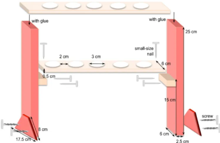
a. Preparation of syringe holders (Figure 2):
Cut the different pieces of wood according to the dimensions given in Figure 2. For example, to hold 6 syringes, the top piece is 37 cm long x 6 cm wide and the one underneath is 32 cm long x 6 cm wide. Adjust and maintain the two pieces together to drill the holes in a two-by-two alignment. Afterwards, assemble all the pieces together as shown in Figure 2.
Figure 2. Homemade holders to support syringes for incubation of mycelia in interaction medium
Copyright © 2017 The Authors; exclusive licensee Bio-protocol LLC. 6
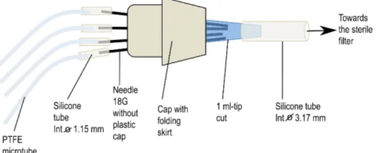
b. Preparation of connectors (Figure 3)
First, take an 18 G needle with a pair of tweezers, heat its collarover a flame to soften the glue and pull it with another pair of clamps. After cooling, adjust a small piece of silicone tube (1.14 mm internal diameter) of about 3 cm long on the top of the needle. Take a skirt cap and stitch 6 needles into its top and close its bottom by a 1 ml pipette tip previously cut at its finest end. If necessary, add silicone paste to fragile places to strengthen the airproof of the system. To the cut end of the blue pipette tip, add a piece of silicone tube (3.17 mm internal diameter) about 20 cm long that will be plugged later into a sterile air filter placed between the air pump and the connector (see Figure 3). Finally, place the whole system [connector + PTFE tubes + large diameter silicone tube] in an autoclavable bag and sterilize it by autoclaving (115 °C, 40 min).
Figure 3. Homemade connector to aerate syringes during the incubation of mycelia in interaction medium
c. Preparation of syringes: remove the plunger of each syringe and discard it. Using a hot nail, drill a hole at the top of the syringe opposite to the graduations giving the volume. This hole will be used to place the microtube for aeration inside the syringe. Place all drilled syringes in an autoclavable bag and sterilize it by autoclaving (115 °C, 40 min).
B. Preparation of fungal cultures
Note: This is done in sterile conditions, in a laminar flow cabinet. The cork-borer and the clamps are flame-sterilized and allowed to cool before use.
1. Step 1: Growing new cultures in solid medium from fungal stock cultures
For experiments, we use stock cultures renewed approximately every 6 months as sources of fungal material. These fungal stock cultures are grown in the dark at 25 °C on agar nutrient medium (solid N6 medium, see Recipes below) in 90 mm Petri dishes. The nitrogen (N) source in the medium (nitrate) is adapted to this fungal species as it does not grow well on ammonium, in contrast to most ectomycorrhizal species.
At the time of experiments, it is necessary to establish new fungal cultures by transferring one agar plug (7.5 mm diameter) on the centre of a new Petri dish (90 mm diameter) containing 25 ml of solid N6 complete medium, the mycelium face down on the nutritive medium to enhance
Copyright © 2017 The Authors; exclusive licensee Bio-protocol LLC. 7
the growth of the fungus. New cultures are grown for 3 weeks, in the dark, at 25 °C in an incubator.
2. Step 2: Growing the fungus in glass jars from solid cultures
First, prepare the fungal plugs with the cork-borer by punching holes in the 3-week old mycelium agar culture.
Then, unscrew the lid of a jar and hold it in one hand; take a pair of clamps in the other hand and remove a plug of fungal culture from the solid medium; prick the plug on the tip of the hook of the nichrome wire. Screw the lid properly on the jar and put the fungal plug just on the surface of the medium by slipping the nichrome wire (Figure 1A). The fungus is grown at 25 °C for two weeks.
3. Step 3: Growing the fungus in glass jars from liquid cultures for labelling experiments
The 2-week old mycelia grown in liquid medium are then used to produce the fungal material for labelling experiments. For that, disks of the fungus are punched from the mycelium and placed on the tip of the hook of the nichrome wire, as described in step B2. This procedure avoids the presence of agar-agar in the fungal culture, as it can interfere with labelling experiments by accumulating 32P from the labelled solution and releasing 32P into the non-labelled solution. The agar-free mycelia are grown for 7 days, at 25 °C, without shaking.
C. P pre-treatments
1. Preparation of nutritive solution
Prepare +P and -P nutritive liquid media for fungal pre-treatment that are N6 medium with P (supplied as NaH2PO4 1 mM, see Recipes) or without P (by omitting the addition of NaH2PO4 in
the N6 medium), 40 ml per mycelium. Prepare additional N6 medium without P (100 ml per mycelium) to rinse the mycelia (see Figure 4B).
2. Preparation of jars
Pour 40 ml of liquid medium, either +P or -P, in new jars. Screw lids without nichrome wire on the jars and autoclave them as described before. Pour also additional -P N6 medium in 1 L bottles and autoclave.
3. Transfer of mycelia to new media
Bring the flasks containing the seven-day-old mycelia into the flow laminar cabinet and the mycelium is withdrawn from the medium by slipping the nichrome wire up through the lid. The nutritive medium is allowed to drip from the fungus before the lid is unscrewed (Figure 4A). a. For the -P treatment only, rinse each mycelium in -P medium by transferring it successively
into a 35 mm-Petri dish containing 20 ml of fresh -P medium (Figure 4B) for 3 min. This operation is repeated four times, meaning that 5 Petri dishes are prepared for each mycelium as illustrated in Figure 5. This step is essential to eliminate the carry-over solution that would otherwise contaminate the -P solution with orthophosphate (Pi). After rinsing, transfer each mycelium into a new flask containing 40 ml of -P medium and allow them to grow for 5 additional days at 25 °C, in the dark, to get P-depleted mycelia.
Copyright © 2017 The Authors; exclusive licensee Bio-protocol LLC. 8
b. For the +P treatment, transfer the mycelia just allowed to drip dry into new flasks containing 40 ml of +P medium, without rinsing. They are allowed to grow under the same conditions as -P conditions.
Figure 4. Transfer of the mycelia to new media. A. the nichrome wire is first pulled through the lid to allow the solution to drip from the fungus before the lid is unscrewed. B. Then the lid is unscrewed and the mycelium rinsed five times in 20 ml P-deficient medium contained in 35 mm-Petri dishes.
Figure 5. Picture of Petri dishes arranged in the laminar flow cabinet for rinsing the mycelia
D. Labelling with 32P
1. Safety precautions: take care to perform the experiments following safety precautions for the handling of Radioactive materials that are principally (i) wear gloves, a lab coat and a dosimeter to control the radiation dose received by the body, (ii) place the radioactive solutions or material behind plexiglas screens, (iii) use pipette tips with filters, (iv) throw away the radioactive solutions or materials in specific containers, (v) keep these containers behind
Copyright © 2017 The Authors; exclusive licensee Bio-protocol LLC. 9
Plexiglass screens, (vi) eliminate the waste after a period of time of at least 10 times the decay period of 32P namely 140 days and only if the radioactivity of the waste is not higher than twice the background (generally 0.8 to 1.6 counts per second).
2. Prepare a set of jars with lid without nichrome wire filled with 40 ml of +P medium and autoclave them at least the day before the labelling experiment. Place a Plexiglass screen and protective paper on the bench and open the jars behind the Plexiglas screen. Label the medium with 32P orthophosphate by quickly adding KH232PO4. Dilute the radio-labelled source
so as to obtain an initial activity of 3.3 x 10⁴ Bq per µmol Pi in the +P medium. Homogenize the solution by hand shaking of the jars and take a subsample (150 µl) to measure the actual radioactivity in the medium. Store the subsample in 1.5 ml Eppendorf tubes at -20°C, in a Plexiglass box. Use 50 µl for radioactivity measurement.
3. Bring the jars with mycelia next to the jars with labelled medium. Allow the fungus to drip dry (as in Figure 4A). Put the lid with the fungus in the jar with the radio-labelled medium, screw it and slip the nichrome wire down until the fungus touches the solution surface. Allow the fungus to incubate for 16 h at 25 °C, in the dark.
E. Rinsing the mycelia in syringes
1. Place the syringes in the holders on the bench, and equip them with a PTFE microtube for aeration (Figure 6). In each syringe, pour 60 ml of interaction medium (IM) and place a collection vessel under the tap. Plug the connectors for aeration into an air pump and check whether or not IM is well aerated. However, turn off the air pump during the rinsing of mycelia. Place the Plexiglass screens in front of the holders.
2. Bring the jars with mycelia in the labelled solution behind the screen, one jar in front of one syringe. Allow the labelled solution to drip from each mycelium (as in Figure 4A) for about 5 min. Unscrew the cap, slip the nichrome wire with the fungus down from the cap and place it into a syringe. Bend the nichrome wire so as to hang the mycelium at a level corresponding to 20 ml (see Figure 6).
3. After all the mycelia are suspended in the syringes, open the tap to allow the IM to flow into the collection vessel placed under the syringe. Close the tap and pour about 30 ml of new IM in each syringe. After 5 min, open the tap again to allow the IM to flow into the collection vessel. Repeat the same operation four times, with the tap open after 10, 15, 20 and 40 min after addition of new IM. On the whole period, each mycelium is rinsed with 150 ml of IM. At each rinsing time, collect 1 ml of IM and placed it into 6 ml liquid scintillation vial to measure radioactivity.
4. After the 90 min-rinsing period, you can sample 6 mycelia to measure their fresh weight and their 32P contents. You can also usethe mycelia for incubation with plant to measure fungal 32P efflux in the IM over time (see Becquer et al., 2017).
Copyright © 2017 The Authors; exclusive licensee Bio-protocol LLC. 10
Figure 6. Complete device used to rinse the mycelia after the labelling period. A. Syringe containing the interaction medium; B. Polycarbonate valve; C. Collection vessel; D. Mycelium; E. Nichrome wire; F. Microtube Teflon PTFE for individual aeration; G. Home-made connector for gathering 4 to 7 Teflon tubes; H. Sterile filter; I. Air pump.
F. Radioactivity measurements
1. Add 4 ml of scintillation liquid into each 6 ml-vial containing IM or labelled medium, close the vial and mix it several times upside down by hand. Make blank samples by pouring 4 ml of scintillation liquid in a vial. Measure the radioactivity using a Liquid Scintillation Counter, following manufacturer’s instructions.
2. To measure radioactivity in the fungus, blot each mycelium between filter paper sheets and record its fresh weights. Depending on the size of the mycelium, put it in a 6 ml- or a 20 ml- scintillation vial and add 4 or 12 ml of scintillation cocktail for counting, respectively.
3. Correct raw data for decay occurring during radioactivity measurement using the equation written in Excel:
cpmt0 = Exp(Ln(cpmt-cpmbackground)) + (0.00003366099x t), with
cpm = counts per minute
t0 = time of the beginning of experiment
t = time of cpm measurement of the sample in counter in minutes, cpmbackground = cpm of solvent (blank sample),
cpmt = radioactivity of the sample at time t,
cpmt0 = radioactivity of the sample at time t0 corresponding to the first measurement. This
value is taken as the reference one for all measurements. 4. Transform cpm in Becquerel (Bq) using the formula:
Copyright © 2017 The Authors; exclusive licensee Bio-protocol LLC. 11
5. Calculate the specific activity (SAi) of phosphate supplied to the fungus (replicatei) as follows,
assuming that the concentration of phosphate is 1 mM:
SAi (Bq/µmole Phosphate) = [(cpmt0 of labelled medium/volume used for measurement (ml)) x
0.033]
6. Calculate the amount of P taken up by the fungusi (replicate i) during the exposure to 32P as
follows:
P accumulation in fungusi (µmoles) = (cpmt0 in the fungusi x0.033)/SAi
Data analysis
Express amounts of 32P (in Bq) released by the mycelia or accumulated in the fungus either per mycelium or per gram of fresh weight. You can also express the data as µmoles of phosphate accumulated by the fungus during the 16 h incubation period and calculate the corresponding uptake rate of Pi. If you compare several treatments, test the normality of data using the Kolmogorov Smirnov test and, where necessary, transform the data either square root or log10 prior to analysis to meet the assumptions of ANOVA. To compare the effect of treatments, use an ANOVA analysis.
Notes
1. As the growth of the fungus in liquid medium may vary, we prepare up to 4 additional culture flasks inoculated with the fungus to discard those with poor growth.
2. Aeration provided through PTFE microtubes could collapse because of liquid meniscus. They can be pulled out from the microtubes by branching a 60 ml syringe completely filled with air and strongly pushing the air through the microtubes.
3. The height of the microtubes in the syringes may need to be adjusted to allow the air to exit. If they are too deep in the medium, the air will not flow.
Recipes 1. Trace elements (1,000 ml) 3.08 g MnSO4·H2O 4.41 g ZnSO4·7H2O 2.82 g H3BO3 0.98 g CuSO4·5H2O 0.29 g Na2MoO4·2H2O Add ddH2O to 1,000 ml, store at 4 °C
Copyright © 2017 The Authors; exclusive licensee Bio-protocol LLC. 12
2. Mineral salt base solutions (100 ml each) Solutions For making100 ml 1 M KNO3 10 g
1 M NaH2PO4·H2O 13.8 g
1 M MgSO4·7H2O 24.6 g
1 M KCl 7.45 g 1 M CaCl2·2H2O 7.35 g
1% ferric ammonium citrate 1 g
3. Thiamine solution (100 ml) 0.01 g Thiamine-HCl
4. N6 complete liquid solution (1,000 ml)
a. Add in a 1 L-beaker approximately 500 ml of deionized water and the following volumes of mineral base solutions:
6 ml KNO3
1 ml NaH2PO4
1 ml MgSO4
4 ml KCl 0.5 ml CaCl2
0.5 ml ferric ammonium citrate
b. Add also trace elements 0.2 ml and thiamine solution 0.5 ml
c. Complete the volume to 1 L and check the pH which should be adjusted to 5.5 d. Finally, add 5 g of D-glucose and shake until complete dissolution
e. This solution is distributed in glass jars 5. Solid N6 complete liquid solution (1,000 ml)
Take two 1 L glass bottles:
a. Add 7.5 g of Agar-agar and pour 500 ml of liquid N6 medium in each bottle b. Sterilize the medium at 121 °C for 20 min
c. Pour the cooled medium (55-60 °C) in Petri dishes 90 mm diameter 6. Interaction medium (3,000 ml)
a. Add in a 3 L-beaker approximately 1.5 L of deionized water and the following volumes of mineral base solutions:
0.6 ml MgSO4
1.5 ml CaCl2
3.2 g of MES (final concentration of 5 mM) 1.82 g of TRIS (final concentration of 5 mM)
b. Complete to 3 L with deionized water and adjust the pH to 5.5 with 1 N H2SO4
Copyright © 2017 The Authors; exclusive licensee Bio-protocol LLC. 13
Acknowledgments
This research was supported by INRA (France) through annual funding devoted to their researchers and a fellowship through a Contract for Young Scientist (CJS) granted to Adeline Becquer, and by CONACYT (Mexico) through a Ph-D fellowship granted to Margarita Torres-Aquino. The protocol is adapted from our previous work (Torres-Aquino et al., 2017). We also thank the three anonymous reviewers for their comments that helped us to improve the protocol.
References
1. Becquer, A., Torres-Aquino, M., Le Guernevé, C., Amenc, L.K., Trives-Segura, C., Staunton, S., Quiquampoix, H. and Plassard, C. (2017). Establishing a symbiotic interface between cultured ectomycorrhizal fungi and plants to follow fungal phosphate metabolism. Bio Protoc 7(20): e2577.
2. Cairney, J. W. G. (2011). Ectomycorrhizal fungi: the symbiotic route to the root for phosphorus in forest soils. Plant Soil 344: 51-71.
3. Plassard, C. and Dell, B. (2010). Phosphorus nutrition of mycorrhizal trees. Tree Physiol 30(9): 1129-1139.
4. Smith, S. E. and Read, D. J. (2008). Mycorrhizal symbiosis. 3rd edition. Academic Press, London.
5. Smith, S. E., Anderson, I. C. and Smith, F. A. (2015). Mycorrhizal associations and phosphorus acquisition: from cells to ecosystems. Annual Plant Reviews 48: 409-440.
6. Torres-Aquino, M., Becquer, A., Le Guerneve, C., Louche, J., Amenc, L. K., Staunton, S., Quiquampoix, H. and Plassard, C. (2017). The host plant Pinus pinaster exerts specific effects on phosphate efflux and polyphosphate metabolism of the ectomycorrhizal fungus Hebeloma cylindrosporum: a radiotracer, cytological staining and 31 P NMR spectroscopy study. Plant