HAL Id: hal-02434404
https://hal.archives-ouvertes.fr/hal-02434404
Submitted on 17 Jan 2020
HAL is a multi-disciplinary open access
archive for the deposit and dissemination of
sci-entific research documents, whether they are
pub-lished or not. The documents may come from
teaching and research institutions in France or
abroad, or from public or private research centers.
L’archive ouverte pluridisciplinaire HAL, est
destinée au dépôt et à la diffusion de documents
scientifiques de niveau recherche, publiés ou non,
émanant des établissements d’enseignement et de
recherche français ou étrangers, des laboratoires
publics ou privés.
Boron doped diamond biotechnology: from sensors to
neurointerfaces
C. Hébert, Emmanuel Scorsone, A. Bendali, R. Kiran, M. Cottance, H.
Girard, J. Degardin, E. Dubus, Gaelle Lissorgues, Lionel Rousseau, et al.
To cite this version:
C. Hébert, Emmanuel Scorsone, A. Bendali, R. Kiran, M. Cottance, et al.. Boron doped diamond
biotechnology: from sensors to neurointerfaces. Faraday Discussions, Royal Society of Chemistry,
2014, 172, pp.47-59. �10.1039/C4FD00040D�. �hal-02434404�
Faraday
Discussions
www.rsc.org/faraday_d
This manuscript will be presented and discussed at a forthcoming Faraday Discussion meeting.
All delegates can contribute to the discussion which will be included in the final volume.
Register now to attend! Full details of all upcoming meetings:
http://rsc.li/fd-upcoming-meetings
This is an Accepted Manuscript, which has been through the
Royal Society of Chemistry peer review process and has been
accepted for publication.
Accepted Manuscripts are published online shortly after
acceptance, before technical editing, formatting and proof reading.
Using this free service, authors can make their results available
to the community, in citable form, before we publish the edited
article. We will replace this Accepted Manuscript with the edited
and formatted Advance Article as soon as it is available.
You can find more information about Accepted Manuscripts in the
Information for Authors
.
Please note that technical editing may introduce minor changes
to the text and/or graphics, which may alter content. The journal’s
standard
Terms & Conditions
and the
Ethical guidelines
still
apply. In no event shall the Royal Society of Chemistry be held
responsible for any errors or omissions in this Accepted Manuscript
or any consequences arising from the use of any information it
contains.
Journal Name
RSC
Publishing
ARTICLE
This journal is © The Royal Society of Chemistry 2013 J. Name., 2013, 00, 1-3 | 1
Cite this: DOI: 10.1039/x0xx00000x
Received 00th January 2012, Accepted 00th January 2012 DOI: 10.1039/x0xx00000x
www.rsc.org/
Boron doped Diamond Biotechnology: from sensors
to neurointerfaces
C. Hébert
1, E. Scorsone
1, A. Bendali
3, R. Kiran
1,4, M. Cottance
2, H.A. Girard
1, J.
Degardin
3, E.Dubus
3, G. Lissorgues
2, L. Rousseau
2, S. Picaud
3, P. Bergonzo
1Boron doped nanocrystalline Diamond (B-NCD) is known as a remarkable material for the fabrication of sensors taking advantage of its biocompatibility, electrochemical properties, and stability. Sensors can be fabricated to directly probe physiological species from biofluids (e.g. blood or urine) as will be presented. In collaboration with electrophysiologists and biologists, the technology was adapted to enable structured diamond devices, such as MicroElectrode arrays (MEAs), i.e. common electrophysiology tools enabling to probe the neuronal activity distributed over large populations of neurons or of embryonic organs. Specific MEAs can be also used to build neural prostheses or implants to compensate function losses due to lesions or degeneration of part of the Central Nervous System (CNS) such as for retinal implants, those latter exhibiting real interests as biocompatible neuroprostheses for in-vivo neuronal stimulations. New geometries of electrodes enable high performance electrodes to surpass more conventional materials for such applications.
A Introduction
Boron doped diamond (BDD) electrodes are extremely promising in the field of biomedical applications as they exhibit a unique combination of properties. Doped diamond synthesis has become a major focus of research and development: undoped diamond exhibits a high band gap and is at room temperatures an electrical insulator. When doped with boron, diamond results in an electrode with remarkable electro-analytical properties which can be used to detect species in solution before oxygen and hydrogen evolution interfere with the analysis. Thus, conductive diamond electrodes take electrochemical detection into new levels and extend their usefulness to analytes which are not measurable with conventional electrode materials. This opens the field of direct analytical detection with diamond, and in addition to applications where analytes can be detected in biofluids, reversely current spikes can be used to stimulate neural tissues. There is a real interest for the development of novel Neuroprostheses and brain-machine interfaces: those applications witnessed an exponential growth since the success of cochlear implants and of deep brain stimulation for Parkinsonian patients1. Neuroprostheses can either be surface electrodes, meaning the electrodes are simply in contact with the glial surface of the neuronal tissues such as the cortex, or penetrating electrodes reaching deeper neuronal structures in the brain. Although cortical implants have shown a gain of function after a few months, this was unfortunately frequently followed by a complete loss of activity2. This loss of functionality could be attributed to a major gliosis occurring around the prostheses or its electrodes in the months following implantation3. Another challenge for neuroprostheses is to
increase the resolution of individual electrodes to stimulate more precisely discrete neuronal areas. These objectives motivate the development for new biocompatible materials limiting glial reactions and improving direct interactions with neurones.
Recently, the concept of retinal prostheses was validated in clinical trials showing that such prostheses can enable blind patients to read short words, identify contrasted objects or follow lanes on the ground 4,5. These retinal prostheses aiming at restoring vision in patients having lost their photoreceptors are either placed in the subretinal space or on the epiretinal side in direct contact with either the outer or the inner limiting membrane both produced by glial Muller cells. In the first configuration, the subretinal implant will stimulate retinal bipolar cells, neurones normally postsynaptic to photoreceptors5 (Zrenner et al., 2011) whereas epiretinal implants are targeting retinal ganglion cells, spiking neurones sending visual information to the brain via their axon through the optic nerve4. Although these implants have already restored
some visual functions in patients 4,5, an increase in electrode resolution is required to further improve the restored visual performances to achieve face recognition, text reading or independent locomotion. Different 3D design were already proposed to reach this objective using either pillars6 or wells7 on the implants.
Among potential new biomaterials, diamond has raised great attention for its use in neuroprostheses because Boron-doped diamond exhibits semiconductive properties. Diamond biocompatibility was already demonstrated on osteoblast cultures8 and even embryonic cortical neurons 9,10. Embryonic neurons were found to grow selectively on protein patterns
Page 1 of 7
Faraday Discussions
Faraday
Discussions
Accepted
Manuscript
Faraday
Discussions
Accepted
Manuscript
ARTICLE Journal Name
stamped on diamond surfaces 9 but they could not grow directly on a polished polycrystalline diamond layer or on a nanocrystalline diamond layer unless these layers were coated with peptides or with dispersed nanodiamond particles 10. The use of diamond as electrodes has further been demonstrated by recording neuronal activities using cell lines11.
B Boron doped diamond (BDD) electrodes and
microelectrode arrays for biosensing
BDD electrode possesses unique electrochemical properties namely, low capacitive background current, wide potential window in aqueous media, poor adsorption of polar molecules and corrosion resistance in harsh environments making them highly promising as electrochemical sensor when compared to other conventional electrode materials 12,13,14. BDD can be grown using microwave plasma enhanced chemical vapour deposition techniques on substrates from 2 to 4 inches using 2.45GHz plasma excitation. Such layers have demonstrated bio-inertness and long term stability that make them ideal candidates for biomedical applications 15. Due to its carbon nature, BDD also opens the way to immobilization of functional groups such as DNA, proteins, enzymes etc. onto the electrode surface,16 thereby bringing high selectivity to sensing process.
We developed a novel process for the fabrication of diamond microelectrodes that involves the selective growth of diamond over silicon substrates, followed by the deposition of metal contacts and passivation layers. This process includes initially the fixing of detonation diamond nanoparticles over a pre-oxidized 4 inches silicon wafer using a protocol described elsewhere 17. Then an aluminum hard mask consisting of a disc of 100µm in diameter was selectively deposited, over the areas where the BDD micro-electrodes have to be grown. Photolithography enabled to partially pattern the Al mask thus used to protect areas where diamond nanoparticles were protected from being etched in a reactive ion etching (RIE) process in oxygen. The Al hard mask was finally removed and the diamond electrode was grown. The dimensions of the resulting diamond disc electrodes were typically of 300 nm in thickness and 100 µm in diameter. Then an assembly of Ti (50 nm) /Pt (150 nm) metal tracks was deposited over the substrate with a metal ring going around the edge of the diamond disc in order to take electrical contact from the diamond electrode. Then a silicon nitride (Si3N4) passivation layer was deposited
by CVD over the substrate. Finally an opening can be made over the diamond electrodes by using local etching of the Si3N4
layer by RIE with SF6 gas. Typical dimensions range between
10 to 100µm (Figure 1).
Figure1: image of typical BDD diamond micro electrodes arrays.
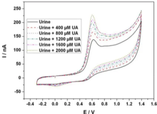
Further to the use of boron doped diamond (BDD) electrodes for the electrochemical detection and quantification of e.g., explosive traces in real media like sea water18, the novel ability to fabricate diamond microelectrode arrays has further enabled the possibility to detect redox active compound directly in unprocessed media such as urine, wine, milk, blood etc 19. e.g.,
uric acid in real urine 20. In fact, conventional BDD electrodes are prone to fouling when used in biological fluids (urine, blood plasma), and synthetic fluids. Kiran et al 19 have recently proposed an electrochemical (EC) treatment where a train of short cathodic and/or anodic pulses are applied to clean fouled electrodes. This technique can be used to retrieve the lost reactivity, characterized by electron transfer rate k0 of the boron
doped diamond electrodes, thereby enhancing their reusability over long period of measurements without degradation of the signal, thus significantly extending the field of monitoring and surveying applications. With this approach, Uric acid detection in real urine samples was made feasible, with a good discrimination of Ascorbic acid. Human urine samples were collected from volunteers as real samples for direct BDD uric acid analysis and were compared to spectrophotometric method, and measured values using the proposed model were observed to be very close to the spectrophotometric results with a maximum difference of 13%. The interest for continuous monitoring of UA in urine in particular for patients admitted in intensive care units (ICU), where the early diagnostic of acute renal failure (ARF) can have a major impact on the survival rate of those patients 21. In this context electrochemical (EC) detection techniques are seen as a promising alternative to conventional optical methods due to their good sensitivity, fast measuring time, portability, low power consumption and cost effectiveness, thus enabling for example direct bedside monitoring.
Figure 2 : Cyclic voltammograms of urine diluted by 2 fold and that of diluted urine containing added uric acid (250 – 1250µM) scanned at 20 Vs-1. The electrodes were activated
electrochemically in the same solution in between two successive scans.
C Boron doped diamond microelectrode arrays for
recording of neuronal signals
Electrophysiology is the study of electrical properties of cells and tissues, which involve the measurement of the voltage change of a biological entity. It is a powerful approach not only
Faraday
Discussions
Accepted
Manuscript
Faraday
Discussions
Accepted
Manuscript
Journal Name ARTICLE
This journal is © The Royal Society of Chemistry 2012 J. Name., 2012, 00, 1-3 | 3 to study the electrical activity of animal cells to understand the
working of the nervous system, brain, hypothalamus, etc. but also to diagnose and treat nervous system disorders. The electrical activity of neurons can be measured directly using extracellular microelectrode arrays. The potential changes in the vicinity of the electrode, caused by currents flowing across neuronal membranes of multiple neurons, can be detected. The same microelectrode arrays as presented in the previous section can be used in order to record the invoke activity of cell signals. For example, arrays of 64 pixel diamond microelectrodes have successfully been used by Maybeck et al., 22 for the recording of the cardiomyocyte-like cell line HL-1. The study demonstrated that BNCD electrodes were able to detect cardiac cell action potentials as well as gold electrodes, while electrodes performed up to a factor of fourfold better than planar metal electrodes of the same diameter. BNCD electrodes survived the mechanical stresses of contractile cells without the defects commonly observed in other nanostructured materials after cell culture (loss of platinum black, removal of CNTs 23, tipping of pillars, D. Brüggemann unpublished results). This enabled to conclude that the diamond layer remained on the surface during cell contraction by the lack of thinning-induced defects after multiple cultures and the contractile tension generated in mature HL-1 cells (cells did not pull themselves off of the surface), thus demonstrating that the diamond MEA’s physical robustness makes it a durable and easy to handle device for repeated or long-term use, with promising interests for recording electrical signals from biological samples in addition to their established role in electrochemistry.
D Boron doped diamond microelectrode arrays for
neurostimulation and implants
The main constraint when in-vivo tissue stimulation is concerned is that the microelectrode array must conform to the size and shape of the tissues to be stimulated. This requires specific developments to enable the fabrication of devices that further to provide the advantages of the former ones must also be flexible and exhibit a long connecting foil enabling the electrical signals to be taken from outside the body to the in-vivo neuronal tissues. This is particularly demanding in the case of retina implants since the curvature is particularly high and the surgery constraints are imposing very strict specifications. In spite of this, the past decade has seen an explosion of research efforts into retinal prostheses aimed at restoring sight to patients blinded by retinitis pigmentosa or age-related macular degeneration. In addition to classical approaches, novel technologies are being explored, often in the context of newly founded commercial enterprises. Also, for the devices to be offering a real breakthrough to blind people, theoretical studies have demonstrated that the ability to reach stimulation structures of 600 pixels would be of great benefit as it should be sufficient for patients to read 24. This extreme number is today not reachable with the current technologies. Several approaches have been proposed,with systems offering typically 60 25,26,27 to 49 28,29,30pixel electrode arrays, with often partial clinical success on short term tests.
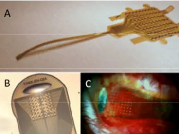
In a similar approach as for the fabrication of diamond MEAs used for recording applications, here nanocrystalline diamond was processed using standard nanotechnology approaches for the fabrication of pixelized microelectrode arrays, but where the structure is grown on a sacrificial layer (e.g. SiO2). This
processed will be later illustrated in a 3D approach on Figure 4. In brief, after diamond electrodes to be deposited on such a sacrificial layer, standard processing of a soft material such as polyimide is made enable on the substrate. Processing of feed lines to contact each electrode can then be embedded in several layers of soft polyimide, to lead after removal of the sacrificial layer to a flexible microelectrode array with high flexibility for retinal stimulation as shown on figure 3A. The preparation of such implants has been made feasible on polyimide as well as on parylene. We typically fabricated matrices of 64 independent pixels for the stimulation of tissues, that were evaluated in the form of preliminary prototypes of diamond implantable electrode devices on laboratory animals (Figure 3).
Figure 3 : (A): view of a non-3D diamond MEA implant for retinal stimulation. The left part consists of 64 diamond electrodes as visualised on (B). (C) In-vivo implanted MEA array as implanted in P23H rats at the Paris Vision Institute.
E Non-planar diamond electrode matrices towards
the improvement of spatial resolution.
Further, a study driven by the Vision Institute in Paris rendered the specifications more challenging, as Djilas et al.7 demonstrated a very high interest in fabricating 3D wells where a small group of retinal cells could be stimulated, in order to improve the stimulation precision. In fact, the major challenge in visual rehabilitation using neuroprostheses aims at improving the spatial resolution of each individual electrode stimulated area: a direct consequence being the density of independent pixels: it has been demonstrated that the electrode resolution can be improved using bipolar stimulations between two neighbour electrodes rather than with a configuration based on a stimulating electrode and a distant common ground31. Using a retinal implant where each pixel exhibits a 3D geometry, one could further improve such bipolar stimulations by locally maintaining neurons between both stimulating electrodes (Palanker et al., 2004). The success of such 3D designs implies that the residual blind retina remains sufficiently plastic to shape itself wherein the electrodes. This plasticity of the blind residual retina was demonstrated using pillars penetrating the
Page 3 of 7
Faraday Discussions
Faraday
Discussions
Accepted
Manuscript
Faraday
Discussions
Accepted
Manuscript
ARTICLE Journal Name
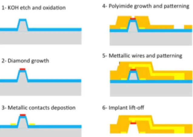
tissue or cavities filled with cells32. However, the neuronal nature of cells surrounding pillars or located in the cavities was not fully assessed since cell immunolabelling could not be performed within this approach. In addition, bipolar stimulation would require two wires per pixel thus significantly increasing the complexity of the implant design when the number of pixels is increased. An alternative approach proposed the use of a returning ground grid surrounding each individual stimulating electrodes thus enabling an improved localisation of the stimulating current, this provided the grid material exhibits a low impedance31. Electrical stimulations have to be constrained to small tissue areas, as proposed in three-dimensional arrays aiming at confining the current within a small volume around the stimulating electrode6. A previous study of our group validated in vivo a model of 3D well-shaped electrodes demonstrating the favourable integration of the retina cells within such cavities when designed at the optimized sizes7. This modelling was based on the hypothesis that the neuronal tissue would shape itself into the 3D wells located within the implant. To address those challenges, Rousseau et al.33 have proposed novel issues enabling the synthesis of nanodiamond 3D shaped microelectrode arrays. The goal is to achieve an implant with each electrode placed at the bottom of a typical 30 µm deep recess and ground located at the surface of the implant. To fabricate this 3D structure, we have chosen to use silicon moulds fabricated by KOH etching on silicon <100> substrates. A specific design with compensation structures was added to obtain theses molds. Initially a simple structure without electrodes was made for histological testing as well as to check the plasticity of the retina. A silicon wafer was oxidized in a furnace at 1050°c in a wet environment with a gas mixture of hydrogen and oxygen during 50 min in order to get 500 nm of silicon dioxide. A photolithography step is done to define the patterns for KOH etching. Silicon dioxide not covered by photoresist was etched away in a solution of Buffer HF(10%). Then the wafer was placed in KOH solution kept at 80°c during approximately 30 minutes so as to obtain a structure 30 µm high. After cleaning, the wafer was oxidized another time in the same conditions to obtain a thick silicon dioxide layer (1µm). Selective growth of diamond could then be initiated as described above using the spreading of a colloidal solution of diamond nanoparticles in deionised water (Di-Water) onto the substrate followed by spin coating34. Nevertheless due to the topology of the 3D substrate used here, spin coating cannot be used, and an alternative approach based on electrostatic grafting of the diamond nanoparticles was used instead35. This latter method is based on electrostatic interactions between diamond nanoparticles and a substrate coated with a polyelectrolyte. Thus the substrates were dipped into Poly DiallylDimethyl-Ammonium Chloride (PDDAC) polymer aqueous solution. The substrates were then washed with water and dried prior to be immersed in the colloidal solution of nanoparticles. Finally, the substrates were washed again with water and dried under N2 flux, resulting in 3D structures completely covered by diamond nanoparticles. A short diamond growth was then performed to fix all the diamond nanoparticles over the surface. The patterning of the diamond electrodes was ensured from the direct diamond nanoparticles patterning before growth36. A 500 nm thick aluminium film was sputtered over the substrate and patterned by photolithography followed by chlorine plasma etching so as to define the electrode areas. The diamond particles not covered by the aluminium film were etched away by classical Reactive Ion etching (RIE) in a gas mixture of
Argon and Oxygen with a power of 200 W for 20 minutes. Lastly aluminium protecting the nanoparticles was removed by wet etching and the wafer was placed in a SEKI 6500 CVD diamond growth reactor to selectively grow the diamond electrodes on a 4-inch substrate. Next polyimide 2611 was poured on the wafer and spin coated to obtain a 10 µm thick layer. The polyimide was cured at 450 °C under Nitrogen during 6 hours. Then a metal mask (aluminium 500nm) was deposited onto the substrate by sputtering. Photolithography with thick photoresist (AZ 4562) was carried out to define the shape of the implant. Aluminium was then etched away by RIE in chlorine and the polyimide layer was etched with oxygen plasma (RIE). Aluminium was removed and the sacrificial layer (thick silicon dioxide layer) was etched by HF. The implants were finally rinsed and dried.
Figure 4: Process for the fabrication of 3D nanocrystalline diamond implants.
A complete surgical procedure was developed to implant in the sub-retinal position a complete 3D diamond implant. 3D structures were implanted in rats P23H on the sub-retinal position during 11 weeks. The retina was then explanted along with the implant and cell labelling was performed. Cell nuclei were labeled by the DAPI stain (blue) while glial cells and retinal bipolar neurons were immunolabelled by the GFAP antibody (grey) and the Goα and PKCα antibodies (green and red), respectively. (In figure 5), the x20 magnification view of the retina/implant wholemount (A) allows visualizing the presence of nuclei stain in all 25 cavities.Confocal analysis was carried out to check the plasticity of the retina. In Figure 5, the blue cell nuclei are well localised within the implant cavities, demonstrating that the retina has followed the shape of the implant. Cell labelling revealed that a small group of bipolar cells is present inside the recess.
Further tests were carried out in order to check the biocompatibility of the BDD diamond electrode on the retina. A 3D implant with diamond on the whole surface was fabricated and implanted in a rat subretinal region. As before, cell labeling was performed after several months. The confocal image (figure 6) shows that no specific reaction occurs on the tissue and no glial proliferation is visible on the surface. No significant difference is observed here when comparing the sample with our reference (polyimide implant). Moreover some bipolar cells can be observed inside each cavity.
Faraday
Discussions
Accepted
Manuscript
Faraday
Discussions
Accepted
Manuscript
Journal Name ARTICLE
This journal is © The Royal Society of Chemistry 2012 J. Name., 2012, 00, 1-3 | 5 Figure 5: Result of histology study on the plasticity of the retina
Figure 6 : Biocompatible test of BDD material
F Boosting diamond electrochemical properties for
implants using nanostructured electrodes.
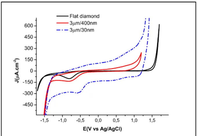
Although diamond exhibits very attractive properties when used as an electrode material and particularly in terms of its electrochemical window, biocompatibility, and resilience to fouling, the material does not match its competitors in terms of impedance and double layer capacitance, which limits the amount of injectable charges 37. In fact, values for flat diamond electrodes generally are expected to range in the 5µF.cm2 range, extremely low values, comparable to that of flat Pt, although black Pt that exhibits a 3D texture can display capacitance values up to 100µF.cm-2.
One way to increase the double layer capacitance of the material is to increase its specific surface area. We propose to use vertically aligned carbon nanotubes with high surface areas as a template onto which boron doped diamond is grown. The resulting composite was found to exhibit a double layer capacitance as high as 0.58 mF.cm-2 and very low impedance when compared to planar diamond electrodes in phosphate buffer saline solution. The influence of the CNTs length as well as of the thickness of the diamond coatings were shown to play a very significative role on the electrode performances as discussed by Hebert et al. 38 All the approach relies on the ability to control the seeding and nucleation stage to avoid the etching of the CNT during the first initial stages of growth.
Figure 7: Cyclic voltammetry (100 mV s-1) electrochemical characterization of BDD coated vertically aligned CNTs in PBS (electrode surface 1 cm2).
Figure 8 (scale bar 1µm) : Cross section of vertically aligned CNT scaffolds covered with diamond.
It is to be noted that such structures exhibit mechanical resiliences that are close to that of diamond and far better than CNT which are known to be very fragile. As an example, scratching experiments performed with stainless steel tweezers showed that it was possible to not damage the diamond on tip structures. We are currently in the process of implementing such very highly structured electrodes in the shape of microelectrode arrays for neurophysiology evaluation as well as histology experiments.
Conclusions
From diamond planar electrodes to highly structured surfaces, diamond can take a wide range of forms with interest for neurointerfacing and particularly for stimulation purposes. Recent works have been dedicated to the development of retinal interfacing structures for the stimulation of the retina in blind patients. The high progress over the last few years will hopefully enable the validation of diamond interests and its convincing development. In neuroprostheses focal stimulation is a real challenge, especially for retinal implants, and we believe this challenge can be successfully taking advantage of diamond properties. As presented, we have recently investigated a new approach to obtain focal stimulation for sub-retinal implants, based on the fabrication of a 3D implant with electrodes localized at the bottom of a cavity and a common return ground electrode placed above at the surface of the implant. This geometry has previously been modelled to evaluate the efficiency of this 3D implant. With this structure the stimulation current is confined within the cavity. Furthermore we have worked on a new material for long term in-vivo stability, namely BDD material. A specific process was
Page 5 of 7
Faraday Discussions
Faraday
Discussions
Accepted
Manuscript
Faraday
Discussions
Accepted
Manuscript
ARTICLE Journal Name
optimised to realize a specific implant for histological tests (3D and 3D with diamond). A complete process was also developed to include BDD electrodes on the soft implant. During in-vivo experiments, we have seen that the retina is soft enough to follow the shape of the implant. Using specific cell labelling of the tissues, confocal pictures showed that bipolar cells were present on each cavity of the implant. The tests have so far revealed that BDD material is well accepted by the tissue. A complete surgical procedure of 3D soft implant was tested to place this implant in the sub-retinal region. The combination of 3D shape and BDD electrode electrochemical performances are expected to lead to significant progress for the next generation of implant. The ultimate limit for diamond to withstand its competing materials was the fact that nanocrystalline diamond still exhibits a relatively low capacitance, much lower than that of e.g. black Pt. With the advances of creating a nano-textured material at the hundreds of nanometre scale, recent progress open up the route for diamond to match the performances of other materials, while it surpasses them with its extreme stability and biocompatibility.
Acknowledgements
We gratefully acknowledge the funding authorities that have supported tis work, and namely the Agence Nationale de la Recherche Franc aise via the MEDINAS ANR07TECSAN014 project, as well as the AVIESAN agency for the ITS-ITMO IMPLANTS, and more recently the FP7 European commission via the NEUROCARE project n° 280433).
Notes
Affiliations
1- CEA, LIST, Laboratoire Capteurs Diamant, CEA-Tech, F-91191 Gif-sur-Yvette, France.
2- ESIEE, Université Paris Est, Noisy le Grand, France 3- INSERM, U968, Institut de la Vision, Paris, France 4- MED-EL Research Center, Feodor-Lynen-Srasse 35, 30625 Hannover, Germany
Contact author, P. Bergonzo philippe.bergonzo@cea.fr
References
1 Lebedev MA, Nicolelis MAL Brain-machine interfaces: past, present and future. Trends in Neurosciences , 2006, 29, 536.
2 Dobelle WH, Mladejovsky MG, Girvin JP Artificial vision for the blind: electrical stimulation of visual cortex offers hope for a functional prosthesis. Science, 1974, 183, 440-444.
3 Maynard EM, Fernandez E, Normann RA A technique to prevent dural adhesions to chronically implanted microelectrode arrays. Journal of Neuroscience Methods 2000, 97, 93.
4 Humayun MS, Dorn JD, Ahuja AK, Caspi A, Filley E, Dagnelie G, Salzmann J, Santos A, Duncan J, Dacruz L, Mohand-Said S, Eliott D, McMahon MJ, Greenberg RJ, Preliminary 6 month results from the argus II epiretinal prosthesis feasibility study. Conf Proc IEEE Eng Med Biol Soc 2009, 1, 4566-4568.
5 Zrenner E, Bartz-Schmidt KU, Benav H, Besch D, Bruckmann A, Gabel VP, Gekeler F, Greppmaier U, Harscher A, Kibbel S, Koch J, Kusnyerik A, Peters T, Stingl K, Sachs H, Stett A, Szurman P, Wilhelm B, Wilke R Subretinal electronic chips allow blind patients to read letters and combine them to words. Proc Biol Sci, 2011, 278, 1489-1497.
6 Butterwick A, Huie P, Jones BW, Marc RE, Marmor M, Palanker D Effect of shape and coating of a subretinal prosthesis on its integration with the retina. Exp Eye Res, 2009, 88, 22-29.
7 Djilas M, Oles C, Lorach H, Bendali A, Degardin J, Dubus E, Lissorgues-Bazin G, Rousseau L, Benosman R, Ieng SH, Joucla S, Yvert B, Bergonzo P, Sahel J, Picaud S, Three-dimensional electrode arrays for retinal prostheses: modeling, geometry optimization and experimental validation. J Neural Eng 2011, 8, 046020.
8 Grausova L BL, Kromka A, Potocky S, Vanecek M, Nesladek M, Lisa V. Nanodiamond as promising material for bone tissue engineering. J Nanosci Nanotechnol 2009, 9, 3524-3534.
9 Specht CG, Williams OA, Jackman RB, Schoepfer R Ordered growth of neurons on diamond. Biomaterials 2004, 25, 4073.
10 Thalhammer A, Edgington RJ, Cingolani LA, Schoepfer R, Jackman RB The use of nanodiamond monolayer coatings to promote the formation of functional neuronal networks. Biomaterials 2010, 31, 2097-2104.
11 Ariano P, Lo Giudice A, Marcantoni A, Vittone E, Carbone E, Lovisolo D A diamond-based biosensor for the recording of neuronal activity. Biosensors and Bioelectronics 2009, 24, 2046.
12 Yano, T.; Tryk, D. A.; Hashimoto, K.; Fujishima, A. Journal of The Electrochemical Society 1998, 145, 1870–1876.
13 Tian, R.; Zhi, J. Electrochemistry Communications 2007, 9, 1120– 1126.
14 Swain, G. M. Analytical chemistry 1993, 65, 345–351.
15 Panizza, M.; Cerisola, G. Electrochimica Acta 2005, 51, 191–199. 16 Agnès, C.; Ruffinatto, S.; Delbarre, E.; Roget, A.; Arnault, J.-C.;
Omnès, F.; Mailley, P. IOP Conference Series, Materials Science and Engineering 2010, 16, 1–11.
17 Scorsone, E.; Saada, S.; Arnault, J. C.; Bergonzo, P. Journal of Applied Physics 2009, 106, 014908.
18 De Sanoit, J.; Van Hove, E.; Bergonzo, P. & Mailley, P. Electrochemical diamond sensors for TNT detection in water Electrochimica Acta, 2009, 54, 5688-5693
19 Kiran, R.; Scorsone, E.; de Sanoit, J.; Arnault, J.-C.; Mailley, P. & Bergonzo, P. Boron Doped Diamond Electrodes for Direct Measurement in Biological Fluids: An In Situ Regeneration Approach Journal of the Electrochemical Society, 2013, 160, H67-H73
20 Kiran, R.; Scorsone, E.; Mailley, P. & Bergonzo, P. Quasi-Real Time Quantification of Uric Acid in Urine Using Boron Doped Diamond Microelectrode with in Situ Cleaning Analytical Chemistry, 2012, 84, 10207-10213
21 Lameire, N.; Van Biesen, W.; Vanholder, R. Nephrology, dialysis, transplantation : official publication of the European Dialysis and Transplant Association - European Renal Association 1999, 14, 2570–2573.
Faraday
Discussions
Accepted
Manuscript
Faraday
Discussions
Accepted
Manuscript
Journal Name ARTICLE
This journal is © The Royal Society of Chemistry 2012 J. Name., 2012, 00, 1-3 | 7 22 Maybeck V, Edgington R, Bongrain A, Welch JO, Scorsone E,
Bergonzo P, Jackman RB, Offenhäusser A., Boron-doped nanocrystalline diamond microelectrode arrays monitor cardiac action potentials. Adv Healthc Mater. 2014 Feb;3(2):283-9.
23 Hanein Y., presented at IBN-2, Forschungszentrum Juelich GmbH, Juelich, Germany (6 Apr 2011). (unpublished results)
24 Sommerhalder J, Rappaz B, de Haller R, Fornos AP, Safran AB and Pelizzone M, Vision Res 2004, 44, 1693-1706.
25 Chader GJ, Weiland J, Humayun MS. Artificial vision: needs, functioning, and testing of a retinal electronic prosthesis. Prog Brain Res 2009, 175, 317-332.
26 Horsager A, Boynton GM, Greenberg RJ, Fine I. Temporal interactions during paired-electrode stimulation in two retinal prosthesis subjects. Invest Ophthalmol Vis Sci 2011, 52: 549-557 27 Fernandes RA, Diniz B, Ribeiro R, Humayun M. Artificial vision
through neuronal stimulation. Neurosci Lett 2012.
28 Zeitz O, Keseru M, Hornig R, Richard G. [Artificial sight: recent developments]. Klin Monbl Augenheilkd 2009, 226, 149-153. 29 Velikay-Parel, Ivastinovic D, Georgi T, Richard G, Hornig R.
Perceptual Threshold And Neuronal Excitability As Long-term Safety Evaluation In Retinal Implants. Invest Ophthalmol Vis Sci 2011, 52, E- Abstract 2590.
30 Keseru M, Feucht M, Bornfeld N, Laube T, Walter P, Rossler G, Velikay-Parel M, Hornig R, Richard G. Acute electrical stimulation of the human retina with an epiretinal electrode array. Acta Ophthalmol 2011.
31 Joucla S, Yvert B, Plos One, 2009, 4(3), e4828
32 Palanker D, Vankov A, Huie P, Baccus S. Design of a high-resolution optoelectronic retinal prosthesis. J Neural Eng 2005, 2:S105-20 33 Bergonzo P., Bonnauron M., Lissorgues. G., Rousseau L., Scorsone
E., Patent IPC: A61N1/05;C30B1/00; C30B29/04, Application number: FR20100054550 20100609, 2011.
34 E. Scorsone, S. Saada, J.C. Arnault, P. Bergonzo, Journal of Applied Physics, 2009, 106 014908
35 Girard, H.A.; Perruchas, S.; Gesset, C.; Chaigneau, C.; Vieille, M.; Arnault, J.-C.; Bergonzo, P.; Boilot, J. & Gacoin, T. Electrostatic grafting of diamond nanoparticles: a versatile route to nanocrystalline diamond thin films ACS Applied Materials and Interfaces, 2009, 1, 2738-2746
36 Bongrain, A., Scorsone, E., Rousseau, L., Lissorgues, G., Bergonzo, P. (2011). Realisation and characterisation of mass-based diamond micro-transducers working in dynamic mode. Sensors & Actuators: B. Chemical, 154(2), 142–149. doi:10.1016/j.snb.2009.12.067 37 Garrett DJ, Ganesan K, Stacey A, Fox K, Meffin H, Prawer S.
Ultra-nanocrsytalline electrodes : optimization towards neural stimualtion applications. J Neur Eng 2012, 9(1), 016002
38 Hébert C., Mazellier J-P., Scorsone E., Mermoux M., Bergonzo P. Carbon 2014, 71, 27–33