HAL Id: tel-00858895
https://tel.archives-ouvertes.fr/tel-00858895
Submitted on 6 Sep 2013HAL is a multi-disciplinary open access
archive for the deposit and dissemination of sci-entific research documents, whether they are pub-lished or not. The documents may come from teaching and research institutions in France or abroad, or from public or private research centers.
L’archive ouverte pluridisciplinaire HAL, est destinée au dépôt et à la diffusion de documents scientifiques de niveau recherche, publiés ou non, émanant des établissements d’enseignement et de recherche français ou étrangers, des laboratoires publics ou privés.
Mécanismes de photo-commutation réversible des
protéines fluorescentes
Aline Regis Faro
To cite this version:
Aline Regis Faro. Mécanismes de photo-commutation réversible des protéines fluorescentes. Autre [cond-mat.other]. Université de Grenoble, 2012. Français. �NNT : 2012GRENY056�. �tel-00858895�
1
THÈSE
Pour obtenir le grade de
DOCTEUR DE L’UNIVERSITÉ DE GRENOBLE
Spécialité: Physique pour les Sciences du Vivant
Arrêté ministériel : 7 août 2006
Présentée par
Aline REGIS FARO
Thèse dirigée par Dominique BOURGEOIS
préparée au sein du L’institut de Biologie Structurale JP. EBEL dans l'École Doctorale de Physique
Mécanismes de
photo-commutation réversible des
protéines fluorescentes
Thèse soutenue publiquement le 27 Septembre 2012, devant le jury composé de :
Mme Fabienne MEROLA
Dr au Laboratoire de Chimie Physique (Université Paris-Sud), Paris, Rapporteur Mr Jean Denis PEDELACQ
Dr à l’Institut de Pharmacologie et de Biologie Structurale, Toulouse, Rapporteur Mme Isabelle DEMACHY
Dr au Laboratoire de Chimie Physique (Université Paris-Sud), Paris, Examinateur Mr Ranieri BIZZARRI
Dr à Scuola Normale Superiore and Istituto Nanoscienze, Pise - Italie, Examinateur
Mr Martin WEIK
Dr à l’Institut de Biologie Structurale, Grenoble, Examinateur Mr Dominique BOURGEOIS
Ces quatre années en France ont été pour moi une très riche expérience, tant au niveau scientifique que personnel. Des nombreuses personnes ont participé directement et indirectement à réalisation de ce travail et je souhaiterai les remercier pour leurs conseils, leur aide et leur soutien.
Tout d’abord je voudrais remercier ma famille.
«Obrigada Manda por esta tão perto mesmo estando tão longe, pai pelo seu apoio e incentivo e eu serei eternamente grata à minha mãe por tudo que ela fez por mim, sua dedicaçao e amor. Ao todo resto da minha familia obrigada pela compreenção pela minha ausência. Uma lembraça muito especial à minha querida tia Cida».
Ensuite, je souhaiterai exprimer mes plus sincères remerciements à mon Directeur de thèse Dominique BOURGEOIS. Dominique m’a donné l’opportunité de réaliser cette thèse, il
m’apporté son enthousiasme, sa vision de la science et ensemble nous avons relevé le
« chalenge » d’écrire cette thèse en anglais. Je remercie également l’ensemble des personnes avec lesquelles j’ai travaillé pendant ces années: Eve DE ROSNY, Philippe CARPENTIER, Virgile ADAM, Sébastien VIOLOT, Claudine DARNAULT, Guillaume POMPIDOR, Delphine ARCIZET, Chenxi DUAN, Martin BYRDIN et Laurent GUYON. Je remercie Eva PEBAY–PEYROULA Directrice de l’Institut de ψiologie Structurale de Grenoble, pour
m’avoir permis d’effectuer ma thèse dans cet établissement. Je remercie aussi l’ensemble des
membres du jury pour avoir accepté d’endosser leurs fonctions respectives.
Je souhaiterai adresser un grand merci tout spécial à Antoine MASURE pour sa patience et les bons moments passés ensemble. Merci aussi à la famille MASURE et BATHILY, vous êtes merveilleux. Merci à tous les amis que m’ont beaucoup aidé avec ses conseilles et ses corrections pendant la rédaction de cette thèse: Cécile TRON, Eleni POLYMENOPOULOU,
Yiannis GEORGIOU « ,
», Alex VIENNE, Alexia ESSERY, Paul LEDBETTER, Fleur CHAPPELLE et Marcel NOGUEIRA.
En dernier lieu, je voudrais exprimer ma reconnaissance aussi envers mes anciens collègues de Labo (IFSC) et spécialement à Lucas BLEICHER pour m’avoir aidé à choisir mon sujet de thèse. Merci aussi à Glaucius OLIVA, Otávio THIEMANN pour vos recommandations et à mes amis Funda KOZANOGLU et à Oumaima BOUSLAMA pour m’avoir aidé à préparer mon entretien à Paris.
XXXXXXXXXXXXXXXXXXXXXXXXXXXXXXXXXXXXXXXXXXXXXXXX XXXXXXXXXXXXXXXXXXXXXXXXXXXXXXXXXXXXXXXXXXXXXXXXXXXX XXXXXXXXXXXXXXXXXXXXXXXXXXXXXXXXXXXXXXXXXXXXXXXXXXXX XXXXXXXXXXXXXXXXXXXXXXXXXXXXXXXXXXXXXXXXXXXXXXXXXXXX XXXXXXXXXXXXXXXXXXXXXXXXXXXXXXXXXXXXXXXXXXXXXXXXXXXX XXXXXXXXXXXXXXXXXXXXXXXXXXXXXXXXXXXXXXXXXXXXXXXXXXXX XXXXXXXXXXXXXXXXXXXXXXXXXXXXXXXXXXXXXXXXXXXXXXXXXXXX XXXXXXXXXXXXXXXXXXXXXXXXXXXXXXXXXXXXXXXXXXXXXXXXXXXX XXXXXXXXXXXXXXXXXXXXXXXXXXXXXXXXXXXXXXXXXXXXXXXXXXXX XXXXXXXXXXXXXXXXXXXXXXXXXXXXXXXXXXXXXXXXXXXXXXXXXXXX XXXXXXXXXXXXXXXXXXXXXXXXXXXXXXXXXXXXXXXXXXXXXXXXXXXX XXXXXXXXXXXXXXXXXXXXXXXXXXXXXXXXXXXXXXXXXXXXXXXXXXXX XXXXXXXXXXXXXXXXXXXXXXXXXXXXXXXXXXXXXXXXXXXXXXXXXXXX XXXXXXXXXXXXXXXXXXXXXXXXXXXXXXXXXXXXXXXXXXXXXXXXXXXX XXXXXXXXXXXXXXXXXXXXXXXXXXXXXXXXXXXXXXXXXXXXXXXXXXXX XXXXXXXXXXXXXXXXXX XXXXXXXXXXXXXXXXXXXXXXXXXXXXXXXXXXXXXXXXXXXXXXXX XXXXXXXXXXXXXXXXXXXXXXXXXXXXXXXXXXXXXXXXXXXXXXXXXXXX XXXXXXXXXXXXXXXXXXXXXXXXXXXXXXXXXXXXXXXXXXXXXXXXXXXX XXXXXXXXXXXXXXXXXXXXXXXXXXXXXXXXXXXXXXXXXXXXXXXXXXXX XXXXXXXXXXXXXXXXXXXXXXXXXXXXXXXXXXXXXXXXXXXXXXXXXXXX XXXXXXXXXXXXXXXXXXXXXXXXXXXXXXXXXXXXXXXXXXXXXXXXXXXX XXXXXXXXXXXXXXXXXXXXXXXXXXXXXXXXXXXXXXXXXXXXXXXXXXXX XXXXXXXXXXXXXXXXXXXXXXXXXXXXXXXXXXXXXXXXXXXXXXXXXXXX XXXXXXXXXXXXXXXXXXXXXXXXXXXXXXXXXXXXXXXXXXXXXXXXXXXX XXXXXXXXXXXXXXXXXXXXXXXXXXXXXXXXXXXXXXXXXXXXXXXXXXXX XXXXXXXXXXXXXXXXXXXXXXXXXXXXXXXXXXXXXXXXXXXXXXXXXXXX XXXXXXXXXXXXXXXXXXXXXXXXXXXXXXXXXXXXXXXXXXXXXXXXXXXX XXXXXXXXXXXXXXXXXXXXXXXXXXXXXXXXXXXXXXXXXXXXXXXXXXXX XXXXXXXXXXXXXXXXXXXXXXXXXXXXXXXXXXXXXXXXXXXXXXXXXXXX XXXXXXXXXXXXXXXXXXXXXXXXXXXXXXXXXXXXXXXXXXXXXXX
Mécanismes de photo-commutation réversible des protéines fluorescents
(RSFPs)
FARO, A. R. 2012. 232 pages. Thèse – Institut de Biologie Structurale Jean-PierreEbel, CEA, Université e Joseph Fourier, Grenoble, FRANCE 2012.
La propriété d’être réversiblement commutable de certaines protéines fluorescentes homologues à la ύόP ouvre un vaste champ d’applications possiblesμ notamment le
bio-stockage de données à haute densité et la microscopie à super résolution. Parmi ces protéines, on trouve plusieurs variantes de la GFP, notamment la protéine jaune YFP, et des protéines fluorescentes issues d'espèces marines Anthozoaires, comme Dronpa ou Padron. Plusieurs études structurales indiquent que ces protéines fluorescentes photochromiques commutent par isomérisation et protonation couplées du chromophore. Cependant, la synchronisation entre ces deux événements, le détail des mécanismes de photo-commutation, et le rôle de la dynamique conformationelle restent incomplètement compris. Par l'utilisation combinée de la cristallographie cinétique et de la spectroscopie optique in cristallo à basse température, nous avons comparé le comportement des protéines YFP, Dronpa et IrisFP, et nous avons étudié en détail le mécanisme photo-physique de commutation chez la protéine Padron. Contrairement à Dronpa et IrisFP, la photo-commutation d’YόP est plus efficace à basse température qu’à
température ambiante. σos résultats suggèrent que le mécanisme de commutation d’YόP
n'implique pas de changement conformationel majeur, mais plutôt une protonation photo-induite du chromophore ne nécessitant pas d'isomérisation. Au contraire, les études réalisées
sur la protéine Padron nous ont permis de montrer que, dans ce cas, l’isomérisation du
chromophore peut se produire indépendamment de sa protonation, et, étonnamment, à température cryogénique. De plus, deux états intermédiaires ont pu être caractérisés au cours du processus de photo-commutation. La protéine Padron a permis de mettre à jour le premier marqueur codable génétiquement qui soit efficacement photo-commutable à température cryogénique.
Mot-clés:
protéines fluorescentes, photo-commutation, RSFPs, états intermédiaires, protonation photo-induite, cryo-nanoscopie.R
ESUMO
Mecanismo de foto comutação reversível de proteínas fluorescentes
(RSFPs)
FARO, A. R. 2012. 232 páginas. Tese – Institut de Biologie Structurale Jean-PierreEbel, CEA, Université e Joseph Fourier, Grenoble, FRANCE 2012.
A propriedade de ser reversivelmente comutável de algumas proteínas fluorescentes homólogas à GFP abre um vasto campo para possíveis aplicações: principalmente a bioestocagem de dados de alta densidade e a microscopia de super-resolução. Entre estas proteínas se encontra diversas variantes da GFP, em especial variante amarela YFP, e proteínas fluorescentes provenientes de espécies marinhas Antozoárias, como Dronpa e Padron. Diversos estudos estruturais indicam que estas proteínas fluorescentes fotocrômicas comutam por meio da união da isomerização mais a protonação do cromóforo. No entanto a sincronização entre estes dois eventos, detalhes do mecanismo, e o funcionamento da dinâmica conformacional permanecem desconhecidos. Através da combinação da cristalografia cinética e da espectroscopia óptica in cristallo à baixas temperaturas, nos comparamos o comportamento das proteínas YFP, Dronpa e IrisFP e estudamos em detalhe o mecanismo foto-físico de comutação da proteína Padron. De forma contrária à Dronpa e IrisFP, a foto-comutação da YFP é mais eficaz à baixa temperatura que a temperatura ambiente. Nossos resultados sugerem que o mecanismo de foto-comutação da YFP não envolve grandes mudanças conformacionais, mas uma protonação induzida do cromóforo que não necessita da isomerização. Inversamente, os estudos realisados com Padron nos permitiu mostrar que, neste caso, a isomerização do cromóforo pode ser produzida independentemente da protonação e surpreendentemente à temperatura criogênica. No mais, dois estados intermediários puderam ser caracterizados durante o processo de foto-comutação. A proteína Padron permitiu desenvolver o primeiro marcador geneticamente codável que foto-comuta eficazmente à temperatura criogênica.
Palavras-chave:
proteínas fluorescentes, fotocomutação, RSFPs, estados intermediários, protonação foto induzida, crio-nanoscopiaReversible photoswitching mechanism of the Fluorescent Proteins (RSFPs).
FARO, A. R.2012. 232 pages. Thesis – Institut de Biologie Structurale Jean-Pierre Ebel, CEA, Université e Joseph Fourier, Grenoble, FRANCE 2012
The property to be reversible switchable of some homologues fluorescents protein of GFP open a large field for possible applications: such as, high-density data bio-storage and super-resolution microscopy. Between these proteins, we find several variants of GFP, such as yellow fluorescent protein, YFP, and fluorescents protein from marine Anthozoary species, as Dronpa or Padron. Several structural studies suggest that these fluorescent proteins switch via isomerization coupled with the protonation of the chromophore. However, the synchronization between these processes, the detail about the photo-switching mechanism, and the role of conformational dynamics remains unclear. In combination of the kinetic crystallography and the optic spectroscopy in cristallo at low temperature, we have compared the YFP behavior, Dronpa and IrisFP, and we have studied in detail the photo-physic mechanism of Padron switching. In contrast to Dronpa and IrisFP, the YFP photoswitching is more efficient at low temperature than at room temperature. Our results suggest that theYFP switching is not associated to large structural rearrangements, but mostly a photo-induced protonation of the chromophore without isomerization. On the contrary, the studies done with Padron allowed us to show, in this case, the chromophore isomerization can be produced independently of the protonation, at cryo-temperatures. Moreover, two intermediate states were revealed in the photo-pathway. Padron fluorescent protein allows to advance the first genetically inserted dye, being photo-switchable at cryogenic temperature.
Keywords:
fluorescent proteins, photoswitching, RSFPs, intermediate state, photo induced protonation, cryo-nanoscopy.________________________________________________________C
HAPTER
1
I
NTRODUCTION1.1 FLUORESCENT PROTEINS (FPS) : WHO ARE THEY ? ... 23
1.1.1 FLUORESCENT PROTEINS IN NATURE ... 24
1.1.2 FLUORESCENT PROTEINS IN SCIENCE ... 25
1.2 HOW DO FLUORESCENT PROTEINS WORK ... 27
1.2.1 CHROMOPHORE FORMATION ... 27
1.2.2 SPECTROSCOPIC BEHAVIOR OF GFPS ... 29
1.2.3 EXCITED STATE PROTON TRANSFER (ESPT) ... 30
1.3 REVERSIBLE PHOTOSWITCHING OVER THE YEARS ... 31
1.3.1 PHOTOSWITCHING AT THE SINGLE MOLECULES LEVEL ... 31
1.3.2 PHOTOSWITCHING AT ENSEMBLES OF MOLECULES LEVEL ... 32
i. Photoswitching at low and ultra-low temperature ... 32
ii. Partial photoswitching at room temperature ... 34
1.3.3 DISCOVERY OF THE FLUORESCENT PROTEINS FROM CORALS ... 36
i. Photo-Activatable FPs (PAFPs) ... 36
ii. Photo-Convertible FPs (PCFPs) ... 38
iii. Reversibly photoswitchable FPs (RSFPs) ... 39
1.3.4 POSITIVE PHOTOSWITCHING: ASFP595 ... 40
i. Padron ... 43
1.3.5 NEGATIVE PHOTOSWITCHING:DRONPA ... 43
1.3.6 DECOUPLED PHOTOSWITCHING:DREIKLANG ... 46
1.3.7 PARTIAL PHOTOSWITCHING FPS OUT OF THE RSFPS GROUP ... 47
1.4 APPLICATIONS OF RSFPS ... .48
1.4.1 DATA-STORAGE ... 48
1.4.2 SUPER-RESOLUTION MICROSCOPY ... 48
i. Stimulated Emission Depletion (STED) ... 49
ii. Photo-Activated Localization Microscopy (PALM) ... 52
1.5 GOALS OF THIS THESIS ... 55
1.5.1 METHODOLOGY OF RESEARCH ... 55
________________________________________________________C
HAPTER
2
R
ESULTS ANDD
ISCUSSIONS2.1 NEW VIEW ABOUT ENHANCE FLUORESCENT PROTEIN ... 59
2.1.1 OUTLOOK OF EYFP CHAPTER ... 59
2.1.2 ROOM TEMPERATURE DYNAMICS OF EYFP ... 60
2.1.3 LOW TEMPERATURE DYNAMICS OF EYFP ... 64
i. Fluorescence emission ... 64
ii. Absorbance ... 67
iii. Quantitative evaluation ... 68
iv. Thermal relaxation ... 70
2.1.4 COMPARISON OF PROTONATED STATES OF EYFP. ... 72
2.1.5 COMPARISON BETWEEN EYFP,DRONPA AND IRISFP AT LOW TEMPERATURE ... 74
2.1.6 THE FIRST EXPERIMENT WITH PADRON FLUORESCENT PROTEIN ... 77
2.2 MECHANISTIC INVESTIGATION OF PADRON, AN INTRIGUING PHOTOSWITCHER ... 79
2.2.1 OUTLOOK OF PADRON CHAPTER ... 79
2.2.2 PADRON CHARACTERIZATION ... 80
i. Biomolecular sequence of Padron ... 80
ii. Crystalline packing of Padron ... 81
2.2.3 PHOTOSWITCHING MECHANISM OF PADRON AT ROOM TEMPERATURE ... 84
i. Fluorescence emission and absorbance of Padron in the equilibrium state at room temperature ... 84
ii. Padron behavior upon photo-activation at room temperature ... 86
iii. Comparison of Dronpa and Padron ... 87
iv. Padron0.9: Brakemann et al. ... 90
2.2.4 PHOTOSWITCHING MECHANISM OF PADRON AT LOW TEMPERATURE ... 92
i. Motivation to continue Padron’s study at low temperature ... 92
ii. Walking in the energy landscape and scrutinizing Padron mechanism ... 93
2.2.5 THE PHYSICAL MODEL OF PADRON PHOTOSWITCHING ... 106
2.2.6 QUANTITATIVE EVALUATION OF PADRON MECHANISM ... 107
i. Spectral deconvolution of Btrans, Icis and Bcis,LT ... 108
ii. Rates of off to on transformations at low temperature ... 110
iii. Quantum yield of photoactivation ... 112
iv. Watching the protonation of Padron ... 114
v. Spectroscopic results on solution samples ... 115
vi. One-photons process ... 119
2.2.7 EXPLORING THE PADRON MECHANISM ... 120
i. Effects of the laser power on Padron photoactivation ... 120
2.3 EITHER PHOTOISOMERIZATION,PHOTOPROTONATION OR DEHYDRATION
ARE IMPLICATED IN THE SWITCHING ... 129
2.3.1 SUBCHAPTER OUTLOOK ... 129
2.3.2 PHOTOSWITCHING MECHANISMS ... 129
2.3.3 STRUCTURAL POINT OF VIEW ... 130
i. Isomerization of the chromophore ... 130
ii. Photo induced protonation ... 134
iii. Imidazolinone-ring hydration (“Dreiklang mechanism”) ... 136
2.3.4 KINETIC POINT OF VIEW ... 137
i. Power- dependent active intermittency ... 138
ii. Time- dependent active intermittency ... 141
2.3.5 ENDPOINTS ... 146
i. Cryo-nanoscopy ... 146
ii. Parameters- dependent active intermittency ... 147
________________________________________________________C
HAPTER
3
C
ONCLUSION ANDP
ERSPECTIVE 3.1 GENERAL CONCLUSION ... 151i. eYFP and the photo-induced protonation ... 151
ii. Padron and the photo-induced isomerization ... 152
3.2 PERSPECTIVES ... 154
i. Partial photoswitching of fluorescent proteins ... 154
ii. The intermediate state of Padron ... 154
iiI. τlsen’s insight ... 154
iv. asFP595 ... 156
________________________________________________________C
HAPTER
4
M
ATERIAL ANDM
ETHODS 4.1 OUTLOOK OF MATERIAL AND METHODS ... 1594.2 MOLECULAR BIOLOGY ... 160
4.2.1 THE EYFP GENE ... 160
4.3 BIOCHEMISTRY TO PRODUCE FPS ... 162
4.3.1 EYFP HETEROLOGOUS TRANSFORMATION OF ESCHERICHIA COLI BACTERIA .... 162
4.3.2 WORKING IN THE DARK ... 162
4.3.3 EXPRESSION OF FLUORESCENT PROTEINS ... 163
4.3.4 PROTEIN PURIFICATION ... 164
4.3.5 FLUORESCENT PROTEIN STORAGE ... 165
4.4 CRYSTAL GROWTH ... 167
4.4.1 PROTEIN CRYSTALLIZATION ... 168
4.4.2 OPTIMIZING PADRON CRYSTALLIZATION BY SEEDING ... 169
4.4.3 CRYSTALLIZATION CONDITIONS ... 169
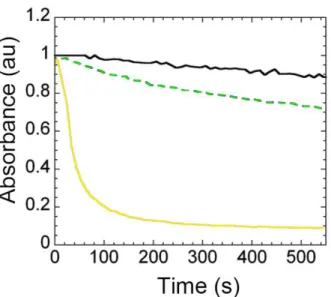
4.4.4 SPONTANEOUS CRYSTAL BLEACHING OVER TIME ... 170
4.5 CRYSTALLOGRAPHY ... 171
4.5.1 SOME CRYSTALLOGRAPHY CONCEPTS ... 171
4.5.2 MODEL BUILDING AND MODEL REFINEMENT OF FPS ... 172
4.5.3 KINETIC X-RAY CRYSTALLOGRAPHY ... 174
4.5.4 X-RAY DATA COLLECTION ... 175
4.6 SPECTROSCOPY ... 178
4.6.1 SPECTROSCOPIC SETUP ... 178
_______________________________________________________R
EFERENCES
__________________________________________________________A
NNEX
1
L
OW-
TEMPERATURE PHOTOINDUCED PROTONATION IN PHOTO-CHROMIC FLUORESCENT PROTEINS
.
F
ARO,
A
LINER
EGIS;
A
DAM,
V
IRGILE;
C
ARPENTIER,
P
HILIPPE;
D
ARNAULT,
C
LAUDINE;
B
OURGEOIS,
D
OMINIQUE;
DER
OSNY,
E
VE.
P
HOTOCHEMICAL&
P
HOTOBIOLOGICALS
CIENCES,
2008.
__________________________________________________________
A
NNEX
2
L
OW-
TEMPERATURE CHROMOPHORE ISOMERIZATION REVEALS THE PHOTOSWITCHING MECHANISM OF THE FLUORESCENT PROTEINP
ADRON.
F
ARO,
A
LINER
EGIS;
C
ARPENTIER,
P
HILIPPE;
J
ONASSON,
G
ABRIELLA;
P
OMPIDOR,
G
UILLAUME;
A
RCIZET,
D
ELPHINE;
D
EMACHY,
I
SABELLE;
FPs: Fluorescent Proteins;
YFP: Yellow Fluorescent Protein;
PTFPs: Photo-Transformable Fluorescent Proteins; RSFPs: Reversible Switchable Fluorescent Proteins; QC/MM: Quantum Chemical /Molecular Mechanics. PALM: PhotoActivated Localization Microscopy; STED: STimulated Emission Depletion;
RESOLFT: Reversible Saturable Optical Linear Fluorescence Transitions; OLID: Optical Lock-In Detection;
pcFRET: photochromic Förster Resonance Energy Transfer; SSIM: Saturated Structured Illumination Microscopy; EM: Eletron Miscrocopy;
ESPT: Excited State Proton Transfer;
L
IST OF FIGURES
1.1 Size comparison between GFP, E. coli cell and Fluorescent jellyfish ... 23
1.2 Unrooted tree for the four phyla in which homologues of GFP were found ... 24
1.3 The Nobel Prize in Chemistry 2008 ... 25
1.4 Schematic diagram of formation the chromophore in the GFP ... 27
1.5 Spectroscopic behavior of GFP... 29
1.6 Excited state proton transfer in the GFP ... 30
1.7 Chromophore of YFP ... 31 1.8 Scheme of RS-GFP model ... 33 1.9 Photo-Activatable FPs (PAFPs) ... 37 1.10 Photo-Convertible FPs (PCFPs) ... 38 1.11 Positively photo-switchable FPs ... 41 1.12 Negatively photo-switchable FPs... 44 1.13 STED microscopy ... 50
1.14 STED based on RESOLFT microscopy... 51
1.15 PALM microscopy ... 53
2.1 Outlook of eYFP subchapter. ... 59
2.2 Absorbance and fluorescence emission spectra of eYFP at room temperature. ... 60
2.3 Scheme of the protonated and deprotonated states of EYFP. ... 61
2.4 Comparison of the photoswitching cycle of IrisFP, Dronpa, eYFP at room temperature. 62 2.5 Photoswitching cycle of eYFP at 100 K following the fluorescence emission evolution. 65 2.6 Fluorescence emission spectrum during photoswitching cycle of eYFP at 100 K. ... 66
2.7 Time series of absorption spectra during photoswitching of eYFP at 100 K. ... 67
2.8 Low-temperature photoswitching of EYFP proceeds via a 1-photon absorption process. 70 2.9 Spectral evolution during temperature-driven back-switching of EYFP. ... 71
2.10 Spectral signatures of the different protonated forms of eYFP. ... 72
2.11 Comparison of spectral evolution of protonated state of eYFP. ... 73
2.12 Decay of anionic absorption peak of IrisFP, Dronpa and eYFP at 100 K. ... 75
2.13 Time series of absorption spectra during photoswitching of Dronpa at 100 K. ... 75
2.14 Time series of absorption spectra during photoswitching of IrisFP at 100 K. ... 76
2.15 Absorbance spectra of Padron flash cooled at 100 K. ... 77
2.16 Outlook of Padron subchapter . ... 79
2.17 Amino acid sequence alignment of Dronpa, Padron, Padron* and Padron0.9. ... 81
2.18 Contacts interface between different oligomers of Padron. ... 82
2.22 Padron positive photoswitching mechanism... 86
2.23 Crystal structure of Padron in fluorescent and non-fluorescent . ... 87
2.24 Superposition of Padron and Dronpa in the bright and in the dark states. ... 88
2.25 Scheme of the hypothetical deconvolution of Dronpa and Padron absorption spectrum. ... 89
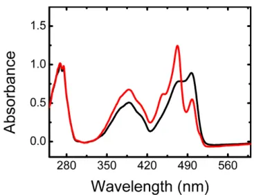
2.26 Absorbance spectra of Padron after few seconds of irradiation at 100 K. ... 92
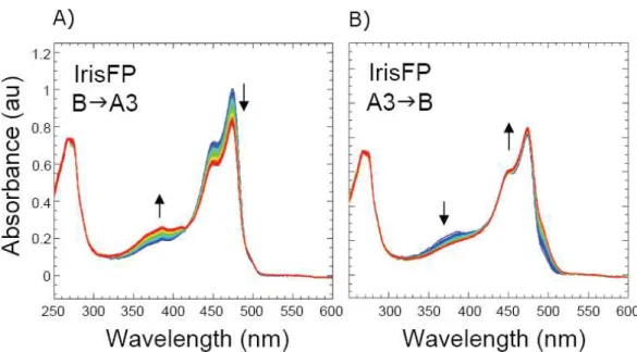
2.27 Spectroscopic signature of Padron along with its off-on photoswitching pathway, recorded in crystallo. ... 95
2.28 Absorbance spectra before and after actinic illumination at 100 K. ... 96
2.29 Crystal structures in states Btrans and after actinic illumination at 100 K Icis /Bcis,LT. ... 99
2.30 Other residues view of Crystal structures in states Btrans and after actinic illumination at 100 K Icis /Bcis,LT ... 99
2.31 Chromophore of Padron CYG and the dihedral angles and . ... 100
2.32 Crystal structure of Dronpa. ... 102
2.33 The average difference electron density map in the crystal structures in states Icis /Bcis,LT. ... 105
2.34 Photo-physical model of Padron photoswitching ... 107
2.35 Absorption spectra for the states Icis (blue line and Bcis,LT ... 110
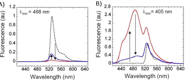
2.36 Evolution of the peak absorbance of Btrans and Icis during actinic illumination at 523 nm at 100 K. ... 111
2.37 Photoactivation at 488 nm of the crystalline sample of Padron at 100 K. ... 112
2.38 Evolution of Padron absorption spectrum during temperature increase... 114
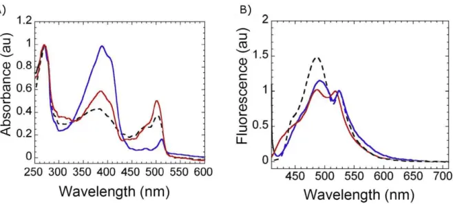
2.39 Spectroscopic signature of Padron along its off-on photoswitching pathway, recorded in solution samples. ... 116
2.40 Evolution of Icis to Bcis,LT and ABcis,LT thermally-induced by a temperature ramp from 100 K to 220 K in solution. ... 117
2.41 Arrhenius representation of the thermally-induced relaxation of Icis to Bcis,LT in solution. ... 118
2.42 Photoactivation of Padron occurs via a 1-photon absorption process at low to room temperature. ... 119
2.43 Comparison of Padron photo-activation from Btrans to Icis/Bcis,LT at different temperatures... 121
2.44 Fluorescence emission spectrum of Padron photoactivation at 488 nm at low temperature. ... 122
2.45 Scheme example of the overlapped spectra in Padron’s case. ... 123
2.46 Padron kinetic model emphasizing possible back-switching rates to Btrans. ... 124
2.48 In crystallo Padron photoswitching at cryo–temperature . ... 126
2.49 Simulation of Padron photoswitching of crystalline samples ... 127
2.50 Padron photo switching at cryo temperature.. ... 127
2.51 Simulation of Padron photoswitching of solution samples ... 128
2.52 Comparison of crystal structures of E222 (cyan and Q222 (green . ... 132
2.53 Illustration of the hypothetic chromophore twisting into GFP cavity. ... 133
2.54 EYFP structure: Wacther pdb, ph3.6, photoinduced. ... 135
2.55 Dreiklang structure. ... 137
2.56 Simulation of eYFP switch alternating between with and without illumination and varying the power intensity illumination. ... 141
2.57 Comparison between fluorescence emission collected upon continuous illumination and with interval without illumination... 142
2.58 Simulation of actinic illumination with different relaxation time. ... 143
2.59 Thermal relaxation recovery at room temperature of Citrine. ... 143
2.60 Simulation of eYFP photobleaching with different periods of illumination. ... 144
2.61 Simulation of eYFP photobleaching with different periods of illumination and relaxation time... 145
3.1 Digital letter (e-mail) written by Olsen sent for us.. ... 155
4.1 Outlook of material and methods. ... 159
4.2 pET-15b vector representation. ... 160
4.3 Scheme about the eYFP insertion into pET-15b. ... 161
4.4 Protein crystallization. ... 168
4.5 Strategy and the programs used in model building and refinement. ... 173
4.6 Microspectrophotometry setup. ... 179
4.7 Scheme of microspectrophotometry components. ... 179
4.8 Scheme of the light source in the Pixel laboratory. ... 180
Chapter 1
23
1.1 F
LUORESCENT
P
ROTEINS
:
WHO ARE THEY
?
Fluorescent proteins (FPs) are the homologous proteins of Green Fluorescent Protein (GFP), the first noticed fluorescent protein. The GFP was discovered in the mid 1970s, when O. Shimomura (Shimomura et al., 1962; Shimomura, 1995) took an interest in understanding the nature of the brightness of the jellyfish Aequorea Victoria, and isolated what he considered to be the source of the light. After four decades, this protein family continues to be extensively studied due to its qualities in the dyes world. Two factors are advantageous with FPs:
compared to synthetic dyes, the FPs have the advantage that they can be genetically
inserted in the organism of interest, providing non-invasive and specific labeling.
compared to other proteins that can also fluoresce, they do not require any cofactor except
molecular oxygen.
The structure responsible for light emission is the chromophore, p-hydroxybenzylideneimidazolinone (HBDI), positioned in the center of the protein scaffold (Figure 1.1).
Figure 1.1) Size comparison between (i) Cartoon representation of the Green Fluorescent
Protein (GFP) (A) top view and (B) in overall (the chromophore is represented in stick), (ii) E. coli cell and (iii) Fluorescent jellyfish.
1 INTRODUCTION
_________________________________________________________________________________________
24
1.1.1 F
LUORESCENT
P
ROTEINS IN NATURE
Homologues of GFP were identified in several marine organisms in the phylum Cnidaria, including Hydrozoa and reef-corals of the class Anthozoa (Matz et al., 1999; Labas et al., 2002). These proteins were also identified in evolutionarily distant species from Arthropoda (Shagin et al., 2004), Chordata (Deheyn et al., 2007; Li et al., 2009), and Ctenophora (Haddock et al., 2010) (Figure 1.2).
Figure 1.2) Unrooted tree for the four phyla, Cnidaria, Arthropoda, Chordata, Ctenophora, in
which homologues of GFP were found. Cnidaria phylum groups the two biggest classes, Anthozoa and Hydrozoa.
The biological function of fluorescent proteins remains unclear. It is possible that the GFP function is not necessarily associated with light emission, since non-fluorescent GFP homologues have been described in Hydrozoa and Anthozoa (Alieva et al., 2008; Gurskaya et al., 2003). Several authors proposed different hypotheses to explain the biological function of FPs, based on experimental evidences that are not completely accepted. Alieva et al have reported an evolutionary study using 40 different fluorescent proteins from reef corals (class Anthozoa) (Alieva et al., 2008). Based on a probabilistic sampling approach, they proposed that FPs play a biological role in the context of a symbiosis with other marine organisms, for example a photoprotection role (Field et al., 2006). An alternative explanation was given by Agmon and co-authors (Shinobu et al., 2010). They have published the high-resolution GFP
25 structure at 0.9 Å, where they have identified several proton wires including a proton-collecting apparatus. In their view, GFP works as a proton pump capable of direct light conversion into proton gradients. Lukyanov and co-authors (Bogdanov et al., 2009) have also suggested a biological function of GFPs. Upon 488 nm illumination in the presence of electron acceptors, GFP undergoes green-to-red photo conversion through a two-electron oxidation process. Based on oxidative redding of GFPs from diverse species, they suggested that the function of GFP is to induce a light-driven electron transfer. The three hypotheses described above are only some of the possibilities for the biological function of FPs. In addition, the possibility that FPs may act in more than only one biological role in nature is not excluded.
1.1.2
F
LUORESCENT
P
ROTEINS IN SCIENCE
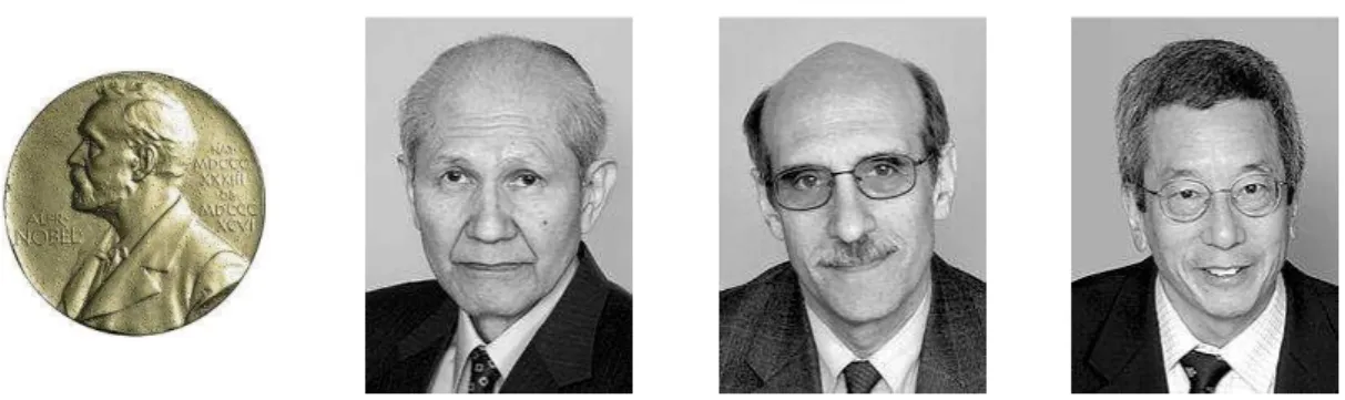
It is thanks to its scientific applications that FPs won their important reputation. The Nobel prize of Chemistry was awarded to Osamu Shimomura, Martin Chalfie and Roger Tsien in 2008 (Nobelprize.org, 2008) for the discovery and development of GFP and its variants (Figure 1.3). These proteins then quickly became widely used markers that allow the spatio-temporal tracking of the dynamic behavior of living systems at the molecular level (Ehrenberg, 2008).
Figure 1.3) The Nobel Prize in Chemistry 2008: Osamu Shimomura, Martin Chalfie, Roger
1 INTRODUCTION
_________________________________________________________________________________________
26
The wide rainbow of FP biosensors offers the possibility to perform multicolor imaging
experiments (Livet et al., 2007) and to develop pairs of dyes suitable for studies based on
Föster resonance energy transfer (FRET) (Chudakov et al., 2010). The development of red fluorescent proteins (RFP), triggered by the discovery of homologues GFP from Anthozoa, was an important improvement for applications in imaging living cells and tissues that require low spectral activity in UV-to-green spectral region (Subach et al., 2011a). The fluorescent proteins from Anthozoa also provide more sophisticated imaging techniques through the development of Photo-Transformable Fluorescent Proteins (PTFPs), which possess the advantage to display spectral properties that can be modulated by actinic illumination. Within the PTFPs group, photo-activatable fluorescent proteins (PAFPs) and photo-convertible fluorescent proteins (PCFPs) are particularly useful to achieve single molecule localization-based super resolution microscopy and to probe dynamic cellular events in living cells via pulse-chase imaging. For example, performing powerful variants of the fluorescence recovery after photobleaching techniques (FRAP). In this thesis, we have studied the photo-switchable fluorescent proteins (RSFPs). These proteins can switch between a fluorescent and a non-fluorescent form repeatedly. RSFPs are promising nano-devices for the development of rewritable data storage (Grotjohann et al., 2011). Moreover, they allow photochromic FRET and facilitate super-resolution microscopy techniques, such powerful variants of stimulated emission depletion (STED) microscopy (Hell and Wichmann, 1994; Klar et al., 2000; Willig et al., 2011) and structured illumination microscopy (SIM) (Rego et al., 2012). όurther details about these RSόP’s applications are described later in the text (see subchapter 1.4).
27
1.2 H
OW DO
F
LUORESCENT
P
ROTEINS
WORK
?
1.2.1
C
HROMOPHORE
F
ORMATION
The molecular structure of GFP is constituted of 230 amino acids, which adopt a secondary structure essentially made of -strands that fold into a nearly cylindrical -barrel, with 4.2 nm and 2.4 nm dimensions (Tsien, 1998) (Figure 1.1). Concomitantly with protein folding, a series of chemical reactions called maturation generate the chromophore that will be buried in the middle of the central helix and formed from the combination of three amino acids residues, Ser65-Tyr66-Gly67 (Figure 1.4).
Figure 1.4) Schematic diagram of the autocatalytic process of formation of the chromophore
from the amino acids Ser65-Tyr66-Gly67 in the GFP (Zhang et al., 2006).
The maturation of the chromophore is an autocatalytic process, where in the first step the cyclization of Gly and Ser is favored by the confinement inside of the -barrel. Kinetic studies of hydrogen peroxide release, combined with mass spectroscopy analysis, have revealed that the second and limiting step during in vitro GFP maturation is an oxidation step
1 INTRODUCTION
_________________________________________________________________________________________
28
(Zhang et al., 2006). The hydrogen peroxide is the product of the oxidation reaction that requires molecular oxygen to occur. During oxidation, a hydroxylated cyclic imine is formed (Pouwels et al., 2008). Finally, dehydration converts the imine to the double-bonded
imidazolinone ring yielding the fully conjugated mature chromophore. This step of reaction is probably assisted by the nature of Tyr66, since the mutation of this residue does not interfere with the cyclization or oxidation steps, but only with the dehydration (Zhang et al., 2006).
The maturation of the chromophore is a common process between all fluorescent proteins that may vary from hours to a few days depending on FP. Indeed, depending on the group of proteins other steps occur, as Verkhusha and other authors have demonstrated recently (Bravaya et al., 2012). In particular, they have shown that red-variants of fluorescent proteins, such as DsRed, TagRFP, fluorescent timers and PAmCherry, pass by a blue anionic intermediate structure, through a single oxidation step, before the mature chromophore is formed.
Once synthesized, the mature chromophore is relatively well shielded from the environment by the -barrel. It is stabilized by a complex network of electrostatic, H-bonding, van der Waals or stacking interactions with its surrounding residues. This stabilization of the chromophore is strongly responsible for the high fluorescence emission. Due to this, the chromophoric moiety in the absence of the -barrel does not fluoresce under normal conditions (Niwa et al., 1996). Similarly, the denaturation product of fluorescent proteins is not fluorescent although FPs recover their fluorescence when they refold (Ward and Bokman, 1982).
Several residues have a central importance in both the maturation process and the ability to fluoresce. In the chromophore triad, only the amino-acid Gly67 is essential to ensure maturation. Tyr66 can be mutated to a tryptophan or to a histidine, giving origin to two other
29 fluorescent proteins, the cyan variant (CFP) and the blue variant (BFP) (Tsien, 1998). The GFP brightness is improved ≈ 10 times upon excitation at 488 nm, when Ser65 is mutated into a threonine (Heim et al., 1995). The F64L is known to improve the folding efficiency, which is advantageous for living cell experiments (Cormack et al., 1996). The S65T and/or F64L mutations in the fluorescent proteins from Aequorea victoria (AFPs) yields the
enhanced fluorescent proteins (eGFP, eBFP, eCFP and eYFP). Other residues are
fundamental in changing the behavior of GFP, such as E222 and S205 and their roles will be described later in the text.
1.2.2
S
PECTROSCOPIC BEHAVIOR OF
GFP
S
The GFP absorption spectrum is constituted of two broad bands: one band with a maximum at ~ 398 nm and another with a maximum at ~ 478 nm (Heim et al., 1994, 1995; Tsien, 1998). At physiological pH, the ratio between these bands is six to one. The 398 nm band is associated to the protonated form of the chromophore (A form). The 478 nm band is associated with the anionic form of chromophore (B form). The chromophore in its anionic conformation is highly fluorescent. It exhibits a strong emission peak at 503 nm. The protonated chromophore is intrinsically weakly fluorescent. However, when it is excited near its absorption maximum, it exhibits an emission peak close to the B fluorescence peak (Heim et al., 1994).
1 INTRODUCTION
_________________________________________________________________________________________
30
1.2.3
E
XCITED
S
TATE
P
ROTON
T
RANSFER
(ESPT)
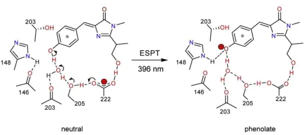
Boxer and co-worker in 1996 were the first to explain why excitation of the protonated band produces a fluorescence emission similar to that of the anionic band (Chattoraj et al., 1996). Their hypotheses are based on the analysis of steady-state absorbance and fluorescence emission spectra and also on ultrafast time-resolved fluorescence excitation with and without deuterium isotopic labeling at low temperatures. The phenomenon is being caused by a proton transfer occurring in the excited state (ESPT) between the hydroxybenzylidene moiety of the chromophore and the nearby H-bond network, leaving the chromophore in an anionic intermediate I state which is structurally close to A and spectroscopically close to B (Fang et al., 2009) (Figure 1.6).
Figure 1.6) Excited state proton transfer between hydroxybenzylidene moiety of the
chromophore and the nearby H-bond network in the GFP.
This first mention of ESPT was done without knowing the precise molecular structure of GFP. Some months later, two different teams published the first molecular structure of GFP (Ormo, 1996; Yang et al., 1996). Together with the GFP structure, the Tsien group also introduced a yellow variant of eGFP, called eYFP (Ormo, 1996) .
31
1.3 R
EVERSIBLE
P
HOTOSWITCHING OVER THE
YEARS
1.3.1 P
HOTOSWITCHING AT THE SINGLE MOLECULES LEVEL
The first time that the reversible photoswitching of a fluorescent protein was described in the literature was through single molecules experiments on YFP, done by Dickson et al in 1997 (Dickson et al., 1997). At physiological pH, the YFP absorption spectrum displays only a deprotonated band at 514 nm corresponding to the anionic chromophore (B form). The YFPa differs from GFP by three amino acid residues S65G/T203Y/S72A (Figure 1.7). Thehighly polarizable phenol of Tyr203 assumes an almost coplanar – stacking with the
chromophore allowing a red shift of ≈ 20 nm of the fluorescence emission ( em = 527 nm)
(Wachter et al., 1998).
Figure 1.7) Chromophore of YFP (PDB: 1YFP) (Wachter et al., 1998).
Dickson et al. showed that single immobilized YFP proteins are light-driven to a long-lived non-fluorescent state (off-state) after long exposure ( 106 photons) at 488 nm. The
protein can recover its emission (on-state) after some minutes in the dark, or upon illumination at 405 nm. This means that, at the single molecule level, the on-off switching cycles can be repeated in a controlled way by alternation of illumination with 488 nm and
a YFP used in Dickson (Dickson et al., 1997) et al is a variant of the YFP 10C described in Wachter et al
1 INTRODUCTION
_________________________________________________________________________________________
32
405 nm light. The photoswitching was also demonstrated for the variant S65G/S72A/T203F (denoted T203F) of GFP, but it exhibits a less efficient activation/inactivation of the fluorescence than YFP (Dickson et al., 1997).
In this work the authors discuss the blinking: another process that also involves the reversible transformation from a fluorescent state to the non-fluorescent state. In contrast to the photoswitching, photoblinking is the stochastic fluctuation of the fluorescence, is essentially uncontrolled, and takes place on a much faster timescale than the switching. Blinking can be observed upon continuous illumination with a single wavelength, reminiscent of spectral and amplitude fluctuation, noticed also in other fluorophores, at the single molecule level (Lu and Xie, 1997).
1.3.2
P
HOTOSWITCHING AT ENSEMBLES OF MOLECULES LEVEL
i.
Photoswitching at low and ultra-low temperature
On and off photoswitching in fluorescent proteins at the ensemble level was observed at ultra-low temperature on GFP and some red-shifted derivatives, using hole-burning spectroscopy (Creemers et al., 1999, 2000; Creemers, TM H Lock et al., 2002). Many of the thermally induced conversions are blocked at low temperature and, therefore, discrimination amongst individual species is facilitated (Creemers et al., 2000). The structured spectra obtained at low temperature allow to identify the protonated (A), the anionic (B) and the intermediate (I) states and to propose the pathway of photo-conversion between them.
The intermediate state I between B and A states, proposed to explain the ESPT, was first observed for GFP. It was also found in the photo-mechanism of red-shifted derivatives: RS-GFP (F64M, S65G and Q69L) and YFP (S65G, V68L, S72A and T203Y). Summarizing,
33
the authors suggest that illumination at the anionic excitation peak (≈ 488 nm) can excite the
B and I forms. When excited, B can fluoresce or eventually be converted to the I state. In turn, if the I state is excited it can fluoresce or be converted to the B or A forms. At wavelengths
less than the protonated excitation peak maximum (≈ 44θ nm), the A state can be exited to
fluoresce or be re-converted to the I form. The reaction can be described by:
As shown in Figure 1.7, RS-GFPb exhibits an excitation peak at 495 nm (blue line)
and an emission peak at 503 nm (red line). These bands are ascribed to the intermediate state I due to its spectroscopic similarities with that of GFP. Upon illumination at 495 nm, we observe the intermediate state reduction with concomitantly an increase of the B and A states and a reduction of the fluorescence (Figure 1.8a). Upon illumination at 434 nm, the A-peak decreases whereas the I-peak increases (not shown). Subsequent illumination into I increases the population of A again (Figure 1.8b).
Figure 1.8) Scheme of RS-GFP model at 1.6 K. Reprinted from (Creemers, TM H Lock et al.,
2002). (A) Interconversion from I to A and B upon illumination 495 nm with concomitantly reduction of fluorescence. (B) Decrease of the A band upon illumination 434 nm and subsequent increase upon illumination at 495 nm.
b In the paper Creemers and co-authors have described RS-GFP as Red Shifted-GFP (Delagrave et al., 1995), not
to be confounded with rs-GFP published recently (Grotjohann et al., 2011).
A
k1I
k2B
1 INTRODUCTION
_________________________________________________________________________________________
34
The approach based on low temperature spectroscopy, used to decipher the reaction pathway in these works (Creemers et al., 1999, 2000; Creemers, TM H Lock et al., 2002), is close with the approach that will be used in the Results chapter of this Thesis (see Chapters 2.1 and 2.2).
ii.
Partial photoswitching at room temperature
Miyawaki and Tsien have observed that in cells expressing eYFP and in pure protein solutions after bleaching at 540 nm, it is possible to recover partially the fluorescence. About 20% of the initial fluorescence is retrieved by illuminating at 340 nm at room temperature (Miyawaki and Tsien, 2000). Furthermore, the recovery can be induced by thermal relaxation, as well as by actinic illumination. However, in these bulk experiments, photoswitching is not induced with the same illumination scheme as in single-molecule experiments. Surprisingly, illumination at 405 nm does not recover the fluorescence, in contrast, it induces further decay (McAnaney et al., 2005; Sinnecker et al., 2005).
Based on stopped-flow, pressure-jump and time-resolved experiments, McAnaney et al. have detailed the kinetics of protonation, photobleaching and photoactivation in YFP 10C (McAnaney et al., 2005). At room temperature, they observed only a weak recovery of the fluorescence, similar to the observation by Miyawaki and Tsien (Miyawaki and Tsien, 2000). They described two absorption peaks at 340/460 nm that would be responsible for fluorescence recovery after illumination, and differ from the protonated peak at 390 nm. In the same year, another observation of photo-switching upon illumination at various wavelengths of samples of eYFP, citrine, CFP and GFP, was described in Sinnecker et al. They showed that a dynamical equilibrium between a reversibly and a non-reversibly photo-bleached population is established after prolonged exposure followed by thermal relaxation (Sinnecker et al., 2005).
35 The common feature of all these processes is the fact that photoswitching in these GFP derivatives is only weakly reversible, 10 – 40% depending on the experimental conditions. A possible interpretation for these results would be the presence of non-interchangeable populations in the photo-pathway of these proteins: one reversibly photoswitchable and the other not. For example, a first anionic fluorescent state, B1, could undergo reversible
switching to a weak or non-fluorescent state A, and concomitantly another B2 anionic state
would only be able to undergo irreversible bleaching to a dark state, D.
The structural mechanisms that could be responsible for these effects are not clear. In all cases, protonation of the chromophore can be observed by inspecting the absorption spectrum. However, the sole protonation cannot explain the long-lived dark state, and this process needs to couple to at least a minimal structural rearrangement (McAnaney et al., 2005). Isomerization of the chromophore has been hypothesized as being the responsible structural rearrangement causing this effect (Weber et al., 1λλλν σifosı et al., β00γ). Although Raman results have showed spectroscopic signatures suggesting such cis-trans isomerization in other variants of YFP (EYQ1) (Luin et al., 2009), further evidence is missing to understand the partial photo-switching. To date, for AFPs, there are no structural results showing isomerization of the chromophore (trans conformation). It is, however, a common consensus that the matrix of the chromophore should play a fundamental role in switching mechanisms (Follenius-Wund et al., 2003; Maddalo and Zimmer, 2006; Fang et al., 2009).
k1 k2
k-1
B
2D
1 INTRODUCTION
_________________________________________________________________________________________
36
1.3.3
D
ISCOVERY OF THE
F
LUORESCENT
P
ROTEINS FROM
C
ORALS
In 1999 Lukyanov and co-authors announced the discovery of GFP homologues isolated from reef corals (Matz et al., 1999). Although these proteins display a -barrel tertiary structure similar to GFP, the new chromophore and amino acid interactions not only extend the existing FP color palette, but also reveal unprecedented photo-physical properties (Lukyanov et al., 2000). These new fluorescent proteins are “photo-transformable” (PTFPs): proper actinic illumination can modulate their fluorescence behavior. This property introduces unique possibilities for precision labeling and tracking of objects of interest in living systems, enhancement of signal-to-noise ratio, and, most importantly, super-resolution fluorescence imaging (Chudakov et al., 2010). Because of these unprecedented developments, anthozoan PTFPs led to a second revolution in the FP field. They can be classified in three groups: Photo-Activatable Fluorescent Proteins (PAFPs) and Photo-Convertible (PCFPs) exhibit non-reversible photo-transformations, whereas Photo-Switchable Fluorescent Proteins (RSFPs), our objet of study, display reversible photoswitching between a fluorescent on state and a non-fluorescent off state.
i. Photo-Activatable FPs (PAFPs)
Photo-Activatable FPs are initially in a non-fluorescent state and are transformed into a fluorescent state upon actinic illumination with violet light (405 nm). The FPs belonging to this group are: PAmRFP1 (Verkhusha and Sorkin, 2005), PA-GFPc (Patterson and
Lippincott-Schwartz, 2002), PA-mCherry (Subach et al., 2010a), PS-CFP, PS-CFP2 (Chudakov et al., 2004), and PA-TagRFP (Subach et al., 2010b).Typically, PAFPs, before illumination, are in a protonated state and are converted to an anionic state upon relatively strong UV- illumination.
37 Attenuated photochromatic behavior is also observed in WT-GFP and it is potentialized in PA-GFP with the T203H substitution, that affects the side chain conformation of Glu222 (Figure 1.9). Glu222 is rotated away from His203 and occupies a slightly different position than that in WT (Henderson et al., 2009). Both mass spectroscopy and crystallographic data suggest that photo-activation in PA-GFP is caused by oxidative decarboxylation of E222 driven by high-energy UV light (Henderson et al., 2009). This is followed by a reorganization of a H-bond network, resulting in favoring the anionic state of the chromophore. A similar decarboxylation reaction is observed in PA-mCherry (Subach et al., 2010a), and in PS-CFPs (Chudakov et al., 2004). The relative efficiencies of decarboxylation are explained in terms of the Kolbe-type mechanism in which the excited state of the chromophore acts as an oxidant by accepting an electron from E222 (Bell et al., 2003). Thanks to their ability to be irreversibly activated, PAFPs are appropriate for tracking experiments in the cell (Chudakov et al., 2010).
Figure 1.9) Photo-Activatable FPs (PAFPs). Irreversible photo-transformation from the
non-fluorescent protonated state to the non-fluorescent anionic state of chromophore based on oxidative decarboxylation of Glu222 upon illumination with high-energy UV light. Representative spectra of this class are based on PA-GFP photo-activation, excitation (solid lines) and emission (dashed lines).
1 INTRODUCTION
_________________________________________________________________________________________
38
ii.
Photo-Convertible FPs
Photo-Convertible FPs are initially in a first, typically green, fluorescent state and upon actinic illumination are irreversibly converted to another state, typically red, fluorescent state (Figure 1.10).
Figure 1.10) Photo-Convertible FPs (PCFPs). Irreversible photo-transformation from the
green fluorescent anionic state to the red fluorescent anionic state of chromophore based on backbone cleavage, via a -elimination reaction, between σ and ω of His62 upon illumination with high-energy UV light. Representative spectra of this class are based on Kaede photo-conversion, excitation (solid lines) and emission (dashed lines).
The FPs belonging to this group are: Kaede (Matz et al., 1999), EosFP (Wiedenmann et al., 2004), mEosFP (Gurskaya et al., 2006), mEos2 (Mckinney et al., 2009), KikGR (Tsutsui et al., 2005), Dendra2 (Gurskaya et al., 2006), IrisFP (Adam et al., 2008, 2011), Nijid (Adam
et al., 2011), PSmOrange (Subach et al., 2011b). Kaede was the first fluorescent protein to exhibit this green to red photo-conversion property upon illumination with UV-violet light (Matz et al., 1999). To date the conversion was observed typically from green to red, but it was observed also from cyan to green (PS-CFP2) (Evrogen, 2007). The mechanism
d Iris and Niji FPs are a special characteristic to posses both photo-conversion and photoswitching behavior
39 accounting for photo-conversion is based on protein backbone cleavage, via a -elimination reaction, between σ and ω of Hisθβ (Mizuno et al., 2003; Wiedenmann and Nienhaus, 2006; Lelimousin et al., 2009), that follows from photon absorption in the protonated state of the chromophore. This irreversible peptide cleavage results in a subsequent extension of the chromophoric conjugated electron system, inducing a red-shifted emission peak.
iii.
Reversibly-Photoswitchable Fluorescent Proteins (RSFPs)
Reversibly-Photoswitchable Fluorescent Proteins (RSFPs) are initially in a fluorescent/fluorescent state, and upon illumination they are reversibly switched to a non-fluorescent/fluorescent state. The FPs belonging to this group and some of their photo-physical properties are shown in Table 1.1.
RSFPs that switch to the non-fluorescent state upon illumination in the absorbance
band of the fluorescent state are called “negatively switchable”μ they show a decrease of
fluorescence upon excitation. Dronpa is the most well-known representative of this subclass. In contrast, RSFPs that switch to the fluorescent state upon illumination in the absorbance band of the fluorescent state are called “positively switchable”μ they show an increase of fluorescence upon excitation. Well-known members of this subclass are asFP595 and Padron. RSFPs have representative proteins in both Hydrozoan and Anthozoan classes.
Several molecular structures have suggested that the structural mechanism accounting for the photoswitching is a cis-trans isomerization of the chromophore. Spectroscopically, photoswitching manifests itself by a light induced interconversion between the anionic and the protonated absorption bands. However, recently, a completely different mechanism than coupled chromophore isomerization and protonation was observed for a new RSFP, named Dreiklang. In the following, we detail further each RSFP sub-class by reviewing asFP595 and Dronpa.
1 INTRODUCTION
_________________________________________________________________________________________
40
Table 1.1) Properties of principal RSFPs developed to date.
*mutation M159A; †other structures of Dronpa: 2IVO, 2IE2, 2GXO, 2GX2, 2POX, 2Z1O, 2Z6X, 2Z6Z; other structures of asFP595: 2A50, 2A52, 2A53; not deposited in PDB (see Material and Methods Chapter 4.5.4). Chromophore triad (Chr. triad), excitation and emission wavelengths ( exc/ em), extinction coefficient ( ), fluorescence quantum yield ( fluo), photoswitching quantum yield ( sw), References (Ref): (1) (Henderson et al., 2007); (2) (Ando et al., 2004; Andresen et al., 2007); (3, 4) (Stiel et al., 2007); (5, 7, 8, 9, 16, 17, 18) (Adam et al., 2011); (6, 15) (Adam et al., 2008); (7)(Fuchs et al., 2010); (10, 12) (Bizzarri et al., 2010); (11) (Grotjohann et al., 2011); (13, 20) (Bogdanov et al., 2009); (14) (Subach et al., 2010c); (19) (Brakemann et al., 2010); (21) (Lukyanov et al., 2000); (22) (Chudakov et al., 2003a); (23) (Brakemann et al., 2011).
________________________________________________________________________________________________________________________________________________________
1.3.4
P
OSITIVE PHOTOSWITCHING
:
AS
FP595
The fluorescent protein asFP595 from Anemonia sulcata was discovered in 2000, in the course of investigations to find homologues of GFP in organisms other than Hydrozoans (Lukyanov et al., 2000; Andresen et al., 2005). Amongst the fluorescent proteins from reef corals, asFP595 was the first observed to exhibit reversible photoswitching (Chudakov et al., 2003b). Upon actinic illumination at 568 nm, asFP595 was observed to dramatically increase its fluorescence (Chudakov et al., 2003b). Due to this property it was called a “kindling
Protein (Class) - Chr. Struct. Strct. exc/em fluo pKa sw sw Actinic Ref
Source organism triad (on ) (off ) (M-1cm-1) (on off ) (off on) Ligh
mTFP0.7 (A) - Clavularia sp. AYG 2OTB 2OTE 453 / 488 60 000 0.50 4.0 ND ND 458/405 1
Dronpa (A) - Echinophy sp. CYG 2Z6Z † 2POX 503 / 517 94 100 0.67 5.3 3.0 X 10 -4 7.0 X 10 -1 458/405 2
rsFastLime (A) - Echinophy sp. CYG ND ND 496 / 518 39 094 0.77 ND ND ND 458/405 3
bsDronpa (A) - Echinophy sp. CYG ND ND 460 / 504 45 000 0.50 ND ND ND 458/405 4
mEosFP * (A) - L. Hemprichii HYG ND ND 487 / 512 98 600 0.52 4.3 2.6 X 10 -3 1.5 X 10 -1 458/405 5
Iris (A) - L. Hemprichii HYG 2VVH 2VVI 488 / 516 57 800 0.48 5.7 3.2 X 10 -3 1.5 X 10 -1 458/405 6 mIrisFP (A) - L. Hemprichii HYG ND ND 486 / 516 74 000 0.60 5.7 2.2 X 10 -3 1.5 X 10 -1 458/405 7 NijiFP (A) - Dendronephthya sp. HYG ND ND 469 / 507 41 100 0.64 7.0 1.8 X 10 -3 1.5 X 10 -1 458/405 8
Drenda2 * (A) - Dendronephthya sp. HYG ND ND 471 / 504 51 100 0.55 6.5 1.1 X 10 -3 8.0 X 10 -2 458/405 9
Mut2q (H) - A. victoria AYG ND ND 496 / 507 54 000 0.28 6.0 4.7 X 10 -3 2.6 X 10 -2 478/405 10
rsGFP (H) - A. victoria TYG ND ND 493 / 510 47 000 0.36 6.5 ND ND 458/405 11
EYQ1 (H) - A. victoria SYG ND% ND 510 / 524 73 000 0.72 6.9 1.8 X 10 -4 6.0 X 10 -2 514/405 12
rsCherryRes (A) - Dicosoma sp. MYG ND ND 572 / 608 84 000 0.01 5.5 ND ND 550/450 13
rsTagRFP (A) - E. quadricolor MYG 3U8C 3U8A 567 / 585 36 800 0.11 6.6 ND ND 570/445 14 Iris (A) - L. Hemprichii HYG 2VVJ ND 551 / 580 27 000 0.50 6.8 2.0 X 10 -3 5.0 X 10 -2 561/440 15
mIrisFP (A) - L. Hemprichii HYG ND ND 546 / 578 26 000 0.44 7.0 4.0 X 10 -4 1.5 X 10 -1 561/440 16
NijiFP (A) - Dendronephthya sp. HYG ND ND 526 / 569 42 000 0.65 7.3 1.0 X 10 -3 1.5 X 10 -1 561/440 17
Drenda2 * (A) - Dendronephthya sp. HYG ND ND 528 / 562 45 000 0.75 6.8 3.2 X 10 -3 1.0 X 10 -2 561/440 18 Padron (A) - Echinophyllia sp. CYG 3LS3 3LSA 503 / 522 43 000 0.64 ND ND ND 405/488 19
rsCherry (A) - Dicosoma sp. MYG ND ND 572 / 610 80 000 0.02 6.0 ND ND 450/550 20
asFP595 (A) - A. sulcata MYG 2A56‡ 2A53 572 / 595 56 200 0.001 ND ND ND 450/568 21
KFP1 (A) - A. sulcata MYG ND ND 590 / 600 59 000 0.07 ND ND ND 458/532 22 Dreiklang (H) - A. victoria GYG 3ST4 (2) 3ST3 511 / 529 83 000 0.41 7.2 ND ND 405/365 23
Negative swicthing
Positive swicthing
41
fluorescent protein” (Figure 1.11). The kindling phenomenon can be slowly reversed by
thermal relaxation or instantly by irradiation with blue-light (Chudakov et al., 2003b).
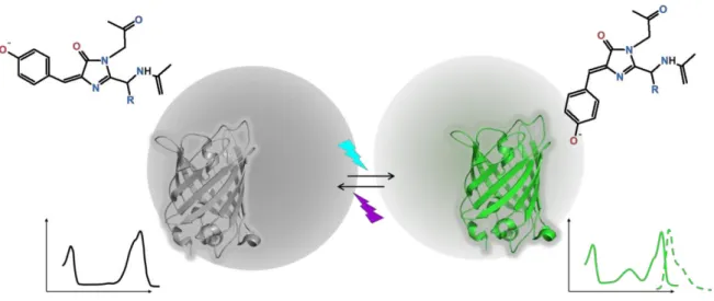
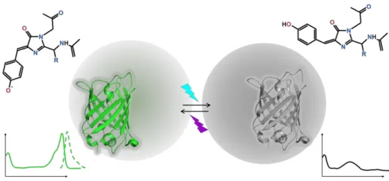
Figure 1.11) Positively photo-switchable FPs. Reversible photo-transformation from the
non-fluorescent state to the non-fluorescent state of chromophore based on trans-cis isomerization of the chromophore upon illumination at 488 nm to switch on and at 405 nm to switch off. Representative spectra of this class are based on Padron photo-switching, excitation (solid lines) and emission (dashed lines).
The first application in fluorescence microscopy taking advantage of reversible switching employed asFP595 to introduce a point-scanning technique relatively similar to STED, and called RESOLFT (REversible Saturable Optical Linear Fluorescence Transitions) (Hofmann et al., 2005). However, asFP595 has a very weak fluorescence quantum yield (<0.001) and it is an obligate tetramer, which considerably limits its practical use for biological applications.
AsFP595 has been subjected to a number of mechanistic investigations. Like for AFPs, a cis-trans isomerization of the chromophore was soon proposed to account for kindling and back-relaxation to the dark state. The cis (fluorescent) to trans (non-fluorescent) isomerization of the chromophore was confirmed by X-ray crystallography for the wild-type
1 INTRODUCTION
_________________________________________________________________________________________
42
asFP595, as well for the variants S158V (Andresen et al., 2005) and asFP595-A143S (designated KFP) (Andresen et al., 2005; Quillin et al., 2005; Wilmann et al., 2005). The single mutation A143S confers to the protein a long-lived bright state and an enhancement of the fluorescence quantum yield, likely because the side chain of Ser143 stabilizes the cis conformation of the chromophore (Quillin et al., 2005).
Based on molecular dynamics calculations, a Hula-Twist motion was suggested as the isomerization mechanism (Andresen et al., 2005). Unfavorable steric clashes were predicted for simple one-bond rotations of the chromophore (Quillin et al., 2005). Contrary to negative RSFPs, only very limited rearrangements of the chromophore pocket were noticed between the trans and the cis configurations, consistent with the observation of kindling at 150 K, below the glass transition temperature of solvent (Schüttrigkeit, 2006).
Schäfer and co-authors have studied how the chromophore protonation state controls photoswitching in asFP595 by performing ab initio calculations and QC/MM molecular dynamics simulations (Schäfer et al., 2007, 2008). It was suggested that cis-trans isomerization occurs in the neutral state of the chromophore, followed by a dark state equilibration to a zwitterionic fluorescent cis state (Schäfer et al., 2008). However, this study did not rule out the possibility that the chromophore may also photo-isomerize in its anionic state.
Other fluorescent proteins exhibit positive photoswitching, for example rsCherry and Padron. RsCherry (and its negative-switching counterpart rsCherryRev) was rationally engineered from the known structure of mCherry (Stiel et al., 2008). It emits at 610 nm and can be switched on/off using blue/yellow illumination, respectively. In contrast with asFP595, rsCherry is monomeric, extending possible applications using red-RSFPs.
43
i.
Padron
Padron was introduced by Andresen et al. in 2008 as being the positive switching counterpart of Dronpa (Andresen et al., 2008). Padron was generated by site-directed saturation mutagenesis of rsFastLime (Dronpa: Val157Gly) at eight positions. Notably, the sole exchange of the Met residue at the position 159 to a Tyr residue is sufficient to confer the positive switching mechanism (Andresen et al., 2008). The scientific interest of this protein stems from the fact that, together with Dronpa, Padron allows the implementation of, multi-label far-field fluorescence nanoscopy (Andresen et al., 2008; Willig et al., 2011). Recently, dual-label monochromatic STED nanoscopy of living cells using Padron and Dronpa reached
70 nm (Padron) and 90 nm (Dronpa) spatial resolution (Willig et al., 2011). The positive
photoswitching mechanism of Padron will be extensively studied in the Results Chapter, leading to the central results of this thesis work (see Results and Discussion Chapter 2.2).
1.3.5
N
EGATIVE PHOTOSWITCHING
:
D
RONPA
Dronpa is a mutant of fluorescent protein cloned from a coral Pectiniidae. This RSFPs satisfies most requirements for being a suitable dye for imaging applications: it is monomeric, displays a high fluorescence quantum yield (0.67 compared to 0.001 for asFP595), has a high extinction coefficient (94100 cm-1M-1 versus 56200 cm-1M-1 for asFP595), exhibits a good on-off contrast of the fluorescence (≈ κ0%) and shows a moderate photoswitching quantum yield of 10-4e (Habuchi et al., 2005). The ensemble of studies carried out with Dronpa forms the basis of our present knowledge of the RSόP’s mechanisms (Bourgeois and Adam, 2012).