HAL Id: tel-02426208
https://tel.archives-ouvertes.fr/tel-02426208
Submitted on 2 Jan 2020HAL is a multi-disciplinary open access archive for the deposit and dissemination of sci-entific research documents, whether they are pub-lished or not. The documents may come from teaching and research institutions in France or abroad, or from public or private research centers.
L’archive ouverte pluridisciplinaire HAL, est destinée au dépôt et à la diffusion de documents scientifiques de niveau recherche, publiés ou non, émanant des établissements d’enseignement et de recherche français ou étrangers, des laboratoires publics ou privés.
Lipid Flippases from Plasmodium Parasites : from
Heterologous Production towards Functional
Characterization
Anaïs Lamy
To cite this version:
Anaïs Lamy. Lipid Flippases from Plasmodium Parasites : from Heterologous Production towards Functional Characterization. Biochemistry, Molecular Biology. Université Paris-Saclay, 2018. English. �NNT : 2018SACLS447�. �tel-02426208�
Lipid flippases of Plasmodium parasites:
from heterologous production towards
functional characterization
Thèse de doctorat de l'Université Paris-Saclay
Préparée à l’Université Paris-Sud
Et à l’I2BC, CEA-Saclay École doctorale n°568
Signalisations et réseaux intégratifs en biologie (Biosigne) Aspects Moléculaires et Cellulaires de la Biologie
Thèse présentée et soutenue à Gif-sur-Yvette, le 23/11/2018, par
Anaïs Lamy
Composition du Jury : Philippe Minard
Professeur, Université Paris-Sud (UMR 9198) Président
Bruno Miroux
Directeur de Recherche, INSERM (UMR 7099) Rapporteur
Renaud Wagner
Ingénieur de recherche (hors classe), CNRS (UMR 7242) Rapporteur
Isabelle Florent
Professeure, MNHN (UMR 7245) Examinatrice
Rosa Laura López-Marqués
Associate Professor, University of Copenhagen Examinatrice
José Luis Vázquez-Ibar
Chargé de Recherche, CNRS (UMR 9198) Directeur de thèse
NNT : 20 18 S A C LS 44 7
Remerciements
Je voudrais tout d’abord remercier l’école doctorale Biosigne pour m’avoir accordé un financement de thèse.
Je veux remercier également les membres de mon jury pour avoir accepter d’être présent et pour leur bienveillance.
Je tiens à remercier Francis Haraux pour m’avoir accueillie au sein du laboratoire et pour sa super affiche pour ma soutenance de thèse !
Je voudrais remercier tout particulièrement mon directeur de thèse José Luis Vázquez-Ibar pour sa patience et son soutien durant ces 3 années. Pour avoir supporté mon caractère un peu « bitchy » à certains moments, mes longues phases de déprime et de m’avoir dopé à la charcuterie espagnole ! Et surtout pour son optimisme (qui n’a pas déteint sur moi malheureusement !). Je remercie également Christine Jaxel, ma co-directrice de thèse, pour sa gentillesse, son écoute, nos discussions scientifiques ou personnelles, son aide précieuse lors de la rédaction et les chocolats anti-déprime.
Je voudrais remercier Cédric Montigny et Guillaume Lenoir pour leurs conseils et leur aide pour les manips , et les moments passés tous ensemble lors des déjeuners.
Je remercie Thibaud « Titi » Dieudonné pour son aide sur les manips surtout sur les derniers moments de cette thèse, pour les fous rire, les concours de qui fera le pus peur à l’autre en sortie de la pièce de culture et les partages de vidéos ridicules. Je te souhaite une bonne fin de thèse (courage, le pire reste à venir), et on se revoit pour ta soutenance.
Je remercie Maylis Lejeune qui comme moi est restée 3 années au labo pour son apprentissage et qui a brillamment réussi son master. Je la remercie pour sa bonne humeur et pour les pauses thé. Sa voix douce et le son feutré de ses pas dans le couloir vont me manquer ☺ !
Je remercie Valentine « Valentaïne » Guinot pour sa gentillesse et sa bonne humeur. Et je lui souhaite le meilleur pour la suite.
Je remercie Thomas Barbot et le félicite pour sa thèse obtenue quelques mois plus tôt. Merci pour les marrades au labo mais je n’ai pas oublié le vieux spoil tout pourri de Game of Thrones, mon cœur en est meurtri à jamais …
Je tiens également à remercier Marc le Maire pour sa gentillesse, ses conseils et pour m’avoir accueillie lors du concours de l’ED et pour les partages de connaissances sur les loutres.
Je remercie Phillipe Champeil d’avoir partagé son bureau avec moi et d’avoir participer à la relecture de cette thèse sur son temps de retraité et de papi.
Les autres membres du LPSM, Manuel Garrigos, pour nos discussions et nos échanges de film et série ! Nadège Jamin, Véronica Beswick, Claire Lemaire, Stéphane Orlowski, Michel Roux, Anthony Rignani, et Ewerton Bruzaferro qui a fait son M2 avec nous et nous a aidé sur le projet.
Les deux personnes sans qui on est vite perdu au labo (en tout cas moi je l’étais) ; Pascale Filoche qui s’occupe de nous et de nos commandes et Josiane Piles qui faisait en sorte qu’on ait toujours notre matériel propre et à disposition. Surtout merci pour votre gentillesse et votre bienveillance envers moi.
Merci à tous pour votre gentillesse, pour l’ambiance au laboratoire sérieuse et moins sérieuse, Pour être venu lors de mes différentes répétitions, pour avoir jouer le jeu lors des repas de Noël et pour les anniversaires.
Je remercie également toutes les personnes des autres labos du cea ou I2BC que j’ai pu rencontrer durant ma thèse.
Je remercie Sandrine Lecart de la plateforme Imagerie-gif de l’I2BC, qui m’a aidé et conseillé pour les images de microscopie confocale.
Je remercie toutes les personnes que j’ai pu rencontrer lors des deux années en tant qu’enseignante à l’Université Paris-Sud, particulièrement Aurélie Hua-Van, Marielle Valerio-Lepiniec, Sylvie Nessler et Agathe Urvoas.
Je remercie aussi les personnes que j’ai rencontré lors de mes différents stages en Licence et Master, l’équipe « Mollicutes » à l’INRA de Bordeaux et l’équipe « Métabolisme intermédiaire des Trypanosomes » au CNRS de Bordeaux, qui m’ont donné envie de continuer en thèse et faire de la recherche.
Hors du cadre du travail, je remercie énormément Sandra alias Rocky, la meilleure sparring partner que j’ai pu avoir. Mon mentor, merci pour son soutien, pour les fous rires et les soirées Hawaï 5-0 ! Merci d’avoir été là pour moi pendant cette thèse et pour longtemps encore !
Je remercie le club de Krav Maga Combat Evolution qui m’a permis de me défouler et d’apprendre beaucoup aussi ! Je remercie les instructeurs Franck, Bauris, Ilker, Christophe, Fabrice, Benjamin et bien sûr toutes celles et ceux que j’ai rencontré, qui m’ont permis de leur taper dessus (et qui m’ont tapé dessus en retour !).
Je remercie bien évidement les girls Elodie D et Charlotte (qui sont aussi folles que moi et à qui je souhaite bon courage pour leurs thèses respectives), Les bananes : Marie et Elodie B ; Jeanne, Clara, Cécile et Léonie, qui étant loin ou moins loin ont toujours été d’un soutien sans faille depuis de nombreuses années maintenant ! Merci d’être toujours là !
Je remercie également ma famille qui s’est agrandie cette année avec les naissances de mon petit cousin Léon et ma petite Cacahuète, ma nièce Olivia.
2
Table of contents
Abbreviations ... 5
I. Introduction ... 8
I-1 Malaria ... 8
I-1-1 General information on the disease ... 8
I-1-2 Plasmodium parasites and their complex life cycle ... 9
I-1-3 Treatments and resistance ... 13
1. Symptoms and diagnosis ... 13
2. Protection: chemoprophylaxis, vaccines and vector control ... 15
3. Treatments ... 16
4. Mechanisms of action of antimalarial drugs and resistance to them ... 18
I-2 Membrane transport proteins from Plasmodium falciparum ... 20
I-2-1 Membrane transporters in the infected erythrocyte ... 21
I-2-2 Genetic studies on the membrane transporters ... 23
I-2-3 Heterologous expression of Plasmodium membrane transporters ... 24
I-3 P4-ATPases ... 26
I-3-1 P-type ATPases ... 26
1. General information and subfamilies ... 26
2. Structural characteristics: ... 27
3. Catalytic cycle of P-Type ATPases ... 28
I-3-2 P4-ATPases ... 30
1. Overview of the P4-ATPases ... 30
2. Regulation of the P4-ATPases ... 32
3. Transport mechanism of P4-ATPases: two different models. ... 32
I-3-3 P4-ATPases and Cdc50 proteins in Plasmodium parasites ... 34
I-4 Project ... 36
II. Objectives ... 38
III. Results and discussion ... 39
III-1 Analysis of ATP2 and Cdc50 sequences present in the genome of Plasmodium species ... 39
III-2 Expression of ATP2 and potentially associated Cdc50 subunits in S. cerevisiae ... 45
III-2-1 Construction of expression plasmid vectors ... 47
III-2-2 Expression tests in small-scale cultures ... 50
III-2-3 Expression tests of PcATP2/ PcCdc50.1 and PcATP2/ PcCdc50.3 in large-scale cultures. 56 III-2-4 Deglycosylation analysis of the PcCdc50.1 and PcCdc50.3 ... 58
III-2-5 Functional complementation assay in S. cerevisiae ... 60
3
III-3 Detergent solubilisation of PcATP2, PcCdc50.1 and PcCdc50.3 ... 64
III-3-1 Screening detergents ... 65
III-3-2 Improving PcATP2/Cdc50.1 and PcATP2/PcCdc50.3 solubilisation: membrane stripping and addition of cholesteryl hemisuccinate ... 67
III-3-3 Discussion ... 71
III-4 Introducing the GFP for the study of the PcATP2/PcCdc50 complex ... 73
III-4-1 Analysis of the cellular localisation of PcATP2 and PcCdc50.1 subunit expressed in S. cerevisiae by confocal microscopy ... 74
III-4-2 Fluorescence-detection size-exclusion chromatography of detergent-solubilized PcATP2 and PcCdc50 subunits... 78
III-4-3 Determination of the interaction by co-immunoprecipitation ... 82
III-4-4 Discussion ... 85
III-5 Purification and functional characterization of PcATP2/PcCdc50.1 complex ... 87
III-5-1 Purification of PcATP2/PcCdc50.1 ... 87
III-5-2 Towards the functional characterization of purified PcATP2/PcCdc50.1 complex in detergent micelles. ... 91
1. Construction of non-functional mutants of PcATP2... 91
2. Phosphorylation assay of purified PcATP2 ... 93
3. ATPase activity of purified PcATP2 ... 95
III-5-3 Discussion ... 97
III-6 Pichia Pastoris: an expression host ... 100
III-6-1 Vectors construction and clones selection ... 101
III-6-2 Expression tests in P. pastoris ... 105
III-6-3 Discussion ... 105
IV. Summary and prospects ... 106
V. Material and methods ... 111
V-1 Material ... 111
V-1-1 Chemicals products ... 111
V-1-2 Buffers and culture Media ... 112
V-2 Methods ... 114
V-2-1 Cloning strategy and vectors design for expression ... 114
1. Optimisation of the gene sequence ... 114
2. Construction of the expression vectors for S. cerevisiae ... 115
3. Construction of the expression vectors for Pichia pastoris ... 124
4. Bacterial strains ... 127
5. General molecular biology ... 127
4
1. Saccharomyces cerevisiae strain ... 130
2. Transformation by the lithium acetate method ... 131
3. Functional complementation (cold-sensitive assay) ... 131
4. Protein expression in S. cerevisiae ... 131
5. Membrane preparation ... 132
6. Deglycosylation of PcCdc50.1 and PcCdc50.3 subunits ... 134
V-2-3 Expression in the yeast Pichia pastoris... 134
1. Pichia pastoris strain ... 134
2. Transformation by electroporation ... 135
3. Clones selection ... 135
4. Protein expression and membrane preparation ... 136
V-2-4 Protein detection ... 137
1. Determination of total protein concentration ... 137
2. SDS PAGE and western blot ... 137
3. General procedure for membrane protein solubilisation ... 138
4. Fluorescence size exclusion chromatography (FSEC) ... 139
5. Co-immunoprecipitation ... 139
6. Purification of PcATP2-GFP/PcCdc50.1 complex by affinity chromatography ... 140
a. Expression and purification of nanobodies targeting the GFP (nanoGFP): ... 140
b. NanoGFP coupling to agarose beads: ... 141
c. 3C protease production ... 142
d. Purification of the PcATP2/PcCdc50.1 complex ... 143
V-2-5 Activity assays of the purified PcATP2/PcCdc50.1 complex ... 144
1. ATPase activity assay ... 144
2. Phosphorylation assay ... 145
V-2-6 Fluorescence Microscopy of S. cerevisiae cells expressing PcATP2-GFP and Cdc50.1-GFP 145 VI. Bibliography ... 147
Article ... 174
5
Abbreviations
ADP: adenosine diphosphate
AP5-A: Di-adenosine pentaphosphate ATP: adenosine triphosphate
BAD: Biotin Acceptor Domain BCA: Bicinchoninic acid BSA: Bovine Serum Albumin
C12E8: octaethylene glycol monododecyl ether CHS: cholesteryl hemisuccinate
CIP: Calf Intestinal Phosphatase
CYMAL-5: 5-Cyclohexyl-1-Pentyl- -D-Maltoside
DDM: n-Dodecyl- -D-maltopyranoside DM: n-Decyl- -D-maltopyranoside DNA: deoxyribonucleic acid
dNTPs: deoxynucleotides triphosphates
DOPC: 1,2-dioleoyl-sn-glycero-3-phosphocholine DTT: dithiothreitol
EB: ethidium bromide
ECL: enhanced chemiluminescent
EDTA: Ethylene diamine tetraacetic acid eGFP: enhanced Green Fluorescent Protein ER: endoplasmic reticulum
FosC12: n-dodecyl phosphocholine 12
6 GlcNAc: N-acetylglucosamine
HEPES: N-(2-Hydroxyethyl) piperazine-N′-(2-ethanesulfonic acid) His: Histidine
HRP: horseradish peroxidase IMC: inner membrane complex LB: Luria-Bertani medium LDAO: Laurydimethylamin-oxid
LDH: lactate dehydrogenase
LMNG: Lauryl maltose neopentyl glycol Mw: molecular weight
NADH: Nicotinamide adenine dinucleotide acid
NaPi: sodium phosphate buffer Ni-NTA: Ni2+-nitrilotriacetic acid NPP: new permeation pathways
OD: optical density
OG: n-Octyl- -D-glucopyranoside OTG: n-Octyl- -D-thioglucopyranoside PBS: Phosphate Buffer Saline
PCR: Polymerase Chain Reaction PEG: polyethylene glycol
PEP: phophoenol pyruvate
PGK: phosphoglycerate kinase Pi: inorganic phosphate
PI4P: Phosphatidylinositol 4-phosphate PIC: Protease Inhibitor Cocktail
PIP2: Phosphatidylinositol 4,5-bisphosphate
7 PK: pyruvate kinase PMSF: Phenylmethanesulfonyl fluoride POPC: 1-palmitoyl-2oleoyl-sn-glycero-3-phosphocholine POPE: 1-palmitoyl-2oleoyl-sn-glycero-3-phosphoethanolamine POPS: 1-palmitoyl-2oleoyl-sn-glycero-3-phosphoserine PVDF: Polyvinylidene difluoride
PVM: parasitophorus vacuole membrane
SDS: sodium dodecylsulfate
SDS-PAGE: polyacrylamide gel electrophoresis (with SDS) SERCA: sarco-endoplasmic reticulum Ca2+ ATPase
sGFP: superfolded Green Fluorescent Protein
RDT: Rapid Diagnosis Test SSD: Salmon sperm DNA TCA: Trichloroacetic acid
TEV: Tobacco Etch Virus TGN: Trans-Golgi Network
TM: transmembrane segment spanning Tris: Tris(hydroxymethyl)aminomethane
UDM: n-Undecyl- -D-maltopyranoside w/v: weight/volume
8
I.Introduction
I-1 Malaria
I-1-1 General information on the disease
Malaria is a parasitic disease caused by different species of the genus Plasmodium and transmitted by the bite of a female mosquito of the genus Anopheles. This disease is considered as a global health problem, as it occurs in 91 tropical and subtropical countries worldwide (fig 1). Malaria is mostly prevalent in countries with particular climate conditions necessary for the habitat of the mosquito Anopheles: high temperatures, high humidity and seasonal rainfalls. However, the incidence of this disease is also influenced by different factors related to the man's activities as agriculture, migration movements or even wars. Indeed, in many areas, the high incidence of malaria is closely related with the socio-economic environment of the country, so under-developed countries are more susceptible to have a higher incidence. Other elements that can affect the incidence of malaria are the susceptibility of the parasite against treatments, the population density of the mosquito vector, and longevity or insecticide susceptibility, among others (Autino et al. 2012; Machault et al. 2011).
In 2016, there was about 216 million cases of malaria reported worldwide with 90% accounted for the African regions, 7% to the South-East Asia regions, and 2% to the Eastern Mediterranean regions. 15 countries carry 80% of the global burden among them, 14 situated in sub-Saharan Africa and India. With regard to the mortality, malaria caused 450 000 deaths, 91% occurring in African regions and 6 % in the South-East of Asia (World Health Organization 2017b). Importantly, about 70% of the total deaths were children under the age of 5, estimating that one child died every two minutes (World Health Organization 2016). In fact, the populations with a high risk acquiring malaria are those ones with a low immunity, like children under 5 years old, pregnant women or travellers visiting endemic countries (World Health Organization 2018).
Due to the efforts to control malaria and to the economic development, between 2000 and 2015 the incidence rate decreased globally 41%, the mortality rate decreased
9 globally of 62%, and 17 countries eliminated malaria completely reporting zero indigenous cases1 for 3 years or more (fig 1 . Although malaria s incidence and mortality have globally declined over the past 18 years, this decline has started to stall since 2014 (World Health Organization 2017b). There are still many efforts to do on the prevention part, including the access to the public health services.
Figure 1: Evolution of the malaria endemic countries between 2000 and 2016 (World Health Organization 2017a).
In 2000, 108 countries were reported endemic for malaria, and in 2016, 91 countries still remained endemic. Kyrgyzstan and Sri Lanka were certified malaria free in 2016, countries with zero indigenous cases over the past 3 consecutive years are eligible to request certification of malaria free status from WHO (World Health
Organization 2017b).
I-1-2 Plasmodium parasites and their complex life cycle
Plasmodium species, the parasites responsible of malaria belong to the Apicomplexa
phylum. About 200 Plasmodium species exist and can infect mammals, birds and reptiles. Five species of Plasmodium infect humans: P. falciparum, P. vivax, P. malariae, P.
ovale and P. knowlesi. The two most prevalent parasites are P. falciparum and P. vivax, P. falciparum is responsible of ~99% of malaria cases in Africa, while P. vivax is
1 Indigenous cases: malaria acquired by mosquito transmission in an area where malaria is regular occurrence.
10 responsible for 64% of malaria cases in the Americas, 30% in South East Asia, and 40% in the Eastern Mediterranean regions (World Health Organization 2017b)
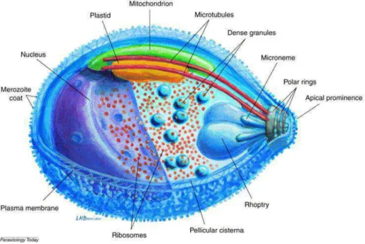
Plasmodium parasites are obligate intracellular parasites and, like other parasites of the Apicomplexa phylum, the infective forms are characterized by an apical complex (fig 2).
In this apical complex region, we find the polar rings (the organizing center of microtubules). In addition, we find the apical secretory organelles, rhoptries and micronemes, which release their contents sequentially during parasite motility and host cell invasion (Baum et al. 2008; Counihan et al. 2013). They also possess an inner membrane complex (IMC) or pellicular cisterna (Harding and Meissner 2014), made of flattened membrane sacs (alveoli), supported by the thin filaments of the subpellicular network. A unique mitochondrion is found just under the IMC, closely linked to a vestigial plastid related to the plant chloroplast, the apicoplast, which is surrounded by four membranes (Mcfadden et al. 1997). The food vacuole is formed during the intraerythrocytic stage of the parasite when haemoglobin is proteolyzed.
Figure 2: Schematic organisation of a Plasmodium falciparum merozoite.
Three-dimensional organisation of a P. falciparum merozoite. The characteristic organelles of a merozoite are represented with the apical complex composed of the polar rings, the two rhoptries, the micronemes and the dense granules. Under the plasma membrane is found the pellicular cisterna attached by the filament of the subpellicular network. The unique mitochondrion is closed to the pellicular cisterna and linked to the apicoplast, a plastid related to chloroplasts. (Bannister et al. 2000)
11 The malaria disease was already known during the Antiquity; however, its causative agent, the Plasmodium parasite, was only discovered in 1880 by Alphonse Laveran, a French military doctor who received the Nobel Prize for his discovery in 1907 (Sherman 2005; Cox 2010). The transmission of the parasite by the mosquito was described by Ronald Ross, a surgeon-major in the Indian medical service, working first on human malaria and later, on bird malaria. He was awarded the Nobel Prize in 1902 (Sherman 2005).
The life cycle of Plasmodium parasite is divided in two parts, one occurring in the animal host and the other in the insect vector, the female mosquito of the Anopheles genus (Sherman 2005; Heussler et al. 2016) (Fig 3). The Plasmodium parasite is first transmitted into the host by an infected female mosquito during blood meal. At this moment, less than sporozoites are released from the mosquito s salivary glands into the skin of the host. Not all the sporozoites succeed to migrate from the skin to the blood vessel (Amino et al. 2006). However, once in the bloodstream, it takes less than an hour for the sporozoites to reach the liver and invade the hepatocytes. The liver stage of the parasite, after hepatocyte invasion, lasts from 2 to 10 days. During this time, the parasite multiplies asexually, releasing between 10 000 and 40 000 merozoites per hepatocyte. Then, the merozoites leave the hepatocyte inside a membranous bud called the merosome and enter the blood system. In two Plasmodium species, P. vivax and P. ovale, a proportion of liver-stage parasites, named hypnozoites, can remain dormant in the hepatocytes for months or several years without clinical manifestations (Richter et al. 2010). Thereby, they can initiate a cycle of asexual reproduction in the absence of a new mosquito bite. Within 2 hours after reaching the blood circulatory system, the merozoites invade red blood cells (erythrocytes or reticulocytes, depending on the
Plasmodium species), starting another cycle asexual reproduction or schizogony. After
invasion of the red blood cell, the merozoite goes through different stages or forms inside the host, evolving from an initial state called ring stage to the trophozoite and, then, to the schizont. Each mature schizont gives a new generation of merozoites that are released into the blood system after the lysis of the red blood cell. These merozoites can invade other red blood cells and repeat more schizogony cycles, resulting in up to a 10-fold increase of the parasite population. Most of clinical symptoms in malaria are caused after red blood cell destruction. After a new invasion of a red blood cell, some parasites switch to a sexual differentiation leading to male and female gametocytes.
12 These gametocytes are released into the blood system and ingested by a female mosquito during blood feeding. In the mosquito, the male gametocyte divides into eight flagellated microgametes that escape from the red blood cell in a process called exflagellation (Sinden et al. 2010). Then, the microgamete fertilizes one female macrogamete to form the ookinete, which due to its motility, moves through the cells of the stomach wall and differentiate into an oocyst. Into the oocyst, sporozoites are formed after asexual multiplication; then, the oocyst bursts and the released sporozoites migrate to the mosquito salivary glands. Finally, when the female mosquito takes a new blood meal, it injects the sporozoites in the new host, completing, therefore, the cycle.
Figure 3: Scheme of the life cycle of Plasmodium falciparum.
The female Anopheles mosquito injects sporozoites in the host during a blood meal. The sporozoites move to the liver and invade hepatocytes, in which they asexually multiply to produce merozoites that are released into the blood stream from the merosome. Merozoites invade erythrocytes and go through different stages: an initial ring stage followed by a trophozoite form, and, finally, the mature schizonts that evolve to merozoites. Merozoites, released by the lysis of the erythrocyte, can reinvade new red blood cells and perform new schizogony cycles. Some merozoites after erythrocyte’s invasion differentiate into either male or female gametocytes that, after being released to the blood systems, are ingested by a mosquito. Into the mosquito’s gut, the mature male microgamete fertilizes the mature female macrogametes, leading to the fertilized zygote forms known as ookinetes due to their high motility. Ookinetes develop into an oocyst where the sporozoites are formed by asexual reproduction. Finally, these sporozoites are released and migrate to the
13
salivary glands, where they will be transmitted to a new host after a new blood feeding. (Cowman, Berry, and
Baum 2012)
In some cases, the malaria infection is not due to the bite of a mosquito. The parasite can be also transmitted from mother to child during pregnancy or even after a blood transfusion with infected blood (Abdullah and Karunamoorthi 2016; Poespoprodjo et al. 2010).
I-1-3 Treatments and resistance
1. Symptoms and diagnosisSymptoms:
After infection by the bite of the female mosquito, the incubation period between the infection and the apparition of the symptoms varies between 9 to 30 days depending on the Plasmodium species. P. falciparum has the shortest period and P. malaria the longest one (Bartoloni and Zammarchi 2012). This period can vary depending on several factors, such as the immunity of the person, the use of chemoprophylactic drugs or the parasite density inoculated. The clinical symptoms are fundamentally associated to the erythrocytic stage of the parasite. The release of the red blood material and accumulated wastes from the parasite after erythrocyte's rupture and merozoite release, induces a reaction by the immune system, resulting in periods of high fever. Usually, the infection leads to a so-called uncomplicated malaria, defined by non-specific symptoms such as fever, chills, sweat, body-aches, headache, cough, diarrhoea, nausea and vomiting, which can be related to other infections like flu, rendering complicated the diagnosing. The fever is irregular and appears after each of the schizogony cycles. Thrombocypenia is also common in uncomplicated malaria. Importantly, the delay in the diagnosis, lack of treatment, and the immunological background of the host can lead to a severe form of malaria.
Severe malaria can lead to different complications as cerebral malaria, respiratory failure, pulmonary oedema, kidney injuries, hypoglycaemia or severe anaemia (Trampuz et al. 2003). One feature of severe malaria caused by P. falciparum is the sequestration of infected erythrocytes in the microvasculature contributing to metabolic acidosis and organ damages. Indeed, cerebral malaria, the most severe neurological complication of
14 cerebral blood vessels (Idro, Jenkins, and Newton 2005). Clinical symptoms of severe malaria are, among others, prostration, impaired consciousness, coma, convulsion, jaundice, anuria and hypoglycaemia. Moreover, severe malaria is propitious for invasive bacterial infections in children. In addition, HIV infection is a major risk after severe malaria development (Ashley, Pyae Phyo, and Woodrow 2018; Bartoloni and Zammarchi 2012; Cowman et al. 2016).
In the case of non-immune pregnant women, malaria can cause a severe pathology for the mother (like pulmonary oedema and hypoglycaemia) and can lead to stillbirth or premature labour. For women with a certain degree of immunity, the infection with malaria normally results in anaemia for the mother and a low birth weight for the child. Sequestration of parasites in the placenta can also occur (Cowman et al. 2016).
Diagnosis:
The diagnosis is mostly based on a parasitological test by microscopic examination or Rapid Diagnosis Tests (RDT) of a blood sample. In addition, and if the resources are available, the parasite s nucleic acid can be detected by PCR. It is very important to do the tests and have a diagnosis as fast as possible, especially for children and non-immunized population for whom the infection can be rapidly fatal (Moody 2002; Wongsrichanalai et al. 2007; Mathison and Pritt 2017).
The microscopy analysis is based on a stained blood film to directly visualise the parasites and to quantify the parasitemia M -A Laboratory Diagnosis of Blood-Borne Parasitic Diseases; Approved Guideline . This widely used technique is called the gold standard to diagnose Plasmodium infection, and it requires trained people. Fluorescence microscopy is also used but it needs a specific microscope and trained people and, sometimes, it is not accessible in endemic areas.
RDT are based on the detection of parasite-specific antigens or enzymes that are genus or species-specific. This test gives a result in 15-30 min and requires no laboratory or trained people. They are immune chromatographic tests that use monoclonal antibodies targeting three parasite's enzymes. In P. falciparum, they target the histidine rich protein HRP-2, a water-soluble protein present during the asexual and the early gametocyte, stages of the parasite and presented at the surface of the erythrocyte. The HRP-2 antigen can be found even after successful treatment so it will not inquire on the disease s
15 evolution. The parasite s lactate deshydrogenase, pLDH, from the glycolytic pathway is also used and it is a pan-species marker. The parasite s enzyme aldolase is also used as pan-species marker and it is mostly used in combination with HRP-2 detection (Wilson 2012).
The parasite's nucleic acid detection by PCR is also used as diagnosis tool since it is a very sensitive technique which can detect very low parasitemia. Detection of the 18S rRNA and the circumsporozoite gene allows to identify the different Plasmodium species (Vasoo and Pritt 2013). The downside of this technique is that it can detect also dead parasites after successful treatment, so it is not suited for monitoring the level of parasitemia after treatments.
2. Protection: chemoprophylaxis, vaccines and vector control
In order to decrease the incidence of malaria, prevention measures were applied. This includes chemoprophylaxis, control of the mosquito vector, and, most importantly, the development of vaccines.
Chemoprophylaxis: For pregnant women and children, an intermittent preventive treatment consists on a minimum dose of sulfadoxine–pyrimethamine (SP) (Ashley, Pyae Phyo, and Woodrow 2018). In the areas with seasonal malaria, children receive a combination of SP and amodiaquine. In the case of travellers, the preventive treatment is the combination of atovaquone and proguanil, sold under the name Malarone®. Doxycycline, mefloquine and primaquine are also used for traveller s prophylaxis.
Control of mosquito vectors and/or bite prevention: The insecticide dichlorodiphenyltrichloroethane (DDT) was discovered at the beginning of the 1940s and was a powerful tool for eliminating the mosquito vector until mosquitoes started to develop resistance against this molecule. Nowadays, in high transmission areas, the most effective measures for controlling the malaria vector, and, therefore, infection are the combination of long-lasting insecticidal nets and indoor residual spraying (Ashley, Pyae Phyo, and Woodrow 2018).
Vaccination: To radically eliminate malaria it is mandatory to design an effective vaccine to provide immunity to high risk population as children and pregnant women. Different vaccines are currently under development, targeting different stages of the parasite s life cycle (Coelho et al. 2017). To this day, the most developed one is the RTS,S/ AS01
16 vaccine that is already in phase IV of clinical trials (Gosling and von Seidlein 2016). This vaccine contains the central repeat region of the P. falciparum circumsporozoite protein (CSP) fused to the Hepatitis B surface antigen, HBS. Other vaccines are composed by whole attenuated parasites by either radiation (PfSPZ) (Greenwood 2017; Mordmüller et al. 2017), genetic attenuation (GAP) or chemical attenuation (CVac). Vaccines targeting the blood stage include the vaccine containing the Plasmodium Apical Membrane Antigen (AMA1) together with the rhoptry neck protein 2 (RON2), or the one containing the parasite s Reticulocyte Binding Protein Homolog 5 (PfRH5) (Douglas et al. 2015). All these vaccines contain essential components of the merozoite s invasion machinery and, therefore, they are designed to block infection. Some vaccines are designed to block the transmission by the mosquito, targeting antigens present in the sexual stage parasite inside the mosquito. After vaccination, the antibodies generated by the host s immune response are taken by the mosquito during blood feeding and, therefore, they can reach their target. Finally, and since placental malaria caused by P.
falciparum is a major cause of maternal, foetal and infant mortality, important efforts
have been made to understand the mechanisms of this type of infection, representing a promising path for vaccine development (Fried and Duffy 2015).
3. Treatments
One of the first isolated molecules with antimalarial properties was the quinine, a molecule extracted from the tree Cinchona succirubra in 1817 by the French pharmacists Pelletier and Caventou (Lee 2002). Quinine remained the mainstay of malaria treatment until the 1920s when more effective synthetic anti-malarials became available like chloroquine. Quinine is still used in combination with antibiotics mainly for uncomplicated malaria and pregnant women (Achan et al. 2011). Chloroquine attained a worldwide use as antimalarial drug at the end of the 1940s due to its low toxicity, efficacy and affordable price (Slater 1993). It was commonly used together with the insecticide DDT, and its use has had a huge impact on malaria elimination in low transmission-rate countries. However, from the 1960s onwards, use of chloroquine progressively succumbed to the appearance and spread of resistant strains of
Plasmodium around the world (Trape 2001) that stalled the elimination of malaria
(Fidock et al. 2004). Nowadays, chloroquine still maintains some efficacy in areas where patients have acquired partial immunity to malaria (Petersen, Eastman, and Lanzer
17 2011). Currently, the most effective treatment for malaria are artemisinin-based combination therapies (ACTs), that combine a semi-synthetic derivative of artemisinin, a chemical compound isolated from the Chinese wormwood Artemisia annua, with a partner drug. Artemisinin was first isolated in the 1970s by Chinese scientists and more particularly by Youyou Tu who was awarded the Nobel Prize in 2015 (Youyou 2015). At present, the most common derivatives of artemisinin are artesunate, artemether, arteether and dihydroartemisinin (Meshnick 2002).
Antimalarial drugs in the current arsenal have been classified in three broad categories according to their chemical structure and mechanism of action (Arrow, Panosian, and Gelband 2004). (1) Aryl aminoalcohol compounds: quinine, quinidine, chloroquine, amodiaquine, mefloquine, halofantrine, lumefantrine, piperaquine and tafenoquine; (2) Antifolate compounds: pyrimethamine, proguanil, chlorproguanil and trimethoprim; and (3) Artemisinin compounds: artemisinin, dihydroartemisinin, artemether and artesunate. Atovaquone is an antimalarial on its own class, commonly combined with proguanil (Malarone®) (Flannery, Chatterjee, and Winzeler 2013).
The ACTs consist in using two compounds that, theoretically, interact with two different targets in the parasite. This strategy aims at decreasing the appearance of resistant strains to this treatment since the probability of generating resistance to both compounds at the same time while preserving parasite s fitness is very low. In addition, ACTs combine a compound with short half-life and rapid action (artemisinin and derivatives) with slowly-eliminated compounds which remain active much longer (amino alcohol compounds) (Eastman and Fidock 2009; Ashley, Pyae Phyo, and Woodrow 2018). The current ACTs used as first-line treatment of uncomplicated malaria are (table 1): artemether/lumefantrine, artesunate/amodiaquine, artesunate/mefloquine, artesunate/sulfadoxine/pyrimethamine, dihydroartemisinin /piperaquine. As to severe malaria, the treatment consists in artenusate, quinine or artemether. Finally, in the case of pregnancy, the treatment consists in a combination quinine/clindamycin or, in some cases, chloroquine.
Several antibacterial drugs (e.g., tetracycline, clindamycin) have also anti-malarial activity although, in general, their action is slow, so they are recommended only in combination with other antimalarial drugs.
18 Table 1: Artemisinin-based combination therapies with their target and the associated resistance to the partner drug (Ouji et al. 2018)
4. Mechanisms of action of antimalarial drugs and resistance to them
To develop new antimalarial drugs outwitting the capacity of the parasite to develop resistance against them, it is very important to have a deep knowledge of the mechanisms of action of the current drugs and also to understand the mechanisms that the parasite develops to fight back while keeping its physiological roles (also called, fitness cost).
The physical chemistry properties of chloroquine allow this drug to concentrate in the interior of the acidic food vacuole, since once on its interior the drug is protonated and cannot cross back the membrane. The food vacuole is involved in the enzymatic digestion of the host haemoglobin and, at the same time, in detoxifying the group heme resulting from haemoglobin digestion, which is dimerized to -haematin and crystallizes as the chemically inert malaria pigment, hemozoin (Rosenthal 2005). Chloroquine interferes with the heme dimerization process and therefore, avoids heme detoxification. The most common way for the parasite to develop resistance to this drug is by simply expelling it out from the food vacuole. In P. falciparum, this is achieved
19 through different mutations in the gene that encodes the essential food vacuole transporter PfCRT (initially named chloroquine resistant transporter because the physiological substrates were unkown), giving it the capacity to recognize chloroquine and to transport it outside the food vacuole (Sidhu, Verdier-Pinard, and Fidock 2002). In addition, in chloroquine-resistant strains, point mutation in the encoding gene of another food-vacuole transporter, the P. falciparum multidrug-resistant transporter, PfMDR1 is also implicated in chloroquine resistance (Schlitzer 2007; Duraisingh and Cowman 2005).
Antifolate drugs interfere with the folic acid synthesis by inhibiting the parasites enzymes dihydrofolate reductase-thymidilate synthase (DHFR) and dihydropteroate synthase (DHPS). Mutations in both genes allow the parasite to acquire resistance to those drugs (Antony and Parija 2016).
Atovaquone interferes with the mitochondrial electron transport leading to the inhibition of cellular respiration. Atovaquones resistance is accomplished by the parasite by single-point mutations in the gene encoding the cytochrome b (Antony and Parija 2016).
The mechanism of action of artemisinin is still under considerable debate. It was first proposed that the main mechanism of action of artemisinin is to create toxic free radicals in the parasites cytoplasm. Other proposed mechanisms involve targeting the heme polymerization (as chloroquine does) or inhibiting the respiratory chain of the parasite s mitochondrion (Cui and Su 2009). A few years ago, Krishna and co-workers (Eckstein-Ludwig et al. 2003) proposed that PfATP6, the unique endoplasmic reticulum Ca2+ ATPase homolog of P. falciparum, was indeed the target of artemisinin; however, a direct interaction of PfATP6 and artemisinin was discarded in our laboratory using recombinantly-produced PfATP6 (Arnou et al. 2011; Cardi et al. 2010). Instead, recent studies have provided solid evidences that artemisinin and derivatives are potent inhibitors of the P. falciparum phosphatidylinositol-3-kinase (PfPI3K) (Mbengue et al. 2015). In addition and although the resistance mechanism is still not fully understood, a mutation in the propeller domain of the gene encoding the Kelch protein 13 (K13) was associated with artemisinin resistance (Mbengue et al. 2015). The function of this protein is still unknown but it shares homologies with the human Keap1 protein,
20 involved in the cell response to oxidative stress. K13 is localised in the reticulum endoplasmic of the parasite (Ouji et al. 2018).
Although the mechanism of action of quinine is still unknown, the main resistance is due to an increased number of the gene copies encoding PfMDR1, also implicated in the resistance to other amino alcohol (Schlitzer 2007). In addition, resistance to this drug is linked to simultaneous mutations in different membrane transporters as PfMDR1, PfCRT, and the sodium/hydrogen exchanger , PfNHE1 (Cheruiyot et al. 2014).,
The antibiotics used in combination with other antimalarial drugs are prokaryotic protein-synthesis inhibitors and their target is localized in the apicoplast (Fidock et al. 2004).
Finally, a new class of antimalarial drug molecules, the spiroindolones, were shown to target the P. falciparum Na+/K+ ATPase pump, PfATP4 (Rottmann et al. 2010; Natalie J. Spillman et al. 2013).
I-2 Membrane
transport
proteins
from
Plasmodium
falciparum
The appearance and spreading of P. falciparum strains resistant to practically all the antimalarial drugs urge the development of new treatments, which implies more efforts towards the identification and validation of essential proteins of the parasite that can be used as drug targets. Membrane transport proteins (MTP) encoded by the parasite represent an important, yet little explored, potential source of new drug targets. In fact, in humans, MTPs are considered as one of the most important pharmacological targets due to their essential role in many physiological processes (Giacomini et al. 2010; Rask-Andersen, Almén, and Schiöth 2011). The intracellular mode of living of Plasmodium species requires precise mechanisms to import nutrients and precursors to fuel metabolic pathways, in addition to highly efficient systems to expel metabolites; and MTPs are the main proteins responsible of these tasks. In addition, and as indicated earlier, they are directly involved in the drug-resistance mechanisms against current drugs. Therefore, to identify future candidates for drug targeting, it is very important to study in detail the different membrane transporters encoded by the parasite, to know at which stage of its life cycle they are expressed, where they are localised within the cell, what are their functional characteristics (this include the identification of their
21 physiological substrate), and how the absence or functional depletion of a particular transporter may affect the viability of the parasite.
The genome of the P. falciparum strain 3D7 is composed of ~23 megabases distributed on 14 chromosomes. It is one of the most A+T rich genome, accounting for 80% of its composition. The sequencing of the entire genome in 2002 (Gardner et al. 2002) allowed the identification of about 5 268 protein-coding genes. Among these genes, P. falciparum encodes ~ 140 MTPs distributed in 28 families and superfamilies , and representing about 2.5% of the encoded proteins (Martin et al. 2005; Weiner and Kooij 2016). For most of them, the functional annotation is still not completed, and many cases it has been deduced from homology to genes of other organism, already characterized. Databases like the PlasmoDB (http://plasmodb.org/plasmo/) offers a remarkable tool to study Plasmodium-encoded proteins, where available information of every single gene is presented in a very comprehensible manner. In addition, the TransportDB2 database (http://www.membranetransport.org/transportDB2/index.html) contains the largest dataset for MTPs, and includes phylogenetic classification and functional annotations.
I-2-1 Membrane transporters in the infected erythrocyte
As explained earlier, MTPs are necessary for the physiology and the development of the parasite within the erythrocyte. They mediate the uptake of nutrients through the different membrane compartments of the parasitized cell, as well as the generation and maintenance of the electrochemical gradients across the membranes and the removal of the metabolic wastes, including xenobiotics. MTPs are also associated to the resistance to antimalarial drugs and can be themselves drugs targets (Kirk & Lehane, 2014; Staines et al., 2010).
After invading the erythrocyte, P, falciparum starts developing inside the host, where it multiplies different organelles such as the nucleus, the endoplasmic reticulum, the Golgi apparatus, the food vacuole, the mitochondrion or the apicoplast; all of them surrounded by lipid membranes (see scheme fig 4)(Staines et al., 2010). In addition, P. falciparum radically modifies the properties of its host cell plasma membrane by exporting and inserting in it its own proteins (Maier et al. 2009). The subcellular localisation of a few number of MTPs have been determined by fluorescent tagging in the cell using antibodies (Martin, Ginsburg, & Kirk, 2009), in situ tagging with the GFP or
22 hemagglutinin tags. However, for a large proportion of them, the localisation still needs to be determined. Most of the information we have with respect MTPs localization in the different membranes comes from functional studies or indirect assays.
Figure 4: Membrane transport proteins within the infected erythrocyte.
Schematic representation of membrane transport within the infected erythrocyte. Erythrocyte plasma membrane: transport of the essential nutrients and ions into the infected erythrocyte via the NPP. Parasitophorous vacuole membrane: protein translocon EXP2. Parasite plasma membrane: are found here the hexose transporter (PfHT), the nucleoside transporter PfENT1, amino acid transporter(s) (LATs) still unknown. Food vacuole membrane: are found here the drug transporters PfMDR1 and PfCRT (chloroquine transport). Picture in the upper left corner: schematic representation of the different organelles and membranes found in the infected erythrocyte (Tilley et al. 2007)
Several P. falciparum-encoded MTPs are involved in the establishment of new permeability pathways (NPP) in the plasma membrane of the erythrocyte (fig 4). The identity of all the MTPs participating in the NPP is not known yet, and some transporters from the host s erythrocyte participate as well. The NPPs role is to transport nutrients like sugar, amino acids, nucleosides and vitamins into the erythrocyte s cytoplasm. They also are involved in maintaining the ionic composition of the erythrocyte s cytoplasm, and in extruding wastes from the metabolic reactions like lactate (Kiaran Kirk and Lehane 2014). The parasitophorous vacuole membrane (PVM) is known to be highly permeable to many and quite different solutes, ions and even small peptides and proteins (Spielmann et al. 2012). It is suggested that the Plasmodium-encoded protein
23 translocon EXP2 is the main responsible of this large permeability and poor specificity (De Koning-Ward et al. 2009). Moreover, this year, a cryo-electron microscopy (cryo-EM) structure of the PTEX was revealed, showing seven EXP2 protomers (of one TM) forming a funnel through the PVM; thus helping to understand the PTEX mechanism for protein translocation (Ho et al. 2018). The uptake of sugars, nucleosides and amino acids through the parasite s plasma membrane is catalysed by different secondary transporters as the hexose transporters (PfHT), the nucleoside transporters (PfENT) or neutral amino acid transporters (LATs) (fig 4) (Kiaran Kirk and Lehane 2014).
I-2-2 Genetic studies on the membrane transporters
Different genetic manipulation tools have been recently used to study the role of MTPs in the Plasmodium life cycle. The importance of some Plasmodium MTPs has been uncovered after disrupting the target genes and observing a deleterious effect upon parasite's survival. A transposon mutagenesis approach on P. falciparum showed that 2680 out of 5399 genes (87%), were essential for optimal growth during in vitro asexual blood stages (M. Zhang et al. 2018). A similar screening, although doing a full knockout of 2,578 genes of P. berghei, reported 1,652 essential genes needed for the parasite to grow in mice (Bushell et al. 2017), therefore estimating that almost two-thirds of the parasite genes are required for normal asexual growth during in vivo blood stage. Concerning MTPs, the former study showed that 26 out of 79 MTPs, are essential for the parasite and another 19 from these 79 are required for normal growth (Bushell et al. 2017). Among these essential transporters, there is a considerable number of MTPs belonging to the P-type ATPase family (Weiner and Kooij 2016), implicated on both maintaining ion homeostasis and lipid translocation in membranes. For instance, PfATP4, the P. falciparum Na+/H+ pump is required to maintain Na+ homeostasis in the parasite and, therefore, it is not surprising to find that PfATP4 deletion resulted in a growth defect of the parasite (M. Zhang et al. 2018). Moreover, PfATP4 was identified as the target of the antimalarial drugs spiroindolones (Rottmann et al. 2010), and one of these molecules has already reached clinical trials (Natalie Jane Spillman and Kirk 2015). Another example of an essential P-type ATPase is the Plasmodium homolog of the sarcoplasmic/endoplasmic reticulum calcium pump, PfATP6, which regulates the intracellular Ca2+ concentration of the parasitized cell (Kiaran Kirk 2015). As in other organisms, ion-transporting P-type ATPases encoded by malaria parasites are already
24 considered as potential drug targets (Yatime et al. 2009; Natalie Jane Spillman and Kirk 2015). Importantly, besides these already characterized P-type ATPases, these studies have also shown that four putative aminophospholipid transporters belonging to the P4 subfamily of P-type ATPases are also essential or important for the parasite's normal development (Kenthirapalan et al. 2016; M. Zhang et al. 2018; Bushell et al. 2017). These transporters are still only poorly known and, therefore, their functional characterisation will give insights into their role for the parasite and into their potential as drug target.
I-2-3 Heterologous expression of Plasmodium membrane transporters
Plasmodium MTPs have important physiological roles for the parasite but our
understanding of the Plasmodium membrane proteome is still quite limited: many essential Plasmodium MTPs are still waiting to be functionally characterized to understand their role within the parasite's life cycle (and, of course, to assess their suitability for drug targeting). Unfortunately, biochemical characterization of individual transporters in Plasmodium-infected erythrocyte cultures or in other "in vivo"
Plasmodium cultures is always complicated due to the low amount of protein under
study, it has nevertheless been successful in a recent study with PfATP4 (Rosling et al. 2018). The production of Plasmodium MTPs in heterologous hosts is a valuable tool to assess in detail their functional properties, since it is possible to produce sufficient amount of protein for the desired experimental technique and, in addition, no other proteins from the parasite are present that, presumably, might interfere with the results. Different organisms can be used and the choice depends on the type of protein that will be produced, the resources and experience of the laboratory, and what kind of analysis will be performed with the recombinant MTP. Moreover, one of the difficulties to heterogously produce proteins from P. falciparum is that its genome contains a very high proportion of A+T bases (Weber 1987), hardly compatible with commonly used heterologous hosts. Consequently, to overexpress P. falciparum proteins it is necessary to use synthetic cDNA, where both the whole sequence and the individual codons are optimised for the chosen expression host.
Different expression systems have been already used to produce Plasmodium proteins (reviewed by Birkholtz et al. 2008): E. coli, S. cerevisiae, Pichia pastoris, insect cells, mammalian cells, X. laevis oocytes, single-celled amoeba Dictyostelium discoideum, T.
25 produce and study Plasmodium MTPs are the X. laevis oocytes and yeast (K Kirk et al. 2005). Using Xenopus oocytes has been the method of choice for characterizing
Plasmodium MTPs, particularly because the oocytes appear to be more tolerant than
other eukaryotic cells to the peculiar codon preferences of Plasmodium genes (Penny et al. 1998). The oocyte system has been shown to provide a useful method for the characterisation of several Plasmodium MTPs (Natalie Jane Spillman and Kirk 2015) like the Equilibrative Nucleoside Transporters 1 and 4, PfENT1 and PfENT4 (Frame et al. 2012; Carter et al. 2000; Parker et al. 2000), the Hexose Transporter PfHT (Woodrow, Penny, and Krishna 1999; Woodrow, Burchmore, and Krishna 2000), the Plasmodium aquaglyceroporin PfAQP (Hansen et al. 2001), the Plasmodium Ca2+/H+ exchanger PfCHA (Rotmann et al. 2010), PfMDR1 (Sanchez et al. 2008), the Plasmodium Folate Transporters 1 and 2, PfFT1 and PfFT2 (Salcedo-Sora et al. 2011), the Plasmodium inorganic phosphate transporter PfPiT (Saliba et al. 2006), PfCRT (Martin et al. 2009) and the Plasmodium Formate-Nitrite Transporter PfFNT (Marchetti et al. 2015). Functional characterization of PfATP6 expressed in oocytes has also recently provided further evidence that artemisinin is not interacting with PfATP6 (David-Bosne et al. 2016). Notably, recent expression of PfCRT expressed in E. coli has provided one of the first evidences that PfCRT is a H+-coupled transporter able to transport peptides of different sizes generated after haemoglobin digestion (Juge et al. 2015). The well-known yeast expression hosts Saccharomyces cerevisiae and Pichia pastoris have also been used to characterise a number of Plasmodium MTPs, for instance, the Plasmodium Ca2+/H+ exchanger PfCHA (Salcedo-Sora, Ward, and Biagini 2012; Guttery et al. 2013), PfATP6 (Cardi et al. 2010; Arnou et al. 2011), PfCRT (Baro, Pooput, and Roepe 2011), PfMDR1 (Amoah, Lekostaj, and Roepe 2007), PfFNT (Wu et al. 2015), or the P. falciparum homolog of the tryptophan transporter Tat2p of S. cerevisiae (Tindall et al. 2018). We also find recent examples of Plasmodium MTP expression using cell-free expression, for example for the Plasmodium lactate transporter PfFNT (Holm-Bertelsen et al. 2016; Lim et al. 2010).
Finally, it is very important to note here that among all these heterogously expressed transporters, to date, only three were purified and functionally or structurally characterized: PfAQ (Hedfalk et al. 2008), providing the first atomic-resolution 3D structure of a Plasmodium membrane protein, PfCRT (Tan et al. 2006b; Juge et al. 2015) and PfATP6 (Cardi et al. 2010; Arnou et al. 2011; David-Bosne et al. 2013).
26
I-3 P4-ATPases
I-3-1 P-type ATPases
1. General information and subfamilies
P-type ATPases are primary MTPs that use the energy of the hydrolysis of ATP to pump their substrates against their concentration gradient. One of their characteristics is that, during the catalytic cycle, they are transiently phosphorylated at a conserved aspartate residue (Palmgren and Nissen 2011).
P-type ATPases are classified within five subfamilies:
P1-ATPases, comprising two groups:
• P1A: represented by the bacterial K+ transporters.
• P B: They are heavy metal transporters and are present in all organisms. They transport Cu+, Ag+, Cu2+, Zn2+, Co2+, Pb2+, Cd2+.
P2-ATPases, divided into three groups:
• P2A, which includes the sarco (endo)plasmic reticulum Ca2+ ATPases SERCA (Ca2+/H+), the most studied P-type ATPases from functional and structural points of view.
• P2B: PMCA plasma membrane calcium ATPase; calmodulin binding ATPases, they have one Ca2+ binding site.
• P2C: comprising the Na+/K+ and the gastric H+/K+ ATPases. These P-type ATPases are associated to a subunit.
P3-ATPases, which include the H+ pumps found at the plasma membrane of plants and fungi.
P4-ATPases. Members of this subfamily transport phospholipids rather than ions, and they are only found in eukaryotes. As the P2C members, P4-ATPases are also associated to a β subunit.
27 P5-ATPases. This subfamily has still unknown substrates. There are two subgroups: P5A, whose members are localized in the ER, and P5B which includes transporters found in lysosomes (humans), vacuole (yeast) or plasma membrane (C. elegans).
2. Structural characteristics:
Different structures of the sarcoplasmic reticulum Ca2+ ATPase (SERCA) have defined the molecular architecture of this family, providing a deep understanding of the relations between structure and activity during the transport cycle (for review see (Møller et al. 2010)). Structurally, P-type ATPases are organized into three cytoplasmic domains (A, P, and N) and two membrane-embedded domains (T and S) (fig 5). The transported substrates bind in the middle of the T-domain and alternate between being exposed to the cytoplasm and the extracytoplasmic side. The S-domain is an auxiliary domain with high rigidity that provides structural support to the T-domain and can have specialized functions such as providing substrate-coordinating side chains for additional substrate binding sites (Toyoshima and Nomura 2002). P-type ATPases also have a large cytoplasmic part composed of three domains, named P, N and A. (fig 5). The phosphorylation domain (P domain) contains the conserved sequence DKTG where the initial aspartate is transiently phosphorylated during the transport cycle. In this P domain we also find two other conserved motifs: the TGDN and the GDGXND that are involved in the coordination of the Mg2+ associated to the bound ATP in the N domain. The nucleotide-binding domain (N domain) binds ATP and allows the phosphorylation of the P domain. The N domain is inserted in the P domain and it is the most variable part of P-type ATPases. The actuator domain (A domain) is constituted by the loop between TMs 2 and 3 and a region in the N-terminal of the protein. The A domain contains the TGES motif conserved among all P-type ATPases, and implicated in the dephosphorylation of the aspartate of the P domain. This domain is flexible and acts as a phosphatase . Several P-type ATPases contain also a regulation domain (R domain) (Ekberg et al. 2010), located at the C-terminal, the N-terminal or both extremities of the pump. This R domain can act as an auto-inhibitory element by interacting with a still unknown region of the pump (Azouaoui et al. 2017; Zhou, Sebastian, and Graham 2013a).
28 Figure 5: Schematic overview of a P-Type ATPase: the example of SERCA1a.
Schematic overview of the structural organization of P-type ATPases. Left: Ribbon model of the crystal structure of the sarco(endo)plasmic reticulum Ca2+ ATPase SERCA1a with its two types of ligand bound: one ATP and two Ca2+ (PDB Accession number 1T5S). Right: Structural organization of P-type ATPase domains.
The domains described in the text are indicated with capital letters (A, N, P, T, S). From reference (Palmgren
and Nissen 2011).
Some P-type ATPases possess associated subunits needed during biogenesis, trafficking or function (Kühlbrandt 2004; Palmgren and Nissen 2011). For example, the renal Na+/K+ ATPase has an integral membrane protein -subunit with only one TM and a large cytoplasmic domain. It is suggested that this -subunit is required for targeting the complex to the final membrane destination during biogenesis, and also it has shown that it can modulate the affinity of the pump for the K+ substrate (Geering 2001). In some cells, the Na+/K+ATPase and its -subunit can interact with a -subunit, a peptide from the FXYD family, also containing one TM (Geering et al. 2003). Similarly, in the muscle, SERCA1a is regulated by sarcolipin (Montigny, Decottignies, et al. 2014; Barbot et al. 2016), a small peptide. Likewise, in the heart, SERCA2a can be regulated by phospholamban, a 52-amino acid transmembrane peptide (MacLennan, Asahi, and Tupling 2003).
3. Catalytic cycle of P-Type ATPases
As commented earlier, different atomic-resolution 3D structures of SERCA have provided a framework of different structural conformers of the protein along the
29 catalytic cycle. This information, together with a remarkable amount of functional and computational data, has permitted a good understanding of the catalytic cycle of this pump in particular and, of P-type ATPases in general (Møller et al. 2010).
Figure 6: Scheme of the catalytic cycle of P-type ATPases (Kühlbrandt 2004).
Ion 1 (inside the cell) binds to a high-affinity site in the E1 conformation of the ATPase. Ion binding leads to phosphorylation of the enzyme by Mg2+–ATP, leading to the phosphorylated E1–P conformation. Transition
to the phosphorylated E2–P conformation leads to a reduced affinity for ion 1, which is released outside of the cell. Ion 2 then binds (from outside) and the enzyme dephosphorylates, releasing ion 2 inside the cell. Another ion 1 will subsequently start another cycle. n and m are the numbers of ions transported (between 1 and 3). Pi : inorganic phosphate.
Taking as example an ion exchanger pump like the Na+/K+ P-type ATPase (Kühlbrandt 2004), the transport cycle begins when ion 1 binds the high-affinity fixation site of the pump through a channel open towards the cytoplasmic side and replaces the ion 2 that has been transported from the other side of the membrane and now is released into the cytoplasmic side. The protein is thus in the E1 conformation. The ion 1 induces a rearrangement of the P domain, which brings a Mg2+ binding-site close to the conserved aspartate of this domain, which is then phosphorylated by the ATP previously bound in the N domain. This leads to the E1-P conformation that releases ADP. This conformation is a high energy conformation and, therefore, the addition of ADP will lead to the inverse reaction and the formation of ATP. In the E1-P conformation, the ion 1 has no longer access to the cytoplasm. At this moment, there is a spontaneous transition from the E1-P conformation to the E2-P conformation or low energy conformation . This transition brings the TGES sequence of the A domain close to the phosphorylated aspartate and opens the channel to the extracellular side. In SERCA, this transition is the
30 rate-limiting step of the transport cycle. Then, ion 1 is released in the extracellular side of the membrane and ion 2 binds in the same binding site, since now this site has evolved to a high-affinity fixation site for ion 2. Upon ion 2 binding, the channel gets occluded from the extracellular side, and the P domain changes conformation and gets dephosphorylated, resulting into the dephosphorylated E2 conformation. Finally, domain A moves away from P domain and the protein goes back to the E1 conformation.
I-3-2 P4-ATPases
1. Overview of the P4-ATPases
P4 ATPases (also known as flippases) constitute a particular subfamily of the P-type ATPase family and are present only in eukaryotic organisms. Unlike the rest of P-type ATPase members that pump ions, P4-ATPases mediate the translocation of phospholipids from the exoplasmic to the cytoplasmic leaflet of biological membranes (fig 7) (López-Marqués, Holthuis, and Pomorski 2011). This process enables eukaryotic cells to create and maintain non-random lipid distribution between the two leaflets of the membrane. They mostly transport PS, PE, PC and lysophospholipids, although recently they were shown to transport glycosphingolipids (Roland et al. 2018).
Figure 7: Schematic
flipping of a
phospholipid by a P4-ATPase.
The P4-ATPases drive the active transport of phospholipids from the external leaflet to the internal leaflet of the membrane.
The activity of P4-ATPases has a direct relation with important cellular functions. For instance, the imbalance in phospholipid number between the two leaflets determines the curvature of the phospholipid bilayer inducing membrane invagination, thus facilitating endocytic or secretory vesicle budding, a key step for protein trafficking and membrane biogenesis. In addition, they play an important role in cell signaling, since the
31 exposition of PE in the external leaflet of the cell is a signal that triggers apoptosis and blood clotting. In plants, the transport of lysophospholipids mediated by P4-ATPases is suggested to have an important role in cell signaling (Lisbeth R. Poulsen et al. 2015). In addition, P4-ATPases are also implicated in cell polarity and migration both, in yeast (Koji Saito et al. 2007) and in humans (Kato et al. 2013). Also, P4-ATPases are implicated in the virulence of pathogens as Cryptococcus (Huang et al. 2016; Shor et al. 2016), and they mediate the transport of drugs in the Leishmania parasite (Pérez-Victoria et al. 2003). In humans, mutations in certain P4-ATPases may leads to intrahepatic cholestasis e.g. mutations in ATP8B1 (van der Mark, Oude Elferink, and Paulusma 2013).
P4-ATPases are predicted to have 10 TMs and they are associated to a subunit from the CDC50/LEM family (fig 8). The subunits possess two TMs with a large exoplasmic loop containing 4 conserved cysteines that form two intramolecular disulfide bridges. In addition, this extracellular domain is glycosylated, which is important during biogenesis and for the interaction with the P4-ATPase (Jonathan A Coleman and Molday 2011). Cdc50 proteins are able to interact with more than one type of P4 ATPase: for example the S. cerevisiae Cdc50 protein Lem3p can interact with two P4-ATPases, Dnf1 and Dnf2 (Koji Saito 2004; Furuta et al. 2007). Moreover, different studies have revealed the crucial role of Cdc50 for proper protein folding and membrane localization of the P4-ATPase/Cdc50 functional complex (Van Der Velden et al. 2010; López-Marqués et al. 2010; S. Chen et al. 2006). There are few exceptions of P4-ATPases that do not have a Cdc50-associated subunit; among them, Neo1p in S. cerevisiae or the human ATP9A and ATP9B (López-Marqués, Holthuis, and Pomorski 2011; van der Mark, Oude Elferink, and Paulusma 2013).