HAL Id: inserm-02124678
https://www.hal.inserm.fr/inserm-02124678
Submitted on 9 May 2019
HAL is a multi-disciplinary open access
archive for the deposit and dissemination of sci-entific research documents, whether they are pub-lished or not. The documents may come from teaching and research institutions in France or abroad, or from public or private research centers.
L’archive ouverte pluridisciplinaire HAL, est destinée au dépôt et à la diffusion de documents scientifiques de niveau recherche, publiés ou non, émanant des établissements d’enseignement et de recherche français ou étrangers, des laboratoires publics ou privés.
Re-exploring immune-related side effects of docetaxel in
an observational study: Blood hypereosinophilia
Diaddin Hamdan, Christophe Leboeuf, Christine Le Foll, Guilhem Bousquet,
Anne Janin
To cite this version:
Diaddin Hamdan, Christophe Leboeuf, Christine Le Foll, Guilhem Bousquet, Anne Janin. Re-exploring immune-related side effects of docetaxel in an observational study: Blood hypereosinophilia. Cancer Medicine, Wiley, 2019, Epub ahead of print. �10.1002/cam4.2062�. �inserm-02124678�
Cancer Medicine. 2019;1–8. wileyonlinelibrary.com/journal/cam4
|
11
|
INTRODUCTION
Docetaxel, a semi‐synthetic taxane inhibiting microtubule depolymerization, is approved for breast and lung cancer
treatment. It is frequently responsible for drug‐induced hy-persensitivity reactions in up to 50% of patients,1,2 thus
leading to deleterious treatment interruptions. Rapid drug desensitization protocols are effective in the management of
O R I G I N A L R E S E A R C H
Re‐exploring immune‐related side effects of docetaxel in an
observational study: Blood hypereosinophilia
Diaddin Hamdan
1,2|
Christophe Leboeuf
2|
Christine Le Foll
1|
Guilhem Bousquet
2,3,4|
Anne Janin
2,5This is an open access article under the terms of the Creative Commons Attribution License, which permits use, distribution and reproduction in any medium, provided the original work is properly cited.
© 2019 The Authors. Cancer Medicine published by John Wiley & Sons Ltd. *Guilhem Bousquet and Anne Janin are co‐senior authors.
1Medical Oncology Department, Grand
Hospital of East Francilien‐Marne‐la‐ Vallée, Jossigny, France
2UMR_S1165, Inserm, University of Paris‐
Diderot, Paris, France
3University of Paris13, Villetaneuse, France 4Medical Oncology Department, Hospital
Avicenne, APHP, Bobigny, France
5Pathology Laboratory, Hospital St Louis,
APHP, Paris, France
Correspondence
Anne Janin, U1165, Université Paris‐ Diderot, Inserm, Hôpital Saint‐Louis, Paris, France
Email: [email protected] and
Guilhem Bousquet, U1165, Université Paris13, Inserm, Hôpital Avicenne, Service d'Oncologie Médicale, Bobigny, France Email: [email protected]
Funding information
This work was supported by the University of Paris‐Diderot and the Institut National de la Santé et de la Recherche Médicale (INSERM).
Abstract
Docetaxel is a major anticancer drug that can induce hypersensitivity reactions lead-ing to deleterious treatment interruptions. Blood hypereosinophilia could be a bio-logical sign, potentially lethal, of delayed visceral hypersensitivity reactions. We hypothesized this biological event is probably underreported. In this prospective ob-servational study, we followed up 149 patients treated with docetaxel monotherapy for breast or lung cancer. For each patient, blood eosinophil counts were recorded during docetaxel treatment and up to 3 months after the end of docetaxel treatment. For all patients, blood eosinophil counts significantly increased under docetaxel chemotherapy (P < 0.01). Seven percent had persistent eosinophilia after the end of treatment. Four patients had blood eosinophil counts over 1000/mm3 with severe
cardiac, cutaneous and digestive toxicities, and docetaxel imputability was confirmed using drug‐imputability scales. For two of these four patients, tissue biopsies were performed during the time of hypereosinophilia and of severe toxicities. Specific im-munostainings and electron microscopy found numerous degranulating mast cells and eosinophils. Our study demonstrated that eosinophilia is frequent under doc-etaxel and could lead to severe complications, implicating eosinophils and mast cells, and possibly IgE. One way of treating hypersensitivity reactions could be by target-ing IgEs with omalizumab, an anti‐IgE monoclonal antibody approved for the treat-ment of severe allergic asthma, and successfully used in food and poison‐induced anaphylactic reactions.
K E Y W O R D S
2
|
HAMDAN etAl.nonsevere hypersensitivity reactions, thus limiting treatment interruptions.3–7 However, severe delayed visceral
hypersen-sitivity reactions, potentially lethal from visceral complica-tions, are excluded from desensitization protocols.
We recently reported a case of docetaxel‐induced blood hypereosinophilia with a severe digestive allergic reaction.8
We hypothesized that drug‐induced blood eosinophilia, prob-ably underreported, could be a biological sign of hypersensi-tivity reaction, and could also predict severe delayed visceral hypersensitivity reactions.
In this observational study, we aimed to determine the incidence of docetaxel‐induced eosinophilia, and whether it could be an early biological event predictive for the risk of delayed visceral hypersensitivity reactions.
2
|
MATERIALS AND METHODS
2.1
|
Inclusion criteria, clinical, and
biological data
This study was approved by our local Institutional Review Board ‐IRB 00006477.
One hundred and forty‐nine patients were included over a period of 1 year. All of them were being treated with docetaxel monotherapy for breast or lung cancer, as specified in the inclusion criteria (Table 1).
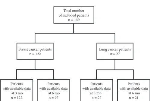
For each patient, blood eosinophil counts were recorded at the beginning of docetaxel treatment, before each cycle, and up to 3 months after the end of docetaxel treatment when data were available. At this last time‐point, data were available for 79% of the patients (Figure 1). For the whole population, the blood eosinophil count was retrieved at 1 week before the beginning of docetaxel, at the end, and at 3 months after the end of docetaxel treatment.
For each patient, we considered that the increase in blood eo-sinophil counts was significant when it was at least twice as high compared to the count before initiation of docetaxel treatment.
For patients with blood eosinophil count >1000/mm3,
various tests were conducted to eliminate other possible causes of eosinophilia (Table S1).
For pharmacological imputability of docetaxel, we calcu-lated an imputability score using the French and the North American validated pharmacovigilance scales.9,10
For each patient, docetaxel‐induced hypersensitivity re-actions of any type (wheal‐and‐flare rere-actions, maculopap-ular eruptions, urticaria, itching, angioedema, local edema, bronchospasm, gastrointestinal symptoms, anaphylaxis, etc) were recorded and graded according to CTCAE‐NCI grading scale version 5. Immediate and delayed hyper-sensitivity reactions were considered, and delayed hy-persensitivity reactions occurred at least 6 hr after each administration of docetaxel.11
Inclusion criteria Exclusion criteria
Breast or lung cancers Cancers of other origins Localized or metastatic cancer
Docetaxel monotherapy Docetaxel combination therapy Available blood analyses before, during and
after docetaxel treatment Blood analyses not available during docetaxel treatment
TABLE 1 Inclusion criteria
FIGURE 1 A flow‐diagram to show the number of patients included in the study and those with unavailable data
Total number of included patients
n = 149
Lung cancer patients n = 27 Breast cancer patients
n = 122
Patients with available data
at 3 mo n = 122
Patients with available data
at 6 mo n = 97
Patients with available data
at 3 mo n = 27
Patients with available data
at 6 mo n = 21
2.2
|
Tissue analyses and characterization of
eosinophils and mast cells
Among the four patients with blood hypereosinophilia >1000/mm3, we were able to perform tissue analyses for
two patients with eosinophil counts for whom tissue samples were obtained at the time of blood eosinophilia.
We used anti‐tryptase and anti‐eosinophil peroxidase (EPO) antibodies to differentiate and count mast cells and eosinophils. These two immunostainings were performed on 5 μm‐thick tissue sections using indirect immunoperoxidase staining, with rabbit polyclonal anti‐human EPO (ab104530, Abcam, Cambridge, UK) and monoclonal mouse anti‐human tryptase (clone G3, Santacruz, Heidelberg, Germany) as pri-mary antibodies. Controls included omitting the pripri-mary antibody and using an irrelevant antibody of identical iso-type. The analysis focused on the number and distribution of mast cells and eosinophils in the different tissue sections. Tissue sections were analyzed using an Olympus AX 70 mi-croscope with a 0.344‐mm2 field size at 400× magnifications
(Olympus, Tokyo, Japan). Images were systematically taken using SAS software for each immunostaining image.
For the ultrastructural analysis, tissue samples were fixed in 2% glutaraldehyde‐buffered 0.1 M. cacodylate and embedded in epoxy resin. Ultra‐thin sections were stained with uranyl acetate and lead citrate and analyzed using a Hitachi‐7650. The images of the distribution of mast cells and eosinophils and their state of degranulation were captured.
2.3
|
Statistical analysis
Categorical variables were summarized as the number (per-centage) and continuous variables were summarized as the mean or the median.
A comparison of the median value of the three matched‐ sample of blood eosinophil count was performed (ie, pre-treatment period, at the end, and at 3 months after the end of docetaxel treatment) using the Wilcoxon's test.
All tests were two‐sided and the threshold for statistical significance was set to P < 0.05. The data were analyzed using the BiostaTGV site (http:\\marne.u707.jussieu.fr/bio-statgv, accessed in Avril 2018).
3
|
RESULTS
3.1
|
Patients
Patients were recruited from January 2017 to December 2017, and inclusion criteria are detailed in Table 1. A total of 149 pa-tients with breast or lung cancers treated with docetaxel mono-therapy were included during this period. Their characteristics are detailed in Table 2:81% had breast cancer, with a median age of 55 and 61 years for breast and lung cancer patients, respectively.
3.2
|
Blood eosinophil counts under
docetaxel chemotherapy
We have compared the median of blood eosinophil counts be-fore, at the end and three months after the end of docetaxel treatment. Among the 149 patients, 73 (49%) had at least a twofold increase in their blood eosinophil counts during the follow‐up period (Table 2). We have compared the median of blood eosinophil counts before, at the end and 3 months after the end of docetaxel treatment. Median blood eosinophil counts significantly increased under docetaxel chemotherapy, from 77/mm3 before treatment, to 135/mm3 and 221/mm3 at 3 and 6 months respectively (P < 0.01) after docetaxel initiation (Figure 2A). Three months after the last cycle of docetaxel, blood eosinophil counts remained higher than 500/mm3 in 7%
of the patients (Figure 2A and Figure S1), with comparable results in the two cancer types (Figures S2 and S3).
3.3
|
Docetaxel‐induced
hypersensitivity reactions
When we looked at all‐grade docetaxel‐induced hyper-sensitivity reactions other than blood eosinophilia, they
TABLE 2 Patients’ characteristic
Patients Breast Lung Whole cohort At least twofold increase in blood eosinophil count during follow‐up period
Number (%) 122 (81) 27 (18) 149 (100) 73 (49)
Mean age (years) 55 61 58 60
Allergic history (%) 18 (13) 2 (7) 20 (13) 9 (12) Mean number of docetaxel cycles 2.89 4.63 3.76 2.58 Mean dose of cycle 1 (mg) 160.72 123 141.86 151.46 HSRs other than blood
eosinophilia G1‐2
a (%) 42 (34) 8 (29) 50 (33) 30 (41)
G3‐4a (%) 9 (7) 2 (7) 11 (7) 7 (9) Bold values are corresponding to percentages.
HSR: hypersensitivity reactions.
occurred for 66 of the 149 patients (40%) (Table 2). Interestingly, among the 52 patients with a significant in-crease in blood eosinophil counts, 50% had a hypersensi-tivity reaction manifestation other than blood eosinophilia (Table 2). Table 3 reports the different types of hypersen-sitivity reactions, and shows that all‐grade hypersensitiv-ity reactions occurred in 21 patients (14%), leading to a premature discontinuation of planned docetaxel treatment.
Four patients (2.6%) had blood eosinophil counts over 1000/mm3 (Figure 2B). We eliminated other possible
etiol-ogies of eosinophilia and thus confirmed the imputability of docetaxel using drug‐imputability scales (Table 4). Patient 1 had an NCI‐CTCAE‐v5 grade II diarrhea without severe complications; Patient 2 had an NCI‐CTCAE‐v5 grade III cardiac flutter at the end of the docetaxel treatment despite the absence of any cardiac risk factor. For Patient 3, blood eosinophilia persisted well beyond 6 months after the discon-tinuation of docetaxel, with severe chronic pruritus justifying a skin biopsy. Patient 4 received docetaxel and had hypereo-sinophilia at the time of breast surgery.
On the skin of Patient 3 and the breast cancer of Patient 4, specific immunostainings (anti‐eosinophil peroxidase, anti‐ tryptase) and electron microscopy found numerous degran-ulating mast cells and eosinophils; for Patient 4, we found clustered tryptase‐expressing mast cells at the invasive front of the tumor (Figure 2B,C).
3.4
|
Literature review
For the literature review of hypereosinophilia cases im-putable to anticancer drugs, we used an ad‐hoc algorithm
composed of both thesaurus and free text terms to search the Medline database up to 2 May 2018. We used the fol-lowing algorithm: ("Eosinophilia"[Mesh] OR "Eosinophilia" OR “eosinophilic” OR “eosinophilic syndrome” OR “hy-pereosinophilia”) AND ("Neoplasms"[Mesh] OR “can-cer”) AND ("Drug Hypersensitivity Syndrome"[Mesh] OR "Antineoplastic Agents"[Mesh] OR "Drug Therapy"[Mesh] OR “chemotherapy” OR "drug‐induced"). With the limits: Species = human and blood eosinophil count >1500/mm3,
818 articles were initially identified. We screened the papers retrieved initially on title and abstract, and finally on full text. Twenty‐three publications on hypereosinophilia imputable to an anticancer agent were identified, 19 were case reports, two others were phase I clinical trials, one was an observational cohort, and the last was a literature review (Table 5).
4
|
DISCUSSION
Our study confirmed our hypothesis that docetaxel‐induced blood eosinophilia is largely underestimated since a two-fold increase in blood eosinophil count occurred in half of the treated population. It was frequently associated with other clinical manifestations of immediate and delayed hypersensitivity reactions, supporting our hypothesis that blood eosinophilia is a biological sign of hypersensitivity reaction. In addition, it was severe, over 500/mm3, and du-rable over time for 7% of the patients, compadu-rable to the 7% of patients with grade 3‐4 hypersensitivity reactions in phase I clinical trials with docetaxel.12,13 It led to visceral
complications for four of the 149 patients, and in all four
FIGURE 2 (A) Blood eosinophil count curves in the course of docetaxel treatment and up to 3 months after discontinuation for all patients included in the study. (B) Blood eosinophil count curves for the four patients who developed blood eosinophilia in the course of docetaxel treatment. (C) Skin biopsy for Patient 3 who had persistent blood eosinophilia well beyond 6 months after the end of docetaxel treatment,
accompanied by severe chronic pruritus. Anti‐eosinophil peroxidase and anti‐tryptase immunostainings show eosinophils and mast cells infiltrating the deep dermis. This was confirmed by electron microscopy showing many degranulating eosinophils and mast cells. (D) Tumor sample for Patient 4 who had hypereosinophilia at the time of breast surgery. Anti‐eosinophil peroxidase and anti‐tryptase immunostainings also show eosinophils and mast cells infiltrating the tumor. This was confirmed by electron microscopy which evidenced numerous degranulating eosinophils and mast cells
TABLE 3 Types of hypersensitivity reactions (HSRs)
HSR events
Delayed Immediate At least twofold increase
in blood eosinophil count during follow‐up period
Visceral Skin Visceral Skin
G1‐2a G3‐4a G1-2a G3‐4a G1-2a G3‐4a G1-2a G3‐4a <500/mm3 >500/mm3
Number 28 2 24 5 8 6 4 1 51 12
Median time to
onset (day) 42 30 30 21 1 hr 1 hr 1 hr 1 hr 76 65
Median duration 1 week 7 months 1 week 2 week 1 hr 2 hr 1 hr 1 hr 9 months 7 months
Docetaxel stopped 2 1 3 4 4 6 1 — 6 1
HSR: hypersensitivity reactions; BEC: blood eosinophil count; N: blood eosinophil count at the initiation of docetaxel treatment.
6
|
HAMDAN etAl.cases the increase in blood eosinophil count preceded the visceral complication. Blood eosinophilia could be thus an early biological sign predictive for the risk of docetaxel‐in-duced delayed visceral hypersensitivity reactions.
Strikingly, eosinophilia is not reported in clinical trials using docetaxel, possibly because of corticoid premedica-tion which limits the increase in blood eosinophil counts, and also because blood eosinophilia can occur after the end of docetaxel treatment when systematic blood counts are no longer performed. Even for other drugs, drug‐induced blood eosinophilia is rarely reported, as our literature review shows.
In case of drug‐induced blood eosinophilia, a desensiti-zation protocol, similar to those implemented for immediate
hypersensitivity reactions,3–7 might be useful to avoid
de-layed visceral complications.
One limitation to our observational study is the limited number of patients and tissue samples analyzed. Despite this, we were able to demonstrate that docetaxel‐induced eosinophilia is a frequent biological sign of hypersensitivity reaction that can predict delayed visceral complications. In addition, with only two biopsy samples obtained at the time of hypereosinophilia with visceral complications, we con-firmed the tissue infiltration by degranulating eosinophils and mast cells, as reported in our previous publication.8
Docetaxel‐induced hypersensitivity reaction is an inflam-matory reaction resulting from the activation of eosinophils and
TABLE 4 Drug imputability scores for the four patients with blood eosinophil counts over 1000/mm3
Drugs
Adverse drug reaction
probability scale9 French imputability score10
Score IS C S Intrinsic imputability
Patient 1 Docetaxel 6 2 3 3 I6 Ondansetrone 0 2 1 2 I2 Prednisone 0 2 1 2 I2 Metoclopramide 0 2 1 2 I2 Paracetamol −2 2 1 2 I2 Loperamide −2 2 0 2 I0 Lansoprazole −2 2 0 2 I0 Phloroglucinol −2 2 0 2 I0 Diosmectite −2 2 0 2 I0 Patient 2 Docetaxel 6 2 3 3 I6 Ondansetrone 0 2 1 2 I2 Prednisone 0 2 1 2 I2 Metoclopramide 0 2 0 2 I0 Esomeprazole −2 2 0 2 I0 Hydroxyzine −2 2 0 2 I0 Sotalol −2 2 0 2 I0 Nicopatch −2 2 0 2 I0 Levetiracetam −2 2 0 2 I0 Clobazam −2 2 0 2 I0 Patient 3 Docetaxel 6 2 3 3 I6 Ondansetrone 0 2 1 2 I2 Prednisone 0 2 1 2 I2 Metoclopramide 0 2 1 2 I2 Patient 4 Docetaxel 6 2 3 3 I6 Ondansetrone 0 2 1 2 I2 Prednisone 0 2 1 2 I2 Metoclopramide 0 2 1 2 I2 Omeprazole −2 2 1 2 I2
Bold values are corresponding to calculated scores according to each pharmacological scales. IS: Informativeness score, C: chronology, S: semiology
mast cells,2 themselves able to enhance their own recruitment
and activation through an autocrine loop.14 IL‐5 and IL‐13 auto‐
secretion boost IgEs and also induce eosinophil activation and degranulation through their low‐affinity IgE receptor. One way of treating hypersensitivity reactions could be by targeting IgEs with omalizumab, an anti‐IgE monoclonal antibody approved
for the treatment of severe allergic asthma, and successfully used in food and poison‐induced anaphylactic reactions.15
In conclusion, our observational study demonstrated that docetaxel‐induced blood eosinophilia is a frequent early bio-logical sign of hypersensitivity reaction that can predict de-layed visceral complication.
ACKNOWLEDGMENTS
We would like to thank Dr. Christine Dosquet for her valuable advice and criticisms. We would like also to thank Ms. Angela Swaine and Sarah Leyshon for revising the English language. CONFLICT OF INTEREST
The authors have declared no conflicts of interest. ETHICS
This work was approved by the local Institutional Review Board ‐IRB 00006477 under the number: 16‐053. It was also approved by the National Committee on private freedoms (CNIL) under the number: 1988828 v 0.
Informed consent from each patient was obtained prior to inclusion in the study.
ORCID
Diaddin Hamdan https://orcid. org/0000-0002-2212-0287
REFERENCES
1. Rive CM, Bourke J, Phillips EJ. Testing for drug hypersensitivity syndromes. Clin Biochem Rev. 2013;34(1):15‐38.
2. Picard M, Castells MC. Re‐visiting hypersensitivity reactions to taxanes: a comprehensive review. Clin Rev Allergy Immunol. 2015;49(2):177‐191.
3. Castells MC, Tennant NM, Sloane DE, et al. Hypersensitivity re-actions to chemotherapy: outcomes and safety of rapid desen-sitization in 413 cases. J Allergy Clin Immunol. 2008;122(3): 574‐580.
4. Castells Guitart MC Rapid drug desensitization for hypersensitiv-ity reactions to chemotherapy and monoclonal antibodies in the 21st century. J Investig Allergol Clin Immunol. 2014;24(2):72‐79; quiz 2 p following 9.
5. Castells M, Sancho‐Serra Mdel C, Simarro M. Hypersensitivity to antineoplastic agents: mechanisms and treatment with rapid desen-sitization. Cancer Immunol Immunother. 2012;61(9):1575‐1584. 6. Makrilia N, Syrigou E, Kaklamanos I, Manolopoulos L, Saif MW.
Hypersensitivity reactions associated with platinum antineoplastic agents: a systematic review. Met Based Drugs. 2010;2010. 7. Rose PG, Fusco N, Smrekar M, Mossbruger K, Rodriguez
M. Successful administration of carboplatin in patients with
TABLE 5 Chemotherapy‐induced hypereosinophilia publications
Name/class of
drug Type References
Check‐point
inhibitor Review Melanoma Res. 2017 Jun;27(3):271‐273 Pemetrexed CR Am J Dermatopathol. 2017
Jan;39(1):e1‐e2
Vismodegib CR Australas J Dermatol. 2017 Feb;58(1):69‐70
Anti‐PD1 CR Eur J Cancer. 2017 Aug;81:135‐137 Pembrolizumab CR Cancer Immunol Res. 2016
Mar;4(3):175‐8
Vemurafinib CR Dermatology. 2016;232(1):126‐8 Pan‐class I
PI3K inhibitor Phase I Oncologist 2015; 20(3): 245‐46 Cisplatin CR Case Rep Pulmonol.
2014;2014:209732 Ipilumumab/
anti‐CTLA4 Multi‐center cohort
PLoS One 2013; 8(1): e53745 Lenalidomide CR Rinsho Ketsueki.
2016;57(12):2502‐2506
Lenalidomide CR Eur J Dermatol 2012;22(6):799‐800 Tosedostat/ aminopepti-dase inhibitor + pa-clitaxel Phase I Br J Cancer 2010;103(9): 1362‐68 Chlorambucil CR Pharmacology 2009;83:148‐149 Imatinib/
dasatinib CR Ann Allergy Asthma Immunol. 2017;119(1):85‐86 Imatinib CR Ann DermatolVenereol 2008;135(5)
:393‐6 CR Ann
DermatolVenereol 2006;133:686‐8 CR Lancet Oncol 2005;6(9):728‐9 Dacarbazine CR Ann DermatolVenereol 2006;133(2)
:157‐60
Fludarabine CR Ann Hematol 2002;81(5):292‐3. CR Ann Hematol 1999;78(10):475‐7 13‐cis‐retinoic
acid CR Med PediatrOncol 1999;32(4):308‐10 Tegafur CR J Gastroenterol 1994;29(1):88‐92 Bleomycine CR Chest. 1985 Jul;88(1):103‐6
8
|
HAMDAN etAl.clinically documented carboplatin hypersensitivity. Gynecol
Oncol. 2003;89(3):429‐433.
8. Hamdan D, Leboeuf C, Pereira C, et al. A digestive allergic re-action with hypereosinophilia imputable to docetaxel in a breast cancer patient: a case report. BMC Cancer. 2015;15:993.
9. Naranjo CA, Busto U, Sellers EM, et al. A method for estimating the probability of adverse drug reactions. Clin Pharmacol Ther. 1981;30(2):239‐245.
10. Arimone Y, Bidault I, Dutertre J‐P, et al. Updating the French method for the causality assessment of adverse drug reactions.
Therapie. 2013;68(2):69‐76.
11. Demoly P, Adkinson Nf, Brockow K, et al. International consensus on drug allergy. Allergy. 2014;69(4):420‐437.
12. Tomiak E, Piccart Mj, Kerger J, et al. Phase I study of docetaxel administered as a 1‐hour intravenous infusion on a weekly basis. J
Clin Oncol. 1994;12(7):1458‐1467.
13. Rosing H, Lustig V, van Warmerdam L, et al. Pharmacokinetics and metabolism of docetaxel administered as a 1‐h intravenous in-fusion. Cancer Chemother Pharmacol. 2000;45(3):213‐218.
14. Vandezande Lm, Wallaert B, Desreumaux P, et al. Interleukin‐5 immunoreactivity and mRNA expression in gut mucosa from pa-tients with food allergy. Clin Exp Allergy. 1999;29(5):652‐659. 15. Shankar T, Petrov AA. Omalizumab and hypersensitivity
reac-tions. Curr Opin Allergy Clin Immunol. 2013;13(1):19‐24.
SUPPORTING INFORMATION
Additional supporting information may be found online in the Supporting Information section at the end of the article.
How to cite this article: Hamdan D, Leboeuf C, Le Foll C, Bousquet G, Janin A. Re‐exploring immune‐ related side effects of docetaxel in an observational study: Blood hypereosinophilia. Cancer Med. 2019;00:1–8. https://doi.org/10.1002/cam4.2062