HAL Id: inserm-00482372
https://www.hal.inserm.fr/inserm-00482372
Submitted on 12 Jul 2010
HAL is a multi-disciplinary open access
archive for the deposit and dissemination of
sci-entific research documents, whether they are
pub-lished or not. The documents may come from
teaching and research institutions in France or
abroad, or from public or private research centers.
L’archive ouverte pluridisciplinaire HAL, est
destinée au dépôt et à la diffusion de documents
scientifiques de niveau recherche, publiés ou non,
émanant des établissements d’enseignement et de
recherche français ou étrangers, des laboratoires
publics ou privés.
and function during mitosis.
Pierre Romé, Emilie Montembault, Nathalie Franck, Aude Pascal, David
Glover, Régis Giet
To cite this version:
Pierre Romé, Emilie Montembault, Nathalie Franck, Aude Pascal, David Glover, et al.. Aurora A
contributes to p150(glued) phosphorylation and function during mitosis.. Journal of Cell Biology,
Rockefeller University Press, 2010, 189 (4), pp.651-9. �10.1083/jcb.201001144�. �inserm-00482372�
The Rockefeller University Press $30.00
Correspondence to Régis Giet: [email protected]
E. Montembault’s present address is Dept. of Pathology, University of Cambridge, Cambridge CB23, England, UK.
Abbreviations used in this paper: DDC, dynein–dynactin complex; dsRNA, double-stranded RNA; IP, immunoprecipitation; MAP, microtubule-associated protein; MBD, microtubule-binding domain; MBP, maltose-binding protein.
Introduction
The Drosophila melanogaster aurora A gene was first identified
through mutations resulting in either female sterility or maternal
effect lethality. In both cases, mutations were associated with
mi-totic defects including failure for centrosomes to separate (Glover
et al., 1995). Subsequently, it was shown to encode a protein
kinase that is widely present in eukaryotic genomes (Giet and
Prigent, 1999). Aurora A is an essential gene in mammals and is
expressed at elevated levels in a wide variety of tumor cells. Its
overexpression is sufficient to trigger genetic instability and
transformation in NIH3T3 mouse fibroblasts but not in normal
cells, suggesting that this protein might behave as an oncogene
under specific genetic backgrounds (Giet et al., 2005; Cowley
et al., 2009). The multiple roles of aurora A protein kinase in
centrosome function and mitotic spindle assembly in Drosophila,
Caenorhabditis elegans
, Xenopus laevis, and human cells have
been extensively studied (Roghi et al., 1998; Hannak et al., 2001;
Giet et al., 2002; Barr and Gergely, 2007). In many systems, the
phenotypes caused by aurora A loss of function suggest that
aurora A
regulates the dynamics of astral microtubules. To do so,
it has been shown that aurora A phosphorylates several
micro-tubule-associated proteins, including the D-TACC subunit of the
D-TACC–Msps microtubule-stabilizing complex. Indeed, after
phosphorylation by aurora A, the D-TACC–Msps complex is
tar-geted to the centrosome component, centrosomin. The Msps
sub-unit of the complex (XMAP215 homologue) binds directly to
microtubules to promote microtubule growth. It is thus proposed
that phosphorylation of the D-TACC–Msps complex favors
stabi-lization of newly nucleated microtubules at the centrosome (Giet
et al., 2002; Terada et al., 2003; Barros et al., 2005; Zhang and
Megraw, 2007). In mitotic X. laevis egg extracts, aurora A
phos-phorylates the kinesin-related protein Eg5, hepatoma up-regulated
protein, and its coactivators, TPX2. These proteins are required
for bipolar mitotic spindle assembly and can be found in a complex
with XMAP215 (Giet and Prigent, 1999; Wong et al., 2008).
Fur-thermore, phosphorylation of the mitotic centromere-associated
kinesin by aurora A induces its redistribution onto spindle
micro-tubules, where it facilitates the establishment of spindle bipolarity
(Zhang et al., 2008). Finally, the aster-associated protein, required
A
urora A is a spindle pole–associated protein
kinase required for mitotic spindle assembly and
chromosome segregation. In this study, we show
that Drosophila melanogaster aurora A phosphorylates
the dynactin subunit p150
gluedon sites required for its
as-sociation with the mitotic spindle. Dynactin strongly
accu-mulates on microtubules during prophase but disappears
as soon as the nuclear envelope breaks down, suggesting
that its spindle localization is tightly regulated. If aurora A’s
function is compromised, dynactin and dynein become
en-riched on mitotic spindle microtubules. Phosphorylation
sites are localized within the conserved
microtubule-binding domain (MBD) of the p150
glued. Although
wild-type p150
gluedbinds weakly to spindle microtubules, a
variant that can no longer be phosphorylated by aurora A
remains associated with spindle microtubules and fails
to rescue depletion of endogenous p150
glued. Our results
suggest that aurora A kinase participates in vivo to the
phosphoregulation of the p150
gluedMBD to limit the
microtubule binding of the dynein–dynactin complex and
thus regulates spindle assembly.
Aurora A contributes to p150
glued
phosphorylation
and function during mitosis
Pierre Romé,
1Emilie Montembault,
1Nathalie Franck,
1Aude Pascal,
1David M. Glover,
2and Régis Giet
11Centre National de la Recherche Scientifique UMR6061, Institut de Génétique et Dévelopement de Rennes, Université de Rennes, 35043 Rennes, France 2Department of Genetics, Cancer Research UK Cell Cycle Genetics Research Group, University of Cambridge, Cambridge CB23EH, England, UK
© 2010 Romé et al. This article is distributed under the terms of an Attribution– Noncommercial–Share Alike–No Mirror Sites license for the first six months after the pub-lication date (see http://www.rupress.org/terms). After six months it is available under a Creative Commons License (Attribution–Noncommercial–Share Alike 3.0 Unported license, as described at http://creativecommons.org/licenses/by-nc-sa/3.0/).
THE
JOURNAL
OF
CELL
BIOLOGY
on July 12, 2010
jcb.rupress.org
Downloaded from
http://jcb.rupress.org/content/suppl/2010/05/17/jcb.201001144.DC1.html Supplemental Material can be found at:(see Materials and methods). We observed a prominent labeled
band of 150 kD, which was analyzed by mass spectrometry
(Fig. 1 B). This protein was identified as p150
glued, a subunit
of the dynactin complex required for several aspects of mitosis
(Goshima and Vale, 2003; Morales-Mulia and Scholey, 2005;
Delcros et al., 2006).
To determine whether aurora A and dynactin might be
physically associated in vivo, we performed
immunoprecipita-tion (IP) experiments in Drosophila S2 cells stably expressing
a tagged aurora A protein kinase (see Materials and methods).
Endogeneous p150
gluedwas able to pull down tagged aurora A
(Fig. 1 C, left) and was found in tagged aurora A
immunopre-cipitates (Fig. 1 C, right), indicating the ability of aurora A and
p150
gluedto interact in S2 cells. We wanted to check whether
aurora A was able to directly phosphorylate p150
glued. For this
purpose, we produced recombinant N-terminal and C-terminal
fragments of p150
gluedin fusion with the maltose-binding
pro-tein (MBP; named MBP-Nt-Gl and MBP-Ct-Gl, respectively;
for spindle assembly, is protected from degradation by the
protea-some during mitosis after aurora A phosphorylation (Saffin et al.,
2005; Venoux et al., 2008).
In this study, we show that aurora A can phosphorylate the
p150
gluedcomponent of the dynein–dynactin complex (DDC) at
the microtubule-binding domain (MBD) to prevent the
accumu-lation of dynactin and its associated protein, dynein, on the
spindle microtubules.
Results and discussion
Most known aurora A substrates are associated with
centro-somes and spindle microtubules (Barr and Gergely, 2007).
Thus, to identify new aurora A substrates, we decided to ask
whether they could be enriched in microtubule preparations.
To this end, we prepared microtubule-associated proteins
(MAPs) from Drosophila embryos (Fig. 1 A). We used these
preparations as substrates for an aurora A in vitro kinase assay
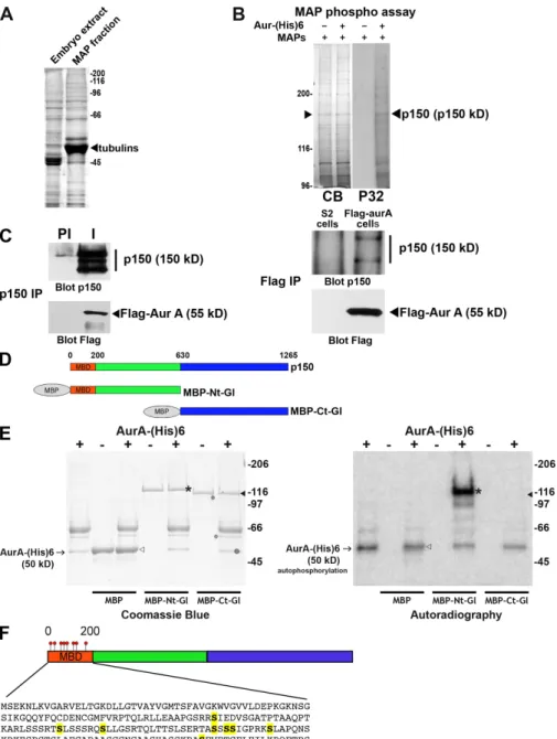
Figure 1. p150glued is an aurora A substrate
in vitro. (A) Coomassie blue–stained gel of the total embryonic extract (left) and the MAPs fraction obtained after sedimentation of polymerized microtubules (right). The strong band corresponds to the tubulins (arrow-head). (B) A kinase assay with (+) or without () aurora A–(His)6 was performed using 20 µg MAPs preparation. (left) The proteins were separated by SDS-PAGE and stained by Coomassie blue (CB). (right) The discrete phosphorylated band (P32) was excised and identified by mass spectrometry as p150glued.
The white line indicates that intervening lanes have been spliced out. (C, left) Extracts from S2 cells stably expressing 3xFlag–aurora A were subjected to IP with preimmune (PI) or affinity-purified immune (I) anti-p150glued
anti-bodies. (right) Extracts from wild-type S2 cells (control) or S2 cells stably expressing 3xFlag–aurora A were subjected to anti-Flag IP. The precipitates were revealed with anti-p150glued (top) or anti-Flag antibodies (bottom).
Note the presence of p150glued in 3xFlag–
aurora A precipitates and, conversely, the presence of aurora A in p150glued
immuno-precipitates. (D) Scheme of the p150glued fusion
proteins used in the kinase assay. N- and terminal fragments of p150glued are displayed
in green and blue, respectively. (E) Recombi-nant MBP, MBP-Ct-Gl, and MBP-Nt-Gl were used for in vitro kinase assays using (+) or not using () aurora A–(His)6 protein kinase in the presence of radio-labeled -[32P]ATP. The
posi-tion of the aurora A–(His)6 band is indicated by arrows (+). MBP and MBP-Ct-Gl, indicated by open and closed arrowheads, respectively, are not phosphorylated, whereas MBP-Nt-Gl (asterisks) is strongly phosphorylated by au-rora A. The Coomassie blue–stained gel (left) and the corresponding autoradiography (right) are shown. (F) Position of the eight phosphory-lated Ser residues (yellow) in the p150glued
MBD (amino acids 0–200).
on July 12, 2010
jcb.rupress.org
Phosphomapping by mass spectrometry revealed
phosphory-lation of eight Ser residues (Ser85, -109, -117, -135, -137, -138,
-144, and -183) within a 200–amino acid N-terminal domain
Fig. 1 D) in Escherichia coli to use in an in vitro kinase assay
(Fig. 1 E). MBP alone and MBP-Ct-Gl were not phosphorylated
by aurora A in vitro, whereas MBP-Nt-Gl was a good substrate.
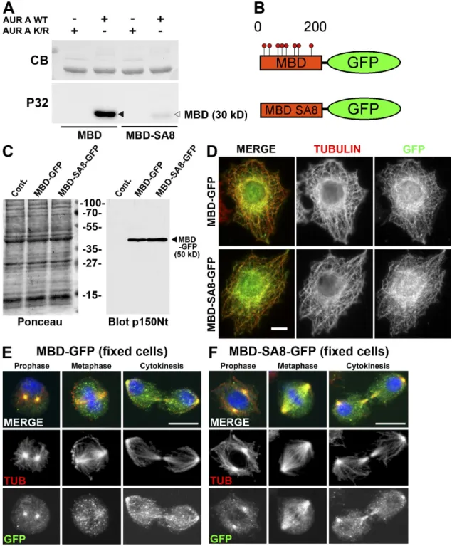
Figure 2. The MBD of p150glued is phosphorylated by aurora A after nuclear envelope breakdown. (A) Wild-type (WT) or mutant MBD fragment in which
the eight Sers (phosphorylated by aurora A) were mutated into Alas (MBD-SA8) were subjected to a kinase assay in the presence of active (AUR A WT) or inactive (AUR A K/R) recombinant aurora A kinase. The Coomassie blue (CB)–stained gel (top) was subjected to autoradiography (bottom). The p150glued
MBD fragment (closed arrowhead) is strongly phosphorylated in the presence of active (but not inactive) aurora A protein kinase, whereas the MBD-SA8 fragment (open arrowhead) remains unphosphorylated. (B) Scheme of the wild-type (MBD-GFP) or mutant (MBD-SA8-GFP) proteins stably expressed in S2-cultured cells. (C) Control (cont) or S2 cell extracts expressing MBD-GFP or MBD-SA8-GFP were separated by SDS-PAGE, transferred onto a nitro-cellulose membrane, and stained by Ponceau S as a loading control (left). The membrane was blotted for p150glued. The position of the 50-kD MBD-GFP
and MBD-SA8-GFP proteins is indicated (arrowhead). (D–F) Interphase S2 cells expressing MBD-GFP (top) or MBD-SA8-GFP (bottom) were fixed and stained for tubulin (red; middle in monochrome) or GFP (green; right in monochrome). Both GFP fusions associate with the interphase microtubule net-work. S2 cells expressing either MBD-GFP (E) or MBD-SA8-GFP (F) were methanol fixed and stained for DNA (blue), tubulin (red; middle in monochrome), and GFP (green; bottom in monochrome). During prophase, the GFP signal was strong at the centrosome region for both proteins. Unlike MBD-GFP, the mutant MBD-SA8-GFP protein remains strongly associated with spindle microtubules during all mitotic steps. Bars, 10 µm.
on July 12, 2010
jcb.rupress.org
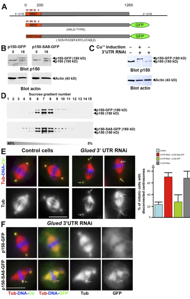
Figure 3. A full-length p150glued that can no longer be phosphorylated by aurora A binds more strongly to microtubules but cannot rescue glued spindle
assembly defect. (A) Scheme of the GFP fusion proteins expressed in S2 cell lines under the control of the metallothionein promoter (inducible by Cu2+). The
constructs lack the glued gene 3UTR (top) targeted by RNAi. (B) p150glued (top) or actin (bottom) Western blot showing p150-GFP (left) and p150-SA8-GFP
(right) protein levels after 0 and 18 h induction on the corresponding cell line. (C) p150glued, p150-GFP, p150-SA8-GFP, and actin protein levels after glued
3UTR RNAi (lanes 2 and 3). Note the knockdown of endogenous p150glued, whereas p150-GFP protein level remains stable. (D) p150glued Western blot
on July 12, 2010
jcb.rupress.org
analysis of the different sucrose fractions after the sedimentation assay (from 5 to 40%) of S2 cell extracts expressing either p150-GFP (top) or p150-SA8-GFP (bottom). (E, left) Fixed control or p150glued-depleted metaphase cells are stained for DNA in blue, microtubules in red (monochrome in bottom), and
centrosomes in green. The arrows point to disconnected centrosomes. (E, right) Quantification of the centrosome disconnection phenotype in control or
glued 3UTR dsRNA-treated S2-cultured cells. Note the rescue obtained after induced expression of p150-GFP protein but not p150-SA8-GFP. (F) Spindle
morphology in p150glued cells after p150-GFP (top) or p150-SA8-GFP (bottom) expression. DNA (blue), microtubules (red; monochrome in the third column),
centrosomes (green), and GFP (green in merge; monochrome on the right) are displayed. p150-SA8-GFP interacts more strongly with spindle microtubules than its wild-type counterpart. The arrows point out the disconnected centrosomes. Error bars indicate mean ± SD. Bars, 10 µm.
corresponding to the MBD (Fig. 1 F). Most of these sites
(ex-cept Ser137 and Ser138) matched the yeast and mammalian
aurora phosphorylation known consensus site RXS/T (where
is a hydrophobic residue; Cheeseman et al., 2002; Ferrari
et al., 2005). These phosphorylated sites were also detected with
other sites after proteomic analyses of p150
gluedisolated from
Kc Drosophila cultured cells in vivo (http://www.sbeams.org/
dev1/sbeams/cgi/Glycopeptide//peptideSearch.cgi). A purified
recombinant MBD in which all phosphorylated Sers were
mutated into Alas had lost its ability to be phosphorylated by
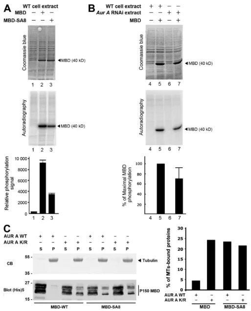
aurora A in vitro (Fig. 2 A). Interestingly, these sites contribute
to 60% of total MBD phosphorylation by S2 cell extracts,
confirming that other phosphorylation sites exist on p150
gluedMBD in vivo (
Fig. S1 A
). Furthermore, phosphorylation assays
using an aurora A–depleted extract showed that aurora A by
itself contributes to 25% of total MBD phosphorylation
ob-served in S2 cells (Fig. S1 B). p150
gluedMBD is responsible for
the microtubule-binding properties of dynactin, and its deletion
affects mitotic but not interphase functions of the protein (Kim
et al., 2007). To analyze the relevance of those phosphorylation
events in vivo, we examined the consequences of expressing
either a GFP-tagged wild-type (MBD-GFP) or
nonphosphory-latable variant of the p150
gluedMBD (MBD-SA8-GFP) in S2
cells (Fig. 2 B). We established stable cell lines in which such
constructs were expressed at equivalent levels (Fig. 2 C). By
immunostaining, both proteins were able to bind microtubules
during interphase when aurora A was not active (Fig. 2 D).
During early mitosis, MBD-GFP and MBD-SA8-GFP were
strongly associated with centrosomal microtubules. However,
the nonphosphorylatable MBD-SA8-GFP mutant protein was
retained on the spindle microtubules during mitosis in all cells
examined during metaphase but not MBD-GFP (n > 200; Fig. 2,
compare E [middle] with F [middle]). We assessed the binding
of recombinant MBD and MBD-SA8 to taxol-polymerized
microtubules in vitro. The p150
gluedMBD affinity for
micro-tubules was five times weaker after incubation with aurora A
protein kinase and ATP. In these conditions, we found that the
mutagenesis of the eight Sers into Alas restored the ability of
p150
gluedMBD to bind to the microtubules (Fig. S1 C). Thus,
the direct interaction of the p150
gluedMBD with the
micro-tubules appears to be negatively regulated by aurora A
phos-phorylation in vitro and in vivo during mitosis.
As p150
gluedMBD is required for mitotic spindle
assem-bly (Kim et al., 2007), it was tempting to speculate that
phos-phorylation at the sites we have mapped is required to control
the overall dynactin accumulation on the mitotic spindle. To
test this hypothesis, we checked whether a full-length p150
gluedGFP fusion protein in which the eight aurora A
phosphoryla-tion sites (Sers) had been replaced by Alas (p150-SA8-GFP)
would (a) accumulate on spindle microtubules and (b) be able
to complement p150
glueddepletion. For this purpose, we
in-duced the expression of either the GFP-tagged p150
gluedwild-type protein (p150-GFP) or a mutant variant (p150-SA8-GFP;
Fig. 3 A). After several hours of induction of their metallothionein
promoter with copper, both GFP-fused proteins were detected by
Western blot analysis (Fig. 3 B). Using a double-stranded RNA
(dsRNA) specific to the glued 3UTR gene (Fig. 3 A; Kim et al.,
2007), we were able to deplete the endogenous p150
gluedbut not
the exogenous GFP-tagged protein (Fig. 3 C). In addition,
sucrose gradient separation analyses revealed that both GFP
fusion proteins were sedimenting together with endogenous
p150
gluedin the 19S dynactin complex (Fig. 3 D). As reported
previously in S2 cells, p150
glued-depleted mitotic cells showed
an obvious disconnection of centrosomes from the mitotic
spin-dle in 67.3 ± 11.6% of the metaphase cells (Fig. 3 E, compare
left with right), whereas only 22.4 ± 4% of the cells showed
this phenotype in control cells (Goshima and Vale, 2003;
Goshima et al., 2005b; Siller et al., 2005). Interestingly, we
com-pletely rescued this defect by expression of p150-GFP (26.7 ±
12%) but not p150-SA8-GFP (69.6 ± 6.9%; Fig. 3 E). In
addi-tion to that, the wild-type protein was weakly localized on
spindle microtubules compared with the mutant, which showed
strong association with these microtubules (Fig. 3 F). This
local-ization of p150-GFP and p150-SA8-GFP was confirmed by
high resolution time-lapse video microscopy. As seen in Fig. 4
(left; and
Video 1
), the p150-GFP protein weakly localized on
the spindle region and microtubule plus ends (Fig. 4, triangles),
whereas p150-SA8-GFP strongly decorated mitotic spindle
fibers and microtubule plus ends (Fig. 4, right; and
Video 2
).
The kinetochore localization (Fig. 4, arrows) remained
identi-cal for both constructs. All of these results led us to investigate
whether aurora A was responsible for the dynamics of dynactin
during mitosis. To do so, we generated a new p150
gluedantibody
against the C-terminal fragment of p150
glued(
Fig. S2 A
) to
localize the p150
gluedprotein during mitosis in Drosophila
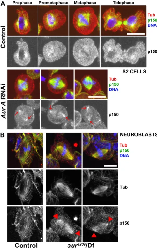
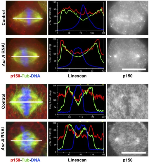
S2-cultured cells. Using this antibody, we observed a
stain-ing of the centrosomal microtubules durstain-ing prophase and the
early stages of spindle formation. The signal decreased
dur-ing spindle maturation before becomdur-ing enriched once more at
telophase, suggesting that spindle pole localization of p150
gluedwas inhibited after nuclear envelope breakdown (Fig. 5 A). We
also found a transient staining of the kinetochore regions of
chromosomes during prometaphase that was replaced after
chromosome congression by a weak staining of spindle fibers,
which is similar to the findings of others (Siller et al., 2005).
After aurora A RNAi, 60% of the metaphase cells (n = 3; 200
cells) showed stronger staining of the spindle pole regions
(Fig. 5 A, arrows; and
Fig. S3
). Interestingly, identical results
on July 12, 2010
jcb.rupress.org
roles during mitotic progression (Karki and Holzbaur, 1999;
Goshima and Vale, 2003; Morales-Mulia and Scholey, 2005;
Delcros et al., 2006). In Drosophila S2 cells, DDC is required
for the centrosome connection to the mitotic spindle and for
the metaphase/anaphase transition (Goshima and Vale, 2003;
Morales-Mulia and Scholey, 2005). Our RNAi experiments in
S2 cells confirm these previous studies, as centrosomes were
disconnected from the poles in 70% of the p150
glued-depleted
mitotic cells. Both RNAi experiments and mathematical models
concur in the notion that kinetochore fibers are captured and
fo-cused at the poles by the DDC, which connects these fibers to
the centrosome-nucleated microtubules. Thus, a lack of DDC
can explain the fact that centrosomes are disconnected from the
spindle (Maiato et al., 2004; Goshima et al., 2005a). Our study
revealed that aurora A participates in the phosphoregulation
of p150
glued, but other kinases can contribute to this process on
p150
gluedMBD itself (Fig. S1) or other DDC subunits (Huang
et al., 1999; Vaughan et al., 2001, 2002). Interestingly, the variant
that can no longer be phosphorylated by aurora A remains
asso-ciated with spindle microtubules and cannot complement the
glued
loss of function. This suggests that DDC levels on spindle
microtubules and at the poles need to be tightly controlled in
Drosophila
cells to avoid centrosome disconnection, a
pheno-type sometimes also observed in aurora A mutant embryos
(un-published data), but not in other cell types in which other kinases
possibly participate in dynactin limitation. The reason why
DDC localization at spindle poles needs to be modulated
re-mains unclear. It is possible that DDC function requires a
mod-erate association of dynactin with spindle microtubules. It is
also possible that overaccumulation of DDC (and cargoes)
could create a traffic obstruction at spindle poles to compete
with other MAPs. Consequently, this would prevent the
connec-tion between kinetochore fibers and astral microtubules. Further
studies will be required to elucidate the complete DDC
regula-tion during mitosis.
Materials and methods
Preparation of microtubules from Drosophila embryos for aurora A phosphorylation assays
500 µl Drosophila early embryos (0–4 h) were collected and lysed in 500 µl BRB80 buffer (80 mM Pipes, pH 6.8, 1 mM MgCl2, and 1 mM EGTA)
sup-plemented with protease inhibitors (Roche) and 0.5% NP-40. The crude extract was centrifuged at 10,000 g for 15 min at 4°C. The supernatant was centrifuged a second time at 100,000 g for 20 min at 4°C. 0.4 ml supernatant containing the soluble proteins was supplemented with 20 µM taxol and 1 mM GTP. Microtubule polymerization was induced for 20 min at 25°C. The microtubules were sedimented on a 0.4 ml BRB80 cushion supplemented with 20 µM taxol, 1 mM GTP, and 40% glycerol. The micro-tubule pellet containing the MAPs was washed twice in BRB80 and resus-pended in BRB80. The MAP fractions were stored at 80°C.
Phosphorylation assay
20 µg MAPs fraction or 3 µg recombinant substrate proteins was incubated with 200 ng recombinant aurora A–(His)6 kinase, 1 µCi radio-labeled -[32P]ATP
(GE Healthcare), and 100 µM ATP in kinase buffer (Giet et al., 2002). The samples were separated by SDS-PAGE and stained by Coo-massie blue. p150glued was identified by mass spectrometry on the excised
band from the gel. To map the phosphorylation sites on the MBP-Nt-Gl pro-tein, the kinase assay was performed without radio-labeled ATP, and the phosphorylated band was excised and sent for phosphopeptide identifi-cation to the Proteomic Platform of the Quebec Genomic Center (http:// proteomique.crchul.ulaval.ca/en/equipment.html).
were obtained after aurora A knockdown in human HeLa cells
(unpublished data), suggesting that DDC regulation is
con-served from Drosophila to humans. We further showed the same
behavior for the dynactin-interacting protein dynein, which
accumulated at the spindle poles after aurora A RNAi in
S2-cultured cells (Fig. S2 B). We also asked whether dynactin
could become mislocalized in larval neuroblasts from aurora A
mutants. As previously described (Siller et al., 2005), dynactin
was faintly distributed along the spindle fibers of wild-type cells
in metaphase. In contrast, 60% of the aurora A mutant cells
with a bipolar spindle showed an accumulation of dynactin
in the spindle pole regions or all over the spindle (six brains;
60 metaphase cells; Fig. 5 B, arrowheads). In these mutant cells,
dynein strikingly followed the same behavior (n = 3; 35
meta-phase cells; Fig. S2 C). Together, these data strongly suggest
that aurora A participates in the control of the dynactin binding
to microtubules, which is an event required for DDC function.
The dynactin complex, composed of p150
gluedand at least
10 other polypeptides, cooperates with dynein to fulfill multiple
Figure 4. p150-SA8-GFP displays higher affinity for microtubules than wild-type p150-GFP protein during mitosis. S2 cells expressing equivalent levels of p150-GFP (left) or p150-SA8-GFP (right) were imaged by time-lapse video microscopy (Videos 1 and 2). The p150-SA8-GFP protein is strongly associated with spindle fibers and microtubule plus ends, whereas p150-GFP shows moderate association with these structures (arrowheads). The kinetochore localization (arrows) is not affected. Bar, 10 µM.
on July 12, 2010
jcb.rupress.org
5-AATAATACGACTCACTATAGGGAAAGGATCDTGTATCGTGGCA-3 and 5-AATAATACGACTCACTATAGGGGAGTTATACAACATCAGCAAA-3 was produced. After isolation, the RNAs were boiled for 20 min and annealed by slow cooling overnight at room temperature. dsRNAs were analyzed by agarose gel electrophoresis and aliquoted at 80°C before use in RNAi experiments. To generate the expression construct encoding the full-length aurora A in fusion with 3xFlag at its N-terminal end, aurora A ORF was first cloned into the pENTR vector (to create an entry clone) using a cloning kit (TOPO; Invitrogen) and recombined using the gateway system (Invitro-gen) into the pHFW containing the 3xFlag tag and a heat shock promoter. An aurora A kinase-dead mutant (K193/R) was generated by mutation of the conserved lysine of the kinase subdomain II into an arginine.
For the MBD expression constructs, each Ser to Ala mutation in the p150glued MBD was obtained by sequential PCRs. The wild-type (MBD)
or nonphosphorylatable (MBD-SA8) domains were cloned directly into For in vivo phosphorylation assay, 3 × 10⁶ control or aurora A–
depleted cells were resuspended in 500 µl of lysis buffer containing protease inhibitors (10 mM Na2VO4 and 50 mM -glycerophosphate;
Giet et al., 2002). 5 µl of the extract was used to phosphorylate 5 µg MBD or MBD-SA8 in the presence of 2.5 µCi radio-labeled -[32P]ATP
(GE Healthcare) and 100 µM cold ATP in kinase buffer. MBD and MBD-SA8 phosphorylation signals were quantified using a phosphoimager (Molecu-lar Dynamics).
dsRNA production and constructs
The aurora A dsRNA production was described previously (Giet and Glover, 2001; Giet et al., 2002). In brief, an 1,000-bp PCR product containing a T7 sequence at each end was used as a template to gener-ate RNA using the Megascript kit (Promega). To obtain the glued tem-plate (for the 3UTR region), an 450-bp PCR product using the primers
Figure 5. p150glued accumulates at spindle
poles of aurora A RNAi-deficient S2 cells and
aurora A mutant neuroblasts. (A) Control S2 cells
(top) were fixed and stained for tubulin (red), p150glued (green), and DNA (blue). The mitotic
phases are displayed at the top. p150glued
localization at spindle poles is clearly detected during prophase and cytokinesis, whereas the signal is very weak during prometaphase and metaphase. In parallel, 60% of aurora A– depleted cells (bottom) exhibit p150glued
accu-mulation at spindle poles (arrows). (B) Wild-type (left) or aurora Ae209/Df(3R)T61 (right) mutant
neuroblasts were fixed and stained for tubu-lin (green; monochrome in middle), p150glued
(red; monochrome in bottom), and DNA (blue). In control metaphase cells, p150glued is faintly
detected along spindle fibers. In aurora A mutant neuroblasts, p150glued accumulates on
spindle fibers and spindle poles (arrowheads). Bars, 10 µm.
on July 12, 2010
jcb.rupress.org
concanavalin A–coated incubation chambers (Labtek; Sigmatek) before imaging. Images were aquired using a spinning-disk system mounted on an inverted microscope (Elipse Ti; Nikon) using a 100× 1.4 NA objective. Images were acquired every 5 or 10 s with a camera (CoolSnap HQ2; Photometrics) controlled by the MetaMorph acquisition software. Antibodies and Western blotting
The YL1/2 rat anti-tyrosinated tubulin antibody (1:1,000) was obtained from Millipore, and the GTU-88 mouse anti–-tubulin antibody (1:1,000) was obtained from Sigma-Aldrich. The mouse dynein clone 1H4 anti-body (1:100) was provided by T. Hays, and the anti-GFP monoclonal antibodies (1:1,000) were obtained from Roche or Invitrogen. Affinity- purified antibodies against aurora A were used at 0.5 µg/ml (Giet et al., 2002). The affinity-purified anti-p150glued antibody was purified as
de-scribed previously (Montembault et al., 2007) and used at 2 µg/ml. Sec-ondary peroxidase-conjugated antibodies were obtained from Jackson ImmunoResearch Laboratories, Inc., and Alexa Fluor–conjugated second-ary antibodies were obtained from Invitrogen. For Western blotting, ECL reagent was purchased from Thermo Fisher Scientific.
Online supplemental material
Fig. S1 shows the contribution of aurora A and other unknown kinases to p150glued MBD phosphorylation in vivo. It also shows that aurora A kinase
inhibits MBD binding to microtubules in vitro. Fig. S2 shows p150glued
anti-body Western blotting in control or aurora A–depleted cells together with dynein localization in control or aurora A–depleted S2 cells. The colocal-ization of dynein and dynactin is also displayed in wild-type or aurora A mutant neuroblasts. Fig. S3 shows the distribution (line scans) of p150glued
and tubulin in control or aurora A–depleted cells. Videos 1 and 2 show the behavior of wild-type and nonphosphorylatable p150glued proteins fused to
GFP during metaphase. Online supplemental material is available at http://www.jcb.org/cgi/content/full/jcb.201001144/DC1.
We thank the laboratory members for stimulating discussions, Clotilde Petretti, Thomas Hays, Pier Paolo d’Avino, Gilles Hickson, and Arnaud Echard for re-agents, and Stéphanie Dutertre for microscopy facilities of the IFR140. R. Giet thanks Kathryn Lilley for initial proteomic studies and the members of David Glover’s group in Cambridge, where this project was initiated (Department of Genetics, University of Cambridge, Cambridge, England, UK). Special thanks are due to Claude Prigent for his full support and to Alex Holmes, Emeric Sevin, and M. Savoian for their critical reading of the manuscript.
R. Giet and N. Franck are funded by the Agence Nationale de la Recherche (programme Jeune Chercheur) and the Ligue Nationale Contre le Cancer (Equipe Labellisée). E. Montembault and P. Romé are doctoral fellows of the French Ministère de la Recherche.
Submitted: 26 January 2010 Accepted: 19 April 2010
References
Barr, A.R., and F. Gergely. 2007. Aurora-A: the maker and breaker of spindle poles. J. Cell Sci. 120:2987–2996. doi:10.1242/jcs.013136
Barros, T.P., K. Kinoshita, A.A. Hyman, and J.W. Raff. 2005. Aurora A acti-vates D-TACC–Msps complexes exclusively at centrosomes to stabi-lize centrosomal microtubules. J. Cell Biol. 170:1039–1046. doi:10 .1083/jcb.200504097
Cheeseman, I.M., S. Anderson, M. Jwa, E.M. Green, J. Kang, J.R. Yates III, C.S. Chan, D.G. Drubin, and G. Barnes. 2002. Phospho-regulation of kinetochore-microtubule attachments by the Aurora kinase Ipl1p.
Cell. 111:163–172. doi:10.1016/S0092-8674(02)00973-X
Clemens, J.C., C.A. Worby, N. Simonson-Leff, M. Muda, T. Maehama, B.A. Hemmings, and J.E. Dixon. 2000. Use of double-stranded RNA inter-ference in Drosophila cell lines to dissect signal transduction pathways.
Proc. Natl. Acad. Sci. USA. 97:6499–6503. doi:10.1073/pnas.110149597 Cowley, D.O., J.A. Rivera-Pérez, M. Schliekelman, Y.J. He, T.G. Oliver, L.
Lu, R. O’Quinn, E.D. Salmon, T. Magnuson, and T. Van Dyke. 2009. Aurora-A kinase is essential for bipolar spindle formation and early de-velopment. Mol. Cell. Biol. 29:1059–1071. doi:10.1128/MCB.01062-08 Delcros, J.G., C. Prigent, and R. Giet. 2006. Dynactin targets Pavarotti-KLP to
the central spindle during anaphase and facilitates cytokinesis in
Drosophila S2 cells. J. Cell Sci. 119:4431–4441. doi:10.1242/jcs.03204 Ferrari, S., O. Marin, M.A. Pagano, F. Meggio, D. Hess, M. El-Shemerly, A.
Krystyniak, and L.A. Pinna. 2005. Aurora-A site specificity: a study with synthetic peptide substrates. Biochem. J. 390:293–302. doi:10 .1042/BJ20050343
pET102/TOPO bacterial expression vector or pENTR to generate entry clones. The latter clones were recombined into pDEST17 to generate bacte-rial expression constructs and in pAWG to allow S2 cell expression of either MBD-GFP or MBD-SA8-GFP under the control of the actin 5c pro-moter. Both pAWG and pHFW were obtained from the Carnegie Institution of Washington.
N- and C-terminus domains of p150glued were amplified by PCR and
cloned into the pMal-C2E expression vector using KpnI and BamHI restric-tion sites. All constructs were verified by sequencing.
Full-length p150glued was cloned into pMT-GFP-Ct (provided by
M. Savoian, University of Cambridge, Cambridge, England, UK) using the gateway system (through an entry clone) to generate p150-GFP expression construct. p150-SA8-GFP was subsequently obtained with the QuickChange Mutagenesis kit (Agilent Technologies). The pAct5C-Cherry–-tubulin construct was provided by G. Hickson (University of California, San Francisco, San Francisco, CA).
Production of the recombinant proteins and antibody purification
Aurora A–(His)6, aurora A–K/R-(His)6, MBD-(His)6, or MBD-SA8-(His)6 were purified as described previously (Giet et al., 2002). MBP-Nt-Gl and MPB-Ct-Gl were expressed in E. coli BL21(DE3) pLysS (EMD) for 4 h at 25°C, purified on an amylose column, and dialysed against PBS and stored at 80°C before use for aurora A kinase assay. The purified MBP-Ct-Gl protein was also used to immunize rabbits. Obtained anti-p150glued antibodies were
affinity purified on a nitrocellulose membrane, and the purified antibodies were stored at 80°C as described previously (Montembault et al., 2007). In vitro microtubule-binding assay
1 µg purified recombinant p150glued MBD or MBD-SA8 proteins in fusion with
a hexahistidine tag was incubated with either 200 ng aurora A or aurora A– K/R proteins in kinase buffer containing 1 mM ATP in a total volume of 10 µl for 20 min. The kinase reaction was then incubated with 25 µl (100 µg) of taxol-stabilized microtubules for 10 min at 37°C (Roghi et al., 1998). The microtubule pellet and the supernatant containing microtubule-unbound proteins were separated by sedimentation at 100,000 g for 20 min at 37°C (TLA-100; Beckman Coulter). The proteins present in the pellets and super-natants were analyzed by Western blotting or Coomassie blue staining. RNAi, transfections, drug treatment, stable line generation, and rescue experiments
Drosophila S2 cells were grown and processed for RNAi as described
previ-ously (Clemens et al., 2000). In brief, 106 cells were incubated with 10 µg/ml
dsRNA in media without serum. Alternatively, 10 µg transfection reagent (Transfast; Promega) was added together with 3 µg dsRNA following the manufacturer’s instructions. After 1 h, fresh media were added to the cells. At 4 d after transfection, the cells were fixed and analyzed for mitotic defects (Giet and Glover, 2001). 100–200 mitoses were scored and analyzed per experiment, and each experiment was repeated at least three times.
To make S2 stable lines expressing 3xFlag–aurora A, MBD-GFP or MBD-SA8-GFP, p150-GFP, and p150-SA8-GFP, 5 µg of each expression construct was cotransfected with 1 µg pIB/V5-His/CAT (Invitrogen). Alter-natively, 5 µg pAct5c-Cherry–-tubulin was cotransfected in those cells. Transfection reagent (Effectene; QIAGEN) was used for transfection. Stable lines were selected and expanded in media containing 25 µg/ml blastici-din S (Invitrogen). For rescue experiments, expression of either p150-GFP or p150-SA8-GFP proteins was induced 18 h before fixation by supple-menting media with CuSO4 (300 µM final concentration).
Immunofluorescence analysis
S2 cells were fixed in PHEM buffer (60 mM Pipes, 25 mM Hepes, 10 mM EGTA, and 4 mM MgCl2) containing 3.7% formaldehyde and 0.1% Triton
X-100. In some cases, the cells were plated on concanavalin A–coated coverslips for 1 h and fixed in methanol at 20°C for 10 min. The fixed cells were briefly washed in PBS and blocked for 1 h in PBS containing 0.1% Triton X-100 and 1% BSA (PBST-BSA). Primary antibodies were incu-bated overnight at 4°C, and secondary antibodies were incuincu-bated for 1 h at room temperature in PBST-BSA. DNA was stained with Hoechst 33258. Slides were mounted in ProLong gold (Invitrogen) and observed with a microscope (DMRXA2; Leica) using a 63× 1.3 NA objective. Images were acquired with a camera (CoolSnap HQ; Photometrics) and processed with MetaMorph software (Universal Imaging). Alternatively, images were ac-quired with an inverted confocal microscope (SP2; Leica).
Time-lapse imaging
S2 cells expressing p150-GFP or p150-SA8-GFP were incubated with CuSO4 (300 µM final concentration) for 18 h and plated for 1 h on
on July 12, 2010
jcb.rupress.org
Zhang, J., and T.L. Megraw. 2007. Proper recruitment of gamma-tubulin and D-TACC/Msps to embryonic Drosophila centrosomes requires Centro-somin Motif 1. Mol. Biol. Cell. 18:4037–4049. doi:10.1091/mbc. E07-05-0474
Zhang, X., S.C. Ems-McClung, and C.E. Walczak. 2008. Aurora A phosphorylates MCAK to control ran-dependent spindle bipolarity. Mol. Biol. Cell. 19: 2752–2765. doi:10.1091/mbc.E08-02-0198
Giet, R., and D.M. Glover. 2001. Drosophila aurora B kinase is required for his-tone H3 phosphorylation and condensin recruitment during chromosome condensation and to organize the central spindle during cytokinesis. J. Cell Biol. 152:669–682. doi:10.1083/jcb.152.4.669
Giet, R., and C. Prigent. 1999. Aurora/Ipl1p-related kinases, a new oncogenic family of mitotic serine-threonine kinases. J. Cell Sci. 112:3591–3601. Giet, R., D. McLean, S. Descamps, M.J. Lee, J.W. Raff, C. Prigent, and D.M.
Glover. 2002. Drosophila Aurora A kinase is required to localize D-TACC to centrosomes and to regulate astral microtubules. J. Cell Biol. 156:437– 451. doi:10.1083/jcb.200108135
Giet, R., C. Petretti, and C. Prigent. 2005. Aurora kinases, aneuploidy and cancer, a coincidence or a real link? Trends Cell Biol. 15:241–250. doi:10.1016/j.tcb.2005.03.004
Glover, D.M., M.H. Leibowitz, D.A. McLean, and H. Parry. 1995. Mutations in aurora prevent centrosome separation leading to the formation of mono-polar spindles. Cell. 81:95–105. doi:10.1016/0092-8674(95)90374-7 Goshima, G., and R.D. Vale. 2003. The roles of microtubule-based motor
pro-teins in mitosis: comprehensive RNAi analysis in the Drosophila S2 cell line. J. Cell Biol. 162:1003–1016. doi:10.1083/jcb.200303022
Goshima, G., F. Nédélec, and R.D. Vale. 2005a. Mechanisms for focusing mitotic spindle poles by minus end–directed motor proteins. J. Cell Biol. 171:229–240. doi:10.1083/jcb.200505107
Goshima, G., R. Wollman, N. Stuurman, J.M. Scholey, and R.D. Vale. 2005b. Length control of the metaphase spindle. Curr. Biol. 15:1979–1988. doi:10.1016/j.cub.2005.09.054
Hannak, E., M. Kirkham, A.A. Hyman, and K. Oegema. 2001. Aurora-A kinase is required for centrosome maturation in Caenorhabditis elegans. J. Cell
Biol. 155:1109–1116. doi:10.1083/jcb.200108051
Huang, C.Y., C.P. Chang, C.L. Huang, and J.E. Ferrell Jr. 1999. M phase phos-phorylation of cytoplasmic dynein intermediate chain and p150(Glued).
J. Biol. Chem. 274:14262–14269. doi:10.1074/jbc.274.20.14262 Karki, S., and E.L. Holzbaur. 1999. Cytoplasmic dynein and dynactin in cell
di-vision and intracellular transport. Curr. Opin. Cell Biol. 11:45–53. doi:10.1016/S0955-0674(99)80006-4
Kim, H., S.C. Ling, G.C. Rogers, C. Kural, P.R. Selvin, S.L. Rogers, and V.I. Gelfand. 2007. Microtubule binding by dynactin is required for micro-tubule organization but not cargo transport. J. Cell Biol. 176:641–651. doi:10.1083/jcb.200608128
Maiato, H., C.L. Rieder, and A. Khodjakov. 2004. Kinetochore-driven formation of kinetochore fibers contributes to spindle assembly during animal mito-sis. J. Cell Biol. 167:831–840. doi:10.1083/jcb.200407090
Montembault, E., S. Dutertre, C. Prigent, and R. Giet. 2007. PRP4 is a spin-dle assembly checkpoint protein required for MPS1, MAD1, and MAD2 localization to the kinetochores. J. Cell Biol. 179:601–609. doi:10.1083/jcb.200703133
Morales-Mulia, S., and J.M. Scholey. 2005. Spindle pole organization in
Drosophila S2 cells by dynein, abnormal spindle protein (Asp), and KLP10A. Mol. Biol. Cell. 16:3176–3186. doi:10.1091/mbc.E04-12-1110 Roghi, C., R. Giet, R. Uzbekov, N. Morin, I. Chartrain, R. Le Guellec,
A. Couturier, M. Dorée, M. Philippe, and C. Prigent. 1998. The Xenopus protein kinase pEg2 associates with the centrosome in a cell cycle-dependent manner, binds to the spindle microtubules and is involved in bipolar mi-totic spindle assembly. J. Cell Sci. 111:557–572.
Saffin, J.M., M. Venoux, C. Prigent, J. Espeut, F. Poulat, D. Giorgi, A. Abrieu, and S. Rouquier. 2005. ASAP, a human microtubule-associated protein required for bipolar spindle assembly and cytokinesis. Proc. Natl. Acad.
Sci. USA. 102:11302–11307. doi:10.1073/pnas.0500964102
Siller, K.H., M. Serr, R. Steward, T.S. Hays, and C.Q. Doe. 2005. Live imaging of Drosophila brain neuroblasts reveals a role for Lis1/dynactin in spindle assembly and mitotic checkpoint control. Mol. Biol. Cell. 16:5127–5140. doi:10.1091/mbc.E05-04-0338
Terada, Y., Y. Uetake, and R. Kuriyama. 2003. Interaction of Aurora-A and centrosomin at the microtubule-nucleating site in Drosophila and mam-malian cells. J. Cell Biol. 162:757–763. doi:10.1083/jcb.200305048 Vaughan, P.S., J.D. Leszyk, and K.T. Vaughan. 2001. Cytoplasmic dynein
inter-mediate chain phosphorylation regulates binding to dynactin. J. Biol.
Chem. 276:26171–26179. doi:10.1074/jbc.M102649200
Vaughan, P.S., P. Miura, M. Henderson, B. Byrne, and K.T. Vaughan. 2002. A role for regulated binding of p150Glued to microtubule plus ends in
organelle transport. J. Cell Biol. 158:305–319. doi:10.1083/jcb.200201029 Venoux, M., J. Basbous, C. Berthenet, C. Prigent, A. Fernandez, N.J. Lamb, and
S. Rouquier. 2008. ASAP is a novel substrate of the oncogenic mitotic ki-nase Aurora-A: phosphorylation on Ser625 is essential to spindle formation and mitosis. Hum. Mol. Genet. 17:215–224. doi:10.1093/hmg/ddm298 Wong, J., R. Lerrigo, C.Y. Jang, and G. Fang. 2008. Aurora A regulates the
activ-ity of HURP by controlling the accessibilactiv-ity of its microtubule-binding domain. Mol. Biol. Cell. 19:2083–2091. doi:10.1091/mbc.E07-10-1088
on July 12, 2010
jcb.rupress.org
JCB
T
H
E
J
O
U
R
N
A
L
O
F
C
E
L
L
B
IO
L
O
G
Y
Supplemental material
Romé et al., http://www.jcb.org/cgi/content/full/jcb.201001144/DC1
Figure S1. Analysis of p150glued MBD phosphorylation in vivo and effect on microtubule binding in vitro. (A) p150glued MBD (lane 2) or MBD-SA8 (lane 3)
were phosphorylated by wild-type (WT) S2 cell extracts. Note the strong decrease of MBD-SA8 phosphorylation compared with MBD, indicating that identi-fied aurora A phosphorylation sites correspond to 60% of the overall in vivo phosphorylation of this fragment (n = 5; compare lane 2 with lane 3). (B) Wild-type (lanes 4 and 5) or aurora A–depleted cell extracts (lanes 6 and 7) were used to phosphorylate wild-type MBD (lanes 5 and 7). After aurora A depletion (compare lane 5 with lane 7), MBD phosphorylation decreases by 25% (n = 6). (C, left) Wild-type MBD or nonphosphorylatable MBD mutant (MBD-SA8) was incubated in the presence of ATP with active (AurA WT) or inactive (AurA K/R) aurora A kinase. The reaction product was incubated with taxol-stabilized microtubules (see Materials and methods) and sedimented at 100,000 g. The microtubule pellets (P) and the supernatants (S) were analyzed by Coomassie blue (CB) staining to reveal tubulins (top) or anti-pentahistidine Western blotting to reveal wild-type MBD or MBD-SA8 (bottom). The arrow-head indicates the position of tubulin (microtubules) in the pellet. In the presence of aurora A K/R, a fraction of wild-type MBD and MBD-SA8 is bound to microtubules. In contrast, in the presence of wild-type aurora A, wild-type MBD cannot bind to microtubules, whereas the MBD-SA8 still does. Thus, specific phosphorylation of MBD by aurora A prevents dynactin binding to microtubules. (C, right) Percentage of total MBD and MBD-SA8 proteins detected in the microtubule pellets. p150glued MBD behavior in this assay was identical in three different experiments. Error bars indicate mean ± SD.
on July 12, 2010
jcb.rupress.org
Figure S2. Analysis of p150glued protein levels in S2 cells, dynein localization in aurora A–depleted S2 cells, and aurora A mutant neuroblasts. (A, right)
Western blot of S2 cell extract probed with affinity-purified anti-p150glued antibody. Western blot showing aurora A (middle), -tubulin (bottom), or p150glued
(top) protein levels in control () or aurora A RNAi-treated cells (+). Note the strong depletion of the aurora A protein, whereas the p150glued protein levels
are unaffected. (B) Control (left) or aurora A RNAi-treated S2 cells were stained for tubulin (green) and dynein (red; right in monochrome). Note the accu-mulation of dynein at spindle poles in aurora A dsRNA-treated cells (right, arrowheads). Bar, 5 µm. (C) Wild-type (WT; top) or aurora Ae209/Df(3R)T61
neu-roblasts (bottom) during metaphase were fixed and stained for tubulin (green) and DNA (blue). Note that left and right panels show the same cells stained for p150glued (red; monochrome in left) and dynein (red; monochrome in right). Dynein and dynactin show similar localization patterns in these cells,
sug-gesting that they remain associated in the mutant. Bar, 10 µm.
on July 12, 2010
jcb.rupress.org
Figure S3. Distribution of p150glued on the mitotic spindle. Control or aurora A dsRNA-treated cells were stained for microtubules (green), DNA (blue), and
p150glued (red). A line scan between the poles was performed to show the relative fluorescence intensity of each fluorophore. Note the strong accumulation
of p150glued at the spindle poles of aurora A dsRNA-treated cells (compare red line with green line). Bar, 10 µm. The two top panels were acquired with a
conventional microscope equipped with a camera. The two bottom cells were also shown in Fig. 5 A (control and aurora A–depleted metaphases).
Video 1. Dynamics of p150-GFP protein in a Drosophila S2 cell. The time (min:s) is displayed at the bottom of the picture.
Video 2. Dynamics of p150-SA8-GFP protein in a Drosophila S2 cell. The time (min:s) is displayed at the bottom of the picture.