HAL Id: inserm-00696195
https://www.hal.inserm.fr/inserm-00696195
Submitted on 23 May 2012
HAL is a multi-disciplinary open access
archive for the deposit and dissemination of sci-entific research documents, whether they are pub-lished or not. The documents may come from teaching and research institutions in France or abroad, or from public or private research centers.
L’archive ouverte pluridisciplinaire HAL, est destinée au dépôt et à la diffusion de documents scientifiques de niveau recherche, publiés ou non, émanant des établissements d’enseignement et de recherche français ou étrangers, des laboratoires publics ou privés.
Differential effects of busulfan on gonadal development
in five divergent anuran species.
Rafal Piprek, Anna Pecio, Jacek Kubiak, Jacek Szymura
To cite this version:
Rafal Piprek, Anna Pecio, Jacek Kubiak, Jacek Szymura. Differential effects of busulfan on gonadal development in five divergent anuran species.. Reproductive Toxicology, Elsevier, 2012, 34 (3), pp.393-401. �10.1016/j.reprotox.2012.05.002�. �inserm-00696195�
Accepted Manuscript
Title: Differential effects of busulfan on gonadal development in five divergent anuran species
Authors: Rafał P. Piprek, Anna Pecio, Jacek Z. Kubiak, Jacek M. Szymura
PII: S0890-6238(12)00079-2
DOI: doi:10.1016/j.reprotox.2012.05.002 Reference: RTX 6694
To appear in: Reproductive Toxicology
Received date: 12-1-2012 Revised date: 13-4-2012 Accepted date: 8-5-2012
Please cite this article as: Piprek RP, Pecio A, Kubiak JZ, Szymura JM, Differential effects of busulfan on gonadal development in five divergent anuran species,
Reproductive Toxicology (2010), doi:10.1016/j.reprotox.2012.05.002
This is a PDF file of an unedited manuscript that has been accepted for publication. As a service to our customers we are providing this early version of the manuscript. The manuscript will undergo copyediting, typesetting, and review of the resulting proof before it is published in its final form. Please note that during the production process errors may be discovered which could affect the content, and all legal disclaimers that apply to the journal pertain.
Accepted Manuscript
Highlights
The effects of busulfan on the gonadal development were investigated in anuran amphibians. The tadpoles treated with busulfan did not display sex reversal signs. The complete germ cell loss was observed in X. laevis. Germ cells are not necessary for the testis formation but are crucial during ovarian development.
Accepted Manuscript
Differential effects of busulfan on gonadal development in five divergent anuran species
Rafał P. Piprek1, Anna Pecio1, Jacek Z. Kubiak2, Jacek M. Szymura1
1Department of Comparative Anatomy, Institute of Zoology, Jagiellonian University,
Grononstajowa 9, 30-387 Kraków, Poland
2CNRS, UMR 6061, Institute of Genetics and Development of Rennes, Cell Cycle Group,
IFR 140, UEB, Faculty of Medicine, F-35043 Rennes, France
Corresponding author : Rafał P. Piprek Department of Anatomy Institute of Zoology UJ Gronostajowa 9 30-387 Kraków Poland Phone : +48126645059 e-mail: rafal.piprek@uj.edu.pl
Accepted Manuscript
Abstract
The aim of this paper was to investigate the effects of germ cell depletion on the sexual differentiation of gonads in five anuran species. We used busulfan to eliminate the germ cells. Our results indicate that germ cells are not required for gonadal ridge
formation or the development of the undifferentiated gonads. We observed a gradual degeneration of gonads in studied species and the transdifferentiation of the whole gonads into large fat bodies in Xenopus laevis. In the latter the sexual differentiation of gonads or seminiferous tubules were not impaired in the absence of germ cells. Thus, the X. laevis may serve as a model to study the human Del Castillo syndrome. Our study shows that in anuran amphibians the germ cells are not necessary for the formation of the testis, but they are crucial for development of the ovaries and are required for the
maintenance of the gonadal structure.
Accepted Manuscript
1. Introduction
Busulfan is an alkylating antineoplastic compound used as a common chemotherapeutic agent [1]. In the intracellular environment, products of busulfan degradation bind the guanine bases of DNA and cause guanine-adenine intrastrand crosslinking [2]. Such damage cannot be repaired and the affected cells undergo
apoptosis, which may exert a teratogenic effect on the organism. Already in 1953, Bollag showed that the intraperitoneal administration of busulfan in rat caused germ cell
depletion [3]. Later, similar results were obtained in chicken and quail [4-6]. This
property is a serious drawback for busulfan use for human chemotherapy, where it causes gonadal failure resulting in a lack of sexual development and infertility [7].
Gonads are unique organs because they are not necessary for somatic life but are crucial for reproduction and gene transfer from one generation to another. Embryonic gonads are composed of somatic cells (epithelial and mesenchymal cells) as well as germ cells that later give rise to the oocytes or spermatozoa. During embryogenesis, the germ cells originate as the primordial germ cells (PGS) in the regions distant from the
embryonic gonads and then migrate towards genital ridges [8,9]. During migration, PGCs divide and for this reason they are very sensitive to the toxins such as busulfan [10,11]. In
Xenopus embryo the PGCs are localized in the endoderm and afterwards actively migrate
from the gut region through the dorsal mesentery to the genital ridges [8].
The sexually undifferentiated gonads are composed of a cortex and medulla. The germ cells are incorporated into the cortex [12]. During ovarian development, female germ cells (oogonia) remain in the cortex where they become enclosed by follicular cells. During development of the testes the basal laminae between the cortex and medulla
Accepted Manuscript
disintegrate and germ cells translocate into the medulla. The testis cords are formed within the central part of the gonad and are composed of Sertoli cells that enclose the germ cells (spermatogonia). The testis cords are rudiments of seminiferous tubules, within which spermatogenesis proceeds after the metamorphosis [13].
Several studies have examined gonadal development after germ cell depletion in various vertebrates [14-16]. In the zebrafish, ablation of germ cells caused
transdifferentiation of the female gonad into the testis [15]. Similar female-to-male sex reversal has been observed in sterile mammals. The precursors of ovarian follicular cells transdifferentiate into clusters of Sertoli cells that form testis cord-like structures [17-19]. On the other hand, the testicular development is not affected in the absence of germ cells and sterile seminiferous tubules are formed [14]. In birds such as quail, sex the germ cell ablation does not result in sex reversal [6]. Similarly, in the reptiles, such as the red-eared slider (Trachemys scripta) the germ cell depletion after busulfan treatment does not alter the sex of the gonads [16]. The effects of germ cell absence on amphibian gonads have been studied only in the early stages of genital ridge formation in X. laevis and
Pelophylax esculentus [20,21]. These studies showed that the germ cells are not required
for the formation of genital ridges.
Several studies showed that different vertebrates response differently to the loss of germ cells in the gonad [14-16]. Because our previous study showed that the
gonadogenesis is different in divergent anuran amphibian species [22]. We assume that the effects of the germ cell depletion should vary within this group of vertebrates. Thus, we studied whether in different anuran species the gonadal sex is independent of the presence of germ cells and if the absence of germinal cells may lead , similar to zebrafish
Accepted Manuscript
and mouse, to sex reversal. To investigate this the 24h exposure of anuran tadpoles to water with 0.12 mM busulfan was carried out. We studied five anuran species
representing distant phylogenetic lineages. The European fire-bellied toad (Bombina
bombina: Bombinatoridae) and the African clawed frog (Xenopus laevis: Pipidae) are
representatives of the most basal branches (Archaeobatrachia) [23]. The more derived group, Neobatrachia, was represented by two sister lineages: Hyloidea (Bufo viridis: Bufonidae, Hyla arborea: Hylidae) and Ranoidea (Rana temporaria: Ranidae). Our choice was determined by the fact that the sex determination as well as the gonadal differentiation patterns vary considerably between these anuran species. In some species the males are heterogametic (Bombina sp., H. arborea, R. temporaria) whereas in others the females are heterogametic (X. laevis, B. viridis) [24-27]. Moreover, sexual
differentiation of gonads can take place at various stages of development: in early larval period (Bombina sp., X. laevis, H. arborea), during the metamorphosis (B. viridis) or postmetamorphosis (R. temporaria) [22,28,29].
2. Materials and Methods 2.1. Animals
Larvae of X. laevis (n=139) were obtained in the laboratory whereas the eggs of
B. bombina, H. arborea, B. viridis and R. temporaria were collected in the wild. The
tadpoles were reared in 10-L aquaria. X. laevis larvae were fed with Sera Micron (Sera, Heinsberg, Germany) twice a day. All specimens used in the experiment were acquired according to Polish legal regulations concerning the protection of wild species (Dz. U. nr 33, poz. 289, 2005). We obtained permission from the Polish Ministry of Environment
Accepted Manuscript
Protection and Forestry and approval from the I Local Commission for Ethics in Experiments on Animals.
2.2 Busulfan treatment
Busulfan (1,4-butanediol dimethanesulfonate (Sigma, Poznań, Poland)) was dissolved in DMSO (0.6 M stock solution) and 2 mL of stock solution was added to 10 L of dechlorinated water to the final concentration 0.12 mM (i.e. 30 mg/L). Tadpoles of X.
laevis were staged according to Nieuwkoop and Faber [30] and the other species
according to Gosner [31]. Larvae at stages proceeding the genital ridge formation, i.e. at the Nieuwkoop stage 45 for X. laevis or the Gosner stage 24 for the rest, were placed in 0.12 mM solution of busulfan for 24 h. Tadpoles kept in water with DMSO (0.2 ml/L) for 24 h were used as a negative control. Afterwards both busulfanized and control animals were reared in water without busulfan or DMSO at temperature of 19oC and 12:12 L:D period. Premetamorphic larvae were staged and anesthetized with MS222 (Sigma, Poznań, Poland) solution once per week at Gosner stages 26, 30, 34, 37, 40, 44 and at Nieuwkoop stages 49, 51, 53, 55, 60, 64 for X. laevis. Postmetamorphic animals were anesthetized six months and one year after metamorphosis (Table 1). The gonads together with the kidneys and fat bodies were dissected and fixed in Bouin’s solution.
2.3 Light microscopy
Fixed organs were dehydrated and embedded in paraplast (Sigma, Poznań, Poland). Then 6 m sections were stained with Debreuill trichrome [32]. Images were taken with a Nikon Eclipse E600 light microscope and processed with Corel Photo-Paint
Accepted Manuscript
11. Numbers of cells were counted in 20 subsequent optical sections and compared using Student’s t-test in Statistica 6 Pl software. The germ cells were recognized due to the large, pale nuclei and the Sertoli cells were defined as the cells located inward from the basal laminae of the testis cords or seminiferous tubules after germ cell ablation [33].
3. Results
3.1. The influence of busulfan on anuran tadpoles
Busulfan treatment impacted anurans’ survival as well as the shape and size of the body (Tables 1, 2). The most noticeable effect was observed in the larval body length in
B. bombina and B. viridis (Table 2). H. arborea larvae (37%) had impaired
osmoregulation manifested by the storage of a large amount of fluid within the body. Some individuals (2%) showed malformations such as additional forelimbs. Because the busulfan had only a minor effect on the mortality and phenotype of larvae of X. laevis we have chosen this species to study the fate of germ cell depleted gonads.
3.2. Effect of busulfan on X. laevis gonads 3.2.1. Undifferentiated gonads
X. laevis was the only species in which busulfan caused complete germ cell
ablation, while the soma was almost unchanged (Table 3). Due to the disappearance of germ cells, the gonads of busulfanized individuals were smaller in comparison to the control (Tables 2,3). The formation of the genital ridges proceeded normally and began at the Nieuwkoop stage 49. The absence of the primordial germ cells during the formation of genital ridges indicated the apoptosis of PGCs during their extragonadal migration. At
Accepted Manuscript
the Nieuwkoop stage 51, the beginning of the medulla formation was visible due to the appearance of a somatic cell cluster in the gonadal hilus (the rudiment of medulla) (Fig. 1A,B). Despite of the absence of the germ cells the somatic cells of the gonad were more abundant than in control. At the Nieuwkoop stage 53, a well-separated medulla, which is the sign of the ovarian differentiation, was discernible in the center of gonad. The gonads devoid of germ cells had a larger amount of extracellular matrix in stromal space, i.e. between the cortex and medulla. Melanophores and fibroblasts were visible within the relatively extensive stroma. The cortex and medulla of the ovary were lined with the folded basal laminae.
3.2.2. Ovary differentiation
At the Nieuwkoop stage 55, during sexual differentiation of the ovary, a
secondary cavity appeared within the medulla due to dispersion of cells as in the control (Fig. 1C, D). In the absence of germ cells, the ovarian follicles or the germ cell nests in the cortex did not form (Fig. 1D). Somatic cells of the cortex were arranged in a thin layer covering the gonad and thus no follicular epithelium was observed. We did not identified any soma damages after busulfanization, i.e., exposure to busulfan, using the light microscopy (Fig. 1D). During the metamorphosis (the Nieuwkoop stage 64) the ovaries assumed the shape of a sac filled with an extensive cavity enclosed by two thin layers of somatic cells (cortex and medulla) separated by a thick sheath of extracellular matrix.
Accepted Manuscript
At the Nieuwkoop stage 55, the sexual differentiation of the testis was recognizable owing to gathering of somatic cells into groups enclosed by the basal laminae in both the control and the busulfanized tadpoles (Fig. 1E,F). Thus the testis cords were formed regardless of the germ cell presence. The shrunken gonadal cortex transformed into the tunica albuginea enclosing the whole testis and a lumen appeared within the testis cords during metamorphosis as in the control. In the absence of germ cells, the seminiferous tubules were aligned with the Sertoli (epithelial) cells forming the pseudostratified epithelium. There were 22 ± 4.4 Sertoli cells per cross section of the testis cord in the busulfan-treated tadpoles whereas the control testis cords contained 5 ± 1.5 Sertoli cells per section (P<0.05; n=50). Thus the number of Sertoli cells increased over four times in comparison to the control, which was clearly visible in the histological sections (Fig. 1E,F). Typical connections between the testis tubules and the tubules of extragonadal system were observed in busulfanized and the control animals. Although the gonads attained a smaller size in comparison to the control, no signs of developmental retardation were noticed at the premetamorphic stages (Table 2).
3.2.4. Long term effects of busulfan
Six months after metamorphosis only a small remnants of testes were observed among extensive fat bodies in 7 among 12 busulfanized individuals (Fig. 2A). In the other 5 individuals no gonads were found at all. The number of the seminiferous tubules was drastically reduced within such residual testes and some abnormal vesicles were noticeable at their periphery. It can be supposed that the sterile seminiferous tubules transformed into such extensive vesicles in juveniles. One year after metamorphosis
Accepted Manuscript
gonads could not be detected during the macroscopic dissection of busulfanized frogs (n=10). The extremely large fat bodies (corpora adiposa) filled the space of abdominal cavity. The remnants of testes were found embedded in the fat bodies in 4 individuals among 10 studied and were composed of a few sterile testis tubules lined with monolayer epithelium (Fig. 2B). The fat bodies were attached along the entire lengths of the kidneys. In 6 animals no signs of gonads could be noticed and no remnants of ovaries were
discerned.
3.3. Effect of busulfan on the gonads in B. bombina
In busulfanized individuals of the European fire-bellied toad, gonads were
distinctly smaller and retarded in development than the control (Table 2, Fig. 3A,B,C). A reduction in germ cell number was apparent in busulfanized individuals, however, these cells were not completely eliminated and 15.95% of germ cells survived until the Gosner stage 34 (Table 3). The genital ridges were formed at the Gosner stage 26 similar to the control. The cluster of somatic cells in the gonadal hilus appeared at the Gosner stage 30 (Fig. 3B), thus the rudimentary medulla was formed in spite of the germ cell absence. In the control gonads such a distinctive medulla was not noticeable at all (Fig. 3A). The visibly decreased number of somatic and germ cells resulted in alternation of the cortex and medulla differentiation and thus the connection of the medulla with the surface of the gonad, which was not observed in the control (Fig. 3A,B). The retardation of
development was significant during both the pre- and postmetamorphic periods since the gonads found in six months old busulfan-treated toads were in the form of ridges
Accepted Manuscript
3.4. Effect of busulfan on H. arborea gonads
In the gonads of busulfan treated larvae of Hyla arborea, the significant number of germ cells survived, i.e. up to 16.92% of germ cells until the Gosner stage 34 (Table 3, Fig. 3D,E,F). However, only 1.71% of germ cells survived until the metamorphosis, which may be a consequence of the impairment of the somatic part of the gonad. The size of the gonads in busulfanized tadpoles was similar to control (Table 2). The gonads in busulfanized tadpoles contained a larger amount of extracellular matrix distributed between germ and somatic cells (Fig. 3E). The separation of the cortex and medulla was not visible, which indicated a distortion of the cellular arrangement within the gonads after busulfanization (Fig. 3D,E). The presence of germ cells in both the cortical and medullar region, which is a sign of partial sex reversal, may resulted from the disruption of general structure of the gonad. Gonadal development after busulfanization was not visibly retarded in H. arborea before metamorphosis, which is probably related to the fact that the relatively high number of germ cells survived. After six months the gonads of busulfan-treated individuals were small and the medullary cells were embedded in abundant extracellular matrix (Fig. 3F). Some persisted gonial cells were present only in the peripheral region of the gonad.
3.5. Effect of busulfan on B. viridis gonads
The busulfan treatment of Bufo viridis resulted in the drastic decrease in the number of germ cells and the impairment of somatic cells (Table 3, Fig. 3H). Only 5.27% of germ cells persisted to the Gosner stage 34. The size of gonads was visibly reduced
Accepted Manuscript
(Table 2). Somatic cells in the gonads did not form cortico-medullary arrangement, which was present in the control (Fig. 3G,H). Six months after metamorphosis, only a streak of somatic cells persisted under the vena cava and there was no sign of the two gonadal layers in busulfanized individuals (Fig. 3I). Germ cells were encountered sporadically in such gonadal remnants.
Visible reduction of the germ cell number was the only sign of busulfan effect in the Bidder’s organ. This organ is an ovary-like structure differentiated from the anterior part of the gonad in both males and females of all bufonids. The comparison of Bidder’s organ structure before and after metamorphosis did not indicate the progressive
degeneration after busulfan exposure, which is probably related to the high number of surviving germ cells (Table 3, Fig. 3J,K,L); about 25% of germ cells persisted in the Bidder’s organ throughout the development (Table 3). Our observations showed a stronger cytotoxic effects of busulfan on the proper gonads than on the Bidder’s organ.
3.6. Effect of busulfan on R. temporaria gonads
The genital ridges at Gosner stage 26 in busulfanized larvae of Rana temporaria were comparable to control. The number of surviving germ cells was highest among five tested species and was 16.92% at the Gosner stage 34 and significantly decreased to 7.24%around metamorphosis (Table 3, Fig. 3M,N,O). Before metamorphosis the cortico-medullary arrangement of the busulfanized gonad was comparable to control. The
amount of extracellular matrix was extremely large after busulfanization, nonetheless, the separation of the cortex and medulla was not evident (Fig. 3N). Persisting germ cells were located usually in the peripheral region of the gonad. Numerous small cavities were
Accepted Manuscript
present in the gonadal centre instead of one large secondary cavity developing in the control. The somatic cells formed a thick peripheral layer covering the gonad.
Busulfanized gonads just before metamorphosis resembled gonads of the Gosner stage 34 indicating a developmental retardation. Six months after metamorphosis, the gonads of busulfan-treated individuals were filled with the extensive space enclosed by a thick multilayer cortex containing germ cells (Fig. 3O). This indicates a strong retardation of the gonadal development after metamorphosis as well as the disappearance of the gonadal medulla.
4. Discussion
Busulfan is a potent anti-cancer agent, however, it has damaging side effect on normal cells. The most sensitive are proliferating primordial germ cells and therefore busulfan treatment leads to infertility. In amphibians busulfan treatment affects both germ and somatic cells of the gonad. In the majority of species investigated in this study (B.
bombina, H. arborea, B. viridis, R. temporaria) the busulfan treatment resulted in a
partial depletion of germ cells and somatic malformations as well as the increased mortality during metamorphosis. At early stages (i.e. Nieuwkoop stage 49 or Gosner stage 26) the animals were the most sensitive to busulfan, which was reflected in the highest mortality. The decreased survival was also observed at metamorphosis, which was also typical for control. The highest survival was characteristic for X. laevis while the highest mortality for B. viridis. In B. bombina, B. viridis and R. temporaria busulfan caused a significant impairment in the somatic part of the gonads and a retardation of their embryo development. In H. arborea the development of gonads was slightly altered.
Accepted Manuscript
Structural distortion such as an excessive deposition of extracellular matrix visible in gonads of this species probably were the result of the germ cell loss during the
development. X. laevis was the only tested species in which busulfan led to a total lack of germ cells without any degenerative effects on the seminiferous tubules and did not cause somatic damage in tadpoles resulting in the development of mature but sterile
individuals. The specific resistance of the soma in X. laevis to busulfan may be related to its ploidy. This species is allotetraploid and evolved from interspecific hybridization [34], which can cause its unusual vigor. The persistence of gonadal structure despite the
absence of germ cells as well as the high survival make X. laevis a good model species for studies of the role of germ cells on the gonad development. This model may facilitate the understanding of mechanisms responsible for germ cell aplasia in the human Del Castillo syndrome, also termed Sertoli cell-only syndrome (SCO). This syndrome is characterized by the total absence of germ cells in male patients (SCO type I) or by the presence of few germ cells in a minority of tubules (SCO type II). Patients with Del Castillo syndrome usually bear mutated Y chromosome, particularly deletions of the AZFa region containing gene USP9Y [35,36].
The most visible effect of busulfanization in anurans, besides germ cell loss, was the reduction of gonad size, a drastic developmental retardation and a tendency to gonad degeneration after metamorphosis. The small size of the gonads is the result of a lack of germ cells, similar to the Del Castillo syndrome in humans [35]. We found that in all studied species the early genital ridges were normally formed. We observed the total lack of primordial germ cells during the formation of genital ridges only in X. laevis. This indicates that the primordial germ cell migration into the sites of gonadal formation did
Accepted Manuscript
not induce formation of the genital ridges. Several other studies also have shown that the genital ridges can develop in the absence of germ cells [20,21]. Wylie and coworkers (1976) analyzed the earliest stage of X. laevis genital ridge development and concluded that only unorganized masses of somatic cells are formed when germ cells are depleted by UV-irradiation. However, this research did not extend to later stages of Xenopus development. The latter study [21] examined the interspecific hybrid Pelophylax
esculentus in which germ cells often disappear during development and a few individuals
exhibited an unaltered gonadal arrangement after germ cell apoptosis.
We found that in all studied species the cortex and medulla of the gonad began the development in spite of the absence of germ cells. Basal laminae appeared between these two gonadal parts. However, before metamorphosis the gonadal structure was disrupted, which was accompanied by the intensified deposition of extracellular matrix and basal lamina. We observed that the matrix was overdeveloped especially when a lot of somatic cells persisted. Thus the somatic cells are responsible for the formation of basal lamina and extracellular matrix components between the cortex and medulla and the germ cells are dispensable for the establishment of the cortico-medullary arrangement of the gonad. The thick layer of stroma between the cortex and medulla did not inhibit the movement of germ cells since some surviving germcellswere often present in the gonadal medulla in R. temporaria and H. arborea. The presence of germ cells in the medulla suggests testicular differentiation whereas the appearance of a cavity within the medulla is a sign of ovarian development. We often observed the simultaneous occurrence of these two conditions in R. temporaria and H. arborea, which could be a sign of the partial sex reversal. Such an impairment of gonadal structure can be a result of a direct busulfan
Accepted Manuscript
effect on somatic cells rather than the germ cell loss. This is confirmed by the fact that the somatic part of the X. laevis gonad is almost unchanged in spite of the total germ cell lack.
Our data showed for the first time that the complete sex reversal is not observed after germ cell ablation in anuran amphibians. No signs of testicular differentiation were noticeable in developing anuran ovaries after germ cell ablation. Similarly, no sex reversal following the germ cell loss was observed in the slider Trachemys scripta [16]. Such resistance may result from better canalization of the sex determining pathway or from the lack of germ cell contribution to the sex determination in these species. Thus germ cells seem dispensable for sex determination in anurans, while they appear of key importance for the sexual differentiation of gonads in many vertebrates, e.g. in zebrafish (Danio rerio) and mammals. Namely, genetic depletion of germ cells in mouse and zebrafish leads to the development of testis structure in females following the trigger of the expression of male sex determination markers [15,37]. Some mutations leading to the germ cell loss followed by female-to-male sex reversal have been described also in the mouse [38-40]. Likewise, busulfan treatment, in utero irradiation and long term-culture in
vitro trigger the testicular development within the mammalian ovaries [41]. These
observations indicated that germ cells in many mammals and zebrafish are critical for the maintenance of the female pathway in the ovary and repression of the male sex
determination pathway. However, the structure of the ovary in X. laevis is probably more canalized and the male sex determination pathway is unable to take control over the female gonad and thus sex reversal does not occur.
Accepted Manuscript
lack. Importantly, a lumen appeared among Sertoli cells forming seminiferous tubules in the testes during metamorphosis. This indicates that testicular differentiation and
formation of the adult testis structure proceeds in spite of the lack of germ cells. It is also interesting that the number of Sertoli cells in the testes of busulfanized males increased in the absence of germ cells in X. laevis. Moreover, these testicular somatic cells were more numerous already since the beginning of gonadal development. A similar situation was observed in the testis of the slider T. scripta deprived of germ cells [16]. This implies that germ cells control the number of somatic cells in the gonad and that spermatogenic cells influence the somatic cells within the testis cords by inhibition of hyperproliferation of Sertoli cells.
During the ovarian differentiation in busulfan treated Xenopus tadpoles, as a result of germ cell absence, the somatic cells in the cortex formed only a thin layer. Similar condition was observed in busulfanized slider T. scripta [16]. Neither epithelium of ovarian nests nor follicleswere formed in busulfanized animals, suggesting that the development of these structures is induced and/or controlled by germ cells. This shows that germ cells are required in the ovary to form the follicular epithelium that together with oocytes constitute the ovarian follicles. In mammals, oocytes secrete many growth factors (such as GDF9) involved in the promotion of follicle cell proliferation and differentiation (folliculogenesis) [42]. Therefore germ cell depletion resulting in the complete lack of follicles in Xenopus indicates a similar mechanism of folliculogenesis in amphibians.
Surprisingly, degeneration of the gonads was proceeding after the metamorphosis in the individuals deprived of the germ cells. Only in H. arborea signs of gonad
Accepted Manuscript
degeneration was relatively mild, which suggested that the persisting germ cells prevent the progression of degeneration. The most significant degeneration of gonads after metamorphosis was observed in Xenopus . The females were probably deprived of the gonads altogether, however, the male gonads were small six months after metamorphosis and highly reduced in one year old frogs. The abdominal cavity of frogs was filled with extremely developed fat bodies (corpora adiposa) within which remnants of testicular tubules were found. Probably, the somatic cells of anuran gonads could transdifferentiate into fat cells (adipocytes) in the absence of germ cells, leading to the transition of sterile gonads into fat tissue after metamorphosis. We observed such a phenomenon exclusively in X. laevis. Thus it can be assumed that the persisting cells prevent the fat cell
differentiation in other species. In anurans fat is stored in fat bodies that are formed from the anterior part of the genital ridges [9]. This gonadal region loses its germinal function due to the germ cell loss and differentiates into fat bodies at early stages of
gonadogenesis [9,12].It is possible that the molecular program of adipocyte differentiation is initiated in somatic cells in the absence of germ cells. Germ cells probably inhibit adipocyte-promoting factors such as insulin, IGF1, WNT10b, SHH, TGFβ, FGF, BMPs [43]. Thus busulfanized X. laevis provides a good model for the study of the molecular machinery involved in fat tissue differentiation.
5. Conclusion
In summary, our research shows that in many anuran species the germ cells are unnecessary for: (i) the formation of genital ridges, (ii) the cortico-medullary
differentiation of gonads, (iii) the sexual differentiation of gonads. However,
Accepted Manuscript
cells appeared to be a key in the prevention of gonad degeneration after metamorphosis and/or their transition into fat bodies. The most resistant to busulfan treatmentis X. laevis in which the somatic part of the gonad developed normally in the absence of germ cells.
X. laevis may thus serve as a good model of Del Castillo syndrome for further studies of
the molecular and cellular mechanisms participating in the function of the gonad deprived of germ cells.
Acknowledgments
We are grateful to Dr. Malgorzata Kloc for valuable discussions and to Dr. M. Pabijan for English correction. This research was supported by a grant from the MNiSzW (N N303 542938). JZK was supported by a grant from ARC. RPP was supported by START stipend from FNP.
References
[1] Bishop JB, Wassom JS. Toxicological review of busulfan (Myleran). Mutation Research 1986; 168: 15-45.
[2] Iwamoto T, Hiraku Y, Oikawa S, Mizutani H, Kojima M, Kawanishi S. DNA intrastrand cross-link at the 5′-GA-3′ sequence formed by busulfan and its role in the cytotoxic effect. Cancer Science 2004; 95: 54-458.
[3] Bollag W. The effect of myleran on rat gonads. Experientia 1953; 9: 268. [4] Reynaud G. Germ cell population of chick embryos incubated 5 1/2 days after
ultraviolet irradiation of young blastoderms. Comptes rendus hebdomadaires des séances de l’Académie des sciences. Série D 1977; 284(10): 843-846.
[5] Reynaud G. Effect of busulfan on the germ cell line of the quail embryo. Archives d'Anatomie Microscopique et de Morphologie Expérimentale 1981; 70: 251-258.
Accepted Manuscript
[6] Hallett JS, Wentworth BC. The effects of busulphan on gonadal differentiation and development in Japanese quail (Coturnix coturnix japonica). Poultry Science 1991; 70: 1619-1623.
[7] López-Ibor B, Schwartz AD. Gonadal failure following busulfan therapy in an
adolescence girl. American Journal of Pediatric Hematology/Oncology 1986; 8(1): 85-87. [8] Nieuwkoop PD, Sutasurya LA. Primordial Germ Cells in the Chordates:
Embryogenesis and phylogenesis. Cambridge University Press; 1979.
[9] Ogielska M. The undifferentiated amphibian gonad. In: Ogielska M, editor. Reproduction of Amphibians, Science Publishers, Enfield. 2009, p. 1-33.
[10] Jackson H, Partington M, Fox BW. Effect of "Busulphan" ("Myleran") on the spermatogenic cell population on the rat testis. Nature 1962; 194: 1184-1185.
[11] Merchant H. Rat gonadal and ovarian organogenesis with and without germ cells. An ultrastructural study. Developmental Biology 1975; 44: 1-21.
[12] Witschi E. Studies on sex differentiation and determination in amphibians: I. Development and sexual differentiation of the gonads of Rana sylvatica. Journal of Experimenatl Zoology 1929; 52: 235-265.
[13] Ogielska M, Bartmańska J. Spermatogenesis and male reproductive system in Amphibia-Anura. In: Ogielska M, editor. Reproduction of Amphibians, Science Publishers, Enfield. 2009, p. 34-99.
[14] McLaren A. Development of the mammalian gonad: The fate of the supporting cell lineage. BioEssays 1991; 13: 151-156.
Accepted Manuscript
National Academy of Sciences USA 2005; 102: 4074-4079.
[16] DiNapoli L, Capel B. Germ cell depletion does not alter the morphogenesis of the fetal testis or ovary in the red-eared slider turtle (Trachemys scripta). Journal of Experimental Zoology B 2007; 308B: 236-241.
[17] Merchant-Larios H, Centeno B. Morphogenesis of the ovary from the sterile W/Wv mouse. Progress in Clinical Biological Research 1981; 59B: 383-392.
[18] Burgoyne PS, Baker TG. Perinatal oocyte loss in XO mice and its implications for the aetiology of gonadal dysgenesis in XO women. Journal of Reproduction and Fertility 1985; 75: 633-645.
[19] Guigon CJ, Coudouel N, Mazaud-Guittot S, Forest MG, Magre S. Follicular cells acquire Sertoli cell characteristics after oocyte loss. Endocrinology 2005; 146: 2992-3004.
[20] Wylie CC, Bancroft M, Heasman J. The formation of the gonadal ridge in Xenopus
laevis. II. Scanning electron microscope study. Journal of Embryology and Experimental
Morphology 1976; 35: 139-148.
[21] Ogielska M, Wagner E. Oogenesis and ovary development in the natural
hybridogenetic water frog, Rana esculanta L. I. Tadpole stages until metamorphosis Zool Jb Anat 1993; 120: 211-221.
[22] Piprek RP, Pecio A, Szymura JM. Differentiation and development of gonads in the yellow-bellied toad, Bombina variegata L., 1758 (Amphibia: Anura: Bombinatoridae). Zoolog Sci 2009; 27: 47-55.
[23] Roelants K, Gower DJ, Wilkinson M, Loader SP, Biju SD, Guillaume K, Moriau L, Bossuyt F. Global patterns of diversification in the history of modern amphibians. Proceedings of the National Academy of Sciences USA 2007; 104: 887-892.
Accepted Manuscript
[24] Witschi E. Studies on sex differentiation and determination in amphibians: III. Rudimentary hermaphroditism and Y chromosome Rana temporaria. J Exp Zool 1929; 54: 157-223.
[25] Chang CY, Witschi E. Breeding of sex-reversed males of Xenopus laevis Daudin. Proc Soc Exp Biol Med 1955; 93: 140-144.
[26] Kawamura T, Nishioka M. Aspects of the reproductive biology of Japanese anurans. In: Taylor DH, Guttman SI, editors. The reproductive biology of amphibians, New York: Plenum Press; 1977, p. 103-139.
[27] Engel W, Schmid M. H-Y antigen as a tool for the determination of the heterogametic sex in Amphibia. Cytogenet Cell Genet 1981; 30: 130-136.
[28] Eggert C. Sex determination: the amphibian models. Reprod Nutr Dev 2004; 44: 539-549.
[29] Ogielska M, Kotusz A. Pattern and rate of ovary differentiation with reference to somatic development in anuran amphibians. J Morphol 2004; 259: 41-54.
[30] Nieuwkoop P D, Faber J. Normal Tables of Xenopus laevis (Daudin). 1st ed. North-Holland, Amsterdam; 1956.
[31] Gosner KL. A simplified table for staging anuran embryos and larvae with notes on identification. Herpetologica 1960; 16: 183-190.
[32] Kiernan JA. Histological and Histochemical Methods: Theory and Practice. 2nd ed. Oxford, New York, Seoul, Tokyo: Pergamon Press; 1990.
[33] Russel LD, Malone JP, MacCurdy D.Effect of the microtubule disrupting agents, colchicines and vinblastine, on seminiferous tubule structure in the rat. Tissue Cell 1981; 13: 349-367.
Accepted Manuscript
[34] Cannatella DC, de Sa RO. Xenopus laevis as a model organism. Systematic Biology 1993 ; 42(4): 476-5.
[35] Del Castillo EB, Trabucco A, De La Baize FA. Syndrome produced by absence of the germinal epithelium without impairment of the Sertoli or Leydig cells. Journal of Clinical Endocrinology 1947; 7: 493-502.
[36] Vogt PH, Edelmann A, Kirsch S, Henegariu O, Hirschmann P, Kiesewetter F, Kohn FM, Schill WB, Farah S, Ramos C, Hartmann M, Hartschuh W, Meschede D, Behre HM, Castel A, Nieschlag E, Weidner W, Grone HJ, Jung A, Engel W, Haidl G. Human Y chromosome azoospermia factors (AZF) mapped to different subregions in Yq11. Hum Molec Genet 1996; 5: 933-943.
[37] Siegfried KR, Nüsslein-Volhard C. Germ line control of female sex determination in zebrafish. Developmental Biology 2008; 324: 277-287.
[38] Vainio S, Heikkila M, Kispert A, Chin N, McMahon AP. Female development in mammals is regulated by Wnt-4 signalling. Nature 1999; 397: 405-409.
[39] Yao HH, Matzuk MM, Jorgez CJ, Menke DB, Page DC, Swain A, Capel B. Follistatin operates downstream of Wnt4 in mammalian ovary organogenesis. Dev Dyn 2004; 230: 210-215.
[40] Piprek RP. Molecular mechanisms underlying female sex determination antagonism between female and male pathway. Folia Biol 2009; 57(3-4):105-13.
[41] Guigon CJ, Magre S. Contribution of germ cells to the differentiation and maturation of the ovary: Insights from models of germ cell depletion. Biology of Reproduction 2006; 74: 450-458.
Accepted Manuscript
[42] Dong J, Albertini DF, Nishimori K, Kumar TR, Lu N, Matzuk MM. Growth differentiation factor-9 is required during early ovarian folliculogenesis. Nature 1996; 383: 531-5.
[43] Rosen ED, MacDougald OA. Adipocyte differentiation from the inside out. Nature Reviews Molecular Cell Biology 2006; 7: 885-896.
Accepted Manuscript
Figure legends
Fig. 1. Effects of busulfan on Xenopus laevis gonads. A. In the undifferentiated control gonad germ cells (asterisk) and somatic cells are present at the Nieuwkoop stage 51. B. In the undifferentiated busulfanized gonad germ cells are absent while somatic cells become more numerous. C. The control ovary is recognizable owing to germ cells (asterisk) located in the cortex during metamorphosis (at the Nieuwkoop stage 64). The secondary cavity fills the medulla (arrow). D. The busulfanized ovary is totally sterile however, both the cortex and medulla are present and the large amount of extracellular matrix is visible (arrowhead). E. During metamorphosis spermatogonia (asterisk) are located in testis cords and Sertoli cells (Sc) are not numerous in the control testis. The lumen appears within the cords (arrowhead). F. The busulfanized testis at this stage is characterized by not only the total lack of germ cells but also enormously great number of Sertoli cells (Sc). Scale bar 20 m.
Fig. 2. Sterile testes in Xenopus laevis after metamorphosis. A. Six months after
metamorphosis the testes structure is altered and the organ size is lowed. The number of seminiferous tubules is reduced and the vesicles appear due to the extension of the lumen in the peripheral tubules (asterisks). B. In one year old frogs the testes are reduced into a few sterile seminiferous tubules (arrow heads) persisted within the extensive fat body. Scale bar 40 m.
Fig. 3. Effects of busulfan on anuran gonads. A,B. The undifferentiated gonads in
Accepted Manuscript
germ cells (asterisk) and significant retardation of gonadal development. The gonadal medulla is distinctive in the absence of germ cells (arrow). C. Six months after metamorphosis, a few germ cells still persist within the gonad. The cortico-mellulary arrangement is visible. D,E. In Hyla arborea at the Gosner stage 34, the reduction of germ cell number and retardation of development are visible. The signs of structure distortion are observed due to the presence of germ cells (asterisk) in both the cortex and medulla. F. Six months after metamorphosis the structure of the gonad is altered, somatic cells are disorderly located within the abundant stroma and a few germ cells are
observed. Abundant of extracellular matrix is discernible (blue). G. The undifferentiated gonad of Bufo viridis displays the cortico-medullar structure. H. The total lack of germ cells and a significant impairment of gonad structure is discernible at the Gosner stage 34. I. Six months after metamorphosis, only a streak of somatic cells persist under the vena cava. J,K. The number of oocytes (asterisks) in the Bidder’s organ in B. viridis is lower over 4 times after busulfanization (K) in the comparison to the control (J). L. The Bidder’s organ six months after metamorphosis contains shows reduced number of oocytes but no progressive degeneration is visible. M,N. In Rana temporaria at the Gosner stage 34, a high reduction of the germ cell (asterisk) number results in a significant change in gonadal structure visible comparing to the control (M). Small cavities appear within the abundant stroma (bleu). The number of somatic cells covering the gonad is thicker than in the control. O. After six months, the gonad is filled with the extensive space and a few germ cells are visible in the layer covering the gonad. Scale bar 20 m.
Accepted Manuscript
Tab. 1. Number of animals used in experiment. N1 – number of individuals survived after
exposure on busulfan. N2 – number of dead individuals after busulfanization. M1 – mortality
after busulfanization. N3 – number of control individuals. N4 – number of dead control
animals. M2 – mortality in the control.
busulfanized control
species stages N1 N2 M1 (%) N3 N4 M2 (%) sum
Xenopus laevis 49 7 1 12.50 11 0 0 19 51 10 0 0 10 0 0 20 53 9 0 0 9 0 0 18 55 4 0 0 12 1 7.69 17 60 6 0 0 9 0 0 15 64 10 0 0 8 0 0 18 6 months 12 0 0 5 0 0 17 1 year 10 0 0 5 0 0 15 sum 68 1 1.47 69 1 1.45 139 Bombina bombina 26 5 2 28.57 7 1 12.50 15 30 9 1 10 9 0 0 19 34 9 1 10 10 0 0 20 37 8 3 27.27 9 0 0 20 40 7 0 0 9 0 0 16 44 6 5 45.45 6 3 33.33 20 6 months 3 0 0 9 0 0 12 1 year 2 0 0 7 0 0 9 sum 49 12 19.67 66 4 5.71 131 Hyla arborea 26 5 6 54.54 9 1 10 22 30 10 6 37.50 12 0 0 28 34 9 1 10 9 0 0 19 37 5 0 0 6 1 14.29 12 40 10 0 0 13 3 18.75 26 44 13 3 18.75 17 0 0 33 6 months 12 5 27.77 15 4 21.05 36 1 year 4 0 0 8 0 0 12 sum 68 21 23.60 89 13 12.75 191 Bufo viridis 26 10 12 54.55 10 0 0 32 30 6 4 40 10 0 0 20 34 15 6 28.57 9 1 10 31 37 5 1 16.66 14 0 0 20 40 7 0 0 12 2 14.29 21 44 10 4 40 15 1 6.25 30 6 months 15 2 11.76 8 2 20 27 1 year 4 1 25 4 0 0 9 sum 72 30 29.41 82 6 6.82 190 Rana temporaria 26 10 9 47.37 15 2 11.76 36 30 10 1 9.09 7 0 0 18 34 10 6 37.50 13 0 0 29 37 8 0 0 9 0 0 17 40 8 0 0 15 1 6.25 24 44 13 3 18.75 19 3 14.29 38 6 months 2 2 50 15 4 21.05 23 1 year 4 3 42.86 6 0 0 13 sum 65 24 26.97 99 10 9.17 198 Tables
Accepted Manuscript
Tab. 2. Body length (SVL) and gonad size among tadpoles: A - at the Gosner stage 34 (at the Nieuwkoop stage 53 for Xenopus laevis), B - at the Gosner 44 (the Nieuwkoop stage 64 for
Xenopus laevis), C - 6 months after metamorphosis.
A
species
SVL (mm) ± SD gonad diameter (m) ± SD
busulfanized control busulfanized control
Xenopus laevis 30.2 ± 1.9 31.1 ± 1.4 35.3 ± 5.4 91.4 ± 6.7 Bombina bombina 22.4 ± 2.3 30.5 ± 1.8 32.7 ± 5.6 120.9 ± 3.6 Hyla arborea 30.1 ± 1.8 36.8 ± 1.3 56.7 ± 7.8 120.3 ± 5.8 Bufo viridis 12.9 ± 2.4 20.4 ± 1.9 36.4 ± 5.9 77.4 ± 6.9 Rana temporaria 24.3 ± 1.6 26.9 ± 1.2 54.0 ± 3.7 98.2 ± 4.7 B species SVL (mm) ± SD gonad diameter (m) ± SD
busulfanized control busulfanized control
Xenopus laevis 62.2 ± 3.7 63.1 ± 2.7 107.3 ± 4.4 215.9 ± 68.7 Bombina bombina 39.5 ± 2.5 60.5 ± 2.2 33.7 ± 5.6 202.7 ± 19.1 Hyla arborea 31.2 ± 4.8 39.8 ± 3.3 59.7 ± 6.9 141.6 ± 57.7 Bufo viridis 9.3 ± 1.1 10.3 ± 1.8 26.4 ± 5.9 107.7 ± 17.9 Rana temporaria 24.9 ± 2.1 28.1 ± 1.8 84.0 ± 4.1 248.2 ± 24.6 C species SVL (mm) ± SD gonad diameter (m) ± SD
busulfanized control busulfanized control
Xenopus laevis 25.2 ± 2.3 25.3 ± 1.5 41.2 ± 13.9 1507.4 ± 789.8 Bombina bombina 10.4 ± 3.3 19.5 ± 2.6 43.7 ± 6.7 465.9 ± 463.1 Hyla arborea 14.1 ± 1.5 16.8 ± 1.9 62.9 ± 8.5 340.3 ± 196.8 Bufo viridis 9.9 ± 3.4 11.4 ± 0.4 36.4 ± 7.8 273.4 ± 46.9 Rana temporaria 13.2 ± 1.1 14.9 ± 1.8 45.0 ± 14.6 464.2 ± 53.7 Tables
Accepted Manuscript
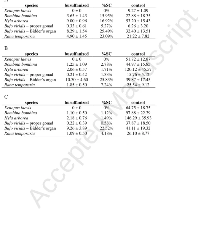
Tab. 3. Number of germ cells per 10 sections (± SD) after busulfanisation and the percentage of surviving cells (%SC): A - in tadpole gonads at the Gosner stage 34 (at the Nieuwkoop stage 53 for Xenopus laevis), B - at the Gosner stage 44 (at the Nieuwkoop stage 64 for
Xenopus laevis), C - 6 months after metamorphosis.
A
species busulfanized %SC control
Xenopus laevis 0 ± 0 0% 9.27 ± 1.09 Bombina bombina 3.65 ± 1.43 15.95% 22.88 ± 18.35 Hyla arborea 9.00 ± 0.96 16.92% 53.20 ± 15.43 Bufo viridis – proper gonad 0.33 ± 0.61 5.27% 6.26 ± 3.20 Bufo viridis – Bidder’s organ 8.29 ± 1.54 25.49% 32.40 ± 13.51 Rana temporaria 4.90 ± 1.45 23.09% 21.22 ± 7.82 B
species busulfanized %SC control
Xenopus laevis 0 ± 0 0% 51.72 ± 12.87 Bombina bombina 1.25 ± 1.09 2.78% 44.97 ± 15.85 Hyla arborea 2.06 ± 0.57 1.71% 120.12 ± 45.57 Bufo viridis – proper gonad 0.21 ± 0.42 1.33% 15.76 ± 5.12 Bufo viridis – Bidder’s organ 10.30 ± 4.60 25.83% 39.87 ± 17.45 Rana temporaria 1.85 ± 0.50 7.24% 25.54 ± 9.12 C
species busulfanized %SC control
Xenopus laevis 0 ± 0 0% 64.75 ± 18.75 Bombina bombina 1.10 ± 0.50 1.12% 97.88 ± 22.39 Hyla arborea 2.18 ± 0.76 1.49% 146.29 ± 35.93 Bufo viridis – proper gonad 0.22 ± 0.39 0.58% 37.87 ± 18.50 Bufo viridis – Bidder’s organ 9.26 ± 3.89 22.52% 41.11 ± 19.32 Rana temporaria 1.09 ± 0.50 4.18% 26.10 ± 8.77