HAL Id: hal-02561075
https://hal.archives-ouvertes.fr/hal-02561075
Submitted on 24 Nov 2020
HAL is a multi-disciplinary open access archive for the deposit and dissemination of sci-entific research documents, whether they are pub-lished or not. The documents may come from teaching and research institutions in France or abroad, or from public or private research centers.
L’archive ouverte pluridisciplinaire HAL, est destinée au dépôt et à la diffusion de documents scientifiques de niveau recherche, publiés ou non, émanant des établissements d’enseignement et de recherche français ou étrangers, des laboratoires publics ou privés.
Early development of the neural plate: New roles for
apoptosis and for one of its main effectors caspase-3
Hugo Juraver-Geslin, Beatrice Durand
To cite this version:
Hugo Juraver-Geslin, Beatrice Durand. Early development of the neural plate: New roles for apop-tosis and for one of its main effectors caspase-3. Genesis, Wiley-Blackwell, 2015, 53 (2), pp.203-224. �10.1002/dvg.22844�. �hal-02561075�
REVIEW
Early Development of the Neural Plate:
New Roles for Apoptosis and for One of its
Main Effectors Caspase-3
Hugo A. Juraver-Geslin1and B!eatrice C. Durand2,3,4,5*
1Department of Basic Science, Craniofacial Biology, College of Dentistry, New York University,
New York, New York
2Institut Curie, Centre Universitaire, Batiment 110, F-91 405, Orsay cedex, France 3Universit!e Paris Sud, Batiment 110, F-91 405, Orsay cedex, France
4CNRS UMR3347, Centre Universitaire, Batiment 110, F-91 405, Orsay cedex, France 5INSERM U1021, Centre Universitaire, Batiment 110, F-91 405, Orsay cedex, France
Received 5 September 2014; Accepted 7 January 2015
Summary: Despite its tremendous complexity, the ver-tebrate nervous system emerges from a homogenous layer of neuroepithelial cells, the neural plate. Its for-mation relies on the time- and space-controlled pro-gression of developmental programs. Apoptosis is a biological process that removes superfluous and potentially dangerous cells and is implemented through the activation of a molecular pathway con-served during evolution. Apoptosis and an unconven-tional function of one of its main effectors, caspase-3, contribute to the patterning and growth of the neuroe-pithelium. Little is known about the intrinsic and extrinsic cues controlling activities of the apoptotic machinery during development. The BarH-like (Barhl) proteins are homeodomain-containing transcription factors. The observations in Caenorhabditis elegans, Xenopus, and mice document that Barhl proteins act in cell survival and as cell type-specific regulators of a caspase-3 function that limits neural progenitor prolif-eration. In this review, we discuss the roles and regu-latory modes of the apoptotic machinery in the development of the neural plate. We focus on the Barhl2, the Sonic Hedgehog, and the Wnt pathways and their activities in neural progenitor survival and proliferation. genesis 53:203–224, 2015. VC 2015 Wiley
Periodicals, Inc.
Key words: organizer; BarH; diencephalon; development; Sonic Hedgehog; Wnt
INTRODUCTION
The vertebrate nervous system is an extraordinarily complex structure. Its formation relies on the coordi-nated progression of genetic programs through time and space. The vertebrate nervous system emerges from the neural plate, a homogeneous layer of neuroe-pithelial cells. The understanding of how developmen-tal programs are encoded and induced during establishment of the neural plate is at present a major goal in developmental biology. The first key steps in neural development are conserved during evolution, including patterning, growth, migration, and wiring mechanisms. Notably, core components of the gene reg-ulatory networks (GRNs) that orchestrate development are evolutionary conserved. Programmed cell death (PCD) allows the efficient elimination of cells and is
* Correspondence to: B!eatrice C. Durand, Team Signaling and Neural Crest Development, Centre de Recherche, Centre Universitaire, B^atiment 110, Orsay, Cedex, France. E-mail: beatrice.durand@curie.fr
Contract grant sponsor: The Centre National de la Recherche Scientifi-que (CNRS UMR8197, INSERM U1024); Contract grant sponsor: The “Association pour la Recherche sur le Cancer”; Contract grant numbers: ARC 4972; ARC 5115; Contract grant sponsor: The Foundation Pierre Gilles de Gennes (FPGG0039); Contract grant number: FRC DOC20120605233; Contract grant sponsor: LABEX Memolife.
Published online 24 January 2015 in Wiley Online Library (wileyonlinelibrary.com). DOI: 10.1002/dvg.22844
implemented through the activation of a molecular pathway conserved during evolution. Observations indi-cate new roles for apoptosis and its key executioners, the caspases, in neural development. Little attention has been devoted to understanding the roles and regula-tory modes of the apoptotic cascade in neural morphogenesis.
Early Events in Neural Plate Patterning and Growth
A highly complex process involving simultaneous and sequential steps subdivides the neural plate into distinct neural territories. Based on the morphological considerations in different species, embryological manipulations, and the expression pattern of regulatory genes, a conceptual model holds that the neural plate is divided into transverse and longitudinal segments that define a developmental grid generating distinct histo-genic fields. Some fields become compartmentalized, specified by a unique pattern of gene expression and maintained by polyclonal cell lineage restriction. In the last two decades, elegant fate-mapping experiments have been undertaken in zebrafish (Woo et al., 1995), Xenopus (Eagleson and Harris, 1990), and chick (Couly and Le Douarin, 1987; Figdor and Stern, 1993; Lumsden and Keynes, 1989) by several pioneering teams. Such fate map analyses prove that the position of the histo-genic fields in the neural plate is a flat representation of their topological relationships in the mature neural tube. Indeed, in the neural plate, the primordia of the forebrain, midbrain, hindbrain, and spinal cord are all already established along the anterior–posterior (AP) axis of the neural plate (Heasman, 2006; Hoch et al., 2009; Wilson and Houart, 2004). Therefore, a construc-tion blueprint of the neural organizaconstruc-tion, and specifi-cally that of the forebrain, is set up during gastrulation. At the stage of neural induction, when neural bulges have not yet appeared, the histogenic fields can be detected as spatially restricted domains of gene expres-sion (Puelles and Rubenstein, 2003; Rubenstein et al., 1994). The analysis of these patterning cues has unveiled some of the GRNs involved in the specification of neural territories (Beccari et al., 2013). The correct development of a histogenic field depends on two criti-cal biologicriti-cal processes that act in parallel and synchro-nously: the specification of neuroepithelial cells into distinct cell types, and the growth of the neuroepithe-lium. The precise coordination of growth and differen-tiation processes is necessary because disturbances in the balance between these two can be harmful to the developing organism (Dreesen and Brivanlou, 2007).
Initial regionalization of the neural tube relies on the synergistic action of at least five major signaling factors secreted by the axial mesoderm that signal in a vertical and planar manner (Niehrs, 2004; Stern, 2002).
The combinatorial activity of various secreted signals both induces the specific expression of identity genes and controls the growth of the different subregions of the central nervous system (CNS). The experiments performed mainly in amphibian, avian, and zebrafish embryos have established crucial roles for bone mor-phogenetic proteins (BMPs), fibroblast growth factors (FGFs), Hedgehog (HH), Wnt, and Nodal proteins in neural induction, neural proliferation, and in the estab-lishment of the future dorsal–ventral (DV) and AP axes of the developing nervous system. The roles of these signaling pathways are highly conserved in vertebrates and invertebrates (Beddington and Robertson, 1999; Jessell and Sanes, 2000). The BMP inhibitors chordin, noggin, and follistatin, which are involved in the deci-sion of ectodermal cells to become epidermal or neural cells, and Sonic Hedgehog (Shh), act as concentration-dependent morphogens to help regionalize the DV axis of the developing nervous system. Wnt signaling ini-tially participates in the establishment of a crude AP axis and later, together with FGFs, participates in a more refined patterning of the anterior neural tube (Stern, 2005; Wilson and Rubenstein, 2000). Besides instructing cell fates, morphogens control the growth of the neural tissue, a process that depends on a deli-cate balance between the proliferation of neural pro-genitor cells and early programmed cell death (EPCD), primarily through apoptosis (Raff, 1998). BMPs, FGFs, Wnt, and Shh are all believed to be involved in control-ling proliferation and apoptosis in the developing nerv-ous system (Chenn and Walsh, 2002; Megason and McMahon, 2002; Mehlen et al., 2005).
Small groups of specialized cells emerge as local sour-ces of secreted patterning factors and hence act as “organizing centers.” In 1924, the first “organizing” cen-ter was discovered by Hans Spemann and Hilde Man-gold. They showed that the dorsal lip of the blastopore, if grafted into the ventral part of a host embryo, was able to induce a secondary axis containing a complete nervous system (De Robertis et al., 2000; Niehrs, 2004; Stern, 2001). Since this discovery, this organizing center has been referred to as “Spemann organizer” and has been found in other model organisms; Hensen’s node in the chick, the node in the mouse, and the shield in zebrafish. This primary organizer is the first to emerge during embryonic development at gastrulation and gives rise to the prechordal plate and the notochord, two tissues that send planar and vertical signals to the overlying prospective neuroepithelium (Niehrs, 2004; Stern, 2002; Wilson and Houart, 2004). The primary organizer induces the neuroectoderm, which gives rise to the neural plate in the dorsal ectoderm through the secretion of inhibitors of the BMP signaling pathway. In contrast, the epidermis forms in the ventral ectoderm, which is still exposed to the high levels of BMP signals (Harland, 2000). The recent studies rendered this
model of neural induction more complex by revealing the importance of crossinteractions with other signal-ing pathways such as Wnt and FGF (Stern, 2006). Sec-ondary signaling centers emerge later in development. These signaling centers are located at molecular fron-tiers that separate territories and are of fundamental importance for the regionalization of the brain as they allow the structures of the CNS to refine their develop-ment (Cavodeassi and Houart, 2012; Echevarri et al., 2003; Kiecker and Lumsden, 2012). The anterior neural ridge (ANR) borders the neural plate anteriorly at the interface between the non-neural ectoderm and the neuroectoderm. The ANR expresses Fgf8 and specifies the forebrain (Houart et al., 1998). The zona limitans intrathalamica (ZLI) is defined by the alar plate expres-sion of Shh, a secreted morphogen that mediates regionalization of the prethalamus anteriorly and the thalamus posteriorly (Juraver-Geslin et al., 2014; Scholpp et al., 2006; Vieira et al., 2005). The midbrain– hindbrain boundary (or isthmic organizer) separates the anterior and posterior neural tube and secretes Wnt1 and Fgf8 (Crossley and Martin, 1995). Finally, the floor plate through the secretion of Shh, and the roof plate through that of Wnts and BMPs ligands also act as signaling centers (Fig. 5a) (Alexandre and Wassef, 2003; Cavodeassi and Houart, 2012; Echevarri et al., 2003).
As morphogenetic gradients are essential for the out-growth and patterning of the neural plate and tube, the number of cells producing these morphogens must be tightly regulated. At least for a signaling structure secreting Shh, the location, size, and shape of the signal-ing center determine the size, shape, and orientation of the target tissue (Agarwala et al., 2001). This is also true for the neural plate whose size and shape are at least partially determined by the secretion of BMP inhibitors from the notochord (Inomata et al., 2013; Khokha et al., 2005) and for the ventral telencephalon, whose correct development relies on local antagonism of Wnt signaling and Fgf-8 secretion from the ANR (Houart et al., 2002; Nonomura et al., 2013). In both Xenopus and mouse, apoptosis is part of the system that controls the correct establishment of signaling centers during the rapid growth of the embryo (Nonomura et al., 2013; Offner et al., 2005).
Moreover the response to a morphogen can be cell specific and, during the period of regionalization, the growth rate of adjacent histogenic fields can vary. In amphibian, the diencephalic (caudal forebrain) primor-dium grows less compared to the telencephalic (rostral forebrain) primordium. Interestingly, this differential growth has been attributed to an unconventional activ-ity of caspase-3, one of the main executioners of the apoptotic cascade. Caspase-3 acts as a proliferation brake by limiting Wnt signaling. Importantly, this role of caspase-3 in limiting Xenopus diencephalon growth does not depend on its apoptosis effector function
(Juraver-Geslin et al., 2011). Taken together, these observations reveal important activities of the apoptotic machinery in early regionalization of the neural plate.
The homeodomain-containing genes are important target genes regulated by morphogens. They encode gene regulatory proteins and specify regional identity by regulating the “effector” genes, which influence cell proliferation, survival, differentiation, adhesion, shape, and migration (Rubenstein and Rakic, 1999). A family of such transcription factors, the Bar-class HD (Barh), con-tains two members, BarH1 and BarH2, which are well conserved in metazoans during evolution. In both Xen-opus and mice, barhl2 is expressed in the developing neuroepithelium from early gastrulation onward, at a time period overlapping with the initial steps of neural plate patterning (Fig. 2A) (Juraver-Geslin et al., 2011, 2014; Offner et al., 2005; Patterson et al., 2000). The investigation of BarH proteins activities in nematode, Xenopus, and mice revealed the roles for these tran-scription factors in the developmental control of cell death and as cell type-specific regulators of caspase-3 nonapoptotic functions (Juraver-Geslin et al., 2011; Li et al., 2002, 2004; Nehme et al., 2010; Offner et al., 2005; Peden et al., 2007; Schwartz and Horvitz, 2007). These activities are strictly dependent on the cellular context and their regulatory modes in vertebrates have not yet been elucidated.
In this review, we discuss the respective contribu-tions of EPCD and the nonapoptotic funccontribu-tions of caspase-3 to early development of the neuroepithelium. We present the activities of BarH proteins in the regula-tion of cell survival. We examine the molecular mecha-nisms underlying the differential growth within the diencephalic field.
Apoptosis and Caspase-3 Activities in Neural Plate Development
Most studies, to date, on the function of the apopto-tic cascade in neural development have focused on the late developmental stages after cell differentiation. Here, we focus on EPCD, which affects proliferating cells during neurulation.
Local apoptosis plays an important role in the formation of organizing centers. PCD was origi-nally documented in the nematode Caenorhabditis ele-gans and more than a decade of extensive work has led to a better understanding of the mechanisms driving cell suicide (Fuchs and Steller, 2011; Hyman and Yuan, 2012; Yi and Yuan, 2009). An emerging body of evi-dence demonstrates that the apoptotic signaling path-way is tightly regulated at different levels (Fig. 1). Current knowledge on the different stages of this cas-cade in vertebrates comes from the studies on the adult immune system. The key players in the regulation of this cascade are the proteins of the Bcl-2 (B-cell
lymphoma 2) family, which include antiapoptotic pro-teins that allow survival (Bcl-2, Mcl1, Bcl-X specifically its long isoform Bcl-XL) and are different from those
which play a proapoptotic role (Bim, Puma, or Bid)
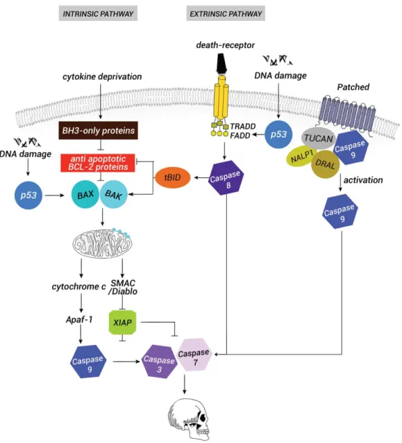
(Happo et al., 2012; Tsuchiya and Yamashita, 2011). Both types of Bcl-2 proteins are present in most cells and a balance between the ratio of pro- versus antiapop-totic proteins decides whether a cell lives or dies (Raff, FIG. 1. Scheme of the vertebrate apoptotic pathway. In a viable cell, the proapoptotic proteins Bax and Bak of the B-cell lymphoma 2 (Bcl2) family are repressed by the antiapoptotic proteins Bcl2, Mcl1 and BclXL. Proapoptotic stimuli such as cytokine deprivation or
endo-plasmic reticulum stress trigger the induction of the BH3-only proteins. BH3-only proteins counteract the effect of Bcl2 and Bcl-XL, a
pro-cess that leads to the release of Bax and Bak. Bax and Bak are then inserted and activated at the outer mitochondrial membrane where they promote the formation of pores. A key event in the induction of death is the release of cytochrome c and SMAC/Diablo from the mito-chondrial intermembranous space to the cytoplasm. SMAC/Diablo inhibits the IAP proteins, whereas cytochrome c recruits APAF-1 to form the apoptosome. Caspase-9 is activated by the apoptosome that leads to the cleavage of the effector caspases, 3 and caspase-7, and to irreversible death. In parallel to this intrinsic pathway, the stimulation of death receptors leads to the recruitment of the adaptor proteins such as TRADD and FADD through their death domain. Caspase-8 is then recruited to the membrane dimerizes and can directly activate caspase-3 and caspase-7. Crosstalk between the extrinsic and the intrinsic pathway depends on the caspase-8 cleavage of the BH3-only protein BH3-interacting domain death agonist BID. The product of this cleavage t-BID (truncated BID) inhibits Bcl2, Mcl1 and Bcl-XLand promotes the accumulation of Bax and Bak. Both pathways can be activated through p53 stimulation resulting from DNA
dam-age. Another pathway is linked to the absence of the survival factor Shh that leads to the stimulation of the proapoptotic function of its receptor patched. In the absence of its ligand, patched forms a protein complex with the adaptor protein DRAL (downregulated in rhabo-muosarcoma LIM-domain protein) and one caspase recruitment (CARD)-domain containing protein, either TUCAN (family member, 8) or NALP (NLR family, pyrin domain containing 1). Apical caspase-9 is then recruited to this complex and activated by patched, which leads to activation of the effector caspases.
1992, 1998). The activation of the apoptotic cascade leads to the activation of a family of cysteine proteases, the caspases, which are very promiscuous enzymes that cleave their substrates at an aspartate residue. The acti-vation of effector caspases such as caspase-3 and caspase-7 by proteolytic cleavage leads to self-activation of caspases and irreversibly to cell death by triggering degradation of the DNA and other vital cell components (Nicholson and Thornberry, 1997) (Fig. 1). From all these studies emerged, the concept that all animal cells contain the proteins required to execute the death pro-gram and that most developing cells, at least in higher organisms, require signals from other cells to avoid apo-ptosis (Raff, 1992, 1998).
EPCD mainly occurs during neurulation, neural crest formation, and eye induction and formation (de la Rosa and de Pablo, 2000; Kuan et al., 2000; Yeo and Gautier, 2004). The targeted disruption of the effector mole-cules of the apoptotic pathway reveals the occurrence and magnitude of EPCD and its early roles during verte-brate CNS development. Specifically, disruption of caspase-3, caspase-9, and apaf-1, and specific ablation of cytochrome c apoptotic function, result in massive dis-organization of the CNS structures (Kuida et al., 1996; Nonomura et al., 2013). Evidence in both mice and Xenopus suggests that, at these developmental stages, defects generated by the lack of apoptosis executioners are indirect effects. TUNEL analysis performed in differ-ent vertebrate species demonstrates that during the regionalization process apoptosis occurs in a develop-mentally controlled manner. During gastrulation and early neurulation, cells undergoing apoptosis are local-ized within the forming axial organizer, which we define as the notochord, the prechordal plate, and the floor plate (Fig. 2A) (Hensey and Gautier, 1998; Offner et al., 2005; Yeo and Gautier, 2003). In Xenopus, inter-ference with endogenous apoptosis interferes with cor-rect signaling from the forming axial organizing center. The neural induction process is largely controlled by the secretion of anti-BMP signals from the Spemann organizer (Inomata et al., 2013).The inhibition of apo-ptosis by overexpression of an antiapoptotic gene of the bcl2 family increases both the size of the neural plate and the number of chordin- and shh-secreting cells from the notochord and floor plate (Offner et al., 2005; Yeo and Gautier, 2003). It has, therefore, been argued that in this context EPCD is part of the system that controls both the size and the correct establish-ment of the axial organizer and indirectly accurate for-mation of BMP and Shh activities gradients within the neural plate (Fig. 2A) (Offner et al., 2005). At later developmental stages, TUNEL-positive cells are detected at the border between the neural plate and the non-neural ectoderm (Juraver-Geslin et al., 2011; Yamaguchi and Miura, 2013). In mouse, apaf1-, and caspase-9-null embryos, the primary cause of brain
mal-formations is a failure to complete cranial neural tube closure and the accumulation of nonproliferative FGF8-producing cells from the ANR. This defect in the elimi-nation of FGF8-producing cells from the ventral part of the ANR by apoptosis generates an accumulation of the FGF8 protein and an increase in FGF8 signaling. This enhanced FGF8 signaling owing to apoptosis deficiency perturbs correct development of the ventral telenceph-alon (Nonomura et al., 2013). Finally, during vertebrate limb development, the zone of polarizing activity (ZPA), a group of cells located at the posterior margin of the early limb bud, produces Shh, which acts as a morpho-gen gradient and progressively specifies anteroposterior digit identities in the wing. The ZPA acts as an organiz-ing center and apoptosis similarly regulates the number of Shh-expressing cells in this region (Sanz-Ezquerro and Tickle, 2000). In this context, apoptosis indirectly contributes to establishing the proper distribution of anti-BMP (chordin), Shh, and FGF8 proteins and hence to correct patterning of the neural plate, the developing forebrain, or the limb. It is worth noting that signaling center cells and their neighboring cells differ in their susceptibility to morphogen-induced apoptosis (Nono-mura et al., 2013; Offner et al., 2005; Sanz-Ezquerro and Tickle, 2000; Storm et al., 2003).
The mechanisms regulating EPCD at these develop-mental stages are largely unknown. The previous stud-ies demonstrated the importance of morphogens, specifically BMP, FGFs, and Shh that are actively involved in regulating apoptosis in the developing brain or limb (Aoto et al., 2002; Furuta et al., 1997; Sanz-Ezquerro and Tickle, 2000; Thibert et al., 2003; Zou and Niswander, 1996). However, as the activation of PCD is dependent on cell lineage or cell location, it remains a challenge to determine whether the growth factors directly induce apoptosis or whether their abil-ity to regulate cellular physiological status has an indi-rect effect on sensitivity to apoptosis (Mehlen et al., 2005). In all cases, although the effector mechanisms controlling apoptotic processes appear to be shared between proliferating neuroepithelial cells and postmi-totic neurons, the genetic controls for apoptosis induc-tion probably differ in both types of cells (Kumar and Cakouros, 2004).
An unconventional activity of caspase-3 limits growth in the anterior neural tube. Disruption of the caspase-3 gene in mice results in disorganized CNS structures associated with a brain overgrowth pheno-type and hyperplasia of the ventricular and/or mantle zone. The first signs of these defects appear around E10.5 and cellular masses in the forebrain are expanded, in particular in the thalamus and the hypo-thalamus, two structures derived from the diencepha-lon (Kuida et al., 1996). Two main routes to caspase activation have been described in vertebrates: activa-tion of death domain receptors and inducactiva-tion of
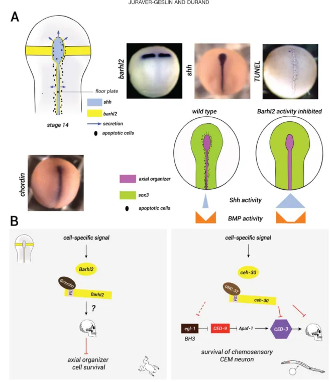
FIG. 2. Barhl2: a regulator of EPCD in the neural plate. (a) Expression patterns of barhl2, shh, and chordin and pattern of endogenous apoptosis in Xenopus neurula embryos. The schematic representation of barhl2, shh, and endogenous apoptosis in the developing neural plate: At stage 14 barhl2 is expressed in the caudal forebrain primordium as two bilateral strips and in cells bordering the floor plate and possibly the notochord (yellow in the scheme). The axial organizer, which we define as the notochord, the prechordal plate and the floor plate, emerge from the Spemann organizer and secrete anti-BMP factors (chordin) and Shh (lavender). The secretion is shown with arrows and cells undergoing apoptosis are shown as black spots. Whole mount in situ hybridization (ISH) using barhl2, shh, or chordin as probes and whole mount TUNEL staining as indicated. Representative neurula embryos are shown in dorsal view, anterior up. Cells undergoing endogenous apoptosis are marked in blue. The schematic representation of the role of Barhl2 on the axial organizer size: In wild-type embryos, Barhl2 has a proapoptotic role (black cells) and negatively regulates the number of axial organizer cells that secrete anti-BMP fac-tors and Shh (pink). As a result, Shh and BMP activity is reduced within the neural plate. When Barhl2 activity is depleted, more cells secret-ing Shh and chordin survive. This leads to enhanced Shh (blue) and BMP (orange) signalsecret-ing activities and subsequently to an increase in the neural plate size marked by Sox3 (green), (after Offner et al., 2005). (b) The BarH survival pathways in amphibians and nematodes. Dur-ing neurulation, Barhl2 regulates EPCD. Barhl2 interacts with Groucho and induces cell death of axial organizer cells expressDur-ing the morph-ogens Shh and chordin through an unknown mechanism. In C. elegans, the Barhl2 orthologue, Ceh-30, regulates the survival of CEM chemosensory neurons in male organisms. In males, ceh-30 is induced by cell-specific signals, and it interacts with UNC-37, the groucho orthologue, and acts as a transcriptional repressor of ced-3 and egl-1 genes.
BH3-only proteins, the proapoptotic members of the Bcl-2 family, that is the extrinsic and intrinsic pathways, respectively (Fig. 1). In the developing CNS, the Bcl-XL
protein, the long isoform encoded by the bcl-x (bcl2l1) gene, and the myeloid cell leukemia 1 Mcl1 protein, act as the main antiapoptotic Bcl2 family members (Motoyama et al., 1995; Arbour et al., 2008; Hasan et al., 2013). The targeted disruption of Bcl2l1 (Bcl-X) causes a dramatic increase in apoptosis of immature neurons throughout the embryonic nervous system. When Bcl2l1 (Bcl-X) mutant mice are crossed with the caspase-3-deficient mice, the double mutation abrogates the ectopic cell death observed in postmitotic neurons, caused by the Bcl2l1 (Bcl-X) deficiency alone. Therefore, in these cells, caspase-3 acts downstream of Bcl-XLas an
effector of cell death. In contrast, Bcl2l1 (Bcl-X) defi-ciency does not affect apoptosis of neural precursor cells and Bcl-XLprotein levels are low in the forebrain
ventric-ular zone. Mcl1 deficient neural progenitors undergo cell death as they commit to a neuronal fate and migrate away from the ventricular zone (Arbour et al., 2008). However overexpression of antiapoptotic proteins of the Bcl2 family, which protect cells against death, does not affect the growth of the forebrain (Martinou et al., 1994). In addition, Bax-deficient embryos have no signs of hyperplasia or malformations of the nervous system, whereas there is a marked hyperplasia of the embryonic nervous tissue in caspase-3-deficient mice. These results
indicate that, though downstream of Bax and Bcl-XLin
the apoptosis of postmitotic neurons, caspase 3 has a unique function in regulating the size of the progenitor pool during early development, before neurogenesis begins (Kuan et al., 2000; Oppenheim, 2001; Roth et al., 2000).
In Xenopus embryos, overexpression of Bcl-XL
decreases the rate of proliferating neuroepithelial cells undergoing apoptosis. However, in agreement with the observations in mice, these embryos do not exhibit any growth phenotype. In contrast, at similar developmen-tal stages caspase-3-depleted embryos exhibit a fore-brain hyperplasia. Indeed, these embryos have a greater number of neuroepithelial cells and this increase is associated with a higher rate of proliferation in cells where caspase-3 is absent. The analysis of apoptotic cell distribution, together with the analysis of the phe-notype of Bcl-XL-overexpressing embryos compare with
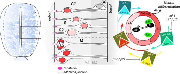
that of caspase-3-deficient embryos, demonstrates that the requirement of caspase-3 for normal growth of these brain regions involves its influence on cell prolif-eration rather than on apoptosis (Juraver-Geslin et al., 2011). In addition, Xenopus caspase-3-depleted embryos exhibit a localized disorganization of the neu-roepithelium architecture, with a disturbance in nuclear migration interkinetic movements (Fig. 3). The proliferation and organization defects of caspase-3-depleted neuroepithelium are similar to those observed FIG. 3. Scheme illustrating cell-cycle dynamics in neural progenitors. The neuroepithelium is composed of a single layer of rapidly dividing NSCs and progenitors. As the cells adjacent to the lumen continue to proliferate, the neural progenitors migrate and form a second layer around the neural tube. This layer becomes progressively thicker and is called the mantle zone (MZ), whereas the germinal neuroepithelium is known as the ventricular zone (VZ), subsequently called the ependyma. Cells in the MZ differentiate into both neurons and glia. In the ger-minal neuroepithelium, neural progenitors undergo different phases of the cell cycle. The nucleus of proliferating cells undergoes a process of interkinetic nuclear migration that corresponds to each phase of the cycle. The nuclei move away from the apical surface during G1. DNA replication (S phase) takes place in the basal VZ. During G2, the nuclei migrate away from the apical ventricular zone where they undergo mitosis (M). After one cycle is completed, the cells choose either to re-enter the cell cycle or to exit and differentiate. Quiescent cells are found in the MZ. Progression through the cell cycle depends on the cyclin/CDK complexes. During G1, phosphorylation of the reti-noblastoma complex (Rb) by cyclins D releases the transcription factor E2F and induces the expression of cyclins E. DNA synthesis during the S phase is followed by the verification of DNA integrity. The progression through mitosis depends on the cyclin B/CDK1 complex. The CDK inhibitors (CKI) p27/Kip1 and p21/Cip1 stop the progression through the cycle and favor exit from the cell cycle (G0).
in mice expressing an activated form of b-catenin (Chenn and Walsh, 2002; Junghans et al., 2005). Indeed, the amounts of total b-catenin and its active form are increased in the brain of caspase-3-depleted
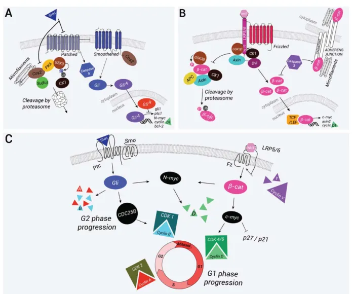
Xenopus embryos. The increase in b-catenin is associ-ated with the upregulation of downstream targets of b-catenin/T-cell factor (TCF)/lymphoid enhancer-binding factor (LEF) signaling, axin2, and the cell-cycle FIG. 4. The Wnt canonical and the Shh pathways and their interactions. (a) Schematic representation of the Shh pathway. In the absence of Shh, patched (Ptc) inhibits smoothened (Smo) and prevents its translocation to the cell surface. Cos2 binding to the microtubules allows the formation of a protein complex with recruitment of GSK3, CK1a, PKA, and Su(Fu). The protein complex phosphorylates the Gli proteins and targets their proteosomal degradation. Truncated Gli forms translocate to the nucleus where they repress target gene expression. In parallel, patched can recruit and activate caspase-9 a process that leads to cell death. Binding of Shh to Ptc initiates the pathway, alleviates repression of Smo, which is translocated to the membrane, and promotes cell survival. Cos2 binds to Smo and inhibits the phosphorylation of Gli proteins by releasing the different kinases. Full activation of the pathway is achieved by phosphorylation of the cytosolic C-tail of Smo by PKA and CK1. Full-length forms of Gli are translocated to the nucleus where they induce the expression of Shh target genes such as Gli-1, Ptc, N-Myc, Bcl-2, and cyclin D1. (b) The schematic representation of the Wnt canonical pathway. In the absence of a Wnt signal, b-catenin is phosphorylated and targeted for proteasome-mediated degradation by a protein complex containing GSK3-b, CK1a, APC, and Axin. After binding of the Wnt ligand to the receptors Frizzled (Fz) and LRP5/6, dishevelled (Dvl) binds to Fz and recruits the destruction complex at the cytosolic tail of LRP5/6. LRP5/6 tail is then phosphorylated by GSK3-b and bound by Axin. In this context, b-catenin is sta-bilized and translocated to the nucleus where in association with TCF/LEF transcription factors it stimulates expression of the target genes c-myc, axin2, and cyclin D1. In parallel,b-catenin is present at the adherens junctions where it associates with cadherins and with the cyto-skeleton, allowing adhesion between neighboring cells. Caspases, and specifically caspase-3, can destabilize the adherens junctions in part by inhibitingb-catenin association with E-cadherin. At note in vitro b-catenin is a substrate for caspases. (c) Crosstalk between the Wnt and the Shh pathways promotes cell-cycle progression. The Shh pathway mostly acts on the G2 and S phases through the regulation of phosphatase CDC25B expression. N-Myc is capable of regulating cyclin D1 expression, which allows progression through G1. In some cases, Shh also regulates the expression of late cyclins A and B. The Wnt canonical pathway also promotes G1 progression by inducing the expression of c-myc, which regulates cyclin D1 expression and inhibits p27/Kip1 and p21/Cip1. A feedback loop is regulated by cyclins Y, which phosphorylate the cytosolic tail of LRP5/6 to activate the Wnt pathway.
regulator, cyclin D1 (ccnd1) (Fig. 4B). b-Catenin is known to participate in adherens junctions and to form a stable complex with the molecules of the cadherin family (Figs. 3 and 4B) (Redies, 2000; Stepniak et al., 2009). The depletion of b-catenin through morpholino injection, or overexpression of glycogen synthase kinase 3 b (GSK3-b) which inhibits b-catenin activation, partially rescues the proliferative and architectural defects observed in caspase-3-depleted embryos. There-fore, increased levels of total and active b-catenin prob-ably account for the proliferative and neuroarchitectural defects observed in caspase-3-depleted embryos (Juraver-Geslin et al., 2011).
b-Catenin is one of the many substrates of caspase-3. When the apoptotic cascade is activated, rupture of cell polarity and loss of cell junctions are two of the har-bingers of the death event (Strasser et al., 2000). In the epithelial cells of Drosophila, drICE, the orthologue of caspase-3, directly interacts with Armadillo, the ortho-logue of b-catenin, and cleaves it at its DQVD peptide motif. This cleavage inhibits its interaction with E-cadherin (epithelial E-cadherin) and destabilizes adherens junctions (Kessler and Muller, 2009). The caspase cleav-age site in the sequence of b-catenin is conserved in vertebrates. However, this ability of caspase-3 to cleave b-catenin is at odds with what is observed in the dience-phalic primordium of Xenopus embryos depleted for caspase-3. In these embryos, depletion of either caspase-3 or b-catenin alters the attachment of cells in mitosis to the apical surface. The depletion of b-catenin partially rescues this defect in caspase-3-depleted embryos and vice versa (Juraver-Geslin et al., 2011). It is therefore tempting to speculate that b-catenin is not the main substrate of caspase-3 in this context and that in caspase-3-depleted embryos, the equilibrium between membrane-bound b-catenin at the adherens junctions and cytosolic b-catenin is disrupted (Nelson and Nusse, 2004). Such disruption is known to dramati-cally alter the internal structure of the neuroepithelium (Kadowaki et al., 2007).
In conclusion, observations in amphibians, chick, and mice indicate that local EPCD plays an important role in the formation of organizing centers. It coordi-nates the emergence and the removal of morphogen-producing cells both spatially and temporally, allowing the accurate growth of the developmental process. In addition, the apoptotic machinery contributes to the differential growth of neighboring histogenic fields, at least partly through caspase-3 unconventional activ-ities. The similarities between the mouse deficient phenotype and that observed in caspase-3-depleted Xenopus embryos suggest that the role of caspase-3 in regulating the proliferation of neuroepi-thelial cells during development could be conserved at least in some vertebrates, irrespective of its role in apoptosis.
Barhl2: A Regulator of EPCD in the Neural Plate Barhl2 regulates survival of axial organizer cells. BarH1 and BarH2 are homeodomain-containing transcription factors of Bar-class HD (BarH) initially dis-covered in Drosophila. The paralogues known as Barh-like (Barhl) have subsequently been identified in fish (zebrafish, medaka), amphibians (Xenopus), birds (chicken), and mammals (mouse). In vertebrates, barhl1 is the closest relative of barhl2, and phyloge-netic analysis shows that Barhl proteins are orthologues of the Drosophila BarH and nematode CEH-30 and CEH-31 proteins and are well conserved in metazoans during evolution (Reig et al., 2007; Schuhmacher et al., 2011). During the embryonic development of verte-brates, barhl1 and barhl2 are predominantly expressed in the CNS and their expression patterns are distinct but partially overlap (Bulfone et al., 2000; Colombo et al., 2006; Patterson et al., 2000). BarH genes are involved in diverse processes such as the acquisition of neural identity in the retina, the specification of com-missural neurons in the spinal cord, and cell migration in the cerebellum and hindbrain (Chellappa et al., 2008; Ding et al., 2009; Li et al., 2004; Mo et al., 2004; Poggi et al., 2004; Saba et al., 2005; Jusuf et al, 2012). Here, we focus on the activities of Barhl2 in early neural development.
In Xenopus, barhl2 is normally expressed from the onset of gastrulation in two longitudinal stripes along the axial organizer (Fig. 2a). Barhl2 promotes apoptosis in the Xenopus neuroectoderm through a mechanism that cannot be attributed to an unspecific cellular stress response. Barhl2 GOF induces a decrease in the size of the neural plate that is rescued by inhibition of apopto-sis. Reversibly, Barhl2 LOF generates a dramatic increase in neuroepithelium size that mimics BMP sig-naling LOF. Indeed, it has been shown that the proapop-totic activity of Barhl2 is essential during normal neural plate formation as it limits the number of chordin- (a BMP inhibitor) and Shh-expressing cells in the prospec-tive notochord and floor plate, which act as organizing centers (Fig. 2a). Therefore, we proposed that Barhl2 is part of a pathway that regulates EPCD in the neural plate. Barhl2-regulated cell death contributes to control both the size and the correct establishment of the axial organizer and indirectly, to the formation of the BMP and Shh activities gradient that control patterning and growth of the neural plate (Offner et al., 2005). The determination of how Barhl2 regulates EPCD during development is unknown. BarH transcription factors are characterized by their homeodomain, and their FIL domains located in the N-terminal region of the protein (Offner et al., 2005). The BarH/Barhl proteins differ from other homeodomain-containing proteins in the presence of a tyrosine (Y) at position 49 in the homeo-domain, which is commonly occupied by a phenylala-nine (F). This residue is located in the third helix,
which is the recognition site for the DNA, suggesting a modification of the specific recognition site. In addi-tion, Barhl proteins possess two highly conserved FIL domains in their N-terminal region that correspond to the engrailed homology 1 (EH1) motif, which is known to interact with the protein Groucho (C. elegans UNC-37, human transducin-like enhancers of split). Although in Drosophila barH genes contain one FIL domain, they contain two in vertebrates. It is believed that through the recruitment of Groucho, Barhl form a protein com-plex that binds at the transcription initiation site and inhibit transcription (Cinnamon and Paroush, 2008). Therefore, Barhl2 could act through transcriptional modulation of the survival pathways controlling apo-ptosis, specifically the Shh and/or the BMP pathway that are produced by axial organizer cells. Alternatively, Barhl2 could transcriptionally modulate the apoptotic developmental program.
Barhl2 and the signaling pathways in neural plate cells survival. At the end of gastrulation, the prechordal mesoderms, together with the notochord, are the primary sources of anti-BMP signals and of Shh. Several BMPs have been shown to promote apoptosis in developing embryos, specifically in the dorsal neuroepi-thelium and in the interdigital mesenchyme (Furuta et al., 1997; Hanel and Hensey, 2006). In the limb, BMPs appear to affect PCD indirectly by regulating the secretion of FGFs from the apical ectodermal ridge, which directly act as a cell survival factor (Pajni-Under-wood et al., 2007). Overall, there is little genetic evi-dence that BMPs directly trigger apoptosis (Hegarty et al., 2013). On the contrary, Fgfs ligands, and most specifically Fgf8, have been demonstrated to act on the survival of neural crest cells and cells of another brain signaling center, the ANR. The conditional or hypomor-phic Fgf8 mutant mice exhibit defective craniofacial development with a significant increased in the rate of apoptosis in the ANR and in migrating neural crest cells (Abu-Issa et al., 2002; Crossley and Martin, 1995). Simi-larly, mice defective in two transcription factors Foxi3 and Sp8 develop dramatic craniofacial malformation that have been associated to reduction in Fgf8 and Fgf17 expression in the neural crest cells and, in the case of Sp8-deficient mice, in the ANR (Edlund et al., 2014; Kasberg et al., 2013). Interestingly, the Wnt canonical pathway participates in the expression of Fgf8, Fgf3, and Fgf17 in the ANR. The conditional mutant mice for b-catenin exhibit a dramatic upregula-tion of apoptosis in the rostral head tissues. This increase in EPCD is associated to a striking diminution of FGF ligands’ expression in the ANR and the adjacent frontonasal ectoderm. Reversibly, conditional gain-of-function of b-catenin signaling causes an upregulation of Fgf8 expression in the ANR, and in the entire facial ectoderm. These data argue that in the ANR, Fgf8 is a target gene regulated by b-catenin signaling and its loss
most probably account for the observed apoptosis in the head tissues (Wang et al., 2011). Both secreted Wnt and Fgf ligands contribute to patterning of the posterior neural plate and could play a part in controlling axial organizer cell survival. However, among the multiple Fgf and Wnt ligands, none has been detected in the axial organizer and it is therefore unlikely that Barhl2 directly act on either the BMP, or the Wnt, or the Fgf pathways in controlling the survival of axial organizer cells. During neurulation, Shh is induced in the noto-chord, the floor plate, and anteriorly in the ventral neu-roepithelium (Fig. 2a) (Placzek and Briscoe, 2005). The activation of the Shh pathway leads to an intracellular response that depends on the zinc finger of the Gli fam-ily of transcription factors. A balance between the pro-duction of activating forms (GliA) and the repressor forms of Gli (GliR) allows modulation of this response. In mammals, there are three Gli proteins (Gli, 1–3). The receiving system of the Shh signal is composed of two proteins: Patched (Ptc), a 12-transmembrane receptor, and smoothened (Smo), a protein with seven transmem-brane domains. Ptc is a constitutive Shh pathway inhibi-tor. In the absence of its ligand, Ptc blocks the activity of Smo. When Smo is inhibited, Gli2 and Gli3 are phos-phorylated, targeted to the proteasome, and then cleaved. The cleaved forms correspond to repressive forms (GliR) and Gli3R is the primary transcriptional repressor of the Shh signaling pathway. The binding of Shh on Ptc lifts its inhibition of Smo, which abolishes the cleavage of Gli proteins. This activates the Shh tar-get genes in full-length forms (GliA). In mammals, Gli2A is the main transcriptional activator of Shh signaling (Fig. 4a) (Briscoe, 2009).
Shh regulates apoptosis in various ways in the neural plate. It acts as a cell survival signal (Charrier et al., 2001; Chiang et al., 1996; Litingtung and Chiang, 2000) but also has an apoptotic promoting activity as demon-strated in chick ventral neuronal precursors and floor plate cells (Oppenheim et al., 1999). The analysis of Shh-defective mice reveals a massive neural cell death mostly in the ventral part of the neural tube (Chiang et al., 1996). In chick embryos, experimental removal of the notochord and the floor plate—the sources of Shh signals—induced a massive apoptosis of the ventral neuroepithelial cells (Charrier et al., 2001; Thibert et al., 2003). Indeed, Shh regulates the apoptosis machinery and bcl-2 is a target of the Shh pathway (Fig. 4a) (Bigelow et al., 2004). The extra toes (Xt) mouse mutant carries a null mutation of the Gli3 gene. Very lit-tle apoptosis is detected in Shh–Gli3 double mouse mutants (Litingtung and Chiang, 2000), and a role for Gli3Ras an effector of a Shh-regulated cell survival path-way has been suggested. Ptc, the Shh receptor, is a dependence receptor. Ptc induces apoptotic cell death of neuroepithelial cells unless Shh is present to block the signal (Thibert et al., 2003). Importantly, Ptc
triggers caspase-9 activation and enhances cell death through a caspase-9 dependent mechanism (Figs.1 and 4a) (Mille et al., 2009).
In summary, Shh survival activity is restricted within a developmental time window and could participate in the survival of axial organizer cells, indirectly contribut-ing to the size and shape of the neuroepithelium. The determination of whether Barhl2 modulates the cellular production of Shh and/or the response of neuroepithe-lial cells to this morphogene is unknown.
Barhl2: a transcriptional regulator of the cell-death switch. The studies in nematode suggest that the temporal and spatial regulation of PCD during development is primarily regulated by transcriptional control of the expression of cell-death execution machinery. In the nematode, the BH3-only protein EGL-1, a proapoptotic member of the Bcl-2 family, is the key effector of developmental cell death and is transcrip-tionally controlled (Nehme and Conradt, 2008). Inter-estingly, three C. elegans genetic screens performed independently highlighted the regulation of the apopto-tic pathway by Barh proteins (Nehme et al., 2010; Peden et al., 2007; Schwartz and Horvitz, 2007). Indeed, the orthologue of Barh, CEH-30, blocks apopto-sis to allow the survival of four specific chemosensory neurons in males (CEM cephalic companion neurons). These neurons undergo apoptosis in the hermaphrodite by inducing the expression of Egl-1, which activates the caspase-3 orthologue, Ced-3. In males, CEH-30 acts as a transcriptional repressor of ced-3 and egl-1 genes (Fig. 2b) (Nehme et al., 2010). The expression of CEH-30 depends on both cell-specific and sex-determination sig-nals. Specifically, the transcription factor Unc-86, a determinant of CEM fate, induces the expression of ceh-30 in male bodies. In contrast, in hermaphrodites, Tra1-A, (the orthologue of Gli proteins) a determinant of sex, inhibits its expression. In C. elegans, the vertebrate barhl1 and barhl2 transgenes rescue the CEM survival defect of male CEH-30 mutants, showing that these genes encode proteins that retain the functions and tar-get specificity of CEH-30 (Fig. 2b) (Schwartz and Hor-vitz, 2007).
As one may expect, the regulation of PCD in verte-brates appears considerably more complex. However, the Hox-dependent regulation of apoptosis seems to be a conserved mechanism during animal development. Hoxa13 may be involved in eliminating cells located between the forming digits in the mouse limb bud (Sta-dler et al., 2001). The targeted deletion of barhl1 caused degeneration of mouse cochlear hair cells. The analysis of this phenotype showed that barhl1 plays an essential role in the maintenance of cochlear hair cells, whereas it had little effect in the specification and dif-ferentiation of these cells (Li et al., 2002). Furthermore, barhl1 plays a role in the survival of cerebellar granule precursor cells and precerebellar neurons, as well as in
the radial migration of these cells (Li et al., 2004). In vertebrates, Egl-1 is the orthologue of proteins that con-tain a single Bcl-2 homology (BH) domain, BH3, for example, Bid, Bim, Noxa, Puma, and Bmf, commonly known as the BH3-only proteins (Happo et al., 2012). The proapoptotic activity of Barhl2 requires the pres-ence of intact FIL domains, reflecting the fact that Barhl2 could act as a transcriptional repressor at these stages of development, (Muhr et al., 2001; Offner et al., 2005). It is therefore possible that, as in the nematode, Barhl2 directly or indirectly affects the decision of axial organizer cells to die by transcriptional upregulation of proapoptotic genes or, more probably by downregula-tion of antiapoptotic genes. However, the identity of such transcriptional targets has not been addressed.
In conclusion, although some transcription factors in vertebrates have been shown to participate in the con-trol of PCD, their relevant targets remain largely unknown. One key question that has not been resolved so far is whether temporal and spatial control of apo-ptosis during vertebrate development is also orches-trated at the transcriptional level. In C. elegans, the transcription factors that turn the survival–death switch on and off are also involved in other pathways. Specifi-cally, CEH-30, the Barhl orthologue, regulates caspase-3 activity (Nehme et al., 2010; Schwartz and Horvitz, 2007).
Developmental Features of the Caudal Forebrain: At Least Three Pathways to Control its Growth
A combinatorial process of growth, patterning, and morphogenetic movements in the neural plate allows the emergence of this three-dimensional complex struc-ture: the mature CNS. The forebrain (telencephalon and diencephalon) is derived from the most anterior part of the neuroepithelium, the prosencephalic neural plate. The cerebral hemisphere, the largest part of the brain, emerges from the telencephalon and contains nuclei controlling perceptual, cognitive, and higher motor functions as well as emotion and memory. The dience-phalon lies between the midbrain and the cortex. It principally generates the thalamus, which processes and distributes almost all sensory and motor informa-tion going to the cerebral cortex. Its key role is to inte-grate all the sensory information from the periphery (with the exception of smell) and to relay it to the cere-bral cortex.
In the developing neural tube, the population of neu-roepithelial cells is rapidly expanding. The cell cycle depends on growth of the cell and replication of its genetic material and includes four finely controlled phases: G1 (gap1), S (DNA replication), G2 (gap 2), and M (mitosis). The location of the nuclei of dividing cells in the pseudo-stratified neuroepithelial ventricular zone depends on their status in the cell cycle. This process is
called nuclear interkinetic migration. A short overview of the cell cycle and of the nuclear interkinetic migra-tion is shown in Figure 3. Apart from the first divisions of the zygote, which are cell-autonomous, the division of any animal cell is dependent on extracellular growth factors produced by neighboring cells. Thus, cell-cycle progression is achieved only in an environment contain-ing mitogenic factors (Sherr, 1994). For neural stem cells (NSC) and their derivative progenitors in the devel-oping neuroepithelium, the secreted factors Wnt and Shh trigger progression through the cell cycle (Fig. 4).
During embryonic development, the neural tube is exposed to a multitude of secreted Wnt canonical ligands. The Wnt canonical pathway is crucial for both patterning and proliferation of neural progenitors and we focus on its mitogenic activity (Wilson and Houart, 2004). The role of Wnt in neural proliferation is sup-ported by the analysis of mice depleted for Wnt ligands or for other effectors of the pathway. Although the Wnt12/2 or Wnt3A2/2 mice lose both the midbrain and the hippocampal areas, double Wnt3A/Wnt1 mutant embryos exhibit an additional reduction in the diencephalon, caudal hindbrain, and rostral spinal cord (Lee et al., 2000; McMahon and Bradley, 1990; Thomas and Capecchi, 1990). These results suggest partially redundant functions for Wnt1 and Wnt3A in brain development. Conversely, ectopic expression of Wnt1 or Wnt3A induces enlargement of the neural tube along the DV axis, without altering the cellular identities of neurons (Megason and McMahon, 2002; Panhuysen et al., 2004).
Many Wnt ligands such as Wnt3, Wnt3A, Wnt8b, Wnt4, and Wnt2b are expressed in the diencephalic alar and roof plates (Fig. 5A). Indeed, the Wnt signaling machinery as well as Wnt target gene expression are enriched in the thalamus, but are absent from the pre-thalamus of all vertebrates analyzed so far from zebra-fish to rhesus monkey (Bluske et al., 2009; Jones and Rubenstein, 2004; Quinlan et al., 2009; Shimogori et al., 2004). However, the early changes in dience-phalic patterning upon manipulation of Wnt activity appear to be primarily owing to the altered fate specifi-cation rather than to changes in proliferation, indicating that other pathways contribute to diencephalic growth control (Houart et al., 2002; Ishibashi and McMahon, 2002).
The development of the caudal part of the forebrain is particularly dependent on Shh signaling. This terri-tory receives the combination of Shh signals from the basal plate and from the ZLI. Both signaling structures contribute to the acquisition of a diencephalic regional identity. The analysis of mutant mice for Shh shows a marked reduction in the size of the forebrain and a marked decrease in the size of the diencephalic terri-tory compared to other areas of the brain. This effect partly reflects the role of Shh, which allows the survival
and proliferation of diencephalic progenitor popula-tions (Chiang et al., 1996; Ishibashi and McMahon, 2002), and controls development of the forebrain ven-tral neuroepithelium (Fuccillo et al., 2006; Gunhaga et al., 2003).
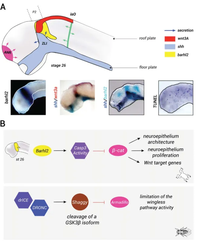
In amphibian, transcripts encoding the barhl2 gene are detected in the diencephalic primordium (Fig. 5a) (Juraver-Geslin et al., 2014). Fate mapping of the dien-cephalic histogenic field at the gastrula stage identifies a wide area similar to the expression pattern of barhl2 in both zebrafish (Staudt and Houart, 2007) and mice (Mo et al., 2004). It has been demonstrated that within the xenopus diencephalic territory, barhl2 acts as a prolif-eration brake via an unconventional activity of caspase-3 that limits the proliferation of neuroepithelial cells. Indeed, comparative analysis of Barhl2-depleted and caspase-3-depleted Xenopus embryos reveals a similar dramatic hyperplasia and cellular disorganization of the caudal forebrain neuroepithelium. The measurement of the relative velocity of the cell cycle in Barhl2 or caspase-3-depleted embryos reveals a similar shortening of cell-cycle length (6 vs. 8 h). The length of the S-phase in these cells remains unchanged (1.5 h). Similarly to what we previously described for caspase-3-depleted embryos, the increase in the speed of the cell cycle observed in Barhl2-depleted neural tube has been corre-lated to b-catenin stabilization and to an increase in the activity of the canonical Wnt pathway. Specifically, both Barhl2 and caspase-3 depletion generate an increase in cyclin D1 expression. Cyclin D1 controls the length of the G1 phase and it is part of a major cell-cycle check-point controlling whether a cell continues to divide or becomes quiescent. It is therefore probable that Barhl2 and caspase-3 normally act as proliferation brakes by lengthening the G1 phase of the cell cycle. The overex-pression of caspase-3 rescues the hyperplasia pheno-type in Barhl2-depleted embryos. Therefore, in the molecular pathway limiting proliferation and Wnt sig-naling, caspase-3 acts downstream of Barhl2 (Fig. 5b) (Juraver-Geslin et al., 2011). It is unknown whether mouse embryos lacking Barhl2 function exhibit a hyper-plasia phenotype of the diencephalic walls.
Taken together, intertwined contributions of the Wnt, the Shh, and the Barhl2 pathways contribute to diencephalic proliferation. We discuss here how these three pathways interact and control diencephalic pri-mordium growth.
The Wnt canonical pathway in early brain growth. Through the control of cell proliferation and cell polarity, the determination of cell identity and axes specification, Wnt functionality covers many aspects of the development (Angers and Moon, 2009; Clevers and Nusse, 2012). Wnt ligands, of which there are 19 in mam-mals, are secreted glycoproteins modified by the addition of a lipid. The key effector molecule of the canonical Wnt pathway is b-catenin. The mechanisms controlling
FIG. 5. Barhl2 acts as a brake on diencephalic progenitor proliferation. (a) barhl2, wnt, and shh ligands are coexpressed in the diencephalic primordium. The scheme of a stage 27 neural tube: barhl2 (yellow) is expressed in the prosomere p2 (p2) within the diencephalon, in a territory giving rise to the ZLI, a secondary organizer that secretes Shh (blue). Shh is also expressed in the anterior neural tube basal plate. Wnt3a (in red) as Wnt3 (data not shown) is expressed in the dorsal (alar plate) part of the prosomere p2, in the midbrain roof plate. The two other major secondary organizers, the ANR (pink) and the isthmic organizer (isO, green), that play roles in patterning the forebrain and the midbrain/hind-brain, respectively, are also shown. The secretion is represented with arrows. Whole mount ISH using barhl2, shh, or wnt3a probes as indi-cated. Representative dissected neural tube of wild-type embryos is shown, dorsal view anterior left. Whole mount TUNEL analysis: cells undergoing endogenous apoptosis appear in blue. (b) Nonapoptotic functions of caspases regulate the levels and activity ofb-catenin. In Xen-opus diencephalic primordium, Barhl2 acts upstream of a nonapoptotic function of caspase-3 that limits both levels and activity ofb-catenin and consequently limits the proliferation of neural progenitors, controls the neuroepithelium architecture, and regulates the transcription of Wnt target genes. In drosophila SOP cells, the caspases orthologues DRONC and drICE limit the activity of the Wingless pathway by cleaving an isoform of GSK3-b, Shaggy, which destabilizes Armadillo (b-catenin orthologue) and consequently limits the Wnt pathway.
the homeostatic balance of Wnt pathway activation are based on the fine regulation of both the expression levels and the activation rate of b-catenin (Fig. 4b).
The Wnt molecular pathway has already been exten-sively reviewed (Arce et al., 2006; Grigoryan et al., 2008; Hur and Zhou, 2010; Kimelman and Xu, 2006). The key step in the activation or inhibition of the Wnt pathway is the modulation of the activity of GSK3-b which targets b-catenin to its degradation by the protea-some. In the absence of Wnt ligands, a protein degrada-tion complex is formed that binds the free b-catenin and leads to its degradation. Cytoplasmic b-catenin lev-els are low, and thus preventing the activation of the pathway (Fig. 4C). The presence of Wnt ligands stabil-izes b-catenin, which is then translocated into the nucleus. In the nucleus, b-catenin displaces Groucho corepressor proteins and in association with LEF and TCF acts as a transcriptional coactivator, allowing the activation of Wnt target genes. Recently, it was demon-strated that Wnt signaling initiates a complex cellular process, leading to GSK3-b sequestration in multivesic-ular bodies into which GSK3-b is sequestered and its activity inhibited (Taelman et al., 2010). Finally, as pre-viously mentioned, b-catenin forms a stable complex with the molecules of the cadherin superfamily and it is part of the adherens junctions that regulate cell–cell adhesion (Fig. 4B) (Redies, 2000).
By stimulating b-catenin accumulation, Wnt activa-tion induces the expression of the proto-oncogene c-myc (He et al., 1998). C-c-myc encodes a bHLH leucine zipper (bHLHZip) transcription factor that has two dis-tinct roles in G1 progression. On the one hand, it increases the expression of cyclins D1 and D2; on the other hand, it represses p27Kip1 and p21Cip1, and hence promoting cell-cycle progression and enhancing cell proliferation (Fig. 4B) (He et al., 1998). In parallel, Wnt stimulates cell growth through a global increase in protein levels during the G1 phase of the cell cycle. Both steps depend on the key event of the inhibition of GSK3-b. The inhibition of GSK3-b induces a general increase in protein stability (Taelman et al., 2010) and stimulates growth through the activation of the TOR pathway, which stimulates protein synthesis. Therefore, the Wnt pathway promotes G1 progression and cell growth, whereas the accumulation of cyclin D1 and c-myc triggers proliferation (Fig. 4B). This activity of Wnt is crucial for the emergence of groups of cells that will help in creating tissue and eventually a functional struc-ture (Davidson and Niehrs, 2010). Besides its role in G1 progression, Wnt signaling is believed to play a role in progression though mitosis. b-Catenin levels are the highest during the G2/M phase. During the M phase, b-catenin allows centrosome separation at the beginning of mitotic spindle formation, regardless of its role in reg-ulating gene transcription (Bahmanyar et al., 2008; Huang et al., 2007). In addition, the Wnt pathway
allows the establishment of mitotic spindle orientation and therefore affects the distribution of cytoplasmic determinants between the two daughter cells, a key process that determines both the specific identity and the spatial positioning of these cells in a tissue (Siller and Doe, 2009; Walston and Hardin, 2006). Importantly, mechanistic regulation and molecular components of Wnt signaling appear to be conserved during evolution (Davidson et al., 2009).
Indeed, the fundamental activity of Wnt in regulating growth of the neuroepithelium is underscored by the analysis of GOF or LOF mutations introduced at the b-catenin locus in mice. The conditional LOF mutants in the spinal cord show a clear reduction in the number of proliferating neurons, associated with an increase in the number of differentiated cells. The reciprocal GOF induces the opposite effect (Zechner et al., 2003). The conditional LOF mutants for b-catenin in the cortex and in the hippocampus also exhibit decreased neuroepi-thelial proliferation and disruption associated with the defects in interkinetic nuclear migration and the loss of adherens junctions. Specifically, the nuclei of proliferat-ing cells are abnormally located outside the ventricular zone (Machon et al., 2003). Furthermore, in mice expressing a stabilized form of b-catenin in the cortex, neural progenitors are kept in a mitotic state and do not differentiate. The brains of these mice are enlarged owing to an expansion of the progenitor pool and a hor-izontal expansion of the ventricular zone with the appearance of convolutions on the surface of the cor-tex, similar to those observed in higher mammals. The process of apoptosis is also increased in these mice. However, the increase in the number of neuroepithelial progenitors cannot be attributed solely to the change in the cell death rate (Chenn and Walsh, 2002).
In conclusion, through its ability to modulate the activity of GSK3-b that promotes a general increase in protein stability, and specifically in b-catenin stability, Wnt signaling promotes concomitantly cell-cycle pro-gression and cell growth.
Shh is a mitogen for neuroepithelial cells. -Through its survival and mitogenic activities, Shh con-tributes to the coherent growth of the neural plate and tube (Jacob et al., 2003; Jessell, 2000; Mehlen et al., 2005; Ribes and Briscoe, 2009). Indeed, Shh regulates both the cell cycle and the apoptosis machinery (Fig. 4a). The two main promoters of G1 progression, cyclin D1 and E, and N-Myc are the targets of the Shh pathway (Katoh and Katoh, 2009; Kenney and Rowitch, 2000; Oliver et al., 2003). In the vertebrate, neuroepithelium electroporation of modulators of Shh activity affects proliferation and apoptosis. In chick’s spinal cord, the expression of a dominant negative form for Ptc induces a reduction in size of the neuroepithelium associated with decreased proliferation and induction of apopto-sis. Electroporation of Gli3R mimics this effect and
causes a cell-cycle arrest associated with a reduction in the expression of cyclin D1. Conversely, overexpression of Gli3A in the neuroepithelium induces hyperplasia linked to a decrease in the length of the G1 phase asso-ciated with an increase in the expression of cyclin D1, an overexpression of cyclins A, B, and E and late pro-gression in G2. Thus, by regulating the ratio of activat-ing and repressive forms of Gli proteins, Shh promotes G1 and G2 progression in the neuroepithelial cells of the spinal cord (Alvarez-Medina et al., 2009; Cayuso et al., 2006). In the Xenopus retina, Shh controls the speed of the neural progenitor cell cycle and stimulates G1/S and G2/M progressions by regulating the expres-sion of the phosphatase Cdc25B, together with late cyclins (B1 and A2) and cyclin D1. Blocking the Shh pathway promotes lengthening of the G1 and G2 phases, whereas Shh overexpression induces premature cell-cycle exit of retinal precursors (Borday et al., 2012). In mouse neuroepithelium, the Shh signal stimu-lates the progression into mitosis. The entry into mito-sis depends on the maintenance of cyclin B/Cdk1 complex in an inactive state by phosphorylation on Thr14 and Tyr15 residues. Cdc25B promotes cycle pro-gression by the activation of the complex (Kristjansdot-tir and Rudolph, 2004) (Fig. 4A). The inhibition of Shh signaling by cyclopamine decreases Cdc25B expression in the neural tube, whereas overexpression of Gli2Aby electroporation leads to its accumulation. Thus, Shh promotes progression in G2/M in mice by inducing the expression of Cdc25B in neuroepithelial progenitors. In agreement with these observations, mutant mice for Shh have a delayed and reduced ventral Cdc25B expres-sion, which argues for a conserved role of Shh in regu-lating the later stages of the cell cycle, G2, and M in mammals (Benazeraf et al., 2006).
In summary, Shh helps control the size of the neural plate and tube by positively regulating progression through the cell cycle, especially during the later stages of the cell cycle.
Shh and Wnt signals cooperate in patterning and growth along the entire neuraxis. At the neu-ral plate stage, Wnt and Shh exhibit opposite patterning activities: Shh contributes to the acquisition of a ventral identity, whereas Wnt promotes dorsal neural progeni-tor fate (Briscoe and Ericson, 2001; Robertson et al., 2004; Zechner et al., 2003). Such antagonism is also observed in the establishment of the spinal cord occur-ring after neural tube closure, where Shh is expressed in the floor plate and Wnt is expressed in the roof plate. The inhibition of the Shh pathway by Wnt signaling is crucial in the regionalization of the spinal cord. In chicken, the Wnt pathway limits the influence of Shh by inducing the expression of Gli3Rin the dorsal part of the neural tube. In mice, the expression of a stabilized form of b-catenin dorsalizes ventral neurons and extends the expression of Gli3Rventrally. This
pheno-type is partially rescued through Gli3 LOF (Alvarez-Medina et al., 2008; Yu et al., 2008). In the Xenopus, postembryonic ciliary marginal zone of the retina, the Shh, and Wnt pathways inhibit each other’s expression, and hence allowing them to segregate in mutually exclusive territories. This inhibition partially depends on the transcriptional regulation of SFRP1, on the one hand, and Gli3 on the other hand, which both contrib-ute to this negative crossregulation. Such antagonistic interplay of Wnt and HH signals appears to regulate the extent of neural stem/progenitor cell proliferation in the Xenopus retina (Borday et al., 2012). Finally, in the chicken caudal forebrain, these two pathways also antagonize one another. Electroporation of Shh in the developing diencephalon induces a decrease in the expression of Wnt4 and Wnt5A. In vivo Wnt4 expres-sion is inhibited by the Shh signal from the ZLI and Wnt4 expression is progressively restricted to the cau-dal part of the diencephalic alar plate, a process that contributes to thalamus regionalization (Quinlan et al., 2009). In addition, Wnt8b regulates Gli3 expression within the diencephalon, and hence contributing to shh induction and dorsal progression of the ZLI within the caudal forebrain (Martinez-Ferre et al., 2013).
The cooperative action of the two signaling pathways also affects cell-cycle progression (Fig. 4c). Shh mutant mice display a reduction of Wnt activity in the dorsal neural tube that is detected by proliferation defects along the entire DV axis of the spinal cord and a reduc-tion in the expression of axin2. This phenotype is com-pensated in mice lacking both Shh and Gli3 (Litingtung and Chiang, 2000). In the chick spinal cord, the prolif-erative activity of Wnt is dependent on Shh signaling. The Shh pathway is epistatic to the Wnt-dependent pro-gression in the G1 phase of the cell cycle: Shh permits transcriptional activation of Tcf4 and Tcf3, which then induces the expression of cyclin D1. Electroporation of a mutated form of Ptc inhibits the proliferative effect of Wnt ligands or of a stabilized form of b-catenin. This phenotype is compensated by the coelectroporation of Tcf3 or Tcf4 (Alvarez-Medina et al., 2009). The study of mutant mice for Shh suggests a conservation of these interactions in the diencephalon. Such mice develop a reduced diencephalon with decreased Tcf4 expression, a phenotype consistent with an epistatic role for Shh in Wnt activity in controlling the growth of the dience-phalic primordium (Ishibashi and McMahon, 2002).
In conclusion, the integration of Shh and Wnt signals appears strictly necessary for correct DV growth and patterning along the entire neuraxis and specifically in the diencephalon.
Crosstalk between Barhl2, Wnt, and Shh within the diencephalic primordium. Little is known about the extracellular signals influencing barhl2 expression and activity, and how the Barhl2, Wnt, and Shh pathways coordinately control diencephalic