HAL Id: hal-00858588
https://hal.archives-ouvertes.fr/hal-00858588
Submitted on 5 Sep 2013
HAL is a multi-disciplinary open access
archive for the deposit and dissemination of
sci-entific research documents, whether they are
pub-lished or not. The documents may come from
teaching and research institutions in France or
abroad, or from public or private research centers.
L’archive ouverte pluridisciplinaire HAL, est
destinée au dépôt et à la diffusion de documents
scientifiques de niveau recherche, publiés ou non,
émanant des établissements d’enseignement et de
recherche français ou étrangers, des laboratoires
publics ou privés.
Design of double-walled carbon nanotubes for
biomedical applications
Vera Neves, Elena Heister, Sara Costa, Carmen Tîlmaciu, Emmanuel Flahaut,
Brigitte Soula, Helen M. Coley, John Mcfadden, Ravi S. R. P. Silva
To cite this version:
Vera Neves, Elena Heister, Sara Costa, Carmen Tîlmaciu, Emmanuel Flahaut, et al.. Design of
double-walled carbon nanotubes for biomedical applications. Nanotechnology, Institute of Physics, 2012, vol.
23, pp. 1-8. �10.1088/0957-4484/23/36/365102�. �hal-00858588�
To cite this version : Neves, Vera and Heister, Elena and Costa,
Sara and Tîlmaciu, Carmen and Flahaut, Emmanuel and Soula, Brigitte andColey,
Helen M. and McFadden, John and Silva, Ravi S. R. P. Design of double-walled
carbon nanotubes for biomedical applications. (2012) Nanotechnology, vol. 23 (n°
36). pp. 1-8. ISSN 0957-4484
O
pen
A
rchive
T
OULOUSE
A
rchive
O
uverte (
OATAO
)
OATAO is an open access repository that collects the work of Toulouse researchers and
makes it freely available over the web where possible.
This is an author-deposited version published in :
http://oatao.univ-toulouse.fr/
Eprints ID : 8758
To link to this article : DOI:10.1088/0957-4484/23/36/365102
URL :
http://dx.doi.org/10.1088/0957-4484/23/36/365102
Any correspondance concerning this service should be sent to the repository
administrator:
staff-oatao@listes-diff.inp-toulouse.fr
Design of double-walled carbon
nanotubes for biomedical applications
V Neves
1,2, E Heister
1,2, S Costa
3, C Tˆılmaciu
4, E Flahaut
4, B Soula
4,
H M Coley
1, J McFadden
1and S R P Silva
21Faculty of Health and Medical Sciences, University of Surrey, Guildford, GU2 7XH, UK
2Nano-Electronics Centre, Advanced Technology Institute, University of Surrey, Guildford, GU2 7XH,
UK
3West Pomeranian University of Technology in Szczecin, Institute of Chemical and Environment
Engineering, Piast´ow 17, 70-310 Szczecin, Poland
4Universit´e de Toulouse, UPS/INP/CNRS, Institut Carnot CIRIMAT, 118 route de Narbonne, F-31062
Toulouse, Cedex 9, France E-mail:s.silva@surrey.ac.uk
Abstract
Double-walled carbon nanotubes (DWNTs) prepared by catalytic chemical vapour deposition were functionalized in such a way that they were optimally designed as a nano-vector for the delivery of small interfering RNA (siRNA), which is of great interest for biomedical research and drug development. DWNTs were initially oxidized and coated with a polypeptide (Poly(Lys:Phe)), which was then conjugated to thiol-modified siRNA using a
heterobifunctional cross-linker. The obtained oxDWNT–siRNA was characterized by Raman spectroscopy inside and outside a biological environment (mammalian cells). Uptake of the custom-designed nanotubes was not associated with detectable biochemical perturbations in cultured cells, but transfection of cells with DWNTs loaded with siRNA targeting the green fluorescent protein (GFP) gene, serving as a model system, as well as with therapeutic siRNA targeting the survivin gene, led to a significant gene silencing effect, and in the latter case a resulting apoptotic effect in cancer cells.
1. Introduction
In recent years, efforts have been dedicated to explore the potential biological applications of carbon nanotubes (CNTs), motivated by their size, shape and structures, as well as their unique physical properties [1–3]. Double-walled carbon nanotubes (DWNTs) bridge the gap between single-walled carbon nanotubes (SWNTs) and multi-walled carbon nanotubes (MWNTs). They are comparable to SWNTs with respect to their small diameter, but their mechanical stability is much higher than that of SWNTs. In addition, the outer wall can be functionalized without changing the mechanical and electronic properties of the inner tube [4]. These characteristics make them attractive for biomedical applications, although there has also been concern about
potential harmful effects on living cells, which seem to depend on a variety of physicochemical properties (e.g. length and rigidity [5], aggregation behaviour [6], and surface functionalization [7]) and should be monitored closely. This study will tackle the challenges set for the field of nanomedicine to create a more efficient and transparent path for nano-product development, including standardization, safety, and the ability to carry and deliver a therapeutic payload [8].
Within the wide range of biomedical applications, CNTs have recently been developed as gene therapy vectors as a consequence of their ability to efficiently enter mammalian cells. Gene therapy can be achieved through the insertion of a gene (via plasmid DNA) into the host genome or through molecules that silence the expression of a pathogenic
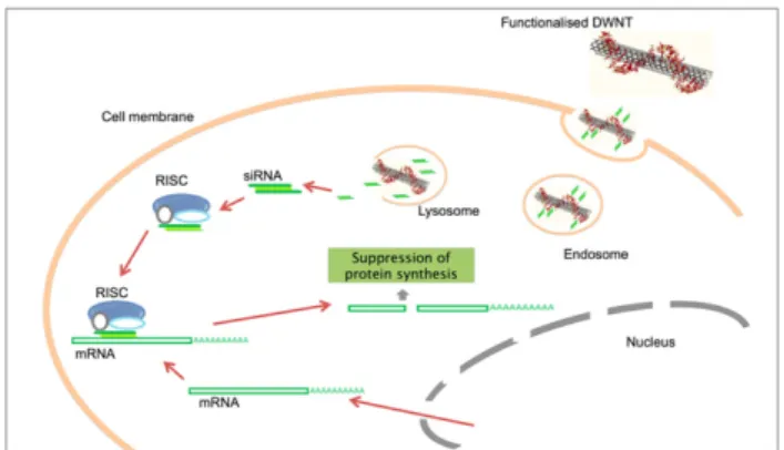
Figure 1. Schematic representation of uptake and release of siRNA inside the cell for gene silencing. After internalization,
DWNT–siRNA complexes are trapped inside endosomes, which maturate into lysosomes, whose acidic environment favours the dissociation of siRNA from the DWNTs. The released siRNA is then free to act as an effector molecule for specific cleavage of target mRNA by RISC. The cleavage of the mRNA prevents the production of a protein. Herein, siRNA is used to prevent expression of survivin, an anti-apoptotic protein that is over-expressed in cancer cells.
gene, such as silencing/small interfering RNA (siRNA). siRNA consists of short double-stranded RNA segments with typically 21–23 bases that are complementary to the mRNA sequence of the protein whose transcription is to be silenced/blocked [9]. Since RNA does not integrate into the genome, it offers greater safety than plasmid molecules. Furthermore, siRNA does not have to transfer through the nuclear membrane for its activity, but exhibits its action in the cytoplasm and therefore requires less sophisticated delivery systems than plasmid DNA, promising faster development and higher efficiencies [10]. The exact intracellular route for siRNA delivered by a suitable carrier, such as DWNTs, is shown in figure1: after uptake, DWNT–siRNA complexes are trapped inside endosomes, which subsequently mature into acidic lysosomes, favouring the dissociation of the siRNA from the polypeptide and its consequent release into the cytoplasm. There, siRNA acts as an effector molecule for specific cleavage of the target mRNA by the RNA-induced silencing complex (RISC), leading to suppression of protein synthesis.
In this study, mRNA of the survivin gene was employed as a siRNA target. Survivin is part of the protein family ‘inhibitors of apoptosis’ (IAPs): it is highly expressed in a number of human tumours and is also involved in tumour cell resistance to anticancer drugs and ionizing radiation. Evasion of apoptosis and the ability to proliferate uncontrollably are two molecular traits found in all human cancers [11]; therefore, anti-apoptotic proteins involved in signalling through specific apoptosis pathways provide attractive targets for possible drug discovery and new anticancer interventions; particularly those based on gene silencing. In this case, elimination of the survivin pathway leads to the lowering of the anti-apoptotic threshold in cancer cells and thus directly causes apoptosis [12], i.e. the suicide of unhealthy, cancerous cells.
Carbon nanotubes have been successfully employed as a delivery system for a variety of genes and other nucleic acids. In one of the pioneer studies, Kam et al reported potent gene silencing by means of siRNA delivery using CNTs. They observed a silencing effect of over 2-fold using SWNTs in comparison with Lipofectamine R, a common transfection
agent used in molecular biology [13]. A follow-up study achieved silencing of a therapeutic target, i.e. the expression of a HIV-specific cell surface co-receptor to block HIV virus entry [14]. In both cases, the applied functionalization scheme was based on cleavable disulfide bonds, which we have adapted for the study presented here. One of the first in vivo studies using siRNA delivered by carbon nanotubes as a therapeutic agent has been carried out by Podesta et al, who used amino-functionalized MWNTs to deliver a proprietary, toxic siRNA sequence to a human lung tumour xenograph model after intratumoral application, which resulted in tumour growth inhibition and prolonged survival of tumour-bearing animals [15]. Overall, this demonstrates that carbon nanotubes present a versatile and promising method for siRNA delivery that can potentially be applied for the treatment of a wide range of illnesses. The aim of our study is to investigate the feasibility of using double wall carbon nanotubes (DWNTs) as a new delivery vector for ‘anticancer’ siRNA targeting the gene survivin, a major factor in contributing to the resistance of cancer cells to apoptosis.
2. Experimental details
2.1. Synthesis of DWNTs
DWNTs were produced by catalytic chemical vapour deposition (CCVD) through decomposition of CH4 over
Mg1−xCoxO solid solution containing small quantities of
molybdenum in the reactor [4]. For purification purposes and to achieve solubility in aqueous media and physiological environments, DWNTs were subjected to a mixture of nitric and sulfuric acid as described in previous studies [16, 17] with subsequent removal of agglomerates and bundles by ultracentrifugation. After oxidation, nanotubes were sterilized by autoclaving at 121◦C for 1 h and maintained under sterile conditions for the duration of the experiment. The final product consists of single or small bundles of shorter nanotubes and will henceforth be called ‘oxDWNTs’.
2.2. Wrapping of oxidized CNTs
oxDWNTs at a concentration of 100 µg ml−1 were mixed with 50 µg ml−1 of Poly(Lys:Phe) in phosphate buffer (20 mM, pH 7.5) by continuous stirring for 2 h at room temperature (∼ 21◦C). To remove unbound polypeptide molecules, the solution was filtered using 100 kDa filter devices (Amicon Ultra-4 centrifugal filter devices from Millipore). Next, Poly(Lys:Phe)-wrapped nanotubes were resuspended in DNase/RNase-free water (Sigma Aldrich, Poole, UK) to a final DWNT concentration of 100µg ml−1.
Figure 2. Attachment of thiol-modified siRNA to Poly(Lys:Phe)-wrapped oxDWNTs using the heterobifunctional cross-linker ‘Sulfo-LC-SPDP’.
2.3. Conjugation with siRNA
oxDWNTs were wrapped with Poly(Lys:Phe) and subse-quently mixed with non-specific RNA for Raman experiments (henceforth called ‘oxDWNT–siRNA’) or siRNA targeting the GFP gene (Silencer R GFP (eGFP) siRNA and negative
con-trol, purchased from Ambion R Life Technologies, UK) for
proof-of-principle silencing experiments (henceforth called ‘oxDWNT–siRNAGFP’), which allowed for siRNA binding
by electrostatic interactions. Excess siRNA was removed by filtration with 100 kDa filter devices (Amicon Ultra-4 centrifugal filter devices from Millipore) and the obtained oxDWNT–siRNA resuspended in DNase/RNase-free water (Sigma Aldrich, Poole, UK) at a final DWNT concentration of 100µg ml−1.
For experiments with therapeutic siRNA targeting the gene survivin (siRNAsurvivin) we decided to pursue
another approach, which allows for intracellular siRNA detachment in a more controlled way, i.e. by cleavage of the disulfide bond by thiol-reducing enzymes aided by the acidic pH in lysosomes (based on a previous study by Kam et al [13]). Here, oxDWNTs were first functionalized with the cationic polypeptide Poly(Lys:Phe) and then conjugated with a heterobifunctional cross-linker containing a disulfide bond (succinimidyl 6-[3-(2-pyridyldithio)-propionamido] hexanoate, henceforth called ‘Sulfo-LC-SPDP’, from Pierce/Thermo Fisher Scientific, UK), which is designed for amine-to-sulfhydryl conjugation and readily reacts with thiol-modified siRNAsurvivin(figure2).
Twentyfive microlitres of Sulfo-LC-SPDP (20 mM) were combined with prepared oxDWNT-Poly(Lys:Phe) diluted 1:1 in PBS (20 mM, pH 7.5) and incubated for 1 h at room temperature (∼ 21◦C) under continuous stirring. Excess cross-linker was removed by filtration using 100 kDa filter devices (Amicon Ultra-4 centrifugal filter device from Millipore). Next, thiol-modified siRNA with the sequence 50-Th CCACUGAGAACGAGCCAGAUU (J-003459-11) to target survivin was mixed with oxDWNT-Poly(Lys:Phe) cross-linker and allowed to react for 1 h at room temperature under continuous stirring. The solutions were again filtered using a 100 kDa filter device (Amicon Ultra-4 centrifugal filter devices from Millipore) to remove excess siRNA and then resuspended in DNase/RNase-free water (Sigma Aldrich,
Poole, UK) at a final DWNT concentration of 100µg ml−1to obtain oxDWNT–siRNAsurvivin.
2.4. Cell culture
Human prostate cancer cells PC3 (ECACC, Porton Down, Salisbury, UK) were cultured in RPMI-1640 medium supplemented with 10% (v/v) foetal bovine serum (FBS), 2 mM GlutamaxTMand 1% (v/v) penicillin–streptomycin (all obtained from Invitrogen, Paisley, UK). Cells were cultured in 35 mm sterile petri dishes (Nunc, Thermo Scientific) containing glass coverslips.
2.5. Raman mapping of single cells
In order to investigate cellular uptake and toxicity, human prostate cancer cells PC3 were incubated with oxDWNT–RNA (30 µg ml−1 diluted in Opti-MEM serum-free cell culture medium) for up to 3 h. Assessment of DWNT uptake was performed at different time points ranging from 0.5 to 24 h. After 3 h of incubation with, the cells were washed twice with ice-cold, sterile PBS, to remove oxDWNT–RNA from the medium and fresh medium containing serum and antibiotics was added. At the end of each time point (0, 0.5, 3, 6, 9, 12, 18 and 24 h) the medium was removed and the cells washed several times with PBS. Cells were subsequently fixed with 4% paraformaldehyde solution (Sigma Aldrich, Poole, UK) to prevent morphological and chemical changes during data acquisition. Finally, the samples were again washed with PBS and slides mounted and hermetically sealed using nail lacquer. Raman mapping was performed using a Renishaw InVia Raman microscope, Elaser=1.59 eV (785 nm excitation
wavelength). The radial breathing modes (RBMs) that are characteristic for carbon nanotubes were used to map the intensity of oxDWNT–RNA in cells and to extrapolate the nanotube diameters present in the mixture using the equation ωRBM =248/dt, with ωRBM being the RBM frequency in
cm−1and dtthe tube diameter in nm [18].
2.6. Raman spectroscopy of whole cell lysates
While the distribution of DWNTs inside cells was monitored by Raman spectroscopy of whole cells, biochemical changes
of cell constituents within this time frame was monitored by Raman spectroscopy of whole cell lysates. For that, PC3 cells were grown in medium as described above and cultured in 25 cm2 tissue culture flasks until 80% confluence was reached. At this point, cells were incubated with oxDWNT–RNA (30µg ml−1final DWNT concentration after dilution in serum-free Opti-MEM medium) for various times ranging from 0.5 to 24 h. After 3 h incubation, the medium containing oxDWNT–RNA was replaced with fresh medium containing serum and antibiotics after two washing cycles with PBS. At each time point, the medium was removed from the cells, followed by several washes with PBS. The cells were subsequently trypsinized, washed with ice-cold PBS, and ruptured via hypotonic shock using a lysis buffer (Tris buffer containing 50 mM Tris HCl and 150 mM NaCl at pH 7.5 and additionally 1% NP-40, 0.2% SDS, 1 mM phenylmethanesulfonyl fluoride (PMSF), 10µg ml−1aprotinin, 10µg ml−1leupeptin, 1 mM sodium orthovanadate (Na3VO4) as cellular protease inhibitors). After
lysis, suspensions were spun down for 10 min at 500g and 4◦C (Eppendorf 5415R, Hamburg, Germany) to remove nuclei and unbroken cells, and supernatants were used to evaluate the DWNT content. For Raman measurements, a droplet of cell lysate (4 µl) was allowed to dry on a glass slide. Spectra of samples were recorded using a NT-MDT NTEGRA Spectra Probe NanoLaboratory inverted configuration microscope (Elaser=2.64 eV or 473 nm).
2.7. GFP knockdown in HeLa and PC3 cells
Stable cell lines expressing green fluorescent protein (GFP) were produced via transfection with pAcGFP-N1 plasmid (Clonetech, Saint-Germain-en-Laye, France) using Lipofectamine R according to instructions in the manual.
GFP-expressing cells were selected using 600 µg ml−1 of Geneticin R (Gibco/Invitrogen, Parsley, UK). After 4 weeks
of selection cells were sorted by fluorescence-activated cell sorting (FACS), whereby 80–90% GFP-expressing cells were obtained. These cells were allowed to recover for 8 weeks and re-sorted once more for further purification. The expression of GFP allows for visualization by confocal microscopy (excitation at 488 nm and 400 mW power emission using an argon laser; detection using a 535 nm ± 15 nm band pass filter).
For oxDWNT–siRNAGFP transfection, the previously
prepared complexes were diluted 1:2 in Opti-MEM R medium
(Invitrogen, Paisley, UK). For comparison with a commer-cially available transfection agent serving as the positive control, Lipofectamine R RNAiMAX, siRNA
GFP complexes
were prepared according to the manufacturer’s instructions (1 or 6 µl Lipofectamine R RNAiMAX, for use in 24-well or
35 mm plates, respectively, were combined with 0.2–1µM siRNAGFP and added to each corresponding well). After
4 h the medium was replaced with fresh complete medium containing serum. The cells were then incubated for further 48 h at 37◦C in a CO2incubator.
To evaluate gene knockdown, cells were fixed with 4% paraformaldehyde (Sigma Aldrich, Poole, UK) in PBS for 1 h
at room temperature, followed by washing with sterile PBS and treatment with 0.1% Triton X100 (Sigma Aldrich, Poole, UK) in PBS for 15 min to permeabilize cell membranes. Cells were then washed three-times with PBS and TO-PRO R-3
iodide (Molecular Probes R, 0.1 µM in PBS), a monomeric
cyanine nucleic acid stain, was utilized to stain the nuclei in all samples. Images were captured at 63× magnification using a Zeiss LSM 510 inverted confocal microscope and analysed using LSM 510 META software. To determine the percentage of GFP knockdown after siRNAGFP delivery, cells were
counted in five different fields. GFP-expressing cells appear green (GFP, emission 505 nm) with the nucleus coloured in blue (TO-PRO R
-3, emission 661 nm), while non-expressing cells only exhibit nuclear staining.
2.8. Survivin knockdown in PC3 cells
To achieve knockdown of the survivin gene in PC3 cells, cells were incubated with oxDWNT–siRNAsurvivin for 4 h
and maintained for 3–7 days to allow for gene silencing to occur. PC3 cells were initially seeded 24 h prior to transfection to obtain 50% confluency at the time of transfection in 35 mm sterile petri dishes. oxDWNT–siRNAsurvivinprepared
as described in the previous section was diluted 1:2 in Opti-MEM R medium (Invitrogen, Paisley, UK). Again, a
commercial transfection agent, DharmaFECT R, was used
to serve as the positive control. DharmaFECT–siRNAsurvivin
complexes were prepared according to the manufacturer’s instructions: 3µl DharmaFECT R
reagent was combined with 400µl of serum-free culture medium and incubated for 5 min at room temperature. Next, siRNAsurvivinwas diluted in 400µl
of media to a final concentration of 0.8 µM per 35 mm plate. The solutions were then combined and incubated at room temperature for a further 20 min, before being added to the corresponding wells. After 4 h the medium was replaced with fresh complete medium (containing 10% serum) for both samples. The cells were then incubated for a further 72 h at 37◦C in a CO2 atmosphere in order to achieve a silencing
effect. After 72 h incubation cells were trypsinized and split for further incubation up to 120 h. Cells were then stained with Trypan Blue and counted using a haemocytometer.
The level of apoptosis induced by silencing of the survivingene was evaluated by means of the Annexin V Biotin Apoptosis Detection Kit (CalBioChem R
) and subsequent analysis by flow cytometry, which permits determination of the percentage of viable cells, cells in early apoptotic stage, late apoptotic stage, and dead cells. Untreated cells, as well as cells treated with transfection agent but without siRNAsurvivin
served as negative controls. For these experiments, cells floating in the medium, as well as cells growing in a monolayer were recovered and combined for an apoptosis assay using the previously described kit. Combined cells were spun down and resuspended in 500µl of medium. Next, 10 µl medium binding reagent and 2 µl Annexin V-FITC were added and the mixture incubated for 30 min at room temperature in the dark. Following incubation, cells were spun down and resuspended in 500 µl of a cold one-time binding buffer. After that, 10µl propidium iodide was added
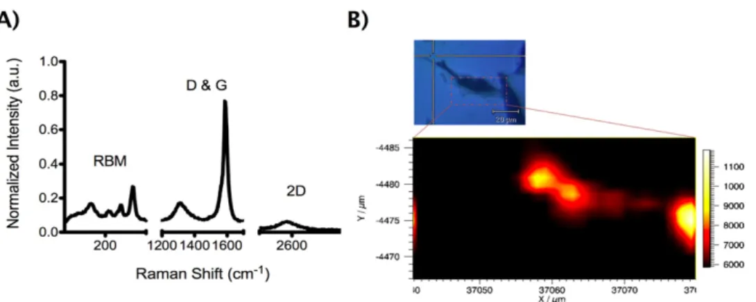
Figure 3. Raman spectrum of DWNTs (A) and mapping of oxDWNT–RNA inside a cell (B). (A) DWNT Raman spectrum featuring the characteristic Raman bands: RBM (150, 200, 230 and 260 cm−1); D-band (1350 cm−1); G-band (1590 cm−1) and 2D-band (2600 cm−1). (B) Raman mapping of RBM mode (160 cm−1) in a single cell (inset). A high intensity is observed in the cell, but not in the surrounding area.
and samples were placed on ice and away from light, before analysis by flow cytometry. Note that this assay can only be performed on live cells and it is unsuitable for fixed material. Measurements were obtained using the Beckman Coulter Epics XL flow cytometer analyser. Data analysis was performed utilizing FlowJo software.
3. Results
oxDWNTs were found to consist of single or small bundles of shorter nanotubes (200–2000 nm length, 296 nm ± 123 nm on average as determined by AFM and phase-analysis light scattering (PALS) [17]). TEM analysis (figure S1 in the supporting information available atstacks.iop.org/Nano/ 23/365102/mmedia) confirmed the Poly(Lys:Phe) coating of Poly(Lys:Phe)-wrapped oxDWNTs. Analysis by Raman spectroscopy revealed distinct peaks characteristic of DWNTs (figure3a): the radial breathing modes (RBMs) at 150, 200, 230 and 260 cm−1 (correlating with diameters of 0.9, 1.1, 1.2 and 1.58 nm [18]), as well as the D-band (1350 cm−1), G-band (1590 cm−1), and the 2D-band (2600 cm−1), which can be clearly observed in the spectrum. Figure3(b) shows a Raman map of the RBM in a mammalian cell, which has previously been exposed to oxDWNT–RNA. A strong, locally confined Raman signal was detected within the whole cell, confirming successful DWNT uptake and possible localization within endosomes, as also reported in previous work by our group, which has looked at the intracellular distribution of functionalized DWNTs in greater detail [19,
20].
An important factor for the design of DWNTs as a nano-vector is that they do not significantly modify the cell environment, so that perturbations due to the payload can be detected. Raman spectroscopy can serve as a powerful tool to assay biochemical changes in cells [19,21]. Indeed, Raman spectra of oxDWNT–RNA inside cells (obtained by analysing whole cell lysates) exhibited several peaks in addition to those observed for DWNTs alone, of which some can be contributed to cell constituents. Figure4 plots three of those
peaks and their variation over time (24 h time frame): DNA backbone stretching at 1090 cm−1 [22], the CH bending mode of proteins and lipids at 1450 cm−1 [23], and amide Iα-helix vibrations of proteins at 1660 cm−1[24]. The DNA backbone stretching (O–P–O) Raman peak has been described as suitable for monitoring the process of cell death due to its integral association with DNA disintegration [22]. When a comparison was made between cells exposed to DWNTs and unexposed controls, it was found that identical forms and patterns were maintained.
Next, we investigated the ability of oxDWNTs to bind and deliver siRNA into cells. siRNA has a vast potential as a therapeutic agent for various disorders, but cannot cross the cell membrane on its own due to its negative charge, and is prone to degradation by nucleases inside the cell. In this case, we used oxDWNTs to deliver two types of siRNA: first, siRNA to silence the GFP gene as a proof of principle (siRNAGFP), and secondly, siRNA to silence the
gene survivin, which has therapeutic value for cancer therapy (siRNAsurvivin).
Figure 5 shows fluorescent micrographs of GFP-expressing cells treated with different concentrations of siRNAGFP. Under the employed conditions, both
lipofectamine R and oxDWNTs showed negligible toxicity.
Control cells and cells treated with non-specific siRNA (siRNAneg) displayed around 80% cells expressing GFP.
The treatment with 50 nM of bound siRNAGFP using
lipofectamine R as delivery agent produced a pronounced
effect in GFP expression in both cells lines, which decreased to a level of about 20% in Hela cells and 45% in PC3 cells. Treatment with 100 nM bound siRNAGFP delivered
by lipofectamine R decreased GFP expression to a level of
about 8% and 21%, respectively. The knockdown effect using oxDWNT–siRNAGFPwas lower than for the positive control
lipofectamine, but nevertheless a significant decrease in GFP expression to a level of about 50% was observed after only 48 h.
We next investigated the delivery of therapeutic siRNA targeting the gene survivin by the developed oxDWNT vectors. Figure6 reveals the effect of siRNAsurvivin delivery
Figure 4. Time-dependent analysis of cell constituents by Raman spectroscopy after exposure to oxDWNT–RNA: DNA, protein/lipids and protein structure (amide Iα-helix). Cell constituents were not disturbed through incubation with oxDWNT–siRNA in comparison with control cells.
Figure 5. Percentage of cells expressing GFP in HeLa and PC3 cells treated with siRNAGFP. GFP stable cell lines were produced via a
protocol of 4-week selection to obtain 80–90% GFP-expressing cells, using fluorescence-activated cell sorting (FACS). GFP knockdown was achieved using either oxDWNTs or a common transfection reagent (Lipofectamine R
) serving as the positive control, and was determined by confocal microscopy by counting GFP-expressing cells (green cytoplasm plus blue nucleus) against non-expressing cells (blue nucleus only). siRNAGFPdelivery by lipofectamine produced around 89% knockdown of GFP in Hela cells and 69% in PC3 cells for
the 100 nM sample, while oxDWNTs–siRNAGFPat 100 nM siRNAGFPconcentration induced about 40% knockdown in Hela cells and 29%
knockdown in PC3 cells in comparison with the negative control. Results are presented as mean ± SEM (n = 5).
using oxDWNTs, again in comparison with a common transfection reagent (DharmaFECT R) and untreated cells
(PC3 cells only and cells treated with DharmaFECT R only).
The results showed similar levels of apoptosis for cells treated with siRNAsurvivin delivered by oxDWNTs (35.89%) and by
DharmaFECT R transfection agent (44.92%), which are both
significantly higher than the two controls; cells only (5.22%) and cells treated with DharmaFECT R
only (18.64%).
4. Discussion
The selective and robust effect of RNA interference on gene expression, mediated by siRNA, makes it not only a valuable research tool, but also a promising therapeutic platform for the treatment of viral infections, dominant disorders, cancer, and neurological disorders [25]. In this paper, we focused on the delivery of siRNA targeting the gene survivin as a potential treatment for a range of cancers, as the survivin
Figure 6. Flow cytometry analysis of apoptosis in cells treated with siRNAsurvivin, showing percentages of cells at different apoptotic
stages for each treatment. PC3 cells only and PC3 cells treated with DharmaFECT R
only served as the negative controls and siRNAsurvivintransfected using the commercial transfection agent
DharmaFECT R
(DharmaFECT–siRNA) as the positive control. A similar apoptotic effect was obtained for oxDWNT–siRNAsurvivin
than for the positive control DharmaFECT–siRNAsurvivin.
protein regulates apoptosis and controls cell proliferation and is expressed highly in most human tumours and foetal tissue, but is completely absent in terminally differentiated cells. The successful delivery of siRNA to target specific genes in mammalian model organisms has been achieved using a variety of methods, including liposomes, cationic lipids, polymers and nanoparticles. However, systemic toxicity remains a great concern with most of these delivery options. Thus, there is an ever-growing need to enhance the available tools that can lead to effective and safe gene delivery. The functionalized oxDWNTs developed in this work present an alternative with reasonable biocompatibility, which can simultaneously serve as an imaging agent due to their unique Raman signature within biological environments (figure 3). DWNTs have an important advantage over SWNTs, for which any structural damage disrupts their mapping and progression over time. Furthermore, as we have shown in previous studies, they are released from the cells within 24 h after having fulfilled their task and do not accumulate within cells [20]. In this study we investigated whether oxDWNT–RNA causes any changes in the cell biochemistry within this time frame, which they did not (figure4). This is in good agreement with a previous study of our group, which has shown that oxDWNT–RNA did not cause phosphorylation of mitogen-activate protein kinases (MAPKs), which are markers for induced cellular stress [19]. In addition, we have also previously investigated the effect of oxDWNTs on cell viability after 96 h incubation by a MTT cytotoxicity assay and found no dose-dependent toxicity, although an overall drop of cell viability of about 5–15% was observed, possibly due to inhibition of cell proliferation or decreased cell adhesion [17]. Overall, this indicates that oxDWNT–RNA does not have a deleterious effect on cells during their passage, although long-term effects remain to be investigated.
In terms of therapeutic efficacy, the achieved silencing effect (‘gene knockdown’) was either lower than commercial transfection agents (GFP–siRNA/lipofectamine R, figure 5)
or at a comparable level (survivin siRNA/DharmaFECT R
,
figure 6). Considering that most commercially available in vitro transfection agents are unsuitable for therapeutic applications due to their systemic toxicity, this indicates that DWNTs may be successfully employed to deliver therapeutic moieties, such as siRNA, to cells for the treatment of cancerous disorders, as demonstrated in this study. Future work will need to be done to study the in vivo performance of our system, which will show whether oxDWNTs do have an advantage over cationic lipid-based transfection agents (e.g. lipofectamine R and DharmaFECT R) due to
improved biocompatibility, and will allow for a comparison of the in vivo performance of oxDWNTs with other carbon nanotube-based delivery systems in the literature.
5. Conclusions
In summary, we show that short oxDWNTs produced by CCVD and subsequent acid treatment and functionalization are suitable for the delivery of therapeutic moieties and do not exhibit the toxic effects that have been attributed to longer CNTs [5]. Their unique Raman spectral features, such as the RBM, allow for their detection inside cells by Raman spectroscopy, while simultaneously enabling the detection of changes in cell biochemistry following oxDWNT uptake. It is demonstrated that during a period of 24 h RNA-wrapped oxDWNTs did not cause detectable changes in the cells’ DNA, protein or lipid content. Our final goal, however, was to use oxDWNTs as a nano-vector for therapeutic payloads to achieve a clinically relevant effect. After loading oxDWNTs with siRNA to block the expression of the anti-apoptotic protein survivin, which is over-expressed in most human tumours, we successfully demonstrated apoptosis of cells after transfection with oxDWNT–siRNAsurvivincomplexes to
a similar level as obtained with siRNAsurvivindelivered by a
standard siRNA transfection agent. It should be noted that although transfection agents are able to efficiently transport siRNA into cultured cells, they are unsuitable for therapeutic applications due to their systemic toxicity. The study thereby advances the goal of developing nano-vectors for safe and efficient delivery of therapeutic payloads.
Acknowledgments
This work has been performed in the framework of the FP6 Marie Curie Research Training Network ‘CARBIO’ (RTN-CT-2006-035616) funded by the European Union.
References
[1] Chen R J, Bangsaruntip S, Drouvalakis K A, Kam N W S, Shim M, Li Y M, Kim W, Utz P J and Dai H J 2003 Noncovalent functionalization of carbon nanotubes for highly specific electronic biosensors Proc. Natl Acad. Sci. USA100 4984–9
[2] Kam N W S, Jessop T C, Wender P A and Dai H J 2004 Nanotube molecular transporters: internalization of carbon nanotube–protein conjugates into mammalian cells J. Am. Chem. Soc.126 6850–1
[3] Bianco A, Kostarelos K, Partidos C D and Prato M 2005 Biomedical applications of functionalised carbon nanotubes Chem. Commun.571–7
[4] Flahaut E, Bacsa R, Peigney A and Laurent C 2003 Gram-scale CCVD synthesis of double-walled carbon nanotubes Chem. Commun.1442–3
[5] Poland C A, Duffin R, Kinloch I, Maynard A, Wallace W A H, Seaton A, Stone V, Brown S, MacNee W and
Donaldson K 2008 Carbon nanotubes introduced into the abdominal cavity of mice show asbestos-like pathogenicity in a pilot study Nature Nanotechnol.3 423–8
[6] Wang X et al 2011 Dispersal state of multiwalled carbon nanotubes elicits profibrogenic cellular responses that correlate with fibrogenesis biomarkers and fibrosis in the murine lung ACS Nano5 9772–87
[7] Crouzier T, Nimmagadda A, Nollert M U and
McFetridge P S 2008 Modification of single walled carbon nanotube surface chemistry to improve aqueous solubility and enhance cellular interactions Langmuir24 13173–81
[8] Sanhai W R, Sakamoto J H, Canady R and Ferrari M 2008 Seven challenges for nanomedicine Nature Nanotechnol.
3 242–4
[9] McManus M T and Sharp P A 2002 Gene silencing in mammals by small interfering RNAs Nature Rev. Genet.
3 737–47
[10] Patil S D, Rhodes D G and Burgess D J 2005 DNA-based therapeutics and DNA delivery systems: a comprehensive review AAPS. J.7 E61–77
[11] Hanahan D and Weinberg R A 2000 The hallmarks of cancer Cell100 57–70
[12] Carvalho A, Carmena M, Sambade C, Earnshaw W C and Wheatley S P 2003 Survivin is required for stable
checkpoint activation in taxol-treated HeLa cells J. Cell Sci.
116 2987–98
[13] Kam N W S, Liu Z and Dai H J 2005 Functionalization of carbon nanotubes via cleavable disulfide bonds for efficient intracellular delivery of siRNA and potent gene silencing J. Am. Chem. Soc.127 12492–3
[14] Liu Z, Winters M, Holodniy M and Dai H 2007 siRNA delivery into human T cells and primary cells with carbon-nanotube transporters Angew. Chem. Int. Edn.
46 2023–7
[15] Podesta J E, Al-Jamal K T, Herrero M A, Tian B, Ali-Boucetta H, Hegde V, Bianco A, Prato M and Kostarelos K 2009 Antitumor activity and prolonged survival by carbon-nanotube-mediated therapeutic siRNA silencing in a human lung xenograft model Small5 1176–85
[16] Heister E, Neves V, Tilmaciu C, Lipert K, Beltran V S, Coley H M, Silva S R P and McFadden J 2009 Triple functionalisation of single-walled carbon nanotubes with doxorubicin, a monoclonal antibody, and a fluorescent marker for targeted cancer therapy Carbon47 2152–60
[17] Heister E et al 2010 Higher dispersion efficacy of
functionalized carbon nanotubes in chemical and biological environments ACS Nano4 2615–26
[18] R A Jishi, Venkataraman L, Dresselhaus M S and
Dresselhaus G 1993 Phonon modes in carbon nanotubules Chem. Phys. Lett.209 77–82
[19] Neves V et al 2010 Uptake and release of double-walled carbon nanotubes by mammalian cells Adv. Funct. Mater.
20 3272–9
[20] Neves V, Gerondopoulos A, Heister E, Tilmaciu C, Flahaut E, Soula B, Silva S R P, McFadden J and Coley H M 2012 Cellular localization, accumulation and trafficking of DWNTs in human prostate cancer cells NanoResearch
5 223–34
[21] Notingher I, Verrier S, Haque S, Polak J M and Hench L L 2003 Spectroscopic study of human lung epithelial cells (A549) in culture: living cells versus dead cells Biopolymers72 230–40
[22] Verrier S, Notingher I, Polak J M and Hench L L 2004 In situ monitoring of cell death using Raman microspectroscopy Biopolymers74 157–62
[23] Krafft C, Knetschke T, Funk R H W and Salzer R 2005 Identification of organelles and vesicles in single cells by Raman microspectroscopic mapping Vib. Spectrosc.
38 85–93
[24] Puppels G J, Garritsen H S P, Segersnolten G M J,
Demul F F M and Greve J 1991 Raman microspectroscopic approach to the study of human granulocytes Biophys. J.
60 1046–56
[25] Ryther R C C, Flynt A S, Phillips J A and Patton J G 2005 siRNA therapeutics: big potential from small RNAs Gene Ther.12 5–11