HAL Id: inserm-00874869
https://www.hal.inserm.fr/inserm-00874869
Submitted on 18 Oct 2013
HAL is a multi-disciplinary open access
archive for the deposit and dissemination of
sci-entific research documents, whether they are
pub-lished or not. The documents may come from
teaching and research institutions in France or
abroad, or from public or private research centers.
L’archive ouverte pluridisciplinaire HAL, est
destinée au dépôt et à la diffusion de documents
scientifiques de niveau recherche, publiés ou non,
émanant des établissements d’enseignement et de
recherche français ou étrangers, des laboratoires
publics ou privés.
New insights into pediatric idiopathic pulmonary
hemosiderosis: the French RespiRare(R) cohort.
Jessica Taytard, Nadia Nathan, Jacques de Blic, Mickael Fayon, Ralph
Epaud, Antoine Deschildre, Françoise Troussier, Marc Lubrano, Raphaël
Chiron, Philippe Reix, et al.
To cite this version:
Jessica Taytard, Nadia Nathan, Jacques de Blic, Mickael Fayon, Ralph Epaud, et al.. New insights into
pediatric idiopathic pulmonary hemosiderosis: the French RespiRare(R) cohort.. Orphanet Journal
of Rare Diseases, BioMed Central, 2013, 8 (1), pp.161. �10.1186/1750-1172-8-161�. �inserm-00874869�
R E S E A R C H
Open Access
New insights into pediatric idiopathic pulmonary
hemosiderosis: the French RespiRare® cohort
Jessica Taytard
1, Nadia Nathan
1, Jacques de Blic
2, Mickael Fayon
3, Ralph Epaud
4, Antoine Deschildre
5,
Françoise Troussier
6, Marc Lubrano
7, Raphaël Chiron
8, Philippe Reix
9, Pierrick Cros
10, Malika Mahloul
1,
Delphine Michon
1, Annick Clement
1, Harriet Corvol
1*and for the French RespiRare® group
Abstract
Background: Idiopathic pulmonary hemosiderosis (IPH) is a rare cause of alveolar hemorrhage in children and its pathophysiology remains obscure. Classically, diagnosis is based on a triad including hemoptysis, diffuse
parenchymal infiltrates on chest X-rays, and iron-deficiency anemia. We present the French pediatric cohort of IPH collected through the French Reference Center for Rare Lung Diseases (RespiRare®, www.respirare.fr).
Methods: Since 2008, a national network/web-linked RespiRare® database has been set up in 12 French pediatric respiratory centres. It is structured as a medical recording tool with extended disease-specific datasets containing clinical information relevant to all forms of rare lung diseases including IPH.
Results: We identified 25 reported cases of IPH in children from the database (20 females and 5 males). Among them, 5 presented with Down syndrome. Upon diagnosis, median age was 4.3 [0.8-14.0] yrs, and the main manifestations were: dyspnea (n = 17, 68%), anemia (n = 16, 64%), cough (n = 12, 48%), febrile pneumonia (n = 11, 44%) and hemoptysis (n = 11, 44%). Half of the patients demonstrated diffuse parenchymal infiltrates on chest imaging, and diagnosis was ascertained either by broncho-alveolar lavage indicating the presence of hemosiderin-laden
macrophages (19/25 cases), or lung biopsy (6/25). In screened patients, initial auto-immune screening revealed positive antineutrophilic cytoplasmic antibodies (ANCA) (n = 6, 40%), antinuclear antibodies (ANA) (n = 5, 45%) and specific coeliac disease antibodies (n = 4, 28%). All the patients were initially treated by corticosteroids. In 13 cases,
immunosuppressants were introduced due to corticoresistance and/or major side effects. Median length of follow-up was 5.5 yrs, with a satisfactory respiratory outcome in 23/25 patients. One patient developed severe pulmonary fibrosis, and another with Down syndrome died as a result of severe pulmonary hemorrhage.
Conclusion: The present cohort provides substantial information on clinical expression and outcomes of pediatric IPH. Analysis of potential contributors supports a role of auto-immunity in disease development and highlights the importance of genetic factors.
Keywords: Idiopathic pulmonary hemosiderosis, Child, Interstitial lung disease, Autoimmune lung disease, Coeliac disease, Down syndrome
* Correspondence:harriet.corvol@trs.aphp.fr
1Pediatric Pulmonary Department, AP-HP, Hôpital Trousseau, Université Pierre
et Marie Curie-Paris6, Inserm U938, 26, avenue du Docteur Arnold-Netter, 75012 Paris, France
Full list of author information is available at the end of the article
© 2013 Taytard et al.; licensee BioMed Central Ltd. This is an open access article distributed under the terms of the Creative Commons Attribution License (http://creativecommons.org/licenses/by/2.0), which permits unrestricted use, distribution, and reproduction in any medium, provided the original work is properly cited.
Introduction
Idiopathic pulmonary hemosiderosis (IPH) is a rare cause of alveolar hemorrhage of unknown etiology in children, leading to chronic infiltrative pulmonary disease [1-4]. It is classically characterized by a triad of hemoptysis, iron-deficiency anemia and pulmonary infiltrates on chest X-rays; and usually occurs before the age of 10 years (yrs) old [5-10]. The diagnosis of IPH is confirmed by bronchoscopy with bronchoalveolar lavage (BAL) sho-wing hemosiderin-laden macrophages called siderophages, pathognomonic of the disease [11]. Lung biopsies are sometimes required to eliminate differential diagnoses such as vasculitis [12]. First line treatments usually include sys-temic corticosteroid therapy. This may be substituted by immunosuppressants in case of corticosteroid resistance or dependence and/or unfavorable outcome [2,3,13,14].
Various hypotheses have been proposed to explain the pathophysiology of IPH: allergic, environmental, genetic and/or auto-immune. The allergic hypothesis is based on the frequent association between IPH and cow’s milk allergy [15]. The environmental theory has been sug-gested after the emergence of IPH in children exposed to Stachybotris chartarum [16,17]. IPH has also been de-scribed in siblings, leading to discuss a genetic theory, but no gene has yet been identified [18,19]. Finally, the auto-immune theory is recognized as the most probable, considering the frequent association with auto-immune diseases such as coeliac disease, glomerulonephritis and/or rheumatoid arthritis [2,5,11,17,20-23].
Pediatric IPH is a rare respiratory disease whose inci-dence varies between 0.24 and 1.26 per million [3]. This explains the limited knowledge on physiopathology and the lack of consensus regarding care. Through the organization of the French Reference Center for Rare Lung Diseases (RespiRare®) with the national network/web-linked database (described previously), we reviewed the IPH cases in the French pediatric population with two main goals: to update management strategies and to provide new insights into disease contributors and physiopathology [24].
Material and methods
Since 2008, the RespiRare® database has been set up in 12 French pediatric respiratory centres. This database is structured as a medical recording tool for the patient, with extended disease-specific datasets containing clinical information relevant to all kinds of rare lung diseases including interstitial lung disease and IPH [24]. The database and the data collection have been approved by French national data protection authorities [24]. Each patient and/or his legal representatives are informed prior to entering their data into the database. This study is a chart based retrospective study. Relevant clinical data of the French pediatric IPH patients were retrospectively collected via the RespiRare® database. Diagnosis is based
on the triad of iron deficiency anemia, respiratory symptoms (including dyspnea, cough and hemoptysis), and pulmonary infiltrates on chest imaging, and is con-firmed by the presence of hemosiderin laden macrophages in the BAL and/or on lung tissue specimens.
At diagnosis, the data collected comprised:
(i) clinical features: age, body mass index (BMI), hemoptysis, dypnea, cough and/or febrile pneumonia; personal and/or familial history of allergy and/or auto-immune diseases;
(ii) imaging data: chest X-ray and chest CT scan; (iii)respiratory function tests: spirometry
measurements (vital capacity (VC), forced vital capacity (FVC), slow vital capacity (SVC) and forced expiratory volume in one second (FEV1)) and
single-breath measure of carbon monoxide diffusion (DLCO);
(iv) BAL: the percentage of hemosiderin laden
macrophages, and the Golde score. The Golde score is a semi-quantitative method for assessing the hemosiderin content of alveolar macrophages after Prussian blue stain: at least 200 alveolar macrophages are examined and, to each macrophage, is attributed a score ranging from 0 (absence of blue staining) to 4 (hemosiderin laden macrophages); the Golde score represents the mean score on 100 macrophages (that could range from 0 to 400). An alveolar hemorrhage is confirmed for a score higher than 20, and characterized as severe for a score above 100 [20]; (v) lung biopsy;
(vi) blood tests: hemoglobin, reticulocytes count, and autoimmune assessments: antitransglutaminase (ATA, IgA and IgG), antigliadin (AGA) and antiendomysium (AEA) antibodies, antinuclear antibodies (ANA), anti-double strand DNA and anti-smooth-muscle antibodies (SMA), rheumatoid factor (RF) and antineutrophilic cytoplasmic antibodies (ANCA).
The data collected during the follow up included treat-ments, side effects and clinical outcome.
Descriptive data analysis (medians and standard devia-tions for quantitative values, percentages for qualitative values) have been realised using the Excel 2010 software.
Results
Population demographic description at diagnosis
Twenty-five patients (20 girls and 5 boys), diagnosed with IPH between 1996 and June 2012, have been included from 12 French pediatric respiratory centers. The main clinical data at diagnosis are described in Table 1.
Median age at diagnosis was 4.3 [0.8-14.0] yrs. Diagnosis was made before the age of 2 yrs in 7 patients, and after Taytard et al. Orphanet Journal of Rare Diseases 2013, 8:161 Page 2 of 7 http://www.ojrd.com/content/8/1/161
the age of 10 yrs in 4 of them. Median BMI was 14.75 [13.3-25.5] with a median Z-score of −0.6 [−2.46-2.16]. The main manifestations at diagnosis were: dyspnea (n = 17, 68%), cough (n = 12, 48%), febrile pneumonia (n = 11, 44%) and hemoptysis (n = 11, 44%). Five patients out of the 25 IPH cases had a concomitant Down syndrome (20%).
Among the 25 patients, 2 had a personal history of coeliac disease and one of arthralgia. Five familial cases of autoimmune diseases were observed: 2 ankylosing polyarthritis, 1 coeliac disease, 1 telangiectasis, 1 type 1 diabetes, and one Minkowski-Chauffard disease. Fur-thermore, 2 patients were dizygote twins and another patient had 2 cousins with an IPH history.
Investigations at diagnosis Blood count
Anemia was present in 64% of cases, with a median hemoglobin of 7.2 g/dl [2.3-12.4]. Nine patients had a severe anemia (<7 g/dl). Reticulocytes, documented in only 4 out of the 25 patients, were systematically elevated, varying between 98,900 and 152,150/mm3.
Thoracic imaging
Interstitial and/or alveolar interstitial patterns were visualized by chest X-rays for 13 patients and by chest CT-scans for 17 patients. The chest CT scans showed ground-glass opacities (12 patients), sub pleural cysts (6 patients), and, less frequently, micronodules (2 patients), thickened interlobular septa (2 patients), honeycomb patterns (1 patient), and traction bronchiectasis (1 patient). Images were often diffuse and bilateral. An illustration is given in Figure 1.
Respiratory function tests
Spirometric analyses at onset were available for 18 patients. The 7 remaining patients had less than 3 yrs old at the time of diagnosis, and, thus, did not per-form functional respiratory tests. The spirometric analyses were normal in 9/18 patients (50%), showed a restrictive pattern for 6 patients (37.5%) and an airway obstruction for 1 patient (6%). Medians of VC, FVC and SVC were re-spectively 69, 70 and 65%. For the patients presenting with
a restrictive pattern, the median age was 9 yrs old [3]. DLCO, measured for 9 patients, was either decreased (4/9: 44%), or normal.
BAL
The diagnosis was ascertained in 19/25 cases by the BAL that showed an accumulation of hemosiderin-laden macrophages. The Golde score, evaluated in 7 cases indicated a median of 158 [52–356].
Pulmonary biopsies
Eight patients underwent a lung biopsy, which documented presence of red blood cells in the alveoli and in the interstitium.
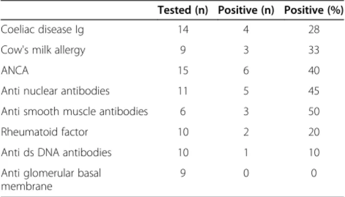
Auto immune assessments
Results of the initial auto-immune screenings are described in Table 2. In screened patients, it principally revealed positive ANCA (n = 6, 40%), ANA (n = 5, 45%), ASM (n = 3, 50%), and specific cow’s milk IgE (n = 3, 33%). Specific coeliac disease antibodies were screened in 14 patients (56%), and found to be positive in 4 (28% of the tested patients): 3 patients had positive ATA IgA, and 1 positive AGA.
During the follow-up, additional auto-antibodies were documented: RF (n = 3) and ANA (n = 2). Interestingly, repeated evaluation of the auto-immune status in the 6 positive ANCA patients showed variable evolution: ANCA disappeared for 4 patients at the end of the follow-up, decreased for one patient and remained stable for one patient.
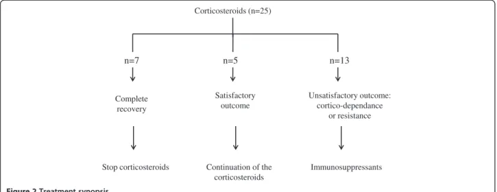
Treatment and follow up
Treatment synopsis is described Figure 2. As highlighted, all the patients received systemic corticosteroids with a variable efficacy. Intravenous corticosteroids were the usual first line treatment with monthly pulses of methylpredniso-lone (300 mg/m2/day for 3 days per month) for 100% of the patients (n = 25). In situations of severe symptoms, daily oral prednisone was added (1 mg/kg/d). The duration of corticosteroids treatment was variable among the patients based on clinical, radiological (alveolar infiltrates) and biological (hemoglobin, reticulocytes) evolution.
Thirteen patients had an unsatisfactory outcome, inclu-ding in 6 of them a Cushing syndrome with important gain of weight, stretch marks and hyperthricosis. For these 13 patients, one or more immunosuppressants were added: hydroxychloroquine (4.5 to 6 mg/kg/day for n = 10 patients), mycophenolate mofetil (600 mg/m2/day for n = 7 patients), or azathioprine (1 mg/kg/day for n = 1 patient). No side effects were reported, allowing either to interrupt the corticosteroids (n = 6 patients), or to reduce their dosage (n=7 patients).
Table 1 Main manifestations at diagnosis
Patients Patients (%) Hemoptysis 11 44 Dyspnea 17 68 Cough 12 48 Febrile pneumonia 11 44 Anemia 16 64 Severe anemia (<7 g/dl) 9 36
The median length of follow-up was 5.5 yrs (2 months to 14 yrs). Respiratory outcome was satisfactory in 23 patients, although 7 of them experienced several relapses. The 2 other patients, who were diagnosed at 5 months old, had a severe expression of the disease: 1 of them developed a pulmonary fibrosis, and the other one (with a Down syndrome) had a fatal pulmonary hemorrhage.
Discussion
Although several pediatric IPH cases have been reported in the literature, few cohorts have been described so far. To our knowledge, this study is one of the largest ever reported, with 25 IPH pediatric cases, and a long median length of follow-up of 5.5 yrs [25].
IPH generally occurs in children below the age of 10 yrs, and commonly between the ages of 1–7 yrs [2,5,8,21]. Although the classical triad of iron deficiency anemia, respiratory symptoms (including dyspnea, cough and hemoptysis), and pulmonary infiltrates on chest imaging is characteristic, clinical presentation is highly
variable; anemia and dyspnea being the most frequent clinical features (64% and 68% respectively in our study). Hemoptysis only occurred in 50% of the patients, but its incidence could be underestimated in young children, who frequently swallow their sputum. So far, number of studies highlighted an important delay between onset of symptoms and IPH diagnostic, ranging from 1 to 6.3 yrs [3,7,11,14,21]. In our study, the highest delay reached 16 months. This delay in diagnosis may be due to several factors: insidious onset, and lack of awareness about the condition. Moreover, iron deficiency anemia can also be misleading, especially if respiratory signs are mild.
IPH pathogenesis remains controversial, with, so far, four main etiological hypotheses described in the literature: the environmental, the allergic, the auto-immune, and the genetic hypotheses. In this cohort, we found no evidence favouring an environmental etiology, with, however, little collected information on the various possible environmen-tal factors. This theory was suggested after the occurrence of acute pulmonary hemorrhages in children exposed to Stachybotris chartarum in Cleveland, but was not further confirmed [16,17]. Similarly, the allergic theory, based on the association with the hypersensitivity to proteins in cow's milk (Heiner syndrome), remains controversial [15]. In the present cohort, 3 patients out of 25 had positive antibodies. As eviction of cow's milk proteins has been shown to benefit the patients with Heiner syndrome, the dosage of cow's milk IgE remains recommended.
Our present study provides support for an auto-immune contribution in IPH physiopathology. First, most of the patients (17 out of the 25) had auto-immune antibodies at onset, and, for 6 patients, additional auto-immune anti-bodies appeared during the follow-up. The most frequent auto-immune antibodies that were found in our cohort were: SMA (50% of the tested patients); ANA (45%) and
a
b
Figure 1 Thoracic imaging. a shows ground-glass opacities on a chest X-ray in a 5 yrs old IPH patient. b shows on a chest CT-scan an interstitial pattern with micronodules and ground-glass opacities in a 5 yrs old IPH patient.
Table 2 Auto-immune assessment at diagnosis
Tested (n) Positive (n) Positive (%)
Coeliac disease Ig 14 4 28
Cow's milk allergy 9 3 33
ANCA 15 6 40
Anti nuclear antibodies 11 5 45
Anti smooth muscle antibodies 6 3 50
Rheumatoid factor 10 2 20
Anti ds DNA antibodies 10 1 10
Anti glomerular basal membrane
9 0 0
Ig: immunoglobulin, ANCA: anti neutrophilic cytoplasmic antibodies, ds: double strand
Taytard et al. Orphanet Journal of Rare Diseases 2013, 8:161 Page 4 of 7 http://www.ojrd.com/content/8/1/161
ANCA (40%). These antibodies are usually associated with primitive vasculitis and systemic diseases, and rarely reported in IPH patients in the literature [26,27]. Furthermore, several authors described that one out of 4 children with IPH who survive develops an immune disorder [21,22]. In their IPH cohort, Le Clainche et al. reported 3 patients out of 15 who displayed rheumatoid arthritis-like symptoms, 6 months to 7 yrs after IPH diagnosis [21]. In our study, the search for RF, performed in 10 patients, gave a positive result in 2 of them. Rheumatoid arthritis is known to be the most frequent systemic disease in the general population (0.5 to 1%) and arthritis is sometimes associated with respiratory symptoms, typically with a diffuse parenchymal lung disease [1]. This would suggest to systematically screen IPH patients for rheumatoid arthritis. Moreover, along with the dosage of RF, we propose to associate the research of anti-citrullinated peptides (anti-CCP). Indeed, several stu-dies have led to suggest that anti-CCPs may be more spe-cific and appear earlier in the course of rheumatoid arthritis than the classical auto-antibodies, even in the absence of clinical manifestations of rheumatoid arthritis or connective tissue disease [28,29]. Of interest, the levels of anti-CCPs were reported to strongly correlate with the variation in DLCO, and possibly to lung disease severity [30].
IPH has frequently been associated with the celiac disease, another auto-immune disorder. This association is well-known as the Lane-Hamilton syndrome, with 14 cases described in the literature [31,32]. In the present study, specific celiac disease anti-bodies (anti-gliadin, anti-endomysium and anti-transglutaminase antibodies) were indeed present in 4 patients among the 14 patients tested (i.e. 28% of the tested patients). As a gluten-free diet has been proven beneficial to the evolution of the celiac disease, as well as to the respiratory outcome of the patients with a Lane-Hamilton syndrome, we recommend
a systematic screening for celiac disease in IPH patients. Some authors even suggest to systematically perform gastrointestinal endoscopies and biopsies in IPH patients for whom the severity of anemia is disproportionate to radiological findings, even in the absence of gastrointes-tinal symptoms [7,31]. In addition, it may be worth to include HLA screening to the panel of tests performed in IPH situations. Indeed, HLA DQ2 is present in 90 to 95% of the patients presenting with a celiac disease [33,34].
Information derived from the present cases analysis supports a role of genetic contribution to IPH expres-sion. Two of the patients were dizogote twins, 2 others had relatives with IPH, and 7 had a personal and/or a familial history of auto-immune disorders. An exhaustive research for family history will help de-veloping appropriate genetic studies. We were sur-prised to find a high prevalence of Down syndromes in our cohort (5 patients out of 25, i.e. 20%), the prevalence of the Down syndrome in the French population being 1/2,000 birth per year. To our knowledge, no study had previously reported such an association, which may provide important insights into IPH physiopathology. Indeed, it is known that Down syndrome patients have more frequent auto-immune diseases, with a particular high frequency of celiac disease [35-38]. In our cohort, among the 4 patients tested positive for celiac disease antibodies, 2 had a Down syndrome. Furthermore, we observed that the Down’s syn-drome patients with IPH had a worst prognosis compared to the others, including the patient with a fatal outcome, and the 4 others who experienced frequent relapses. One possible explanation may be the higher frequency of lower respiratory tract infections in Down syn-drome patients, possibly linked to lower IgG2 serum levels, lower lymphocytes’ count, and/or lower T and NK lymphocytes counts [39]. Corticosteroids (n=25) Stop corticosteroids Complete recovery Continuation of the corticosteroids Satisfactory outcome Unsatisfactory outcome: cortico-dependance or resistance Immunosuppressants n=7 n=5 n=13
Several therapeutic approaches have been reported in IPH with various results [3,5,13]. Disease rarity precludes the set-up of randomized controlled trials to evaluate the efficacy of immunomodulatory and/or anti-inflammatory agents for IPH. As indicated in small case series and/or case reports, the main treatments are corticosteroids with various regimens. Corticosteroids have been reported to be associated with decreased pulmonary bleeding relapses and pulmonary fibrosis progression, as well as with higher survival rates [5]. Therapeutic regimens vary across the studies. Immunosuppressive therapies, mainly azathioprine and hydroxychloroquine, are mostly proposed in situations of patients with steroid-refractory disease [3,14,17,21,40,41]. Similar therapeutical strategies were observed in the present cohort, with mainly corticosteroids at diagnosis, then introduction of hydroxychloroquin or mycophenolate mofetil in situations of cortico-dependence and or resis-tance. In the literature, 2 cases of lung transplantation were reported, but both experienced a recurrence of bleeding within the allograft [40,41].
The major strengths of the present study are the large size of the cohort, with 25 IPH pediatric cases, and the long follow-up, with a median of 5.5 yrs. The largest pediatric cohort published so far was from India, with 26 children with IPH, but with a shorter length of follow-up (mean: 28 ± 27 months) [25]. These strengths have been achieved thanks to the RespiRare® database, which offers a unique opportunity to collect clinical, bio-logical and radiobio-logical data from patients with rare respira-tory disorders followed in French pediatric respirarespira-tory expert centers. This study has, however, limitations, mainly the heterogeneity in investigation protocols and therapeutic regimens. This is a major challenge that will be efficiently addressed with the set-up of the RespiRare® network and the establishment of common procedures of IPH patient management.
To conclude, the review of this large pediatric cohort leads to suggest a probable concomitant auto-immune and genetic etiology of IPH. This implies that a systematic screening for auto-immune diseases should be included in the diagnostic procedure. An important finding is also the high number of IPH patients with Down syndrome, that will need to be further investigated. Furthermore, a struc-tured follow-up including blood tests with reticulocyte counts is critical to improve patient management and to allow an earlier diagnosis and treatment of exacerbations. Competing interests
The authors declare that they have no competing interests. Authors’ contributions
JT and HC designed the study and drafted the manuscript. All the authors contributed to enter patients’ data in the RespiRare® database and to check their data before publication. NN, AC, JdB and MF provided a special contribution to the data and the manuscript correction. All authors read and approved the final manuscript.
Acknowledgments
The authors want to thank all members of the RespiRare® centres, the members of the coordination team and the patients’ parents for their involvement and their enthusiasm to create and to lead parents’ associations: Respirer c’est grandir, RespiRare, Belleherbe and A.F.P.I.E. Author details
1
Pediatric Pulmonary Department, AP-HP, Hôpital Trousseau, Université Pierre et Marie Curie-Paris6, Inserm U938, 26, avenue du Docteur Arnold-Netter, 75012 Paris, France.2Pediatric Pulmonary Department, AP-HP, Hôpital Necker
Enfants Malades, Paris, France.3
CHU de Bordeaux, Centre d’Investigation Clinique (CIC 0005), F-33076 Bordeaux, France.4Pediatric Department, Centre Hospitalier Inter-communal de Créteil, Créteil, France.5Pediatric Department,
Centre Hospitalier Universitaire de Lille, Lille, France.6Pediatric Department,
Centre Hospitalier Universitaire d’Angers, Angers, France.7Pediatric
Department, Centre Hospitalier Universitaire de Rouen, Rouen, France.
8Pediatric Department, Centre Hospitalier Universitaire de Montpellier,
Montpellier, France.9Pediatric Pulmonary Department, Centre Hospitalier
Universitaire de Lyon, Lyon, France.10Pediatric Department, Centre
Hospitalier Universitaire de Brest, Brest, France.
Received: 12 July 2013 Accepted: 11 October 2013 Published: 14 October 2013
References
1. Clement A, Nathan N, Epaud R, Fauroux B, Corvol H: Interstitial lung diseases in children. Orphanet J Rare Dis 2010, 5:22.
2. Ioachimescu OC, Sieber S, Kotch A: Idiopathic pulmonary haemosiderosis revisited. Eur Respir J 2004, 24:162–170.
3. Saeed MM, Woo MS, MacLaughlin EF, Margetis MF, Keens TG: Prognosis in pediatric idiopathic pulmonary hemosiderosis. Chest 1999, 116:721–725. 4. Vrielynck S, Mamou-Mani T, Emond S, Scheinmann P, Brunelle F, de Blic J:
Diagnostic value of high-resolution CT in the evaluation of chronic infiltrative lung disease in children. AJR Am J Roentgenol 2008, 191:914–920. 5. Bhatia S, Tullu MS, Vaideeswar P, Lahiri KR: Idiopathic pulmonary hemosiderosis:
alveoli are an answer to anemia. J Postgrad Med 2011, 57:57–60. 6. Kamienska E, Urasinski T, Gawlikowska-Sroka A, Glura B, Pogorzelski A:
Idiopathic pulmonary hemosiderosis in a 9-year-old girl. Eur J Med Res 2009, 14(Suppl 4):112–115.
7. Keskin O, Keskin M, Guler E, Tutar E, Saygili O, Kucukosmanoglu E, Kor Y, Celik H, Coskun E: Unusual presentation: pulmonary hemosiderosis with celiac disease and retinitis pigmentosa in a child. Pediatr Pulmonol 2011, 46:820–823.
8. Kiper N, Gocmen A, Ozcelik U, Dilber E, Anadol D: Long-term clinical course of patients with idiopathic pulmonary hemosiderosis (1979–1994): prolonged survival with low-dose corticosteroid therapy. Pediatr Pulmonol 1999, 27:180–184.
9. Pedersen FM, Milman N: Idiopathic pulmonary hemosiderosis. Ugeskr Laeger 1996, 158:902–904.
10. Poggi V, Lo Vecchio A, Menna F, Menna G: Idiopathic pulmonary hemosiderosis: a rare cause of iron-deficiency anemia in childhood. J Pediatr Hematol Oncol 2011, 33:e160–e162.
11. Gencer M, Ceylan E, Bitiren M, Koc A: Two sisters with idiopathic pulmonary hemosiderosis. Can Respir J 2007, 14:490–493. 12. Godfrey S: Pulmonary hemorrhage/hemoptysis in children.
Pediatr Pulmonol 2004, 37:476–484.
13. Colombo JL, Stolz SM: Treatment of life-threatening primary pulmonary hemosiderosis with Cyclophosphamide. Chest 1992, 102:959–960. 14. Luo XQ, Ke ZY, Huang LB, Guan XQ, Zhang XL, Zhu J, Zhang YC:
Maintenance therapy with dose-adjusted 6-mercaptopurine in idiopathic pulmonary hemosiderosis. Pediatr Pulmonol 2008, 43:1067–1071. 15. Moissidis I, Chaidaroon D, Vichyanond P, Bahna SL: Milk-induced
pulmonary disease in infants (heiner syndrome). Pediatr Allergy Immunol 2005, 16:545–552.
16. Dearborn DG, Smith PG, Dahms BB, Allan TM, Sorenson WG, Montana E, Etzel RA: Clinical profile of 30 infants with acute pulmonary hemorrhage in Cleveland. Pediatrics 2002, 110:627–637.
17. Nuesslein TG, Teig N, Rieger CH: Pulmonary haemosiderosis in infants and children. Paediatr Respir Rev 2006, 7:45–48.
18. Matsaniotis N, Karpouzas J, Apostolopoulou E, Messaritakis J: Idiopathic pulmonary haemosiderosis in children. Arch Dis Child 1968, 43:307–309. Taytard et al. Orphanet Journal of Rare Diseases 2013, 8:161 Page 6 of 7 http://www.ojrd.com/content/8/1/161
19. Beckerman RC, Taussig LM, Pinnas JL: Familial idiopathic pulmonary hemosiderosis. Am J Dis Child 1979, 133:609–611.
20. Golde DW, Drew WL, Klein HZ, Finley TN, Cline MJ: Occult pulmonary haemorrhage in leukaemia. Br Med J 1975, 2:166–168.
21. Le Clainche L, Le Bourgeois M, Fauroux B, Forenza N, Dommergues JP, Desbois JC, Bellon G, Derelle J, Dutau G, Marguet C, et al: Long-term outcome of idiopathic pulmonary hemosiderosis in children. Medicine (Baltimore) 2000, 79:318–326.
22. Lemley DE, Katz P: Rheumatoid-like arthritis presenting as idiopathic pulmonary hemosiderosis: a report and review of the literature. J Rheumatol 1986, 13:954–957.
23. Reading R, Watson JG, Platt JW, Bird AG: Pulmonary haemosiderosis and gluten. Arch Dis Child 1987, 62:513–515.
24. Nathan N, Taam RA, Epaud R, Delacourt C, Deschildre A, Reix P, Chiron R, de Pontbriand U, Brouard J, Fayon M, et al: A national internet-linked based database for pediatric interstitial lung diseases: the French network. Orphanet J Rare Dis 2012, 7:40.
25. Kabra SK, Bhargava S, Lodha R, Satyavani A, Walia M: Idiopathic pulmonary hemosiderosis: clinical profile and follow up of 26 children. Indian Pediatr 2007, 44:333–338.
26. Casian A, Jayne D: Current modalities in the diagnosis of pulmonary vasculitis. Expert Opin Med Diagn 2012, 6:499–516.
27. Ozen S, Pistorio A, Iusan SM, Bakkaloglu A, Herlin T, Brik R, Buoncompagni A, Lazar C, Bilge I, Uziel Y, et al: EULAR/PRINTO/PRES criteria for henoch-schonlein purpura, childhood polyarteritis nodosa, childhood Wegener granulomatosis and childhood takayasu arteritis: Ankara 2008. Part II: final classification criteria. Ann Rheum Dis 2010, 69:798–806.
28. Nishimura K, Sugiyama D, Kogata Y, Tsuji G, Nakazawa T, Kawano S, Saigo K, Morinobu A, Koshiba M, Kuntz KM, et al: Meta-analysis: diagnostic accuracy of anti-cyclic citrullinated peptide antibody and rheumatoid factor for rheumatoid arthritis. Ann Intern Med 2007, 146:797–808. 29. Fischer A, Solomon JJ, du Bois RM, Deane KD, Olson AL, Fernandez-Perez ER,
Huie TJ, Stevens AD, Gill MB, Rabinovitch AM, et al: Lung disease with anti-CCP antibodies but not rheumatoid arthritis or connective tissue disease. Respir Med 2012, 106:1040–1047.
30. Wilsher M, Voight L, Milne D, Teh M, Good N, Kolbe J, Williams M, Pui K, Merriman T, Sidhu K, Dalbeth N: Prevalence of airway and parenchymal abnormalities in newly diagnosed rheumatoid arthritis. Respir Med 2012, 106:1441–1446.
31. Mayes DH, Guerrero ML: A few good men: a marine with hemoptysis and diarrhea. Idiopathic pulmonary hemosiderosis and celiac sprue. Chest 2008, 134:644–647.
32. Sethi GR, Singhal KK, Puri AS, Mantan M: Benefit of gluten-free diet in idiopathic pulmonary hemosiderosis in association with celiac disease. Pediatr Pulmonol 2011, 46:302–305.
33. Admou B, Essaadouni L, Krati K, Zaher K, Sbihi M, Chabaa L, Belaabidia B, Alaoui-Yazidi A: Atypical celiac disease: from recognizing to managing. Gastroenterol Res Pract 2012, 2012:637187.
34. Nelsen DA Jr: Gluten-sensitive enteropathy (celiac disease): more common than you think. Am Fam Physician 2002, 66:2259–2266. 35. Chand N, Mihas AA: Celiac disease: current concepts in diagnosis and
treatment. J Clin Gastroenterol 2006, 40:3–14.
36. Niewinski MM: Advances in celiac disease and gluten-free diet. J Am Diet Assoc 2008, 108:661–672.
37. Nespoli L, Burgio GR, Ugazio AG, Maccario R: Immunological features of Down's syndrome: a review. J Intellect Disabil Res 1993, 37(Pt 6):543–551. 38. Pellegrini FP, Marinoni M, Frangione V, Tedeschi A, Gandini V, Ciglia F,
Mortara L, Accolla RS, Nespoli L: Down syndrome, autoimmunity and T regulatory cells. Clin Exp Immunol 2012, 169:238–243.
39. Broers CJ, Gemke RJ, Weijerman ME, Kuik DJ, van Hoogstraten IM, van Furth AM: Frequency of lower respiratory tract infections in relation to adaptive immunity in children with Down syndrome compared to their healthy siblings. Acta Paediatr 2012, 101:862–867.
40. Calabrese F, Giacometti C, Rea F, Loy M, Sartori F, Di Vittorio G, Abudureheman A, Thiene G, Valente M: Recurrence of idiopathic pulmonary hemosiderosis in a young adult patient after bilateral single-lung transplantation. Transplantation 2002, 74:1643–1645. 41. Wroblewski BM, Stefanovic CR, McDonough VM, Kidik PJ: The challenges of
idiopathic pulmonary hemosiderosis and lung transplantation. Crit Care Nurse 1997, 17:39–44.
doi:10.1186/1750-1172-8-161
Cite this article as: Taytard et al.: New insights into pediatric idiopathic pulmonary hemosiderosis: the French RespiRare® cohort. Orphanet Journal of Rare Diseases 2013 8:161.
Submit your next manuscript to BioMed Central and take full advantage of:
• Convenient online submission • Thorough peer review
• No space constraints or color figure charges • Immediate publication on acceptance
• Inclusion in PubMed, CAS, Scopus and Google Scholar • Research which is freely available for redistribution
Submit your manuscript at www.biomedcentral.com/submit