HAL Id: hal-02980980
https://hal.archives-ouvertes.fr/hal-02980980
Submitted on 6 May 2021HAL is a multi-disciplinary open access archive for the deposit and dissemination of sci-entific research documents, whether they are pub-lished or not. The documents may come from teaching and research institutions in France or abroad, or from public or private research centers.
L’archive ouverte pluridisciplinaire HAL, est destinée au dépôt et à la diffusion de documents scientifiques de niveau recherche, publiés ou non, émanant des établissements d’enseignement et de recherche français ou étrangers, des laboratoires publics ou privés.
Regulation of coenzyme A levels by degradation: the
‘Ins and Outs’
Philippe Naquet, Evan Kerr, Schuyler Vickers, Roberta Leonardi
To cite this version:
Philippe Naquet, Evan Kerr, Schuyler Vickers, Roberta Leonardi. Regulation of coenzyme A lev-els by degradation: the ‘Ins and Outs’. Progress in Lipid Research, Elsevier, 2020, 78, pp.101028. �10.1016/j.plipres.2020.101028�. �hal-02980980�
Regulation of coenzyme A levels by degradation: the ‘Ins and
Outs’
Philippe Naqueta,*, Evan W. Kerrb, Schuyler D. Vickersb, Roberta Leonardib,*
aAix Marseille Univ, INSERM, CNRS, Centre d’Immunologie de Marseille-Luminy, Marseille,
France
bDepartment of Biochemistry, West Virginia University, Morgantown, West Virginia 26506, United
States of America
Abstract
Coenzyme A (CoA) is the predominant acyl carrier in mammalian cells and a cofactor that plays a key role in energy and lipid metabolism. CoA and its thioesters (acyl-CoAs) regulate a multitude of metabolic processes at different levels: as substrates, allosteric modulators, and via post-translational modification of histones and other non-histone proteins. Evidence is emerging that synthesis and degradation of CoA are regulated in a manner that enables metabolic flexibility in different subcellular compartments. Degradation of CoA occurs through distinct intra- and extracellular pathways that rely on the activity of specific hydrolases. The pantetheinase enzymes specifically hydrolyze pantetheine to cysteamine and pantothenate, the last step in the extracellular degradation pathway for CoA. This reaction releases pantothenate in the bloodstream, making this CoA precursor available for cellular uptake and de novo CoA synthesis. Intracellular degradation of CoA depends on specific mitochondrial and peroxisomal Nudix hydrolases. These enzymes are also active against a subset of acyl-CoAs and play a key role in the regulation of subcellular (acyl-)CoA pools and CoA-dependent metabolic reactions. The evidence currently available indicates that the extracellular and intracellular (acyl-)CoA degradation pathways are regulated in a coordinated and opposite manner by the nutritional state and maximize the changes in the total intracellular CoA levels that support the metabolic switch between fed and fasted states in organs like the liver.
The objective of this review is to update the contribution of these pathways to the regulation of metabolism, physiology and pathology and to highlight the many questions that remain open.
*To whom correspondence should be addressed: Philippe Naquet, Centre d’Immunologie de Marseille Luminy, Avenue L Lachamp,
13288 Marseille cedex 9, France, naquet@ciml.univ-mrs.fr; Roberta Leonardi, Department of Biochemistry, 1 Medical Center Drive, West Virginia University, Morgantown, WV 26506, roleonardi@hsc.wvu.edu.
Publisher's Disclaimer: This is a PDF file of an unedited manuscript that has been accepted for publication. As a service to our
customers we are providing this early version of the manuscript. The manuscript will undergo copyediting, typesetting, and review of the resulting proof before it is published in its final form. Please note that during the production process errors may be discovered which could affect the content, and all legal disclaimers that apply to the journal pertain.
HHS Public Access
Author manuscript
Prog Lipid Res
. Author manuscript; available in PMC 2021 April 01.Published in final edited form as:
Prog Lipid Res. 2020 April ; 78: 101028. doi:10.1016/j.plipres.2020.101028.
A
uthor Man
uscr
ipt
A
uthor Man
uscr
ipt
A
uthor Man
uscr
ipt
A
uthor Man
uscr
ipt
Keywords
coenzyme A; metabolic regulation; Nudix hydrolase; Pantetheinases; Pantothenate; organelles
1. Introduction
Free CoA, or simply, CoA is an essential and universally distributed intracellular cofactor that binds and activates carboxylic acid substrates as CoA thioesters for a variety of metabolic processes in multiple subcellular compartments. CoA and acyl-CoAs are
estimated to participate in 4% of all known biochemical reactions [1]. These include, among others, oxidation of glucose through the tricarboxylic acid cycle, fatty acid synthesis and oxidation, ketogenesis, amino acid metabolism, and acetylcholine synthesis. In addition to controlling substrate availability, CoA and its thioesters modulate metabolism through the allosteric regulation of key metabolic enzymes, such as pyruvate carboxylase and carnitine palmitoyltransferase 1, and through the post-translational acylation of histones and
thousands of other proteins [2–6]. CoA itself directly reacts with cysteine residues of target proteins under conditions of oxidative stress, resulting in protein ‘CoAlation’ [7, 8]. Among the different acyl-CoAs, acetyl-CoA is the most abundant and occupies a strategic position as a central metabolite that regulates the balance between anabolic and catabolic pathways. Furthermore, changes in the CoA/acetyl-CoA ratio and in the acetylation of several proteins regulate a variety of cellular processes including mitosis, autophagy, and cell death [9, 10]. Long-chain acyl-CoAs play a central role in lipid metabolism as they are essential for the synthesis of a variety of lipids, including membrane lipids (i.e. glycerolipids and
sphingolipids), triacylglycerols and cholesteryl esters, and for the generation of energy through fatty acid oxidation [11, 12].
Consistent with the specific action of this cofactor in multiple subcellular compartments, major pools of CoA and acyl-CoAs are found in the cytosol, mitochondria, and peroxisomes [13, 14]. A dedicated pool of acetyl-CoA is also found in the lumen of the endoplasmic reticulum (ER) where it is involved in protein quality control and autophagy [15], while nuclear acyl-CoAs, which can equilibrate with the cytosolic pool across the nuclear pores, contribute to the regulation of gene expression [6]. Total intracellular CoA (free CoA plus acyl-CoAs) levels are regulated and dynamically adjust in response to changes in the metabolic state [16–19]. A net increase in the concentration of total CoA, mainly driven by free CoA, characterizes the fed-to-fasted transition in the liver and is required to support the switch from glucose oxidation to fatty acid oxidation. This increase in fatty acid oxidation stimulates gluconeogenesis preventing fasting hypoglycemia [17]. Conversely, abnormally high total hepatic CoA levels that no longer respond to changes in the nutritional state contribute to excessive gluconeogenesis and hyperglycemia [20]. Dynamic regulation of total CoA levels, at the whole tissue level and in the different subcellular compartments, is thus essential to allow metabolic flexibility and requires both synthesis and degradation of this cofactor.
In eukaryotes, CoA is synthesized from cysteine, ATP and pantothenate (Pan), also known as vitamin B5. Humans have lost the ability to synthesize water-soluble vitamins; however,
A
uthor Man
uscr
ipt
A
uthor Man
uscr
ipt
A
uthor Man
uscr
ipt
A
uthor Man
uscr
ipt
Pan can be efficiently released from the (acyl-)CoA present in the food and from the gut microbiome, before being absorbed in the intestine and distributed through the bloodstream to all the organs. Indeed, CoA itself cannot diffuse across membranes [21–25] and each cell needs to take up Pan to synthesize its own CoA. While it is well established that regulation of the CoA biosynthetic pathway plays a key role in the control of intracellular tissue CoA levels [26], recycling Pan through degradation of extracellular CoA insures that the supply of this precursor does not become a limiting factor in the synthesis of the cofactor.
Furthermore, the identification and characterization of enzymes that specifically hydrolyze CoA and select acyl-CoAs in the mitochondria and peroxisomes in the past few years supports the conclusion that degradation is an important mechanism to modulate intracellular (acyl-)CoA pools and CoA-dependent metabolism in different subcellular compartments.
The main purpose of this review is to summarize the current knowledge on the pathways that lead to extracellular and intracellular degradation of CoA, with particular attention to the classes of enzymes, the pantetheinases and the CoA diphosphohydrolases, which confer CoA specificity to these processes. We will discuss the impact of degradation on the regulation of (acyl-)CoA levels at the whole organ level and in the different subcellular compartments, its effect on organ function and the connection to various pathologies.
2. Extracellular CoA degradation regenerates Pan for CoA synthesis
Pan is mainly present in the form of its derived cofactor CoA in almost all plant- and animal-derived food. This ubiquitous presence explains its name from the Greek “pantothen” meaning “from all sides”. In humans, daily needs of Pan are estimated to be of the order of 5 to 10 mg/day [27]. In animal models, Pan deficiency leads to growth arrest, weight and hair loss, intestinal, endocrine and neurological disorders. In humans, deficiencies in this vitamin cause asthenia, headaches, nauseas and vomiting, as well as some forms of neuropathies. Conversely, Pan administration promotes skin healing and hair development [27, 28]. Once released from the breakdown of the (acyl-)CoA contained in food, Pan is absorbed in the intestine [29]. In individuals with normal feeding habits, this vitamin has traditionally been considered as a non-limiting resource [30]. However, malnutrition or intestinal resection can lead to the combined hypovitaminosis caused by the reduced absorption of B vitamins, including Pan, and vitamins A, C, D, E, and K. Interestingly, antibiotic treatment can also lead to a Pan deficit, suggesting that microbiota contributes to the supply of this vitamin [31]. The microbiota seems to be a major source of vitamins B3, B6, B7, B9, and B12; however, estimates based on systematic bacterial genome assessment suggest that its contribution could be less than 0.1% of daily Pan needs in human, a result that awaits experimental confirmation [32]. Food is likely the major source of Pan, and the ability to extract this vitamin from food depends on the efficacy of (acyl-)CoA degradation in the intestine. Furthermore, CoA species derived from homeostatic cell turnover in vivo also undergoes degradation at the intestinal and systemic levels, contributing to the regeneration of Pan. Enzymes present on plasma membrane preparations can hydrolyze free CoA to phosphopantetheine (see below), but they can also hydrolyze acyl-CoAs to a mixture of products including acyl-phosphopantetheine and non-esterified fatty acids [33, 34].
A
uthor Man
uscr
ipt
A
uthor Man
uscr
ipt
A
uthor Man
uscr
ipt
A
uthor Man
uscr
ipt
Similarly, partially purified pantetheinase preparations have been shown to hydrolyze a pantetheine analog modified at the thiol group [35]. While these reactions occur in vitro, it is presently unclear at what step of the extracellular CoA degradation pathway the acyl group of acyl-CoAs is removed in vivo. For this reason, we will limit our discussion to the degradation of free CoA.
2.1. Conversion of CoA to pantetheine
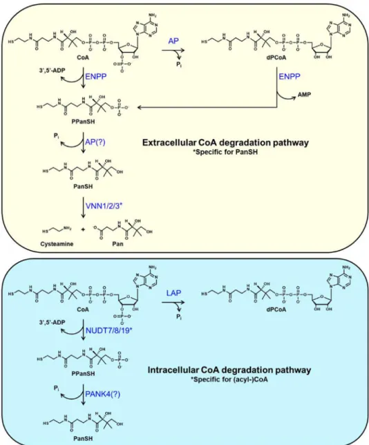
Complete extracellular breakdown of food- or tissue-derived CoA leads to the release of Pan in the bloodstream [23, 36, 37]. In the intestinal lumen, CoA hydrolysis is known to proceed with the production of the intermediates dephospho-CoA (dPCoA), phosphopantetheine (PPanSH) and pantetheine (PanSH) [23]. In particular, Shibata et al. showed that generation of dPCoA could be directly achieved by incubating CoA with partially purified preparations of intestinal alkaline phosphatase [23], an enzyme that, given its high abundance in the intestine, is a good candidate for this activity in vivo (Fig. 1). Intestinal alkaline phosphatase (AP) is one of four mammalian AP isoforms, which are homodimeric, metal-dependent proteins anchored to the plasma membrane via a glycosyl-phosphatidylinositol anchor (GPI) (reviewed in [38, 39]). Two other isoforms are expressed in placenta (placental AP) and testis (germ cell AP), respectively, while a fourth one, tissue nonspecific AP, is abundant in skeletal tissues, liver and kidneys. These enzymes, which require an alkaline pH for optimal activity, can hydrolyze a wide variety of substrates, including nucleotides tri-, di- and monophosphates, pyridoxal phosphate and sugar phosphates, and are thus not specific for CoA.
Formation of PPanSH requires the hydrolysis of the diphosphate bond in either CoA or dPCoA by a nucleotide pyrophosphatase (Fig. 1). This activity, originally detected in plasma membrane preparations from rat liver, was found to be highly promiscuous, being capable of hydrolyzing a variety of diphosphate-containing molecules [40–42]. The responsible enzyme exhibited a significantly lower KM and higher hydrolysis rate for dPCoA compared to CoA,
indicating that the enzyme prefers dPCoA as a substrate [40, 41]. More recently, Srinivasan et al. have shown that members of the ectonucleotide pyrophosphatase/phosphodiesterase (ENPP) family are likely responsible for the hydrolysis of CoA to PPanSH observed in serum obtained from multiple sources, including mice and humans [25]. In these in vitro experiments, formation of PPanSH in serum supplemented with CoA was inhibited by the addition of ENPP-specific inhibitors but not by the addition of the AP inhibitor levamisole [43, 44]. Interestingly, dPCoA was not detected as a degradation product, suggesting that the ENPP enzymes might directly hydrolyze CoA without prior dephosphorylation. Seven structurally related ENPP isoforms are currently known to exist (reviewed in [45–47]) and five of them, ENPP1–5, can hydrolyze the diphosphate bond in a variety of substrates including nucleotide tri- and di-phosphates and sugar-nucleotides. ENPP2 is a secreted enzyme that preferentially hydrolyzes the phosphodiester bond in phospholipids. All other ENPPs are transmembrane enzymes, and soluble forms have been identified for both ENPP1 and ENPP3, which exhibit strong nucleotide-hydrolyzing activity. Interestingly, both the CoA-degrading activity of the serum reported by Srinivasan et al. and that observed in rat liver membranes were refractory to inhibition by sodium fluoride, suggesting that the ENPPs could be involved in both processes [25, 41]. In the intestinal lumen, dephosphorylation of
A
uthor Man
uscr
ipt
A
uthor Man
uscr
ipt
A
uthor Man
uscr
ipt
A
uthor Man
uscr
ipt
PPanSH to PaSH could be catalyzed by a promiscuous phosphatase such as AP [23], which is also present at various levels on the apical cell surface in tissues (Fig. 1). Further
breakdown of PanSH to Pan is then known to be controlled by a class of enzymes called pantetheinases. Unlike the enzymes involved in the first three steps of extracellular CoA degradation, the pantetheinases are specific for PanSH and thus, in a position to regulate this process.
2.2. Pantetheinases specifically hydrolyze PanSH to cysteamine and Pan
Pantetheinases catalyze the last step of extracellular CoA degradation. Initially identified from pig kidneys, these ubiquitous ecto-enzymes hydrolyze PanSH into cysteamine and Pan, thereby providing a key precursor for the intracellular synthesis of CoA [35, 48]. This activity is present in most multicellular organisms tested (mammals, birds, fish, insects) but not in C. elegans [49]. The Vanin (Vnn) genes are clustered in the genome (bands 10A2B1 in mouse and 6q23–24 in human) and code for the VNN1 and VNN3 pantetheinase isoforms in mouse and for the VNN1, VNN2 and VNN3 isoforms in humans [49–51]. Comparative sequence analysis of all mouse and human VNN isoforms indicate the presence of two distinct functional domains: an N-terminal catalytic domain showing homology to biotinidase, another enzyme involved in the metabolism of vitamins, and a C terminal domain unrelated to any other known protein [49]. The catalytic domain of the VNN isoforms belongs to the subgroup 4 of the nitrilase superfamily, which are enzymes that perform the hydrolysis of a large variety of non-peptide carbon-nitrogen bonds in plants, animals, fungi, and some prokaryotes [52]. Nitrilases share a conserved catalytic triad made of a cysteine nucleophile, a glutamate base and an active site lysine in the catalytic domain. The hydrolytic activity of pantetheinases is highly specific for a single amide bond in the PanSH molecule and exclusively produces cysteamine and Pan (Fig. 1) [53]. The crystal structure of the human VNN1 has recently confirmed the predicted domain organization and identified the mechanism of catalysis. In the active site of VNN1, Glu79 and Lys178 orient and activate Cys211 for catalysis [54]. Mutation of any of these residues leads to enzyme inactivation [54, 55]. Additionally, two glutamate residues found at the inter-domain
interface may potentially participate in the allosteric regulation of the enzyme by altering the interaction between the two domains and, consequently, enzymatic activity.
The three pantetheinase isoforms - VNN1, VNN2/GP180, and VNN3 - have been studied in rodents and humans. The VNN1 protein is a broadly expressed GPI-anchored
ecto-pantetheinase. This enzyme is present on the apical side of epithelial cells, such as mature enterocytes in the ileum [56] and the epithelial cells of the proximal tubuli in the kidneys [57]. Vnn1-deficient mice, which lack the predominant pantetheinase activity in the intestine [56, 58], show reduced tissue cysteamine levels but no symptoms of Pan deficiency [57]. This is likely due to compensation by the VNN3 and/or a possible rescue by microbiota-derived Pan and PPanSH (Fig. 2) [59, 60]. During development, VNN1 expression is specifically induced in testis and cartilage [61–64]. In adults, tissues with a high metabolic activity, such as liver, ileum, kidneys and some endocrine glands, show high basal
expression levels of the Vnn1 gene [57]. The VNN1 pantetheinase also exists as a soluble form secreted by hepatocytes into the blood [65]. The Vnn3 transcript is expressed by many tissues, including the spleen and blood, but its expression pattern does not overlap with that
A
uthor Man
uscr
ipt
A
uthor Man
uscr
ipt
A
uthor Man
uscr
ipt
A
uthor Man
uscr
ipt
of Vnn1 [50, 66]. The human VNN3 gene contains a frame shift in the 3’-end region of the gene preventing the expression of a complete protein. The presence of splice variants in human neutrophils suggests that truncated forms of VNN3 might exist, but experimental evidence is currently lacking [67]. Agents provoking oxidative or inflammatory stress boost VNN3 expression, as observed by immunohistochemistry in inflamed psoriatic skin [68]. VNN3 does not have a membrane-anchoring domain and, together with VNN1, contributes to the pantetheinase activity detected in mouse serum [50, 65]. The human VNN2/GPI80, absent in mouse, is a GPI-anchored ecto-enzyme found at the plasma membrane of human neutrophils and monocyte subsets [69] where it regulates neutrophil adhesion [70, 71]. The contribution of pantetheinases to systemic CoA homeostasis will be discussed below.
2.3. Regulation of the systemic pool of Pan
Following intestinal breakdown of CoA, Pan is absorbed by enterocytes [23, 72] prior to entering the bloodstream. In these cells, Pan is taken up via the sodium-dependent
multivitamin transporter (SMVT, encoded by the Slc5a6 gene), which also transports biotin (vitamin B6) and lipoate [29, 73, 74]. Mice that lack SMVT in the intestine show various systemic phenotypes such as growth retardation, decreased bone density and length [75], which can be rescued by oral administration of biotin and Pan in the drinking water [76]. Serum and plasma levels of Pan are also regulated by urinary excretion [27] and normally maintained at a relatively constant concentration in the 0.2–2 μM range [77, 78]. Conditions such as diabetes and fasting, however, lead to a net increase in the levels on Pan in the blood [79–81]. In the liver, this increase is associated with an enhanced rate of Pan uptake and CoA synthesis and with higher total intracellular CoA levels [18, 82].
Although not formally demonstrated in vivo, one may anticipate that the recycling of cellular content would also be a source of Pan in the bloodstream. During tissue development, remodeling or stress, the maintenance of cell activity depends on the redistribution of numerous metabolites. Autophagy plays an important role in this process, beyond its conventional function as a cell-autonomous process in response to starvation and intracellular stress [83]. Autophagy delivers cytosolic metabolites or damaged organelles to the lysosomes, which degrade and recycle building blocks such as amino acids, nucleotides or fatty acids. Interestingly, a fraction of the cellular content will be secreted upon fusion of autophagolysosomes with the plasma membrane, thus contributing to a non-cell autonomous recycling process. This occurs for peptide hormones that regulate the energy balance such as apelin or FGF21, leaderless proteins like the IL-1 cytokine, or ATP. Following extracellular release, ATP may locally accumulate contributing to the triggering of inflammation [84]. In pancreatic tumors, cancer-associated fibroblasts show enhanced autophagy and contribute to the production of several nutrients including amino acids and nucleotides which, through crosstalk with neighboring cells, contribute to tumor growth [85, 86]. In addition to autophagy, it has been shown that numerous phagocytic immune cells release metabolites like nucleotides, which participate in inflammation and tumor development [87] (also reviewed in [88]). Therefore, metabolites generated by the intracellular degradation of CoA and acyl-CoAs may follow similar routes and be secreted in the milieu where the enzymatic activities of the extracellular CoA degradation pathway, including that of the VNNs, would result in Pan release.
A
uthor Man
uscr
ipt
A
uthor Man
uscr
ipt
A
uthor Man
uscr
ipt
A
uthor Man
uscr
ipt
3. CoA is an intracellular cofactor present in multiple subcellular
compartments
3.1. CoA synthesis
In mammals, CoA is synthesized in each cell from Pan, cysteine, and ATP. A variety of acyl-CoAs are then generated by the formation of a thioester bond between the carboxylic group of organic acids and the free thiol in CoA. This ATP-dependent reaction is catalyzed by a number of acyl-CoA synthetases, which are enzymes that reside in different subcellular compartments and exhibit defined specificities for short- (2–4 carbons in length), medium- (6–10 carbons in length), long- (12–20 carbons in length) and very long-chain (≥ 22 carbons in length) fatty acids [89]. Acyl-CoAs can also be formed as intermediates or final products of multiple metabolic pathways including fatty acid synthesis and oxidation, ketogenesis, cholesterol synthesis, and the TCA cycle.
The five universal steps of the CoA biosynthetic pathway and its regulation are extensively reviewed elsewhere [26, 90, 91]. Briefly, the pathway is initiated by the phosphorylation of intracellular Pan to 4’-phosphopantothenate. This reaction is catalyzed by pantothenate kinase (PANK), which often acts as the rate-limiting enzyme of the pathway. Four active PANK isoforms (PANK1α, PANK1β, PANK2 and PANK3) with distinct regulatory properties localize to different subcellular compartments, acting as metabolic sensors in the cytosol, mitochondria and nucleus, and adjusting the rate of CoA synthesis to the cellular metabolic state. 4’-Phosphopantothenate is then condensed with cysteine by
phosphopantothenoylcysteine synthetase (PPCS), followed by decarboxylation by phosphopantothenoylcysteine decarboxylase (PPCDC) to generate PPanSH. PPCS is predicted to localize to the cytosol (https://reactome.org/), which is also where the activity of PPCDC has been detected [92]. The last two steps in the pathway convert PPanSH to dPCoA by forming a phosphoanhydride bond between PPanSH and the AMP moiety of ATP, followed by phosphorylation of the 3’-OH of the ribose to form CoA (Fig. 2). In mammals, both the 4’-phosphopantetheine adenylyltransferase and dephospho-CoA kinase (DPCK) activities are found on the bifunctional enzyme coenzyme A synthase, COASY. The subcellular localization of COASY, which determines the cellular compartment from which CoA needs to be transported to different subcellular pools (see also below), remains debated to this date. COASY has been annotated to associate with the outer membrane of the mitochondria [93, 94], mitochondrial matrix [95–97] and even consists of a cytosolic pool [97, 98]. Additionally, mammals and other eukaryotes possess an unconfirmed
monofunctional DPCK. In humans, this enzyme is encoded by the DCAKD gene [99] and its subcellular localization remains unknown. Genetic mutations in the CoA biosynthetic pathway, specifically in the PANK2 or COASY genes, are responsible for rare
neurodegenerative disorders characterized by iron accumulation in the brain [95, 100]. In addition to Pan, extracellular PPanSH has recently been proposed to be a source of intracellular CoA [25] (Fig. 2). Under normal conditions, the concentration of PPanSH in the serum is undetectable, but the circulating levels of PPanSH can be increased by injecting large doses of CoA in mice. Oral administration of PPanSH to Pank2−/− mice has been shown to rescue the CoA-related abnormalities detected in the globus pallidus of these animals, although CoA levels were not reported in this paper [101]. While these data support
A
uthor Man
uscr
ipt
A
uthor Man
uscr
ipt
A
uthor Man
uscr
ipt
A
uthor Man
uscr
ipt
the conclusion that extracellular PPanSH could be a precursor of intracellular CoA, the ability of this phosphorylated molecule to diffuse across the plasma membrane and enter the CoA biosynthetic pathway as intact PPanSH is still debated [25, 59, 101, 102].
3.2. Subcellular (acyl-)CoA pools and transporters
Processes dependent on CoA and its thioesters, including metabolic reactions and the post-translational modification of proteins, occur in multiple subcellular compartments and pools of CoA and acyl-CoAs exist in the cytosol, mitochondria, peroxisomes, and ER [14, 16, 103–106]. Because of the presence of large nuclear pores, the cytosolic pool likely equilibrates with the nucleus. Nuclear acetyl-CoA can also be generated in situ by the activity of acetyl-CoA synthetase 2, which can translocate from the cytosol to the nucleus in a regulated process involving phosphorylation and binding to an importin [107]. Local acetate for the synthesis of nuclear acetyl-CoA seems to be mostly provided by histone deacetylases [108]. The pool of total CoA found in the mitochondria is considered to be the largest one. Indeed, liver and heart mitochondria are estimated to contain 80–95% of total cellular CoA [13, 14, 103, 109]. In rat liver, the cytosolic pool seems to comprise a little less than 20% under normal conditions, and the peroxisomal pool accounts for the remaining 2– 4% [13, 14]. The estimated concentrations of free CoA, specific acyl-CoAs and total CoA in liver mitochondria, cytosol and peroxisomes are reported in Table 1 [13, 14, 103, 106]. While the accuracy of these estimates is unavoidably limited by the purity and integrity of the individual subcellular fractions, these numbers still provide information about the relative size and concentration of each pool. The lumen of the ER has been recently reported to contain a pool of acetyl-CoA used to acetylate ER-transiting and ER–resident proteins [15, 104], but no quantitative information on the size of this pool is currently available. Fasting, diabetes, a high fat diet, and clofibrate treatment cause an increase in total subcellular CoA levels, with a more robust effect on the peroxisomal pool [13, 14, 82]. Diabetes also increases the concentration of total CoA in the cytosolic and mitochondrial fractions isolated from rat hearts [110]. The above (acyl-)CoA pools are generated and exchanged through the activity of the dedicated transporters discussed below.
3.2.1. Mitochondrial CoA transporters—While long-chain acyl-CoAs are not transported across the inner mitochondrial membrane, the carnitine shuttle allows the fatty acids to cross it as acylcarnitines and to be re-activated as acyl-CoAs in the matrix [111]. This process and several additional reactions, such as those catalyzed by thiolase, pyruvate dehydrogenase and α-ketoglutarate dehydrogenase, require the availability of free CoA in the matrix. Human mitochondria contain SLC25A42, a transporter that can exchange (deoxy)adenine nucleotide derivatives including CoA, dPCoA, 3’,5’-ADP, ATP, (d)ADP, AMP and FAD [112] (Fig. 2). Mutations in this protein are associated with muscle
weakness, lactic acidosis, altered mitochondrial morphology, and decreased levels of CoA in isolated fibroblasts [113–115]. However, beyond these clinical presentations, little is known regarding the effect of these mutations on mitochondrial metabolism. The kinetics
parameters of SLC25A42, determined using the purified protein reconstituted in liposomes, indicate that CoA, dPCoA and 3’,5’-ADP efficiently compete with [14C]ADP with Kis of
112, 108 and 27 μM, respectively. The Kis for AMP, ATP and dADP were around 400 μM,
indicating that, at least in vitro, this transporter has a higher affinity for CoA and dPCoA and
A
uthor Man
uscr
ipt
A
uthor Man
uscr
ipt
A
uthor Man
uscr
ipt
A
uthor Man
uscr
ipt
the highest affinity for 3’,5’-ADP [112]. Inside the mitochondria, 3’,5’-ADP could be produced by the (acyl-)CoA-degrading enzyme NUDT8 (see below). SLC25A42 can bi-directionally exchange CoA with [14C]ADP in vitro, but the direction of CoA transport and the substrate(s) that this transporter exchanges CoA with in vivo are currently not known. Answering this question has become particularly important in light of the conflicting results on the sub-mitochondrial localization of COASY. Indeed, if the conversion of PPanSH to CoA occurs in the cytosol or in a compartment that can rapidly exchange metabolites with the cytosol (e.g. the outer mitochondrial membrane or the mitochondrial intermembrane space), then SLC25A42 could work to import CoA into the mitochondrial matrix.
Conversely, if COASY resides in the mitochondrial matrix, PPanSH produced in the cytosol would have to cross the inner mitochondrial membrane to be converted to CoA, followed by export of the cofactor into the cytosol for distribution to peroxisomes, ER and nucleus. Normally, the intracellular concentration of PPanSH is only a small percentage (1–3%) of the total CoA amount and, notably, PPanSH does not accumulate under conditions that increase CoA synthesis up to 3-fold [82, 116]. This evidence indicates rapid and efficient conversion of PPanSH to CoA, which is currently difficult to reconcile with the low membrane permeability exhibited by PPanSH [25]. Indeed, consistent with its
phosphorylated nature, the ability of PPanSH to cross a lipid bilayer is at the threshold of a parallel artificial membrane permeability assay. Interestingly, SLC25A42 has a low but detectable activity with Pan [112] and it would be interesting to assay this transporter for its ability to exchange PPanSH.
Finally, human mitochondria contain another putative transporter, SLC25A16 (also known as Graves ‘ disease carrier protein), which, similar to SLC25A42, complements the deletion of yeast LEU5, a gene encoding for a protein that imports CoA or a CoA precursor into the mitochondrial matrix [117]. Based on this evidence, SLC25A16 may be another transporter for CoA/dPCoA, but its activity has not been successfully reconstituted in vitro [112, 118].
3.2.2. Peroxisomal transporters of CoA and acyl-CoAs—In mammalian peroxisomes, three members of the ATP-binding cassette subfamily D, ABCD1–3, mediate the import of a variety of acyl-CoAs from the cytosol (reviewed in [119, 120]) (Fig. 2). These transporters exhibit defined but partially overlapping substrate specificities. Human ABCD1 prefers saturated acyl-CoAs with a chain length of 24 and 26 carbons. Mutations in the ABCD1 gene lead to the accumulation of very long-chain fatty acids in plasma and tissues and lead to the development of the pathological features that characterize X-linked adrenoleukodystrophy. ABCD2 preferentially transports polyunsaturated long-chain acyl-CoAs, while ABCD3 prefers 2-methyl branched-chain acyl-CoAs and the CoA thioesters of long-chain dicarboxylic fatty acids and bile acid precursors. It is presently unclear whether these transporters move intact acyl-CoAs across the peroxisomal membrane or whether they possess intrinsic thioesterase activity, like the plant homolog COMATOSE, which
hydrolyzes the acyl-CoAs to non-esterified fatty acids and CoA during the transport [121]. In the latter case, the non-esterified fatty acids would have to be re-activated to acyl-CoAs inside the peroxisomal lumen. The acyl-CoA synthetases ACSL4, SLC27A2 and SLC27A4 have been reported to be peroxisomal and could catalyze this reaction [122].
A
uthor Man
uscr
ipt
A
uthor Man
uscr
ipt
A
uthor Man
uscr
ipt
A
uthor Man
uscr
ipt
Once inside the peroxisomal lumen, these acyl-CoA substrates undergo acyl-chain
shortening through α- and β-oxidation. These pathways require a supply of free CoA, which needs to be imported from the cytosol. In human peroxisomes, a member of the solute carrier 25 family (SLC25), SLC25A17, has recently been shown to catalyze the transport of CoA through a counter-exchange mechanism [123]. Purified recombinant SLC25A17 reconstituted into liposomes can transport CoA, but also FAD, FMN, AMP and, to a lesser extent, NAD+, ADP and 3’,5’-ADP. The latter metabolite can be produced by various enzymes, including sulfotransferases and (acyl-)CoA-degrading enzymes, both inside and outside the peroxisomes. Inside the peroxisomes, 3’,5’-ADP is the product of the hydrolysis of CoA and acyl-CoAs catalyzed by the Nudix hydrolases NUDT7 and NUDT19 (see below). Thus, the ability of the SLC25A17 to exchange 3’,5’-ADP for another substrate, including CoA, could prevent a buildup of this metabolite in the peroxisomes. Based on competition assays with [14C]AMP, the Ki of SLC25A17 for CoA is 20 μM, a value that is
compatible with the estimated cytosolic concentration of this cofactor (Table 1) [16, 106]. In vitro, the transporter can both import and export CoA, exchanging it with [14C]AMP or FAD, which are transported in the opposite direction. It will be important to determine whether this SLC25A17-mediated bi-directional movement of CoA across the peroxisomal membrane could also occur in vivo. Indeed, this could potentially be one of the mechanisms contributing to the regulation of the peroxisomal concentration of CoA. In vitro, SLC25A17 can also transport dPCoA, but shows little or no activity with acetyl-CoA and propionyl-CoA [123].
3.2.3. The ER transporter of acetyl-CoA—In the ER, a transporter originally named coenzyme A transporter 1 or AT-1 (encoded by the SLC33A1 gene), imports acetyl-CoA into the ER lumen exchanging it with acetyl-CoA [124, 125]. Within the ER, acetyl- acetyl-CoA-dependent protein acetylation regulates proteostasis and autophagy, and the essential role played by AT-1 in this processes is underscored by the fact that changes in its expression and activity are linked to human diseases (also reviewed in [126]). Indeed, chromosomal
duplications of the locus containing SLC33A1 are associated with intellectual disability and autism spectrum disorder; furthermore, AT-1 is upregulated in motor neurons of patients with sporadic amyotrophic lateral sclerosis and late-onset Alzheimer’s disease [125–127]. Conversely, the autosomal-dominant mutation, S113R, ablates AT-1 activity and causes autosomal dominant spastic paraplegia-42. The importance of tightly regulating the access of acetyl-CoA to the ER is also recapitulated by the severe phenotypes of mice with both increased and decreased activity of the transporter. Mice heterozygous for the S113R knock-in mutation exhibit neurodegeneration and an knock-increased occurrence of knock-infection, systemic inflammation and malignancies resulting from reduced efficiency of the secretory pathway and hyperactivation of ER autophagy. Conversely, increased efficiency of the secretory pathway and reduced activation of ER autophagy in mice over-expressing AT-1 systemically causes a progeria-like phenotype. The activity of purified AT-1 has been reconstituted in both liposomes and ER vesicles. This transporter has a KM of 14 μM for acetyl-CoA [104],
compatible with the cytosolic concentration of this acyl-CoA (Table 1) [16, 106].
A
uthor Man
uscr
ipt
A
uthor Man
uscr
ipt
A
uthor Man
uscr
ipt
A
uthor Man
uscr
ipt
4. Intracellular degradation of CoA and acyl-CoAs
Intracellular levels of total CoA increase in response to the metabolic changes elicited by fasting, high fat diets, or drugs such as clofibrate [13, 14, 18, 19, 128, 129]. Some of these changes occur rapidly, in just a few hours. For example, when rats are allowed to re-feed after a fast, total hepatic CoA levels drop within 2 hours [18] and we have observed the same rapid decrease in the concentration of total CoA in mouse livers as well (unpublished observations). This rapid decline in total CoA levels requires activation of the degradation pathway, as even inhibiting CoA biosynthesis completely would not achieve a net decrease. The dramatic 80% decrease in the concentration of liver and kidney free CoA in mice treated with HoPan, a Pan analog that inhibits CoA synthesis, provides additional and strong evidence for the existence of CoA turnover in vivo [130]. Interestingly, the rate of CoA turnover differs between organs, with the heart and the brain exhibiting lower rates than the liver and the kidneys [130]. Similar to the extracellular degradation of CoA, intracellular CoA breakdown occurs through a combination of specific and non-specific enzymes. However, intracellularly, degradation likely stops at the PPanSH or PanSH level, as there is no known intracellular pantetheinase (Fig. 1).
4.1. Dephosphorylation of CoA
CoA can be dephosphorylated to dPCoA inside the lysosomes. This activity is sensitive to tartrate inhibition and higher at acidic pH [131]. Although not formally demonstrated, the lysosomal acid phosphatase 2 (also known as LAP) is a likely candidate for this activity, based on its ability to dephosphorylate a variety of metabolites, its pH optimum, and its inhibition by tartrate [132]. Lysosomes are key players in the degradation and recycling of both intracellular and extracellular material and contain over 60 hydrolases and accessory proteins [133]. Among the lysosomal proteins is palmitoyl-protein thioesterase, PPT, a protein that can hydrolyze long-chain acyl-CoAs in vitro [134–136]. Therefore, the combination of LAP and PPT could convert long-chain acyl-CoAs to dPCoA (Fig. 2). Delivery of intracellular (acyl-)CoA to these organelles could occur through autophagy; however, once formed, dPCoA would likely require a transporter to return to the cytosol [137]. No lysosomal dPCoA transporter is currently known, but several of the transporters identified on the lysosome membrane remain with an un-assigned function [138]. Multiple organs also contain detectable levels of select short-chain acyl-dPCoAs and octanoyl-dPCoA [139]. PPT has minimal to no activity against short- to medium-acyl-CoAs, and it is
currently unknown whether these acyl-dPCoAs are dephosphorylation products of LAP in the lysosomes or whether they may be generated by short-chain acyl-CoA synthetases accepting dPCoA as a substrate. Indeed, CoA synthetase can synthesize acetyl-dPCoA and butyryl-acetyl-dPCoA from acetyl-dPCoA in vitro [139].
Following incubation with CoA under acidic conditions, the production of Pi was also reported to occur along the Golgi apparatus of fixed ameoblasts of rat incisor [140]. This activity was significantly lower with dPCoA compared to CoA, suggesting that there was a CoA phosphatase along the Golgi apparatus. The more recent discovery of a Golgi-resident 3’-nucleotidase (gPAPP, encoded by the Impad1 gene) that hydrolyzes 3’,5’-ADP and could also recognize CoA, may explain the production of Pi in the Golgi [141]. The observation
A
uthor Man
uscr
ipt
A
uthor Man
uscr
ipt
A
uthor Man
uscr
ipt
A
uthor Man
uscr
ipt
that (acyl-)CoA can be degraded to dPCoA has not been further investigated, and the physiological relevance of this reaction for the regulation of total intracellular CoA levels is currently unclear. Indeed, one would predict that de-phosphorylation of CoA in liver lysosomes would be activated during a fast, following stimulation of the autophagy pathway [142, 143]. However, this process would occur at the same time as increased CoA synthesis [17], which currently seems counterintuitive.
4.2. Specific Nudix hydrolases split CoA and acyl-CoAs into 3’,5’-ADP and (acyl)phosphopantetheine
Intracellular CoA and select CoA thioesters can be hydrolyzed without prior
dephosphorylation to dPCoA by three Nudix hydrolases, NUDT7, NUDT8, and NUDT19. Similarly to the ENPPs and the membrane activity described by Skrede [41], these enzymes split the CoA molecule into 3’,5’-ADP and phosphopantetheine (Fig. 1). However, unlike the ENPPs, NUDT7, NUDT8 and NUDT19 are highly specific for CoA species and, thus, in a position to regulate intracellular (acyl-)CoA levels [144–147].
Nudix hydrolases are a diverse, widespread family of intracellular enzymes that most commonly hydrolyze the phosphoanhydride bond present in a broad range of substrates, including nucleoside di- and triphosphates, dinucleoside polyphosphates, and nucleotide cofactors such as NADH and CoA [148–150]. As a class, these enzymes are characterized by the presence of a conserved sequence, G[5X]E[7X]REXXEEXGU (where X can be any amino acid and U is a hydrophobic amino acid), which forms a loop-helix-loop structural motif named the Nudix box [148, 150]. This motif is involved in the coordination of a divalent cation, usually Mg2+ or Mn2+, which is essential for activity. Regions outside the Nudix box can also participate in catalysis, confer substrate specificity, and allow for the definition of multiple classes of Nudix hydrolases based on substrate preferences and structural motifs. In the case of NUDT7, NUDT8, and NUDT19, the specificity for CoA is thought to be conferred by another conserved sequence (L/M)(L/F)TXR(S/A)[3X](R/K) [3X]G[3X]FPGG, termed the CoA box [151], which is common to all other validated CoA diphosphohydrolases [152–154]. In support of this notion, mutagenesis studies in NUDT7 and NUDT19 have identified key residues within, but also outside, the CoA box, which are involved in CoA binding and catalysis [146]. Interestingly, although NUDT7, NUDT8 and NUDT19 catalyze the same reaction, these enzymes exhibit strikingly different features including size, regulation, tissue distribution, and subcellular localization.
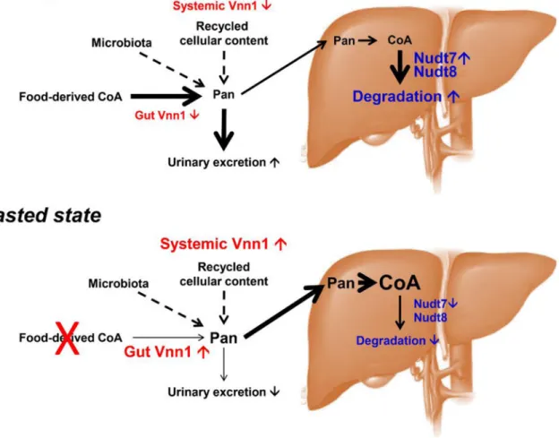
4.2.1. Hydrolysis of CoA and CoA thioesters in the peroxisomes—NUDT7 was the first CoA-specific Nudix hydrolase identified in mammals [145]. This enzyme has a molecular mass of 27 kDa and localizes to the peroxisomes via a tripeptide PTS1 signal at the C -terminus [145, 155]. Mouse NUDT7 exhibits the highest expression in the liver, with significantly lower expression levels in brown and white adipose tissue, and minimally detectable levels in lungs and kidneys [144–146]. Recombinant NUDT7 exhibits the highest activity at pH 8.0, in the presence Mg2+ or Mn2+ and, like all other Nudix hydrolases, is strongly inhibited by fluoride ions [145, 147, 156]. This enzyme readily hydrolyzes a broad range of CoA substrates, including free CoA, bile acid-CoAs, and acyl-CoAs with variable chain lengths from 2–14 carbons. The activity against acyl-CoAs longer than myristoyl-CoA
A
uthor Man
uscr
ipt
A
uthor Man
uscr
ipt
A
uthor Man
uscr
ipt
A
uthor Man
uscr
ipt
declines sharply and becomes almost undetectable against stearoyl-CoA. The specific activity of NUDT7 against butyryl-, hexanoyl-, octanoyl-, choloyl- and
trihydroxycoprostanoyl-CoA is 1.5 to 2 times higher compared to free CoA [144–146]. The fairly modest preference of the enzyme for these substrates in vitro may not be sufficient to alter the ratio between free CoA and these acyl-CoA in liver peroxisomes. Conversely, the poor activity of NUDT7 against stearoyl-CoA and longer acyl-CoAs may lead to a decrease in the ratio between free CoA and select long-chain acyl-CoAs. Any change in this ratio, however, can be buffered by the activity of acyl-CoA thioesterases (ACOTs) and carnitine acyltransferases, which can offload the acyl groups from CoA and release them as non-esterified fatty acids or acylcarnitines, respectively [157]. Indeed, the decrease in
peroxisomal fatty acid oxidation caused by the over-expression of NUDT7 in mouse liver does not cause an accumulation of intracellular long-chain acyl-CoAs but an increase in the concentration of the correspondent acylcarnitines [158]. In mouse liver, NUDT7 expression and activity are regulated by the nutritional state and decrease in the transition from the fed to the fasted state [17, 20, 144], a process thought to be controlled by PPARα [144] (Fig. 3). NUDT19 was identified by proteomic analysis of mouse kidney peroxisomes [159]. Formerly known as RP2p, this enzyme is expressed primarily in the kidneys, with significantly lower but detectable levels in skeletal muscle and brain [146]. Similar to NUDT7, a C-terminal PTS1 signal directs NUDT19 to the peroxisomes. NUDT19 isoforms have a molecular mass of 40–42 kDa in mice and humans, and the larger size compared to NUDT7 and NUDT8 is in part due to the presence of a 45–49 amino acid sequence inserted within the Nudix box, which may play a role in the folding or stability of the protein [146, 159]. When assayed using CoA as a substrate, recombinant mouse NUDT19 displays optimal enzymatic activity at pH 8–9.5 and in the presence of 10 mM Mg2+ or Mn2+. The recombinant enzyme displays the highest activity against free CoA, octanoyl-, lauroyl- and myristoleoyl-CoA and a 2–3 times lower activity against other short- and medium-chain acyl-CoAs. Similar to NUDT7, the activity of NUDT19 against stearoyl-CoA is low to undetectable; however, in striking contrast to the liver isoform, NUDT19 is almost completely inactive against acetyl-CoA [146, 159]. As discussed above, while conditions that increase or decrease the activity of NUDT19 may affect the ratio between free CoA and some of the acyl-CoAs, the effects on the metabolism of kidney peroxisomes are not easy to predict because of the buffering system offered by the ACOTs and the carnitine
acyltransferase in vivo. Mutagenesis studies have revealed that the CoA boxes of NUDT7 and NUDT19 are not interchangeable, as doing so significantly reduced enzymatic activity and interfered with substrate binding [146]. Additionally, NUDT19, but not NUDT7, is competitively inhibited by specific bile acids. This fact is likely not physiologically relevant due to the localization of NUDT19 in kidney peroxisomes, but together with the
mutagenesis studies, these data suggest that the CoA binding sites of NUDT7 and NUDT19 are different. Several crystal structures of human NUDT7 are now available, but they do not have substrates or products bound [160]. Thus, structural data for both NUDT7 and NUDT19 in complex with CoA will be required to determine whether the mode of CoA binding is indeed different between these two enzymes.
A
uthor Man
uscr
ipt
A
uthor Man
uscr
ipt
A
uthor Man
uscr
ipt
A
uthor Man
uscr
ipt
4.2.2. Hydrolysis of CoA and CoA thioesters in the mitochondria—NUDT8 is the most recently discovered (acyl-)CoA diphosphohydrolase and the first found to reside outside the peroxisomes (Fig. 2). NUDT8 localizes to the mitochondria and exhibits a broad tissue distribution, being found in highest amounts in mouse kidneys, heart, brown adipose tissue, and liver [147]. This enzyme also appears to be the major CoA-degrading enzyme in the heart, brain, and skeletal muscle, owing to the lack of expression of the other two isoforms in these tissues. In solution, recombinant mouse NUDT8 is mostly found as a monomer, similar to NUDT7 and NUDT19. In mammals, NUDT8 has a molecular weight of 23–25 kDa, depending on the species, and possesses a predicted N-terminal mitochondrial targeting sequence. In vitro, mouse NUDT8 displays optimal enzymatic activity at pH 8.5 and in the presence of 2 mM Mn2+. In contrast to NUDT7 and NUDT19, whose activity is supported to similar levels by either Mg2+ or Mn2+, NUDT8 is only appreciably active in the presence of Mn2+. This enzyme hydrolyzes free CoA and a broad range of short-and
medium-chain acyl-CoAs substrates, showing a 10-fold difference in specificity constant between free CoA and acetyl-CoA, which are the best and worst substrates for NUDT8, respectively, among those tested. Based on this substrate specificity, Nudt8 may contribute to the regulation of the mitochondrial free CoA/acetyl-CoA ratio in vivo, but experimental evidence is still lacking. Furthermore, mitochondria contain carnitine acetyltransferase, an enzyme that prevents the accumulation of acetyl-CoA by transferring the acetyl group from acetyl-CoA to carnitine [161]. The activity of NUDT8 against long-chain acyl-CoAs remains to be determined, as these acyl-CoAs precipitated in reaction mixtures containing Mn2+. The mitochondrial localization of mouse NUDT8 has been clearly demonstrated via microscopy- and subcellular fractionation-based approaches. Furthermore, evidence from spatially restricted enzyme tagging-based proteomics supports the localization of human NUDT8 to the matrix, although association with the matrix side of the inner mitochondrial membrane cannot be excluded [96, 162]. The presence of NUDT8 in the mitochondrial matrix potentially places the enzyme in the same submitochondrial compartment as COASY, which could recycle the PPanSH generated by NUDT8 back to CoA (Fig. 2). Prevention of such a wasteful cycle of CoA synthesis and degradation would require regulation of NUDT8, COASY or both. COASY has recently been reported to be phosphorylated in an insulin-dependent manner by ribosomal protein S6 kinase, polypeptide 1 (S6K1) [163]. While the exact site of phosphorylation and the effect of this modification on COASY activity have not been directly identified, recombinant mouse COASY expressed in baculovirus exhibits the highest activity when incubated with protein tyrosine phosphatase, non-receptor type 11 (also known as Shp2), suggesting that phosphorylation could be inhibitory [164]. S6K1 is known to associate with the outer mitochondrial membrane [165] and, while S6K1 could probably interact with COASY if this enzyme was localized in the cytosol or outer mitochondrial membrane, it is currently unclear whether the localization of S6K1 would be compatible with the regulation of COASY in the matrix. Similarly, mouse COASY has also been shown to be inhibited by mRNA-decapping protein 4, but this enzyme, which is involved in the DNA damage response, resides in the nucleus and cytosol [166]. With respect to NUDT8, one would expect its activity to decrease in the fasted state to allow for the accumulation of total CoA and the switch to mitochondrial fatty acid oxidation that occurs during a fast. Conversely, NUDT8-mediated (acyl-)CoA degradation would likely be stimulated in the fed state, decreasing the size of the total CoA pool (Fig. 3).
A
uthor Man
uscr
ipt
A
uthor Man
uscr
ipt
A
uthor Man
uscr
ipt
A
uthor Man
uscr
ipt
However, In mouse liver, kidneys and heart, the expression of NUDT8 does not change between the fed and fasted states [147], suggesting that alternative mechanisms such as post-translational modification or regulation by small metabolites may be involved in the regulation of the enzyme.
4.3. PPanSH recycling to CoA and the fate of 3’,5’-ADP
NUDT-mediated hydrolysis of CoA and CoAs produces PPanSH and acyl-phosphopantetheines, respectively. Due to its relatively small size (<400 Da), PPanSH generated inside the peroxisomes is likely able to freely move across the channels of the peroxisomal membrane and reach the cytosol [167]. However, any phosphopantetheine attached to a carboxylic acid longer than acetate would be too large to exit through the same route. These metabolites are likely further hydrolyzed to non-esterified fatty acids and PPanSH by peroxisomal ACOTs. Indeed, ACOT3 and ACOT8 have been reported to exhibit robust activity against lauroyl-phosphopantetheine [157]. The ability to hydrolyze acyl-phosphopantetheines could also be a feature of mitochondrial ACOTs, such as ACOT2, ACOT9 and ACOT10 [168], but evidence is currently lacking. Furthermore, it is still unclear whether any PPanSH potentially produced in the mitochondrial matrix by NUDT8 would be able to diffuse into the cytosol (Fig. 2).
Cytosolic PPanSH could re-enter the biosynthetic pathway downstream of the regulated PANK step. Alternatively, this metabolite could be dephosphorylated to PanSH, and the C-terminal domain of PANK4, an inactive cytosolic PANK [169, 170], has been shown to possess this activity in vitro [171]. PanSH is an excellent PANK substrate [172] and, while de-phosphorylation followed by re-phosphorylation would consume an additional ATP molecule, this process would leave the recycling of PanSH to CoA under the control of PANK, which often is the rate-limiting enzyme in the pathway. Intracellular PanSH is not likely to be an in situ source of pantothenate and cysteamine, as all currently known VNN enzymes are ecto-enzymes.
Given the strong preference of NUDT7, NUDT8 and NUDT19 for CoA over dPCoA, 3’,5’-ADP (also known as 3’-phosphoadenosine 5’-phosphate or PAP) is likely the only other product generated by these enzymes, in addition to PPanSH [145, 147, 159]. 3’,5’-ADP is also produced by sulfotransferases, enzymes that transfer a sulfonic group from 3’-phosphoadenosyl-5’-phosphosulfate to an acceptor molecule [173, 174], and during the transfer of the PPanSH prosthetic group from CoA to proteins such as the mitochondrial acyl carrier protein and the eukaryotic fatty acid synthase [175, 176]. Quantitatively, the
contribution of NUDT7, NUDT8 and NUDT19 to the 3’,5’-ADP pool is currently not known. 3’,5’-ADP is converted to 5’-AMP by the action of two 3’-nucleotidases, the Golgi-resident gPAPP and bisphosphate 3’-nucleotidase 1 (encoded by Bpnt1), which localize to the Golgi lumen and the cytosol, respectively [141, 177, 178]. These enzymes belong to the metal-dependent lithium-sensitive phosphomonoesterases and produce 5’-AMP, which can then re-enter the nucleotide pool. 3’,5-ADP produced in the mitochondrial matrix by NUDT8 or the phosphopantetheinyl transferase reaction [147, 176] could be exchanged with CoA or another substrate by the SLC25A42 transporter (Section 3.2.1). In the extracellular space, 5’-AMP and 3’,5’-ADP formed from the hydrolysis of the diphosphate bond in
A
uthor Man
uscr
ipt
A
uthor Man
uscr
ipt
A
uthor Man
uscr
ipt
A
uthor Man
uscr
ipt
dPCoA and CoA, respectively, can be de-phosphorylated to adenosine by non-specific phosphomonoesterases such as alkaline phosphatase [38, 40, 179, 180]. 5’-AMP can also be dephosphorylated, specifically, by the ecto-5’-nucleotidase (CD73, encoded by the Nt56 gene) [181, 182]. These metabolites modulate purinergic signaling and are involved in multiple processes such as neurotransmission, bone mineralization, inflammation and platelet aggregation [183].
5. Contribution of degradation pathways to (acyl-)CoA regulation and
pathologies
5.1. Contribution of the VNN1 pantetheinase to CoA-dependent metabolism in tissues
Most studies linking pantetheinase activity to CoA-dependent metabolism were performed with the VNN1 isoform. VNN1 expression increases under stress conditions including metabolic, oxidative, cytosolic, mitochondrial and ER stresses [55, 184, 185]. This regulation depends on the presence of several antioxidant response element (ARE) sequences and PPAR-response elements (PPRE) in the proximal promoter of the gene, which recruit transcription factors such as specificity protein 1 SP1, activator protein 1 AP1 [184], PPARα [186, 187], and PPARγ [58]. In the liver, VNN1 is a prototypical PPARα target gene and a key mediator of the adaptation to fasting in centrolobular hepatocytes [55]. Indeed, despite a partial compensation by VNN3, Vnn1-deficient mice tend to develop liver steatosis following a fast, arguing in favor of a decrease in fatty acid oxidation [188]. Furthermore, pantethine, a compound that is rapidly converted to PanSH [189, 190], is a drug approved for the treatment of hyperlipidemia due to its boosting effect on fatty acid oxidation [191, 192]. Analysis of the promoter of the Vnn1 gene also identified a synergistic interaction between PGC1α and HNF4α on Vnn1 gene expression [193]. Analysis of db/db mice or mice fed a high fat diet confirmed that high VNN1 levels correlate with the development of insulin resistance. Furthermore, increased VNN1 expression contributes to the hepatic glucose output, leading to hyperglycemia. Interestingly, many enzymes of the gluconeogenic and glycolytic pathways are sensitive to variations in the redox status and can undergo thiolation by formation of mixed disulfides. More specifically, cyst(e)amine was found to reversibly inhibit hexokinase, phosphofructokinase, glucose-3-phosphate
dehydrogenase, and pyruvate kinase, while activating fructose-1,6-bisphosphatase activity in vitro [194, 195]. Overall, the effect of cyst(e)amine would favor the gluconeogenic flux [193], suggesting a possible mechanism through which VNN1 could contribute to the metabolic adaptation that occurs in the liver, following changes in the nutritional state. A recent study using a VNN1 inhibitor in rat showed that, while inhibition of VNN1 improves insulin sensitivity in mice fed a high fat diet, it has a limited value as an anti-diabetic strategy [196].
A more direct contribution of VNN1 to CoA homeostasis and mitochondrial activity has been documented in a sarcoma model [185]. In this system, VNN1 behaved as a tumor suppressor by reducing cell growth while maintaining tumor cell differentiation and collagen production. In this sarcoma-derived cell line transfected with a catalytically proficient or deficient VNN1, the level of production of Pan and CoA was proportional to the activity of the enzyme. VNN1-expressing tumor cells showed enhanced mitochondrial organization,
A
uthor Man
uscr
ipt
A
uthor Man
uscr
ipt
A
uthor Man
uscr
ipt
A
uthor Man
uscr
ipt
activity and connection to a functional ER. In contrast, VNN1-deficient tumor cells displayed a dilated ER, poorly connected to mitochondria, which could in part explain the reduction in extracellular matrix protein production associated with myofibroblast maturation. Furthermore, the anti-glycolytic effect mediated by cysteamine contributed to the anti-Warburg effect. This study highlights the contribution of pantetheinase to CoA-dependent energy metabolism. It also suggests that the simultaneous production of Pan and cysteamine could synergize to enhance the biological effect of Pan via a cysteamine-mediated redox regulation and inhibition of glycolytic enzymes (see also 5.2). Accordingly, the enhanced expression of VNN1 in myofibroblasts is associated with elevated Pan levels in serum, increased collagen deposition leading to tissue fibrosis, and aggravation of
scleroderma [77].
These observations linking Pan regeneration to increased CoA-dependent cell function might also explain the importance of a local Pan supply for colonic homeostasis. Indeed, dietary Pan deficiency alters the intestinal barrier function in the carp [197]. Mice lacking the SMVT transporter in the intestine die of peritonitis due to the development of a chronic inflammation of the caecum and colon [76]. Alterations in the expression of VNN1 in the intestine impact susceptibility to colitis [56, 58]. Furthermore, combined supplementation of vitamins B and D in humans mitigates the symptoms associated with the irritable bowel syndrome [30]. These results suggest that colonocytes might be particularly sensitive to CoA depletion. Indeed, these cells use the butyrate produced by microbiota for energy production through fatty acid β-oxidation and regulation of stem cell renewal [198, 199].
5.2. Contribution of pantetheinases and their products to pathologies
VNN1 has become a prognostic marker in several human diseases [200]. In many chronic inflammatory diseases, tissues are exposed to persistent metabolic, oxidative, or hypoxic stresses, which limit normal cell activity while re-directing energetic resources towards repair mechanisms, both requiring high levels of protein synthesis and control of redox power. This dual requirement may explain how the simultaneous release of Pan and cysteamine may synergize for an optimal effect in vivo, although their respective contribution is difficult to disentangle. Cysteamine, like glutathione and CoA, has a free thiol group that is sensitive to the redox environment. Under conditions of oxidative stress, the free thiols of cysteamine, glutathione, and CoA can form mixed disulfides with cysteine residues, resulting in protein cysteaminylation, glutathionylation, and CoAlation,
respectively [7, 8, 201]. The degree of protein modification with cysteamine, glutathione, and CoA depends on the local concentration of these molecules and on the pKa values of their thiol groups, which can be influenced by the specific protein environment [202]. These post-translational modifications are thought to protect cysteine residues from irreversible oxidation and have been shown to regulate the activity of key intracellular enzymes [201, 203]. Under metabolic stress, CoAlation in the mitochondria preferentially affected metabolic enzymes, suggesting that there might be a certain degree of specificity [8]. Cysteamine contributes to the mobilization of cysteine pools, a property used to treat the intracellular accumulation of the oxidized form cystine in cystinosis [204]. Under oxidative stress, cystamine, the oxidized form of cysteamine, inhibits gamma-glutamylcysteine synthetase required for glutathione synthesis [205]. Accordingly, the lack of VNN1 causes a
A
uthor Man
uscr
ipt
A
uthor Man
uscr
ipt
A
uthor Man
uscr
ipt
A
uthor Man
uscr
ipt
reduction in tissue cyst(e)amine levels [57] and can lead to a paradoxical increase in the concentration of reduced glutathione [184, 206]. Cysteaminylation has been shown to modulate the activity of several stress-related enzymes such as caspase 3 [207],
transglutaminase [208, 209], glutathione-S transferase A3 [210], or protein kinase Cε [203]. These combined effects might in part explain the protective effect of cysteamine in certain pathological contexts such as neurodegenerative diseases, type 1 diabetes, and
acetaminophen-induced liver damage [211–213]. More generally, increased expression of VNN1 in diseases would sustain CoA-dependent metabolic processes and cysteamine-mediated redox homeostasis, explaining its ability to enhance tissue tolerance to stress [214]. Indeed, the CoA-dependent enhancement of mitochondrial activity would generate more reactive oxygen species, which could be in part buffered by an increase in redox power mediated through cysteamine production, altogether improving energy production while limiting ROS-induced cell damage.
Few studies have identified allelic variants of VNN1 or VNN3 as contributing to quantitative traits in complex diseases [215–217]. In mouse, polymorphisms in the Vnn locus are associated with reduced pantetheinase activity in tissues [66]. Furthermore, a unique nonsense mutation in the A/J strain leads to undetectable Vnn3 transcripts and pantetheinase activity, which enhances the susceptibility to blood-stage or cerebral malaria in various plasmodium infection models. Cysteamine exerts an autonomous anti-plasmodial activity on infected red blood cells and optimizes the efficacy of artemisinin-based anti-malarial therapy [218]. Furthermore, in a mouse model of serum, but not tissue, VNN1 pantetheinase
deficiency, the half-life of erythrocytes was severely diminished due to enhanced oxidative stress, a phenotype that drastically aggravated the severity of malaria [65]. In human malaria, low levels of serum pantetheinase are associated with severe and complicated forms of the disease. While these results argue in favor of a beneficial role of VNN1/3
pantetheinase activity in malaria, it is also known that virulent strains of P. falciparum depend on an extracellular supply of Pan, provided by the host pantetheinase, to make their own CoA [219]. Therefore, the protective versus sensitizing effects of pantetheinase activity on the growth of the malaria parasite might vary at different stages of disease progression. Indeed, pantethine, a pharmacological source of Pan and cysteamine, has a protective effect in cerebral malaria, which is characterized by high levels of inflammation and intravascular microvesiculation [220]. The mechanisms of microvesiculation involve oxidative stress and perturbations in both the lipid composition of the plasma membrane and cytoskeleton integrity. One might speculate that the combined antioxidant effect of cysteamine and the effect of Pan and CoA on lipid catabolism might reduce this process. Another study showed that in mice with diet-induced steatohepatitis, VNN1 could be incorporated in hepatocyte-derived microvesicles and contributed to their pro-angiogenic effect [221]. Further studies are required to understand how VNN1 can regulate the process of microvesiculation and the impact of microvesicles on target cells.
The contribution of VNN2 to CoA homeostasis has not yet been explored. VNN2/GPI80 regulates neutrophil adhesion [70, 71], colocalizes with CD18, a β2 integrin and urokinase-type plasminogen activator receptor, and might undergo conformational/structural changes during neutrophil migration [222, 223]. Mice lack a Vnn2 gene and rather express VNN3, which likely plays a VNN2-like role in hematopoietic cells [49, 50, 224]. Interestingly,
A
uthor Man
uscr
ipt
A
uthor Man
uscr
ipt
A
uthor Man
uscr
ipt
A
uthor Man
uscr
ipt
VNN2 role in cell migration might also be important during hematopoietic stem cell engraftment through cooperation with another integrin, ITGAM [225]. Similarly, VNN2 gene expression is regulated by miRNA-106a, which affects osteosarcoma cell migration [226]. Furthermore, mouse VNN1 was initially discovered as a regulator of cell homing to the thymus [227]. Altogether, these data suggest a more specific regulation of the
engagement of adhesion receptors by VNNs during migration [228]. Migration requires a metabolic adaptation to maintain ATP levels and mitigate the risk of ROS-dependent cell death, a program triggered following detachment from the extracellular matrix [229]. Indeed, cell death caused by detachment from the extracellular matrix, or anoikis, can be rescued by anti-oxidants. Interestingly, survivin, an inhibitor of apoptosis family member, promotes the relocalization of mitochondria to membrane ruffles and, through cooperation with Hsp90 chaperones, regulates the folding of complexes I and II of the electron transport chain, therefore boosting mitochondrial bioenergetics at the migration pole [230]. Thus, one may speculate that the colocalization of VNNs with adhesion foci might be a prerequisite to provide a local source of Pan/CoA to generate ATP that, together with the antioxidant cysteamine, could limit the risk of anoikis.
5.3. Regulatory role of NUDT7, NUDT8 and NUDT19 in CoA-dependent metabolism
Peroxisomal CoA diphosphohydrolases are found in a variety of organisms including yeast, worms, mice and humans [145, 151, 152, 159]. Mitochondrial isoforms have been identified in plants and now humans and mice [147, 231]. A. thaliana also contains a cytosolic isoform [231]. Overall, the presence of CoA-degrading enzymes in all three subcellular
compartments, strongly suggests that these enzymes may play a key role in the local regulation of total CoA levels and CoA-dependent metabolic pathways. Furthermore, it opens the possibility that a yet undiscovered isoform might exist to regulate the cytosolic (acyl-)CoA pool of in mammals.
While measuring CoA and acyl-CoAs in mitochondria-free peroxisomal fractions remains a technical challenge, evidence that NUDT7 participates in the regulation of the peroxisomal (acyl-)CoA pool has recently been provided by the effect that the over-expression of this enzyme has on peroxisomal lipid metabolism in the liver. Indeed, mice injected with an adeno-associated virus carrying the Nudt7 gene under a liver-specific promoter, exhibited a significant decrease in peroxisomal fatty acid oxidation and in the content of bile acids, whose synthesis occurs in the liver and relies on peroxisomes [158]. This phenotype was observed only in the fasted state, when endogenous NUDT7 expression is at its lowest. Furthermore, at the whole liver level, over-expression of mouse NUDT7 was associated with a significant decrease in the concentration of several short-chain acyl-CoAs and, notably, choloyl-CoA, a precursor of bile acids, which is only formed in the peroxisomes and is a good substrate for NUDT7 [144]. Elevated NUDT7 levels caused a slight, but not
statistically significant increase in the hepatic levels of PPanSH, which may be an indication of rapid recycling of its degradation products to CoA (see also section 4.3). Furthermore, NUDT7 over-expression did not prevent the increase in total hepatic CoA levels observed in the fasted state, indicating that activation of the CoA biosynthetic pathway, and not a decrease in NUDT7 expression, is the driving force behind the accumulation of free CoA with fasting [17]. Conversely, the transition from the fasted to the fed state is characterized