CD14 Works with Toll-Like Receptor 2
to Contribute to Recognition and Control
of Listeria monocytogenes Infection
Laure Janot,1,aThomas Secher,1,aDavid Torres,1,aIsabelle Maillet,1Josef Pfeilschifter,2Valerie F. J. Quesniaux,1 Regine Landmann,3Bernhard Ryffel,1and François Erard1
1University of Orléans and Centre National de la Recherche Scientifique Unité Mixte de Recherche (UMR 6218), Orléans, France;2Pharmacologie, University of Frankfurt, Germany;3Zentrum Lehre und Forschung, University Hospitals Basel, Switzerland
Toll-like receptor 2 (TLR2) signaling has been shown to contribute to resistance to Listeria monocytogenes infec-tion, as TLR2-deficient mice have a heightened susceptibility to infection with this organism. Because CD14 may associate with TLR2, we investigated the role of CD14 in Listeria responses. In both CD14-deficient and TLR2-deficient macrophages, nuclear factorB translocation; CD40 and CD86; and the production of interleukin (IL)– 12, IL-6, tumor necrosis factor, and nitric oxide are reduced. The absence of CD14 augmented susceptibility to
Listeria infection, reduced survival, and diminished bacterial clearance, as observed in TLR2-deficient mice.
Com-pared with C57BL/6 control mice, CD14-deficient mice were observed to have a greater number of hepatic micro-abscesses containing abundant neutrophils, these micro-abscesses were larger in size, and there was reduced inducible nitric oxide synthase expression. Further, mice that are both CD14 deficient and TLR2 deficient display suscepti-bility to infection that is comparable to that of mice deficient in either CD14 or TLR2 alone. Therefore, the present data demonstrate the role of CD14 and TLR2 in the recognition and control of Listeria infection and host resistance.
Toll-like receptors (TLRs) recognize a wide range of mi-crobial pathogens, and their products modulate the in-nate immune response that may lead to inflammation and altered host defense [1–3]. We previously reported that TLR2 signaling can be shown to contribute to resis-tance to Listeria monocytogenes infection because TLR2-deficient mice have a higher susceptibility to infection with this organism [4]. The involvement of the TLR sig-naling pathway was further confirmed by the heightened susceptibility of myeloid differentiation primary re-sponse gene (88) (MyD88)– deficient mice [5, 6]. Hav-ing tested several other TLRs, such as TLR3, TLR4, and TLR9, by using gene-deficient mice, we concluded that
they play no role in Listeria-induced immune activation (unpublished data), which emphasizes the role of TLR2 in the immune response to Listeria. Because TLR2 may associate with other membrane receptors for pathogen recognition, we asked whether CD14 might contribute to the TLR2-mediated response to Listeria.
Together with TLR4 and MD2, CD14 is critical for the recognition of lipopolysaccharide (LPS) and/or endotoxin from gram-negative bacteria by different host cells that initiate cell activation and the release of proinflammatory cytokines [7, 8]. CD14 is not an obligatory coreceptor of TLR4. Indeed, in LPS-induced, TLR4-mediated acute lung injury, CD14 is required in response to low doses of LPS but is less critical at higher doses of LPS [9]. CD14 may par-ticipate in MyD88-independent, Toll/interleukin-1 receptor (TIR) domain-containing adaptor inducing interferon-–mediated and/or TIR domain-containing adaptor inducing interferon-–related adaptor mole-cule–mediated signalling of rough LPS or lipid A [10]. CD14 is also involved in corecognition by TLR2 of var-ious TLR ligands, such as peptidoglycan from Staphylo-coccus aureus and StreptoStaphylo-coccus pneumonia [11] and
hu-Received 26 October 2006; accepted 20 November 2007; electronically pub-lished 5 May 2008.
Potential conflicst of interest: none reported.
Financial support: Fondation de la Recherche Medicale (to B.R.); French Ministry of Education (to L.J. and F.E.).
aL.J., T.S., and D.T. contributed equally to the work.
Reprints or correspondence: Bernhard Ryffel, Molecular Immunology and Em-bryology, 3B rue de la Ferollerie 45071 Orleans, France ([email protected]).
The Journal of Infectious Diseases 2008; 198:115–24
© 2008 by the Infectious Diseases Society of America. All rights reserved. 0022-1899/2008/19801-0020$15.00
DOI: 10.1086/588815
man cytomegalovirus [12], and secreted microbial products from group B Streptococcus [13]; lipoteichoic acid–induced cell activation also depends on CD14 [14]. By using neutralizing an-tibodies to CD14 in TLR2-transfected CHO cells, the induction of tumor necrosis factor (TNF) production by killed L. monocy-togenes was shown to depend, in part, on CD14 associated with TLR2 [15]. Typically, this is the case for the synthetic bacterial lipopeptide Pam3CSK4, signaling through heterodimers of TLR2 and TLR1, whereas diacylated Pam2CSK4, MALP2 or zy-mosan that signal through TLR2 and/or TLR6 require CD14 [10, 16]. CD14 may, however, participate to a lesser extent in signal-ling mediated by the TLR2-TLR6 complex than in signalsignal-ling me-diated by TLR4, because the TLR2-TLR6 complex was also shown to associate with CD36 [17]. Recently, activation of TLR3 has been shown to be enhanced by CD14, and an association of TLR3 and CD14 with the activating ligand has been demon-strated [18]. However, several TLR ligands recognize and acti-vate cells independently of CD14.
In this study, we investigated whether CD14 participates in the host response to Listeria infection. There are a number of reasons to think that this is the case. First, CD14-deficient mac-rophages have a reduced proinflammatory response and re-duced nitric oxide (NO) production. Second, CD14-deficient mice display diminished resistance to intravenous Listeria infec-tion, similar to TLR2-deficient mice, and mice that are both TLR2 deficient and CD14 deficient are no more susceptible than mice deficient in either gene alone. Finally a role for TLR4 is excluded in this response. Therefore, the data suggest that CD14 is acting together with TLR2 to mount an efficient innate im-mune response to Listeria.
METHODS
Mice and infection. Mice 8 –12 weeks old that were CD14 deficient (obtained from M.W Freeman [19]), TLR4 deficient [20], TLR2 deficient [21], TLR2 deficient and CD14 deficient (obtained from R. Landmann), or MyD88 deficient [22] and C57BL/6 control mice were used in this study. All mice were backcrossed for 10 generations onto the C57BL/6 background and bred under specific pathogen-free conditions at the Trans-genose Institute (Centre National de la Recherche Scientifique). The animal experiments complied with the French govern-ment’s ethical and animal experiment regulations. L. monocyto-genes (L028 strain; Pasteur Institute) was cultured in LPS-free trypticase soy broth (soybean casein digest medium; Biovalley) and was aliquoted and stored in 30% glycerol at⫺80°C until use [4].
CD14-deficient mice, TLR2-deficient might, TLR4-deficient mice, CD14-deficient and TLR2-deficient mice, MyD88-deficient mice, and C57BL/6 control mice were injected in the caudal vein with 3⫻ 104colony-forming units (cfu) per mouse.
On days 2 and 3, livers and spleens were harvested. The organs were homogenized by use of a disposable homogenization sys-tem (Dispomix; Medictools AG), and the number of viable bac-teria (colony-forming units) in the homogenates was deter-mined by plating serial dilutions on trypticase soy broth agar plates (Biovalley) incubated at 37°C for 24 h as described else-where [4].
Primary macrophage culture. Murine bone marrow cells were isolated from femurs and differentiated into macrophages after culturing 106cells/mL for 7 days in Dulbecco’s modified
Eagle medium (DMEM) (Sigma) supplemented with 20% horse serum and 30% L929 cell-conditioned medium (as a source of macrophage colony-stimulating factor) [23]. Three days after washing and reculturing in fresh medium, the cell prepara-tion contained a homogenous populaprepara-tion of macrophages (⬎97% CD11b⫹ cells). Bone marrow– derived macrophages
(BMDMs) were plated in 96-well microtiter culture plates (at 105
cells/well) by using serum-free DMEM (free of sCD14 and lipopolysaccharide-binding protein) with 100 U of interferon (IFN)␥ and stimulated with LPS (LPS of Escherichia coli, sero-type O111:B4 [Sigma], at 100 ng/mL), and L. monocytogenes (at MOI 1:2, e.g., 2⫻ 106 cfu/mL). The viability of the
macro-phages as tested by trypan blue exclusion was not affected. After 6 h and 18 h of stimulation, the supernatants were harvested for cytokine determination.
Cytokine and nitric oxide (NO) determination. TNF, in-terleukin (IL)–12p40, or IL-6 were quantified in cell-free super-natants from cell culture by use of commercial ELISA with a detection limit of 5 pg/mL (Duoset; R&D Systems). Nitrite (NO2⫺; derived from NO breakdown) concentrations in
super-natants from macrophages were determined by use of Griess reagent (1% sulphanilamide in 2.5% phosphoric acid, 0.1% n-1-napthylethylenediamide dichloric in 2.5% phosphoric acid [24]. After 30 min incubation at room temperature under agitation, the absorbance at 540 nm was measured. NO2⫺ was quantified
using sodium nitrite (NaNO2) as a standard.
Microscopic evaluation of liver tissue. Livers were fixed in 10% buffered formalin (Shandon). Tissues were dehydrated in ethanol and embedded in paraffin. Sections (3m) were cut and stained with haematoxylin and eosin for evaluation of patho-logic changes. The number of microabscesses was quantified by counting in 20 microscopic fields at 100⫻ magnification. The diameter of microabscesses was evaluated at 400⫻ magnifica-tion by using an ocular grid, and 50 microabscesses were mea-sured by 2 independent observers [4].
NO synthase type 2 (NOS2) expression in paraffin-embedded sections of liver tissue. Paraffin-blocked tissue slides were rehy-drated through xylene, 100% ethanol, 96% ethanol, and 70% etha-nol to water. To unmask epitopes, sections were placed in citrate buffer, heated for 10 min in a microwave oven, and fixed for 10 min in acetone. Endogenous peroxidase activity was blocked by 1%
H2O2in methanol for 30 min, and endogenous biotin was blocked.
Slides were incubated for 2 h at room temperature with the rabbit anti–mouse inducible nitric oxide synthase (NOS2) primary anti-body (1:1000 dilution; supplied by J. Pfeilschifter), followed by in-cubation with biotinylated goat anti–rabbit antibody and revealed by use of the ABC Vector Kit for 30 min. Slides were washed and incubated for 10 min in fresh diaminobenzidine substrate. Sections were incubated for 2 min in 1% CuSO4, counterstained with haematoxylin, dehydrated through 70% ethanol, 96% ethanol,
100% ethanol to xylene and mounted in Eukitt (O.Kindler) for semiquantitative analysis by light microscopy.
Flow cytometry analysis of macrophages and cells obtained from liver tissue. After stimulation, macrophages were har-vested, washed once in PBS that contained 0.5% bovine serum albumin (BSA), and incubated on ice at 105cells/50L with
primary antibodies (anti–CD40-PE [clone 3/23], anti–CD86 – fluorescein isothiocyanate [FITC] [clone GL1], and anti– CD11b-PerCP Cy5.5 [clone M1/70]) for 20 min in the dark. All Figure 1. CD14-dependent production of proinflammatory cytokines in Listeria monocytogenes (LM)–infected bone marrow– derived macrophages (BMDMs). BMDMs from C57BL/6 control mice(black bars) and CD14-deficient (⫺/⫺) mice (white bars) were unstimulated (medium) or stimulated for 18 h with live L. monocytogenes (MOI, 1:2 [as described in the Methods section]) or lipopolysaccharide (LPS) (100 ng/mL). The concentrations of interleukin (IL)–12p40 (A), IL6 (B), tumor necrosis factor (TNF) (C) and nitric oxide (NO) (D) in the supernatant were determined by ELISA (for IL-12p40, IL-6, and TNF) and by the Griess method (for NO). Graphs show results of 1 experiment representative of 2 independent experiments, expressed as mean ⫾ SD (*P ⬍ .05, ***P ⬍ .001). E, Bacteriocidal activity of macrophages from CD14⫺/⫺ mice and C57BL/6 control mice assessed in vitro, as described in the Methods section. Graph shows results of 1 experiment representative of 2 independent experiments; number of viable bacilli are expressed as mean⫾ SD (n ⫽ 4). ⫺/⫺, deficient; ND, not detectable.
antibodies were obtained from BD Pharmingen. After washing with PBS that contained 0.5% BSA, cells were analyzed on a Becton Dickinson LSR analyzer.
For the flow cytometric analysis of inflammatory cells in the liver, mice were euthanized and the organs perfused with saline. Spleens and livers were minced with scissors, pressed through a nylon filter, resuspended in Percoll 33% gradient, and centri-fuged at 1000 g for 20 min. Erythrocytes in the pellet were lysed with 155 mmol/L NH4Cl, 10 mmol/L NaHCO3, and 0.1 mmol/L
EDTA for 5 min on ice. Finally, the cells were resuspended in PBS that contained 0.5% BSA for fluorescence-activated cell sorter analysis using the protocol and antibodies described above.
Nuclear translocation of nuclear factor B (NF-B) in macrophages. BMDMs were grown on microscopic slides in the presence of 10 U IFN␥ overnight and then incubated with L. mono-cytogenes (MOI, 1:2) for 1h , washed with PBS, and then fixed in 4% paraformaldehyde and permeabilized with 0.5% Triton X-100. Macrophages were incubated with goat anti–murine NF-Bp65 mAb for 1 h at room temperature, washed and incubated with
swine anti– goat IgG FITC (Sigma). The nuclear translocation of NF-B was assessed by fluorescence microscopy and 200 cells per group were counted. The nuclear translocation was confirmed and documented by confocal microscopy (Leica SP2).
Macrophage killing assay. The bactericidal activity of mac-rophages was determined as described elsewhere [25]. In brief, cells were plated in 24-well tissue culture plates (106cells/well) in
the presence of 100 U IFN␥ overnight and then exposed to L. monocytogenes (MOI, 1:2 [e.g., 106 cells/mL per 2⫻ 106 cfu/
mL]). After 30 min incubation at 37°C, gentamycin (10g/mL) was added, and the cells were harvested immediately or after incubation periods of 1.5 h, 3 h, and 5 h and washed 3 times (each wash was with 1 mL of DMEM that contained 10% fetal calf serum). The supernatant was plated at 10-fold dilutions on trypticase soy broth (Biovalley), as described above. The plates were incubated at 37°C, and the number of colony-forming units was enumerated after 24 h.
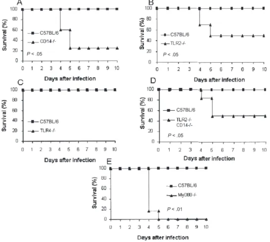
Statistical analysis. The statistical evaluation of differences between the experimental groups was performed by the use of the log rank test for survival studies and the Student’s t test. Figure 2. Reduced resistance of CD14-deficient (⫺/⫺) mice infected with Listeria monocytogenes. We injected 3 ⫻ 104colony-forming units of
Listeria intravenously into CD14⫺/⫺ mice (A), TLR2⫺/⫺ mice (B), TLR4⫺/⫺ mice (C), CD14⫺/⫺ and TLR2⫺/⫺ mice (D), and MyD88⫺/⫺ mice (E).
Survival rates were compared to that of C57BL/6-infected control mice during a 10-day period (for 6 mice per group). Graphs show results from 1 experiment representative of 2 independent experiments (*P⬍ .05, **P ⬍ .01, determined by log rank test).
RESULTS
Reduced production of IL-12p40, IL-6, TNF, and NO in CD14 – deficient macrophages stimulated by Listeria. To characterize the role of CD14 for proinflammatory cytokine production in response to L. monocytogenes, primary bone mar-row– derived macrophages from CD14-deficient mice and C57BL/6 control mice were stimulated with L. monocytogenes. The production of IL-12p40, IL-6, and, to a lesser extent, TNF was significantly reduced after L. monocytogenes activation of CD14-deficient macrophages at 6 h (not shown) and 18 h (fig-ures 1A–1C), compared with cells from control mice, without affecting the cells’ viability. As expected, the cytokine responses to low doses of LPS were abrogated in CD14-deficient phages (figure 1), as was also seen in TLR4-deficient macro-phages (not shown). A similar reduction in cytokine production in response to L. monocytogenes infection is seen in TLR2-deficient and MyD88-TLR2-deficient macrophages, as reported else-where [4].To explore the potential mechanisms of a bactericidal effect, we measured NO production. Macrophages from CD14-deficient mice produced less NO in response to L. monocytogenes
infection, compared with macrophages from control mice (fig-ure 1D) (P⬍ .05). Despite diminished NO production, there was no significant reduction of bacterial killing by macrophages from CD14-deficient mice (figure 1E) (P⬎ .05).
Therefore, the data suggest that CD14 is involved in L. mono-cytogenes–induced macrophage activation. In view of the re-duced proinflammatory cytokine and NO production, we hy-pothesized that host resistance to infection with this intracellular pathogen may be diminished in CD14-deficient mice.
Reduced resistance and bacterial clearance in Listeria-infected, CD14-deficient mice. Because L. monocytogenes activates macrophages in a CD14-dependent and TLR2-dependent manner, we asked whether the CD14 engagement contributes to TLR2-dependent resistance to an in vivo L. mono-cytogenes infection. We compared the resistance of C57BL/6 control mice and CD14-deficient mice to a systemic injection of L. monocytogenes. Intravenous infection with 3⫻ 104cfu per
mouse caused death in 60% of CD14-deficient mice within 5 days, whereas all control mice survived (figure 2A). TLR2-deficient mice display a similar susceptibility to infection, whereas TLR4-deficient mice are resistant (figures 2B and 2C). To test whether there was an additive effect when both CD14 and TLR2 genes were inactivated, we used mice that were both CD14 deficient and TLR2 deficient; their susceptibility to L. monocy-togenes infection was comparable to that of single gene– deficient mice (figure 2D). In contrast, we confirm that MyD88-deficient mice are highly sensitive to L. monocytogenes infection (figure 2E), as we reported before [6]. Therefore, these data clearly show that CD14-deficient mice have heightened susceptibility to L. monocytogenes infection.
In view of their increased sensitivity to infection with this or-ganism, we asked whether the rate of bacterial clearance might be reduced in CD14-deficient mice. The bacterial load in spleen and liver tissue from control and CD14-deficient mice that had been infected with 3⫻ 104cfu of L. monocytogenes per mouse
was analyzed. Three days after infection, CD14-deficient mice displayed⬃2 log higher levels of colony-forming units in the liver and 1 log higher levels in the spleen, compared with wild-type control mice, and MyD88-deficient mice had significantly higher levels than control mice (figures 3A and 3B). TLR2-deficient mice and mice TLR2-deficient in both CD14 and TLR2 dis-played an increase in the number of viable bacteria in the liver and spleen that was comparable to that seen in mice that lacked CD14 expression alone, without evidence of an additive effect (figure 3). Therefore, reduced host resistance in the absence of CD14 and/or TLR2 is associated with a significant increase of bacilli in the liver. As consequence of the higher bacterial load, augmented inflammation with an increased number of hepatic microabscesses was expected.
Augmented hepatic inflammatory response in Listeria-infected, CD14-deficient mice. Microscopic examination of liver tissue at low magnification revealed distinct differences in Figure 3. Reduced rate of bacterial clearance from the liver and
spleen of CD14-deficient mice infected with Listeria monocytogenes. Bacterial load in the liver (A) and spleen (B) of CD14-deficient (⫺/⫺) mice, TLR2⫺/⫺ mice, CD14 ⫺/⫺ and TLR2⫺/⫺ mice, TLR4⫺/⫺ mice, MyD88⫺/⫺ mice, and C57BL/6 control mice 3 days after infection with 3⫻ 104colony-forming units of L. monocytogenes (4 per group). Each
symbol indicates the liver or spleen of 1 mouse. Results are from 1 experiment representative for 2 independent experiments and are ex-pressed as mean⫾ SD (*P ⬍ .05, **P ⬍ .01. NS, not significant.
the hepatic microabscesses observed in CD14-deficient mice, compared with those observed in C57BL/6 control mice (figure 4A) 3 days after infection. The hepatic microabscesses in CD14-deficient mice were generally larger, less confined, and partially confluent. The number of abscesses was significantly increased in CD14-deficient mice (figure 4B). Moreover, the microab-scesses in these mice had less-defined boundaries, contained more abundant neutrophils and necrotic tissue (figure 4C), and were larger (figure 4D), compared with the abscesses observed in control mice. Further, as an indicator of reduced phagocyte ac-tivation and bactericidal activity in vivo, NOS2 expression in infected liver tissue was clearly reduced in CD14-deficient mice, compared with control mice (figure 4E). Therefore, the absence of CD14 is associated with reduced NOS2 expression, enhanced neutrophil recruitment, and formation of large microabscesses in the liver.
Flow cytometric analysis of the inflammatory cells in the liver at 2 days after infection, when the bacterial loads are comparable
between the experimental groups (data not shown), revealed de-creased expression of CD40 by CD11b⫹ cells from CD14-deficient mice, compared with cells from C57BL/6 control mice, which suggested reduced activation of monocytes and/or mac-rophages in the liver (figure 5). In contrast, the number of Gr1⫹ neutrophils is slightly increased at the same time point (not shown), as shown by microscopic examination of the tissue sec-tions.
CD14- and TLR2-dependent activation of NF-B and co-stimulatory molecules. To further dissect potential crosstalk between the TLR2 and CD14 receptor pathways, we assessed the translocation of cytosolic NF-B into the nucleus in BMDMs at several time points after L. monocytogenes infection. Although uninfected macrophages from control mice did not show any significant nuclear staining, incubation with L. monocytogenes induced rapid nuclear translocation of NF-B in macrophages from C57BL/6 mice, which reached a maximum at 1 h with 64% of macrophages showing nuclear staining. In contrast, nuclear Figure 4. More abundant and larger hepatic microabscesses observed in CD14-deficient (⫺/⫺) mice. C57BL/6 and CD14⫺/⫺ mice were infected with 3⫻ 104colony-forming units of Listeria monocytogenes and liver tissue was histologically examined 3 days after infection (4 mice per group).
Sections of liver tissue showing small, confined microabscesses in wild-type mice and spreading infection in CD14⫺/⫺ mice (A) (hematoxylin and eosin staining,⫻100). Increased numbers of microabscesses in CD14⫺/⫺ mice, compared with C57BL/6 controls (B). Histological sections of liver tissue showing small, confined microabscesses in wild-type mice and spreading infection in CD14⫺/⫺ mice (C) (hematoxylin and eosin staining, ⫻400). D, Mean diameter of microabscesses in millimeters. Graphs show results from 1 experiment representative of 2 independent experiments expressed as mean⫾ SD (*P ⬍ .05, **P ⬍ .01). E, Reduced expression of inducible nitric oxide synthase in the liver of CD14⫺/⫺ mice. Immunostaining was performed at 3 days after infection in C57BL/6 control mice and CD14⫺/⫺ mice by immunohistochemical analysis (⫻100 and ⫻400) (n ⫽ 4).
translocation was absent in macrophages from CD14-deficient or TLR2-CD14-deficient mice after L. monocytogenes infec-tion (figure 6A). Therefore, L. monocytogenes induced NF-B activation through CD14 and TLR2 with similar kinetics and magnitude. Further, reduced NF-B activation was found to be associated with diminished upregulation of CD40 and CD86 expression in macrophages from CD14-deficient mice and TLR2-deficient mice infected with L. monocytogenes, compared with macrophages from control mice (figure 6B). Therefore, both NF-B and costimulatory molecule expres-sion are reduced to a similar extent in macrophages from CD14-deficient mice or TLR2-deficient mice. The data suggest that CD14 and TLR2 sense molecular patterns expressed by L. monocytogenes in concert. Reduced macro-phage activation, cytokine production, and NO production may explain reduced in vivo host responses with dimin-ished rates of bacterial clearance and enhanced
inflamma-tion observed in CD14-deficient mice and TLR2-deficient mice.
DISCUSSION
The present data demonstrate for the first time, to our knowl-edge, that CD14-deficient mice have an increased sensitivity to L. monocytogenes infection with reduced survival rates due to impaired activation and killing of phagocytes that results in re-duced rates of bacterial clearance and augmented inflammatory pathology. In fact, in macrophages stimulated by L. monocyto-genes, the production of IL-12p40, IL-6, NO, and, to a lesser extent, TNF that depends in part on TLR2 signaling, but not on TLR4 signaling, is reduced. These results are in agreement with those of Flo et al., who demonstrated that CD14 neutralization reduced TNF production in TLR2-transfected CHO cells acti-vated by heat killed L. monocytogenes [15]. In the present study, we show that cytokine and NO production by primary macro-phages stimulated with live L. monocytogenes is dependent on CD14 and TLR2 [4]. In addition, the nuclear translocation of NF-B and expression of the costimulatory molecules CD40 and CD86 are diminished in the absence of either of CD14 or TLR2. In conformity with our results, reduced cytokine production in macrophages from TLR2-deficient mice has been shown [26] and reduced serum levels of IFN␥, TNF, and IL-12 have been reported in these mice [5]. Furthermore, we extend this obser-vation and demonstrate a role for CD14 in in vivo Listeria infec-tion. Reduced resistance to Listeria infection is seen in both CD14-deficient and TLR2-deficient mice. Moreover, mice defi-cient in CD14 or TLR2 alone and mice defidefi-cient in both CD14 and TLR2 display comparable reduction in host resistance, with increased of bacterial load and enhanced acute hepatic inflam-mation. The discrepancy between the susceptibility reported previously in TLR2-deficient mice [6, 27] and our data here may be explained by different routes of infection (intraperitoneal versus intravenous), the number of backcrosses of the mice, and the strains of Listeria used.
The involvement of CD14 agrees well with our unpublished data that suggest L. monocytogenes signals through TLR2-TLR6 complexes. Indeed, ligands of TLR2-TLR6 heterodimers, such as Pam2CSK4, MALP2 or zymosan, seem to require CD14, whereas ligands of TLR2-TLR1 heterodimers, such as Pam3CSK4, bind and signal independently of CD14 [1, 28, 29]. It should be emphasized that the MyD88-deficient mice are dis-tinctly more susceptible to L. monocytogenes infection than TLR2-deficient or CD14-deficient mice (figure 2D). Previously, a high sensitivity to Listeria infection was reported for MyD88-deficient mice, but interestingly, an adaptive immune response with protective immunity was obtained in the absence of MyD88 [30]. MyD88 is further involved in crosstalk with other pathways such as the focal adhesion kinase [31] that may play a role in the Figure 5. CD14-dependent recruitment and activation of macrophages
in the liver. CD14-deficient mice and control mice were infected with 3⫻ 104colony-forming units of Listeria monocytogenes, and
mononu-clear cells from the liver were isolated 2 days after infection (4 mice per group). The cells were stained with CD11b and CD40 and analyzed by flow cytometry. Numbers are the percentage of double-positive cells in the mononuclear cell population. Graphs show results from 1 experiment that are representative of 2 independent experiments, expressed as mean ⫾ SD (n ⫽ 2).
process of spreading Listeria infection. CD14 has so far not been reported to be involved in receptor complexes other than TLR4 and TLR2, with the exception of TLR3. A TLR3 ligand poly-inositine-cytosine analogue has been shown to activate antigen-presenting cells in a TLR3- CD14 dependent manner, and a ligand- TLR3-CD14 complex has been demonstrated [18].
CD14 seems to be involved in regulating neutrophil influx, because CD14-deficient mice show increased expression of CXCR2, MIP2, and neutrophil transmigration in pneumococcal infection [32], whereas neutrophil migration was delayed in CD14-deficient mice early after peritoneal infection with Salmo-nella, acting through TLR4 [33]. Here, neutrophil recruitment Figure 6. CD14-dependent nuclear factor B (NF-B) translocation and upregulation of CD40 and CD86 expression in Listeria monocytogenes (LM)–infected bone marrow– derived macrophages (BMDMs). NF-B translocation in L. monocytogenes–infected macrophages (A). BMDMs from C57BL/6 mice and CD14-deficient mice were stimulated for 1h with L. monocytogenes (MOI, 1:2), and NF-B staining was assessed. A quantitative evaluation of the percentage of cells showing NF-B translocation is presented here as a bar graph. Reduced upregulation of CD40 and CD86 expression (B) in CD14-deficient macrophages after L. monocytogenes or lipopolysaccharide (LPS) stimulation (2 mice per group). Graphs show results from 1 experiment representative of 3 independent experiments. Counts represent the relative cell number. MFI, mean fluorescence intensity.
after L. monocytogenes infection is enhanced, resulting in large, diffuse microabscesses in the liver of CD14-deficient mice. How-ever, the data suggest that macrophages and neutrophils from CD14-deficient mice and TLR2-deficient mice may be less effec-tive in killing in vivo, as indicated by reduced iNOS expression in the liver, although the macrophage mediated killing was not re-duced in vitro. Reactive nitrogen is a key bactericidal mechanism for mycobacteria [34] and Listeria [35]. However, the killing of Listeria is further supported by reactive oxygen produced by phagocytes oxidase (pg91phox). In fact, mice that are
double-deficient for pg91phoxand NOS2 are more susceptible to Listeria
infection [35] than single-deficient mice, but the existence of alternative mechanisms of killing is discussed [36].
Although we showed reduced resistance in TLR2-deficient mice [5, 6] and CD14-deficient mice, the dramatically height-ened susceptibility of MyD88-deficient mice that has bee de-scribed before [5, 6] cannot be solely ade-scribed to defective TLR sensing of L. monocytogenes. We have tested most of the TLRs by using single-gene deficient mice (data not shown), suggesting that TLR2-CD14 may contribute to this heightened susceptibil-ity, along with other non-TLR pathogen recognition receptors on the membrane, which are yet to be identified.
Previous investigations demonstrated the induction of MCP-1 in macrophages by L. monocytogenes and CCR2-dependent recruitment and activation of monocytes [27]. The induction of MCP-1 was found to be MyD88-dependent and TLR2-independent. Edelson et al. [6] offer data that reinforce the MyD88-dependent control of L. monocytogenes infection, suggesting a contribution by IL-1 and IL-18 receptor pathways, which depend on TLR signaling on the MyD88 adapter protein. This hypothesis is further reinforced by reduced IL-1 production in L. monocytogenes–infected, ASC-deficient macrophages [26]. Therefore, TLR2-CD14 and additional, undefined pattern rec-ognition receptors induce IL-1 and IL-18, which are required, together with IFN␥, IL-1, and TNF, to control L. monocytogenes infection. However, a recent report demonstrated that IL-1 sig-naling is dispensable for the control of systemic Listeria infec-tion, but required to control cerebral listeriosis [37].
In conclusion, Listeria activates macrophages in vitro in a CD14-dependent and/or TLR2-dependent manner. CD14 ex-pression is required for host resistance to L. monocytogenes in-fection in vivo; reduced CD14 expression results in bacterial per-sistence, reduced NOS2 expression in the liver, and heightened mortality. However, there is no additive effect with TLR2; mice deficient in both CD14 and TLR2 have comparably heightened susceptibility to Listeria infection.
References
1. Akira S. Toll-like receptor signaling. J Biol Chem 2003; 278:38105– 8. 2. Janeway CA, Jr., Medzhitov R. Innate immune recognition. Annu Rev
Immunol 2002; 20:197–216.
3. Medzhitov R, Janeway C, Jr. The Toll receptor family and microbial recognition. Trends Microbiol 2000; 8:452– 6.
4. Torres D, Barrier M, Bihl F, et al. Toll-like receptor 2 is required for optimal control of Listeria monocytogenes infection. Infect Immun 2004; 72:2131–9.
5. Seki E, Tsutsui H, Tsuji NM, et al. Critical roles of myeloid differentia-tion factor 88-dependent proinflammatory cytokine release in early phase clearance of Listeria monocytogenes in mice. J Immunol 2002; 169: 3863– 8.
6. Edelson BT, Unanue ER. MyD88-dependent but Toll-like receptor 2-independent innate immunity to Listeria: no role for either in macro-phage listericidal activity. J Immunol 2002; 169:3869 –75.
7. Ulevitch RJ, Tobias PS. Receptor-dependent mechanisms of cell stimu-lation by bacterial endotoxin. Annu Rev Immunol 1995; 13:437–57. 8. Ulevitch RJ, Tobias PS. Recognition of gram-negative bacteria and
en-dotoxin by the innate immune system. Curr Opin Immunol 1999; 11: 19 –22.
9. Jeyaseelan S, Chu HW, Young SK, Freeman MW, Worthen GS. Distinct roles of pattern recognition receptors CD14 and Toll-like receptor 4 in acute lung injury. Infect Immun 2005; 73:1754 – 63.
10. Jiang Z, Georgel P, Du X, et al. CD14 is required for MyD88-independent LPS signaling. Nat Immunol 2005; 6:565–70.
11. Yoshimura A, Lien E, Ingalls RR, Tuomanen E, Dziarski R, Golenbock D. Cutting edge: recognition of Gram-positive bacterial cell wall com-ponents by the innate immune system occurs via Toll-like receptor 2. J Immunol 1999; 163:1–5.
12. Compton T, Kurt-Jones EA, Boehme KW, et al. Human cytomegalovi-rus activates inflammatory cytokine responses via CD14 and Toll-like receptor 2. J Virol 2003; 77:4588 –96.
13. Henneke P, Takeuchi O, van Strijp JA, et al. Novel engagement of CD14 and multiple toll-like receptors by group B streptococci. J Immunol
2001; 167:7069 –76.
14. Ellingsen E, Morath S, Flo T, et al. Induction of cytokine production in human T cells and monocytes by highly purified lipoteichoic acid: in-volvement of Toll-like receptors and CD14. Med Sci Monit 2002; 8: BR149 –56.
15. Flo TH, Halaas O, Lien E, et al. Human Toll-like receptor 2 mediates monocyte activation by Listeria monocytogenes, but not by group B streptococci or lipopolysaccharide. J Immunol 2000; 164:2064 –9. 16. Hoebe K, Georgel P, Rutschmann S, et al. CD36 is a sensor of
diacyl-glycerides. Nature 2005; 433:523–7.
17. Hoebe K, Janssen E, Beutler B. The interface between innate and adap-tive immunity. Nat Immunol 2004; 5:971– 4.
18. Lee HK, Dunzendorfer S, Soldau K, Tobias PS. Double-stranded RNA-mediated TLR3 activation is enhanced by CD14. Immunity 2006; 24: 153– 63.
19. Moore KJ, Andersson LP, Ingalls RR, et al. Divergent response to LPS and bacteria in CD14-deficient murine macrophages. J Immunol 2000;
165:4272– 80.
20. Hoshino K, Takeuchi O, Kawai T, et al. Cutting edge: Toll-like receptor 4 (TLR4)-deficient mice are hyporesponsive to lipopolysaccharide: evi-dence for TLR4 as the LPS gene product. J Immunol 1999; 162:3749 –52. 21. Werts C, Tapping RI, Mathison JC, et al. Leptospiral lipopolysaccharide activates cells through a TLR2-dependent mechanism. Nat Immunol
2001; 2:346 –52.
22. Adachi O, Kawai T, Takeda K, et al. Targeted disruption of the MyD88 gene results in loss of IL-1- and IL-18-mediated function. Immunity
1998; 9:143–50.
23. Muller M, Eugster HP, Le Hir M, et al. Correction or transfer of immu-nodeficiency due to TNF-LT alpha deletion by bone marrow transplan-tation. Mol Med 1996; 2:247–55.
24. Green LC, Wagner DA, Glogowski J, Skipper PL, Wishnok JS, Tannen-baum SR. Analysis of nitrate, nitrite, and [15N]nitrate in biological flu-ids. Anal Biochem 1982; 126:131– 8.
25. Fields PI, Swanson RV, Haidaris CG, Heffron F. Mutants of Salmonella
typhimurium that cannot survive within the macrophage are avirulent.
26. Ozoren N, Masumoto J, Franchi L, et al. Distinct roles of TLR2 and the adaptor ASC in IL-1beta/IL-18 secretion in response to Listeria
mono-cytogenes. J Immunol 2006; 176:4337– 42.
27. Serbina NV, Kuziel W, Flavell R, Akira S, Rollins B, Pamer EG. Sequential MyD88-independent and -dependent activation of innate immune re-sponses to intracellular bacterial infection. Immunity 2003; 19:891–901. 28. Beutler B. TLR4 as the mammalian endotoxin sensor. Curr Top
Micro-biol Immunol 2002; 270:109 –20.
29. Cleveland MG, Gorham JD, Murphy TL, Tuomanen E, Murphy KM. Lipoteichoic acid preparations of gram-positive bacteria induce interleukin-12 through a CD14-dependent pathway. Infect Immun
1996; 64:1906 –12.
30. Way SS, Kollmann TR, Hajjar AM, Wilson CB. Cutting edge: protective cell-mediated immunity to Listeria monocytogenes in the absence of my-eloid differentiation factor 88. J Immunol 2003; 171:533–7.
31. Zeisel MB, Druet VA, Sibilia J, Klein JP, Quesniaux V, Wachsmann D. Cross talk between MyD88 and focal adhesion kinase pathways. J Im-munol 2005; 174:7393–7.
32. Echchannaoui H, Frei K, Letiembre M, Strieter RM, Adachi Y, Land-mann R. CD14 deficiency leads to increased MIP-2 production, CXCR2
expression, neutrophil transmigration, and early death in pneumococ-cal infection. J Leukoc Biol 2005; 78:705–15.
33. Yang KK, Dorner BG, Merkel U, et al. Neutrophil influx in response to a peritoneal infection with Salmonella is delayed in lipopolysaccharide-binding protein or CD14-deficient mice. J Immunol 2002; 169:4475– 80.
34. MacMicking JD, Nathan C, Hom G, et al. Altered responses to bacterial infection and endotoxic shock in mice lacking inducible nitric oxide synthase. Cell 1995; 81:641–50.
35. Shiloh MU, MacMicking JD, Nicholson S, et al. Phenotype of mice and macrophages deficient in both phagocyte oxidase and inducible nitric oxide synthase. Immunity 1999; 10:29 –38.
36. Fehr T, Schoedon G, Odermatt B, et al. Crucial role of interferon con-sensus sequence binding protein, but neither of interferon regulatory factor 1 nor of nitric oxide synthesis for protection against murine liste-riosis. J Exp Med 1997; 185:921–31.
37. Deckert M, Virna S, Sakowicz-Burkiewicz M, et al. Interleukin-1 recep-tor type 1 is essential for control of cerebral but not systemic listeriosis. Am J Pathol 2007; 170:990 –1002.