Images in cardio-thoracic surgery
Intimal sarcoma of pulmonary artery: multi-slice ECG-gated computed
tomography findings with 3D reconstruction
Elena Rizzo
a, Mauro Pugnale
b, Tom Mckee
c, Salah D. Qanadli
a,*
aDepartment of Radiology, CHUV—University of Lausanne, 46 Rue du Bugnon, 1011 Lausanne, Switzerland bDepartment of Radiology CHYC, Yverdon-Les-Bains, Lausanne, Switzerland
cPathology Institute, CHUV—University of Lausanne, Lausanne, Switzerland
Received 17 November 2004; received in revised form 2 January 2005; accepted 10 January 2005; Available online 5 March 2005
Keywords: Intimal sarcoma; Multi-slice ECG-gated CT
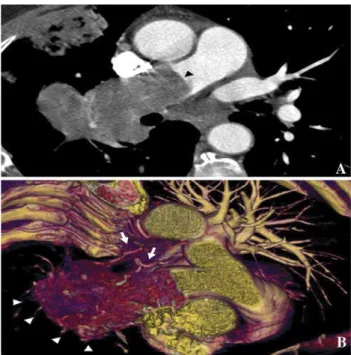
An intra-vascular right pulmonary artery mass was shown
using ECG-gated multi-slice CT in patient with progressive
dyspnea and weight lost (
Fig. 1
). Endovascular biopsy,
guided by virtual angioscopy derived from the CT data,
suggested diagnosis of intimal sarcoma. Pneumonectomy
was done with total resection of right pulmonary artery,
pulmonary trunk and partial resection of the left pulmonary
artery confirming the histopathological diagnosis (
Fig. 2
).
European Journal of Cardio-thoracic Surgery 27 (2005) 919
www.elsevier.com/locate/ejcts
Fig. 1. Multi-slice ECG-gated CT scan (slice thickness, 1.25 mm). (A) Contrast enhanced axial image, the right pulmonary artery was completely occluded by a mass (arrowhead). Endovascular component and extension into the surrounding pulmonary parenchyma were well shown. (B) 3D volume-rendered reconstruction (oblique view) shows the extension (arrowheads) and bronchial artery hypertrophy (arrows) more clearly.
Fig. 2. Histopathology. (A) Low magnification of sample obtained from endovascular biopsy (haematoxylin-eosin stain 40!). (B) High magnification showing histological feature: the tumour cells showed significant cytonuclear atypia (arrows) and a high mitotic index (15 mitoses per 10 hpf—0.177 mm2)
producing a solid tumor without specific architectural features. No foci of heterologous differentiation were identified. The histological feature was compatible with diagnosis of intimal sarcoma of pulmonary artery grade III (according to the FNCLCC-system) with invasion into the surrounding pulmonary parenchyma and adjacent lymphatic nodes (hematoxilin-eosin stain 200!).
1010-7940/$ - see front matter Q 2005 Elsevier B.V. All rights reserved. doi:10.1016/j.ejcts.2005.01.056
* Corresponding author. Tel.: C41 21 314 44 45; fax: C41 21 314 44 88. E-mail address: [email protected] (S.D. Qanadli).