PICTORIAL ESSAY
Noninflammatory fallopian tube pathology in children
Laura Merlini&Mehrak Anooshiravani&Aaron Vunda&Irene Borzani&Marcello Napolitano&
Sylviane Hanquinet
Received: 16 March 2008 / Revised: 2 September 2008 / Accepted: 7 September 2008 / Published online: 21 October 2008 # Springer-Verlag 2008
Abstract Noninflammatory tubal abnormalities are rare in children and usually not well covered by traditional educational material. The presenting symptoms are non-specific and are common to many other conditions, so its preoperative diagnosis is rarely made. The purpose of this study was to review the hospital charts and imaging findings in children and sexually inactive adolescents who showed fallopian tube pathology. Understanding of the pertinent findings of previous imaging examinations might assist radiologists in making the correct preoperative diagnosis and increase the likelihood of preserving the fallopian tubes. The clinical entities described in this article include isolated tubal torsion, paratubal cysts, hydrosalpinx, undescended/ectopic fallopian tube, and tubal inguinal hernia.
Keywords Fallopian tube . Imaging . Children
Introduction
The presenting symptoms of noninflammatory paratubal and tubal abnormalities are nonspecific and are common to
many other conditions, so the preoperative diagnosis is rarely made. The role of imaging is underestimated by clinicians and radiologists because it is usually not well covered by traditional educational material. A limited number of clinical images of most of the paediatric tubal pathology are reported in the literature. In addition, some clinical conditions such as pelvic congestion and endome-triosis, that are well known as predisposing factors of tubal pathology in adults, are not considered by paediatric radiologists because they are not aware of this diagnostic possibility in children. In fact, tubal pathology should be considered as a cause of acute pelvic pain in children and adolescents because of the possibility of salvage surgery.
The purpose of this study was to review the hospital charts and imaging findings in children and sexually inactive adolescents referred to the Pediatric Radiology Unit of the Children’s University Hospital of Geneva who had been identified as having fallopian tube pathology.
Isolated fallopian tube torsion
Torsion of the fallopian tube not associated with an ovarian abnormality is termed isolated fallopian tube torsion. There are only 13 sporadic published case reports and a recent series of eight cases of children with preoperative imaging [1–4], of which one was chronic [1] and five occurred before menarche. In the absence of adnexal disease, three different aetiological factors are mentioned: (1) physical hyperactivity, (2) anatomical predisposition with a mobile tube on a wide ligamentum latum, and (3) immediate premenstrual congestion of the tube.
Ultrasonography has been widely considered as the gold standard imaging technique in the evaluation of suspected adnexal torsion, even if it lacks specificity [1,4], and the
DOI 10.1007/s00247-008-1034-3
L. Merlini (*)
:
M. Anooshiravani:
S. HanquinetUnit of Pediatric Radiology, University Hospital HUG, 6 Willy-Donzé,
1205 Geneva, Switzerland e-mail: [email protected] A. Vunda
Clinic of Pediatric Surgery, University Hospital, Geneva, Switzerland
I. Borzani
:
M. NapolitanoPediatric Radiology, Ospedale Buzzi, Milan, Italy
preoperative diagnosis is only seldom verified. The US appearances of a torsed tube are variable and include a midline cystic mass sometimes extending towards the midline [1,4], a complex mass with free fluid (Fig.1) or
a serpiginous structure with fluid contents, haemorrhage, and thickened walls [2] (Fig. 2) associated with a normal ipsilateral ovary; the diagnosis can be most easily deter-mined in the last mentioned mode of presentation. A
Fig. 1 A 12-year-old postme-narchal female presented with acute pelvic pain. US images show a 3×1.5-cm complex structure (a, b, c arrows) next to the right ovary (c, d arrow-heads), with no flow apparent on the Doppler image (d). Dif-ferentials proposed were hae-morrhagic cyst of the ovary or a teratoma. Two days later she presented with acute abdominal pain and therefore underwent laparoscopy. At surgery there was a threefold twist of the long axis of a largely necrotic fallo-pian tube necessitating salpingectomy
Fig. 2 An 11-year-old preme-narchal female with a 2-day history of pelvic pain. a Pelvic US image shows a serpiginous structure with fluid contents and thickened walls. Ovaries are not seen. a–d MRI: T2-W axial image (b) confirms the presence of a tubular, hyperintense struc-ture of 7×2 cm diameter with separate, normal ovaries (b, c white arrows), with a deposit in the lower part (black arrow-heads); T1-W gadolinium-en-hanced axial image (d) shows that there is no contrast en-hancement of the left tubal walls (black arrow). The correct diag-nosis of fallopian tube torsion was made. However, due to delay in diagnosis, the tube could not be salvaged and sal-pingectomy was performed
complex mass can be mistaken for a haemorrhagic ovarian cyst if the ovary is not well separated and no flow is present on Doppler examination. Colour Doppler can be useful, but the presence of normal flow does not necessarily rule out torsion because of the dual vascular supply to the ovary and tube [5] and differentiation from an uncomplicated hydro-salpinx may be difficult. In this case, MRI seems to be of some value, as it can reveal the bloody nature of the tubal contents and show a lack of gadolinium enhancement, which may be indicative of infarction [2,6] (Fig.2).
The gold standard for diagnosis and treatment is operative laparoscopy with detorsion of the tube unless there is evidence of necrosis or rupture. However, surgical remedy of the torsed tube is controversial [4].
Pre-existing anomalies that might result in swelling of the distal portion of the fallopian tube and subsequently provoke torsion are described in the following paragraphs. The most frequent abnormalities in children are paraovarian or paratubal cysts; hydrosalpinx and tumours are extremely rare. Pelvic congestion syndrome is a cause of tubal torsion in adults and is rarely found in children (Fig.3).
Paraovarian or paratubal cysts
Paraovarian or paratubal cysts in children are mostly observed incidentally on routine US pelvic examinations in adolescents (Fig. 4). They are rarely a cause of pelvic
Fig. 3 A 15-year-old girl with known Hirschsprung disease treated with a pull-through pro-cedure at 1 year of age, pre-sented with acute pelvic pain, pressure, and heaviness. US (a) and colour Doppler (c, d) images demonstrate congested pelvic veins. CT performed the day after (b) demonstrates en-hanced, bilateral tortuous ves-sels, and a cystic structure on the right ovary (arrow). The pain subsided on the same day. This case shows that a cause of tubal torsion in adults can be found also in children
Fig. 4 Asymptomatic paraovar-ian or paratubal cysts, inciden-tally observed in two different patients. a A cyst (arrowhead) is separated from the ovary (arrows). b Three small para-tubal cysts (1, 2 and 3)
pain due to rupture (Fig. 5) or torsion (Fig. 6) and have been reported as the most frequent cause of isolated tubal torsion in the paediatric population, being present in 10 of 20 previously reported cases [4]. Sonographically, the diagnosis depends on identifying the ipsilateral ovary separate from the tube, although this is not always feasible [7]. Paraovarian cysts usually appear as single or multiple cysts separate from the ovary with no septa or folds. Paratubal cysts are also called hydatid cysts of Morgagni; they are round cysts attached to the fimbriated end of the fallopian tube by a pedicle [8]. This aspect was well shown by US imaging in the case we present in Fig.6; this is the second case of isolated tubal fimbria necrosis in the paediatric literature, and uniquely shows the US findings [9].
The role of CT and MRI in diagnosis is still not well established [10].
Hydrosalpinx
Hydrosalpinx develops due to a blocked, dilated fluid-filled fallopian tube. It is a very rare finding in adolescent girls without inflammatory disease and its pathogenesis is not well studied. Sporadic cases of unilateral noninflammatory hydrosalpinx are reported as isolated postsurgical compli-cations [11], or as complications of peritoneal drains [12]. In the only case report in the literature, bilateral hydro-salpinx in an adolescent without inflammatory disease was due to endometriosis [13], but no imaging was provided. A
Fig. 5 A 14-year-old girl presenting with pelvic pain. Abdominal US showed signs of appendicitis (not shown), two small cysts (a thin arrows) and a bigger one of 2.7 cm with thickened walls (a, c thick arrows) next to the normal right ovary (b arrowhead). A tubular but solid structure (b, c short arrows) connects the ovary to the uterus and is interpreted as a possibly torsed fallopian tube. At laparoscopy
appendicitis was confirmed; there was blood-stained fluid in the peritoneal cavity and on the right adnexal region, a torsed and broken 3-cm tubal cyst leaking a light yellowish fluid, and multiple small paraovarian cysts. The right tube was very congested but not torsed. A detorsion after the cyst had broken could have occurred. The right tube was not fixed
Fig. 6 A 13-year-old girl with torsion of a Morgagni cyst and history of recurrent moderate pain in the right iliac fossa. She presented with acute pain of abrupt onset without vomiting or fever. US images show a normal right ovary in the right iliac fossa (a, b arrowheads) and a tubular structure (a short arrows) ending in a 3×1-cm cyst (a, b; thick
arrows) and free fluid in the Douglas pouch (a, b). Colour Doppler imaging was unhelpful. Laparoscopy was performed and a torsed Morgagni cyst was found wrapped around an engorged and bluish fimbria, which was resected. The fallopian tube was not torsed
further case was presented by Durand et al. at the Annual Congress of the French Society of Pediatric Radiology in Rennes in 2000. They reported a case of bilateral hydro-salpinx as a postoperative complication in a 17-year-old female with bladder exstrophy.
At our institution we have observed three cases of bilateral noninflammatory hydrosalpinx in adolescents treated by pull-through surgical procedure in early infancy for Hirschsprung disease (Fig.7) [14].
Abdominal US showing fallopian tubes as fluid-filled tubular structures folded onto themselves to form a C or S shape (Fig.7) and separated from the ovaries is consistent with hydrosalpinx and helps to distinguish the condition from a septated ovarian cystic mass, which is usually round in shape. When the elongated nature of these folds is not recognized, they may be mistaken for mural nodules of an ovarian cystic mass. In this case, MRI helped distinguish between hydrosalpinx and a cystic ovarian mass (Fig.7). T1-W images have also been reported to be useful to rule out
endometrioma, which can be a cause of hydrosalpinx (hyperintense on T1-W images), but there is no such case noted in the paediatric literature. Gadolinium enhancement can be useful in symptomatic patients to differentiate simple hydrosalpinx or pyosalpinx from torsion with infarction [1,15] (Fig.7), and the clinical data are also obviously different.
Undescended/ectopic fallopian tube
An ectopic fallopian tube is thought to occur subsequent to ovarian maldescent during fetal development. Reports of the heterotopic location of a fallopian tube are extremely rare, occurring in between 0.3% and 0.5% of 2,025 gynaecological operations performed for infertility [16]. With only a single exception in the literature, the ovary is always located beneath the fallopian tube. An ectopic fallopian tube can be associated with Müllerian malforma-tions, often with a unicornuate uterus on the contralateral
Fig. 7 Different aspects of un-complicated hydrosalpinx in three adolescent girls treated by pull-through surgical procedure in early infancy for Hirsch-sprung disease. a, b Patient 1. Pelvic US images show a fluid-filled irregular structure, with folds on left side (a), and a beaked protrusion of a cyst continuing in a band-like structure connecting it with the uterus on the right side, which obviously represents the fallo-pian tube (b). The contents are fluid, with a small amount of low-level echoic debris. c, d Patient 2. Transverse US image through the pelvis shows the fallopian tubes on both sides as fluid-filled tubular structures folded on themselves with longitudinal folds (c). T1-W contrast-enhanced MR image does not show high signal intensity suggestive of endome-triosis and a good enhancement of the dilated tube walls, allow-ing torsion to be ruled out (d). e, f Patient 3. US image shows complex structures in the adnexal regions (e) (reproduced with permission from the American Journal of Roentgen-ology). Sagittal T2-W MR image (f) shows the fluid-filled, tubular aspect with longitudinal folds, typical of dilated fallopian tubes
side of the undescended ovary and fallopian tube [17] (Fig. 8). This genital anomaly can be silent before menarche, and its discovery is mostly incidental, by laparoscopy or MRI performed for other reasons (Fig. 8). On the other hand, it sometimes can produce life-threaten-ing conditions such as intestinal obstruction or ischaemic bowel perforation (Fig.9). Thus, the finding of a“missing ovary” during US evaluation of an acute abdomen should prompt a thorough search if surgery is performed to locate a possible ectopic adnexa [17].
MRI, particularly with T2-W sequences, is the best imaging technique to localize the ovary and to rule out other urinary and gynaecological abnormalities; however, its ability to analyse tubal morphology is very limited if the tube is not dilated.
Tubal inguinal hernia
The fallopian tube and ovary are found in 15–31% of sliding hernias in females. They are reported more frequently in premature infants, due to patency of the
processus vaginalis (canal of Nuck) and the incidence decreases with age. Of hernias with fallopian tubes and ovaries, 70% occur in patients below the age of 5 years. Incarceration is the most common complication (30% during the first year of life). Sonography is an easy and accurate preoperative diagnostic procedure (Fig. 10); how-ever, correct recognition of the tubal nature of the hernial sac contents is not essential because surgical repair is recommended shortly after diagnosis in any case [18].
Miscellaneous
Fallopian tube endometriosis in adolescents is seldom reported in the literature [19,20] and none has been imaged so far, explaining the reason why we have no cases to illustrate. In adolescents, the appearance of endometriosis may be subtle and difficult to visualize even at laparoscopy; small red lesions and clear shiny peritoneal vesicles are below the resolution of US and MRI [20].
Tumours are a known cause of isolated fallopian tube torsion; they are, however, exceedingly rare in children and
Fig. 8 A 13-year-old girl with an undescended fallopian tube. US was performed for clinical suspicion of appendicitis. a The appendix is not visible, the right ovary is not visualized and a complex structure is seen in the right paracolic region (CT showed signs of appendicitis but could not explain the signifi-cance of the structure in the right paracolic gutter; not shown). At laparoscopy appen-dicitis was confirmed, the left ovary and tube were normal, and a right morphologically normal ovary was found in the paracolic gutter. The right fallo-pian tube was very elongated, its origin was ectopic from the superior two-thirds of a
uni-cornuate (left) uterus. b–d MR
images obtained after surgery depict well the undescended right ovary in the paracolic gutter (b, c) and the left uni-cornuate uterus (d black arrows); the undilated fallopian tubes are not visible
adolescents. Case reports include rhabdomyosarcoma, mixed Müllerian and alveolar soft-part sarcoma tumours, and sporadic cases of bilateral paraovarian endometrioid cystic tumours in Proteus syndrome [21]. We do not have any case to show, and we were not able to find in the literature any case reports with imaging findings of a paediatric fallopian tube tumour.
Conclusions
Knowledge of the variable US appearances of a torsed tube might assist radiologists in making the correct diagnosis. In our experience, in doubtful cases, MRI is of some value in showing normal ovaries and the absence of gadolinium enhancement. Pelvic congestion syndrome,
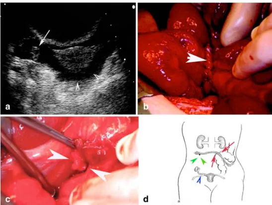
noninflamma-Fig. 9 A 14-year-old prepubertal girl with an ectopic fallopian tube presented with a 10-day history of abdominal pain as well as nausea, vomiting, and progressive abdominal distension. A plain abdominal radiograph showed typical features of a mechanical obstruction. a US image shows free fluid in the Douglas space (arrowheads) but no left ovary can be seen (arrow right ovary). b, c At laparoscopy, no ovary or fallopian tube were found on the left side. One ectopic tube extends next on the lateral sigmoid mesentery and is wrapped around the ileum, provoking a local strangulation and an ischaemic covered
bowel perforation (b arrow). A small calcified ovary is found adjacent to the fimbriae (c arrowheads). The ectopic fallopian tube and rudimentary ovary were resected, the perforated small bowel excised (reproduced with permission from the Journal of Pediatric Surgery).d The schematic drawing illustrates the complex anatomic situation found at laparotomy (blue arrowhead normal right tube, green arrowheads calcified ectopic ovary, red arrows ectopic left fallopian tube crossing the sigmoid mesentery)
Fig. 10 A 2-year-old girl pre-sented with an asymptomatic palpable movable mass in the left groin. She was born prema-turely at 35 weeks and no hernia was noted during the neonatal period. US images show a tu-bular solid structure in the her-nial sac (a) and an
intraabdominal ovary (b). In-guinal surgical exploration con-firmed the suspicion of isolated tubal hernia
tory hydrosalpinx, and paraovarian cysts also occur in children and can be imaged easily. Hydatid cysts of Morgagni can be recognized on US and, in the correct clinical setting, complications such as fimbrial or tubal torsion can be suggested. The finding of a “missing ovary” during US evaluation of an acute abdomen should prompt a thorough search by MRI (or by surgery) to identify a rare case of ectopic tube. The fallopian tube can be the only contents of a hernial sac and can be differentiated from intestinal contents.
References
1. Schollmeyer T, Soyinka AS, Mabrouk M et al (2008) Chronic isolated torsion of the left fallopian tube: a diagnostic dilemma.
Arch Gynecol Obstet 227:87–90
2. Orazi C, Inserra A, Lucchetti MC et al (2006) Isolated tubal torsion: a rare cause of pelvic pain at menarche: sonographic and MR findings. Pediatr Radiol 36:1316–1318
3. Breech LL, Hillard PJ (2005) Adnexal torsion in pediatric and adolescent girls. Curr Opin Obstet Gynecol 17:483–489 4. Harmon JC, Binkovitz LA, Binkovitz LE (2008) Isolated fallopian
tube torsion: sonographic and CT features. Pediatr Radiol 38:175– 179
5. Baumgartel P, Fleischer A, Cullinan J et al (1996) Color Doppler
sonography of tubal torsion. Ultrasound Obstet Gynecol 7:367–370
6. Bader T, Ranner G, Haberlik A (1996) Torsion of a normal adnexa
in a premenarcheal girl: MRI findings. Eur Radiol 6:704–706
7. Barloon TJ, Brown BP, Abu-Yousef MM et al (1996) Paraovarian and paratubal cysts: preoperative diagnosis using transabdominal
and transvaginal sonography. J Clin Ultrasound 24:117–122
8. Salamon C, Tornos C, Chi DS (2005) Borderline endometrioid tumor arising in a paratubal cyst: a case report. Gynecol Oncol
97:263–265
9. Blickstein I, Lancet M, Rozenman D et al (1989) Isolated necrosis
of the tubal fimbriae in a prepubertal girl. Z Kinderchir 44:172–
173
10. Kishimoto K, Ito K, Awaya H et al (2002) Paraovarian cyst: MR imaging features. Abdom Imaging 27:685–689
11. Sacher P, Meuli M (1997) Laparoscopic removal of hydrosalpinx in a girl: case report. Pediatr Surg 7:239–240
12. Washington EC, Holmes M, Haines SJ et al (2002) Ventriculoper-itoneal shunt migration presenting with vaginal discharge and
hydrosalpinx in a 16-year-old patient. Pediatr Emerg Care 18:28–
30
13. Stavroulis AI, Saridogan E, Creighton SM et al (2006) Laparo-scopic treatment of endometriosis. Eur J Obstet Gynecol Reprod
Biol 125:248–250
14. Merlini L, Anooshiravani M, Peiry B et al (2008) Bilateral
hydrosalpinx in adolescent girls with Hirschsprung’s disease –
association of two rare conditions. AJR 190:278–282
15. Outwater E, Siegelman E, Chiowanich P et al (1998) Dilated
fallopian tubes: MR imaging characteristics. Radiology 208:463–
469
16. Trinidad C, Tardaguila F, Fernandez GC et al (2004) Ovarian maldescent. Eur Radiol 14:805–808
17. Bugmann P, Hanquinet S, Brundler MA et al (2001) Intestinal obstruction caused by an ectopic fallopian tube in a child: case report and literature review. J Pediatr Surg 36:508–510
18. Huang CS, Luo CC, Chao HC et al (2003) The presentation of asymptomatic palpable movable mass in female inguinal hernia.
Eur J Pediatr 162:493–495
19. Yamamoto K, Mitsuhashi Y, Takaike T et al (1997) Tubal endometriosis diagnosed within one month after menarche: a case
report. Tohoku J Exp Med 181:385–387
20. Laufer MR, Sanfilippo J, Rose G (2003) Adolescent endometri-osis: diagnosis and treatment approaches. Pediatr Adolesc
Gynecol 16:S3–S11
21. Funk T, Frew P, Rodgers W et al (2006) Unusual fallopian tube neoplasm in a 10-year-old patient with Proteus syndrome: a case