ARTICLE
Yersinia enterocolitica strains associated with human
infections in Switzerland 2001
–2010
M. Fredriksson-Ahomaa&N. Cernela&H. Hächler&
R. Stephan
Received: 3 August 2011 / Accepted: 20 October 2011 / Published online: 10 November 2011 # Springer-Verlag 2011
Abstract Yersinia enterocolitica infections are common in humans. However, very scarce data are available on the different biotypes and virulence factors of human strains, which has proved to be problematic to assess the clinical significance of the isolated strains. In this study, the presence of the ail gene and distribution of different bio-and serotypes among human Y. enterocolitica strains bio-and their possible relation to the genotype and antimicrobial resistance were studied. In total, 128 Y. enterocolitica strains isolated from human clinical samples in Switzerland during 2001–2010 were characterised. Most (75 out of 128) of the Y. enterocolitica strains belonged to biotypes 2, 3 or 4 and carried the ail gene. One of the 51 strains that belonged to biotype 1A was also ail positive. Most of the ail-positive strains belonged to bioserotype 4/O:3 (47 out of 76) followed by 2/O:9 (22 out of 76). Strains of bioserotype 4/O:3 were dominant among patients between 20 and 40 years old and strains of biotype 1A dominate in patients over 40 years. Strains belonging to biotypes 2, 3 and 4, which all carried the ail gene, exhibited a high homogeneity with PFGE typing. Y. enterocolitica 2/O:5,27 and 2/O:9 strains showed resistance to amoxicillin/clavulanic acid and cefoxitin, but Y. enterocolitica 4/O:3 strains did not.
Introduction
Yersiniosis is a zoonotic bacterial disease with high public health relevance, especially in Europe because of its high incidence [1]. It is the third most frequently reported bacterial enteric disease after campylobacteriosis and salmonellosis in most European countries [2]. Human illness is mainly caused by Yersinia enterocolitica, which includes strains of diverse pathogenicity. Y. enterocolitica strains belonging to biotypes 1B and 2–5 are considered pathogenic, whereas strains belonging to biotype 1A are in general considered non-pathogenic. Pathogenicity can also be determined by PCR methods detecting different viru-lence genes [3]. Further characterisation of pathogenic Y. enterocolitica strains is still frequently determined by pulsed-field gel electrophoresis (PFGE). The PFGE types have been shown to correlate with biotype [4].
Y. enterocolitica is considered to be an important food-borne pathogen. Infection is most often acquired by eating raw or undercooked pork [3]. Rarely, this organism is transmitted through contaminated blood during a transfusion. Common symptoms of food-borne infections are diarrhoea, abdominal pain and fever, but sequelae, such as joint pain (reactive arthritis) and skin rash (erythema nodosus), are not uncommon [1, 5]. Infection with Y. enterocolitica occurs most often in young children [2].
Uncomplicated yersiniosis usually resolves on its own without antimicrobial treatment. However, in more severe cases, like septicaemia or focal extra-intestinal infection, and in immune-compromised patients, medication may be required. Y. enterocolitica is a β-lactamase producer and thus is resistant to ampicillin and first-generation cephalo-sporins, but it is mostly susceptible to other antimicrobials, such as aminoglycosides, tetracyclines, trimethoprim-sulfamethoxazole and fluoroquinolones, that are used for M. Fredriksson-Ahomaa
Department of Food Hygiene and Environmental Health, Faculty of Veterinary Medicine, University of Helsinki, Helsinki, Finland
N. Cernela
:
H. Hächler:
R. Stephan (*)Institute for Food Safety and Hygiene, Vetsuisse Faculty, University of Zurich,
Winterthurerstrasse 272, 8057 Zurich, Switzerland e-mail: stephanr@fsafety.uzh.ch
therapy. Multi-drug-resistant strains, which are resistant to streptomycin, sulfonamides, tetracycline and /or nalidixic acid, have occasionally been reported among some Y. enterocolitica strains [6].
The most commonly reported serotype of Y. enter-ocolitica strains isolated from human cases in Europe is O:3 and less commonly O:9 [2]. However, very scarce data on the virulence genes and the distribution of different biotypes among human strains are available. In this study, human Y. enterocolitica strains isolated during 2001 to 2010 in Switzerland were characterised to obtain information about the ail gene, and bio- and serotypes present in human clinical strains. Furthermore, the strains were genotyped and the antimicrobial resistance was studied.
Materials and methods
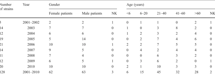
In total, 128 Y. enterocolitica strains collected from human patients in Switzerland during 2001 to 2010 were charac-terised (Table 1). Most of the strains (94%) were from humans suffering from diarrhoea, 4 strains were from blood, 2 from liver, 1 each from gall bladder, ulcer and abscess. Y. enterocolitica strains were almost equal in number in female (62) and male (63) patients. Only 6 strains (5%) were from patients under 6 years of age, but 28 strains (22%) were from patients over 60 (Table 1). Six patients reported having travelled abroad before infection.
All 128 Y. enterocolitica strains were biotyped and serotyped. The biotype was determined using pyrazinamidase and tween activity, esculin hydrolysis, indole production, and salicin, xylose and trehalose fermentation [4]. Serotyping was carried out with slide agglutination using commercial Y. enterocolitica O:3, O:5, O:9 and O:27 antisera (Sifin, Berlin,
Germany). PCR was used to detect the ail gene located in the chromosome of pathogenic Y. enterocolitica strains according to Thoerner et al. [7].
All strains were genotyped with pulsed field gel electrophoresis (PFGE) according to the PulseNet protocol
(http://www.cdc.gov/pulsenet/protocols/yersinia_Apr2006.
pdf) using restriction enzyme XbaI (New England Biolabs, Beverly, MA). DNA fragments were separated with a CHEF-DR III system (Bio-Rad, Hercules, CA, USA) using pulse times ranging from 5 to 40 s for 19 h at an angle of 120°C. Salmonella enterica serovar Braenderup H9812 (New England Biolabs) was used as the standard. GelCompar II software (Applied Maths NV, Sint–Martens–Latem, Belgium) was used for pattern comparison. PFGE patterns were considered clonally related if they had a similarity coefficient >80%, optimisation 0.5% and tolerance 2.5% (the Dice similarity index and the unweighted pair group method with the arithmetic mean).
Antimicrobial resistance analysis was performed by the disc diffusion test according to the Clinical and Laboratory Standards Institute (2009). Fourteen antimi-crobials were tested: ampicillin (10 μg), amoxicillin/ clavulanic acid (20/10 μg), cefalothin (30 μg), cefoxitin (30μg), cefpodoxim (10 μg), ceftazidime (30 μg), cefuroxime (30μg), ciprofloxacin (5 μg), gentamicin (10 μg), kanamycin (30 μg), nalidixic acid (30 μg), streptomycin (10 μg), tetracycline (30 μg) and trimethoprim/sulfamethoxazole (1.25/23.75μg). The reference strain Escherichia coli ATCC 25922 was used as the quality control.
Results
About 60% (75 out of 128) of the human Y. enterocolitica strains isolated from clinical samples belonged to biotypes
Table 1 Y. enterocolitica strains by gender and age collected during 2001 and 2010 in Switzerland from human clinical samples Number
of strains
Year Gender Age (years)
Female patients Male patients NK <6 6–20 21–40 41–60 >60 NK
5 2001–2002 2 2 1 0 1 1 0 2 1 14 2003 7 7 0 1 0 3 8 2 0 12 2004 6 6 0 1 2 3 2 4 0 19 2005 5 14 0 0 2 7 4 6 0 21 2006 10 10 1 2 2 7 5 5 0 14 2007 9 5 0 0 4 2 4 4 0 11 2008 7 4 0 0 0 6 3 2 0 12 2009 6 5 1 0 3 6 2 0 1 20 2010 10 10 0 2 1 10 3 3 0 128 2001–2010 62 63 3 6 15 45 32 28 2 NK = not known
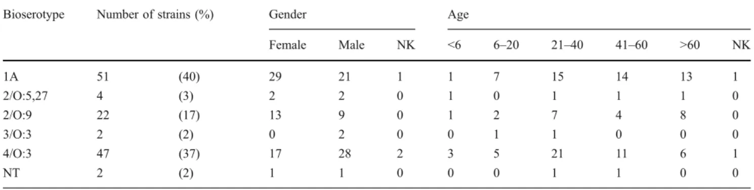
2, 3 or 4 which are considered to be pathogenic to humans (Table2). However, a high number (51 out of 128, 40%) of Y. enterocolitica strains belonged to biotype 1A, which is regarded as non-pathogenic. The dominant pathogenic bioserotype was 4/O:3 (37%) followed by 2/O:9 (17%). Only 4 strains belonged to bioserotype 2/O:5,27 and 2 to bioserotype 3/O:3. Two strains did not belong to any of the known biotypes.
The ail gene was studied by PCR and it was detected in all 75 strains belonging to pathogenic bioserotypes 2/O:5,27, 2/O:9, 3/O:3 and 4/O:3. Surprisingly, ail was also detected in one strain belonging to non-pathogenic biotype 1A. This strain was isolated in 2007 from the faeces of a 62-year-old woman. Both strains of unknown biotype were ail negative. They were isolated from the faeces of a 40-year-old woman and a 50-year-old man in 2005. The two strains, which were not of any known biotype, were ail negative and pyrazinamidase positive, and thus were regarded as non-pathogenic Y. enterocolitica-like strains.
More Y. enterocolitica strains of biotype 1A and bioserotype 2/O:9 were from female than from male patients (Table 2). Strains belonging to bioserotype 4/O:3 were more frequently from males than from female patients. Y. enterocolitica of bioserotype 4/O:3 was isolated more often from patients between 0 and 40 years of age (29) than from patients over 40 (17; Table 2). Biotype 1A strains were the most common type in patients over 40 and they were frequently (18 out of 27, 67%) isolated from female patients.
Eight Y. enterocolitica strains were isolated from extra-intestinal sites: blood (4), liver (2), gall bladder (1), ulcer (1) and abscess (1). Strains of bioserotype 4/O:3 were isolated twice from blood and once from liver and ulcer. The 2 strains of bioserotype 2/O:9 were isolated from blood. Surprisingly, 2 strains belonging to biotype 1A were from extra-intestinal sites: the gall bladder and an abscess. All 128 Y. enterocolitica strains were sensitive to ceftazidim (30 μg), ciprofloxacin (5 μg) and gentamycin (10μg), and resistant to ampicillin (10 μg) and cefalothin Table 2 Y. enterocolitica strains of different bioserotypes according to gender and age
Bioserotype Number of strains (%) Gender Age
Female Male NK <6 6–20 21–40 41–60 >60 NK 1A 51 (40) 29 21 1 1 7 15 14 13 1 2/O:5,27 4 (3) 2 2 0 1 0 1 1 1 0 2/O:9 22 (17) 13 9 0 1 2 7 4 8 0 3/O:3 2 (2) 0 2 0 0 1 1 0 0 0 4/O:3 47 (37) 17 28 2 3 5 21 11 6 1 NT 2 (2) 1 1 0 0 0 1 1 0 0
NK = not known; NT = not typeable
Table 3 Number ofY. enter-ocolitica strains showing resis-tance to antimicrobial agents
NT = not typeable;
I = intermediate; R = resistant
Antimicrobial agent Bioserotype (number of strains)
1A (51) 2/O:5,27 (4) 2/O:9 (22) 3/O:3 (2) 4/O:3 (47) NT (2)
I R I R I R I R I R I R Ampicillin 0 51 0 4 0 22 0 2 0 47 0 2 Amoxicillin/clavulanic acid 4 47 1 3 3 18 0 2 7 0 0 2 Cefalothin 0 51 0 4 0 22 0 2 0 47 0 2 Cefoxitin 22 23 0 4 3 18 0 2 0 0 1 1 Cefpodoxim 16 4 1 1 4 2 2 0 1 0 0 0 Cefuroxime 7 0 0 0 13 0 2 0 5 0 0 0 Kanamycin 0 3 0 0 0 0 0 0 0 0 0 0 Nalidixic acid 0 1 0 0 0 0 0 0 0 2 0 0 Streptomycin 3 0 0 0 0 0 1 0 5 7 0 0 Tetracycline 0 0 0 0 0 0 0 0 1 0 0 0 Trimethoprim/sulfa 0 0 0 0 0 0 0 0 2 1 0 0
(30 μg). Several strains were resistant to amoxicillin/ clavulanic acid (20/10μg) and cefoxitin (30 μg; Table 3). Interestingly, most strains belonging to bioserotypes 2/O:5,27 (3–4/4), 2/O:9 (18/22) and 3/O:3 (2/2) were resistant to amoxicillin/clavulanic acid and cefoxitin, but all 47 strains belonging to bioserotype 4/O:3 were sensitive to these agents. Sporadic resistance occurred to cefpodoxim (10 μg), kanamycin (30 μg), nalidixic acid (30 μg), streptomycin (10 μg) and trimethoprim/sulfamethoxazole (1.25/23.75). Strains belonging to bioserotypes 2/O:5,27 and 2/O:9 showed resistance cefpodoxim and strains of
bioserotype 4/O:3 to streptomycin, nalidixic acid and trimethoprim/sulfamethoxazole (Table 3). Only 1 multi-drug resistant strain was detected. This strain was belonging to bioserotype 4/O:3 and it was isolated in year 2006 from the faeces of a 27-year old woman. It showed resistance to nalidixic acid, streptomycin and trimethoprim/ sulfamethoxazole.
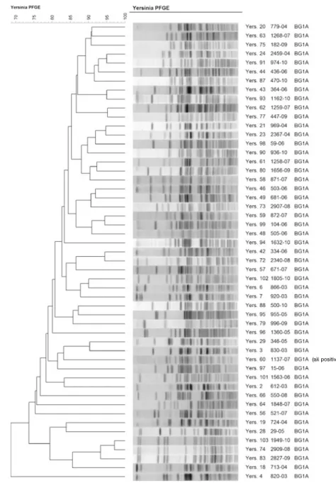
All 128 Y. enterocolitica strains were characterised by PFGE using the XbaI restriction enzyme. Strains belonging to bioserotypes 2/O:5,27 (2), 2/O:9 (22), 3/O:3 (2) and 4/O:3 (47) showed very limited genetic diversity; however, Fig. 1 Dendrogram ofY.
enter-ocolitica strains belonging to biotypes 2, 3 and 4
each bioserotype differed clearly from all the others (Fig.1). The 51 strains belonging to biotype 1A were very heterogeneous (Fig.2). The ail-positive biotype 1A strain was grouped among the other biotype 1A strains (Fig.2) and differed clearly from strains belonging to bioserotypes 2/O:5,27, 2/O:9, 3/O:3 and 4/O:3.
Discussion
Most of the human Y. enterocolitica strains belonged to the bioserotypes associated with human disease (75 out of 128). All these strains belonging to bioserotypes 2/O:5,27, 2/O:9, 3/O:3 and 4/O:3 were ail positive. The ail gene codes the Ail protein, which is involved in the attachment and invasion of the host cells and in the serum resistance of pathogenic Y. enterocolitica strains [8]. Bioserotype 4/O:3 was the most frequently identified pathogenic type (47 out of 75, 63%); however, the prevalence of bioserotype 2/O:9 (22 out of 75, 30%) was surprisingly high. Only 4 strains (5%) belonged to bioserotype 2/O:5,27 and 2 (3%) to bioserotype 3/O:3. In the last annual report of the European Centre for Disease Prevention and Control (ECDC), 8 European countries provided data on serotype; the two most common serotypes were O:3 (91%) and O:9 (7%) [2]. In Germany, of all the notified human cases with information on serotype, 89% were attributed to serotype O:3, and only 6% to serotype O:9 and 0.8% to serotype O:5,27 [1].
Only 6 (5%) patients reported having travelled abroad before infection, which indicates that the infections are domestically acquired. The majority (> 90%) of yersiniosis cases in Europe have also been reported to be domestically acquired [1,2]. In Switzerland, pigs at slaughter have been shown to carry ail-positive Y. enterocolitica bioserotype
4/O:3 frequently in the tonsils [4], which shows that domestic pigs can be an important source of human bioserotype 4/O:3 infection in Switzerland. Pigs have been shown to be an important reservoir and pork an important source of human Y. enterocolitica 4/O:3 infection in Germany and Finland [9]. Bioserotypes 2/O:5,27 and 2/O:9 have been sporadically isolated from Swiss pigs and could thus also be the source of human infections. However, the prevalence of bioserotype 2/O:9 was high in human infections, which indicates that there may be infection sources other than pigs. Two human strains belonged to bioserotype 3/O:3, which can indicate an Asiatic origin because this bioserotype is mainly reported in Asia [10]. However, bioserotype 3/O:3 has also been sporadically isolated from chinchillas in Europe [11].
Some of the Y. enterocolitica strains belonging to bioserotypes 2/O:9 and 4/O:3 (6 out of 69) were isolated from extra-intestinal sites. Extra-intestinal infections are rare and usually described in humans who are immune-compromised or in those with iron overload [12]. Y. enterocolitica 4/O:3 was isolated once from liver and skin lesions. Two strains of type 2/O:9 (9%) and 4/O:3 (4%) were isolated from blood, showing that these bioserotypes are not uncommon causes of bacteraemia in humans. Y. enterocolitica was one of the first recognised causes of post-transfusion sepsis [13]. Serotypes O:3 and O:9 are the most common types attributed to post-transfusion septic shock. Because Yersinia has the property to grow at 4°C, the low number of Y. enterocolitica strains collected from asymptomatic donors can lead to a high bacterial load if the blood products are stored for some weeks.
Y. enterocolitica strains belonging to biotype 1A are considered non-pathogenic to humans because they lack the major virulence determinants [14]. However, the number of strains belonging to biotype 1A was high (40%) among Fig. 1 (continued)
human patients in Switzerland. In Finland, an even higher prevalence (64%) of strains belonging to this biotype was recently reported [15]. One reason might be that in Finland, but not in Switzerland, cold enrichment, which increases the number of biotype 1A strains, is frequently used for human clinical samples. Y. enterocolitica strains belonging to biotype 1A lack the ail gene, which is an important chromosomal virulence gene found only in strains belonging to biotypes 1B and 2–5. Surprisingly, 1 of 51 strains of biotype 1A (2%) was ail-positive. Recently, Sihvonen et al. reported 1 ail-positive strain among 299 human clinical Y. enterocolitica biotype 1A strains, which shows that the ail gene is not a common finding among clinical biotype 1A strains [16].
Y. enterocolitica biotype 1A strains were mostly (49 out of 51) isolated from faeces. Surprisingly, Y. enterocolitica
biotype 1A strains were also isolated from the gall bladder and from skin lesions. The clinical disease with which Y. enterocolitica biotype 1A has been reported to be associ-ated is enteritis [17]. Biotype 1A strains have previously been isolated from blood, but not from other extra-intestinal sites. However, the pathogenic potential and true public health significance of Y. enterocolitica biotype 1A strains are still unclear [17].
The highest notification rate has been in children under 5 years of age in Europe followed by the age group 5– 14 years [2]. However, Y. enterocolitica was seldom isolated from young children under 6 years of age (6 out of 128) and from patients between 6 and 20 (15 out of 128) in Switzerland. One reason might be the high number of biotype 1A strains that were commonly found in patients (ail positive)
Fig. 2 Dendrogram ofY. enter-ocolitica strains belonging to biotype 1A
over 20 years of age. In a recent study, the symptoms of the patients with Y. enterocolitica biotype 1A differed from yersiniosis caused by the pathogenic biotypes [5]. Patients infected with pathogenic biotype strains were younger (mean age: 32 years and 50 years respectively), had fever (67% and 35% respectively) and reactive arthritis more often (10% and 3% respectively) than those with biotype 1A strains, which suggests the lacking virulence of biotype 1A strains. However, more research is needed to study the potential pathogenicity of biotype 1A strains.
Bioserotype 4/O:3 strains were more often isolated from male (28 out of 47) than from female patients (17 out of 47). In Germany, where strains of biotype 4/O:3 strain dominate, the infections occurred more frequently in boys and men than in girls and women [1]; however, there was no explanation for this phenomenon. Surprisingly, biotype 1A strains were more often found in female patients, especially in women over 40, than in male patients. One explanation for the higher prevalence of biotype 1A strains among women over 40 might be that they visit the doctor more frequently than men. Human Y. enterocolitica strains showed susceptibility to most antimicrobials other than β-lactams. All strains were susceptible to ceftazidime, ciprofloxacin and gentamicin and only sporadically resistant to cefpodoxim, kanamycin, nali-dixic acid, streptomycin and trimethoprim/sulfamethoxazole. Interestingly, most of the biotype 2 and 3 strains were resistant to amoxicillin/clavulanic acid and cefoxitin when all biotype 4/O:3 strains were sensitive, indicating an association between susceptibility to amoxicillin/clavulanic acid and cefoxitin and biotype. Only one strain, which belonged to bioserotype 4/O:3, showed resistance to multiple microbial agents: streptomycin, nalidixic acid and trimethoprim/ sulfamethoxazole. Multi-resistance has been shown to be rare among Y. enterocolitica strains in Switzerland and Germany [18, 19]. In a recent study from Finland, multi-resistance was significantly associated with travelling abroad [6]. Surprisingly, 3 Y. enterocolitica strains (1 biotype 1A and 2 biotype 4/O:3 strains) were resistant to nalidixic acid, which has not been reported before in Switzerland.. It has been shown that nalidixic acid resistance correlates with decreased susceptibility to ciprofloxacin in MIC determina-tion [6], which can be problematic in the treatment of complicated yersiniosis cases.
All strains were characterised with PFGE using XbaI. This restriction enzyme was used in the PulseNet protocol and in an earlier Swiss study [19]. PFGE with XbaI distinguished the different biotypes from each other and can thus help to identify ail-positive Y. enterocolitica strains of different biotypes. Strains belonging to biotypes 2, 3 and 4, which all carried the ail gene, exhibited high homoge-neity. However, strains belonging to serotypes O:5,27 and O:9 of biotype 2 could also easily be differentiated from each other. Strains belonging to biotype 1A, including the
one ail-positive strain, appeared to be highly heteroge-neous. High genetic diversity among biotype 1A strains has also been demonstrated by amplified fragment length polymorphism (AFLP) typing [20]. High genetic diversity is a common feature among non-pathogenic, environmental bacteria when most of the pathogenic bacteria are usually genetically monomorphic [21].
Acknowledgements We thank Grethe Sägesser for her help with strain collection. This project was partially funded by the Swiss Federal Office of Public Health, Division of Transmissible Diseases, Section of Infectious Diseases.
References
1. Rosner BM, Stark K, Werber D (2010) Epidemiology of reported Yersinia enterocolitica infections in Germany, 2001–2008. BMC Publ Health 10:337
2. Anonymous (2010) The community summary report on trends and sources of zoonoses and zoonotic agents in the European Union in 2008. European Food Safety Authority Journal. 10:1496
http://www.efsa.europa.eu/fr/scdocs/doc/s1496.pdf. Accessed 19 October 2011
3. Fredriksson-Ahomaa M, Korkeala H (2003) Low occurrence of pathogenic Yersinia enterocolitica in clinical, food, and environ-mental samples: a methodological problem. Clin Microbiol Rev 16:220–229
4. Fredriksson-Ahomaa M, Stolle A, Stephan R (2007) Prevalence of pathogenic Yersinia enterocolitica in pigs slaughtered at a Swiss abattoir. Int J Food Microbiol 119:207–212
5. Huovinen E, Sihvonen LM, Virtanen MJ, Haukka K, Siitonen A, Kuusi M (2010) Symptoms and sources of Yersinia enterocolitica-infection: a case-control study. BMC Infect Dis 10:122
6. Sihvonen LM, Toivonen S, Haukka K, Kuusi M, Skurnik M, Siitonen A (2011) Multilocus variable-number tandem-repeat analysis, pulsed-field gel electrophoresis, and antimicrobial suscep-tibility patterns in discrimination of sporadic and outbreak-related strains of Yersinia enterocolitica. BMC Microbiol 11:42
7. Thoerner P, Kingombe CIB, Bögli-Stuber K, Bissig-Choisat B, Wassenaar TM, Frey J, Jemmi T (2003) PCR detection of virulence genes in Yersinia enterocolitica and Yersinia pseudotu-berculosis and investigation of virulence gene distribution. Appl Environ Microbiol 69:1810–1816
8. Miller VL, Beer KB, Heusipp G, Young BM, Wachtel MR (2001) Identification of regions of ail required for the invasion and serum resistance phenotypes. Mol Microbiol 41:1053–1062
9. Fredriksson-Ahomaa M, Stolle A, Siitonen A, Korkeala H (2006) Sporadic human Yersinia enterocolitica infections caused by bioserotype 4/O:3 originate mainly from pigs. J Med Microbiol 55:747–749
10. Wang X, Cui Z, Wang H, Tang L, Yang J, Gu L, Jin D, Luo L, Qiu H, Xiao Y, Xiong H, Kan B, Xu J, Jing H (2010) Pathogenic strains of Yersinia enterocolitica isolated from domestic dogs (Canis familiaris) belonging to farmers are of the same subtype as pathogenic Y. enterocolitica strains isolated from humans and may be a source of human infection in Jiangsu Province, China. J Clin Microbiol 48:1604–1610
11. Fredriksson-Ahomaa M, Stolle A, Korkeala H (2006) Molecular epidemiology of Yersinia enterocolitica infections. FEMS Immunol Med Microbiol 47:315–329
12. Bottone EJ (1997) Yersinia enterocolitica: the charisma continues. Clin Microbiol Rev 10:257–276
13. Leclercq A, Martin L, Vergnes ML, Ounnoughene N, Laran JF, Giraud P, Carniel E (2005) Fatal Yersinia enterocolitica biotype 4 serovar O:3 sepsis after red blood cell transfusion. Transfusion 45:814–818
14. Bhagat N, Virdi JS (2011) The enigma of Yersinia enterocolitica biovar 1A. Crit Rev Microbiol 37:25–39
15. Sihvonen LM, Haukka K, Kuusi M, Virtanen MJ, Siitonen A (2009) Yersinia enterocolitica and Y. enterocolitica-like species in clinical stool specimens of humans: identification and prevalence of bio/serotypes in Finland. Eur J Clin Microbiol Infect Dis 28:757–765
16. Sihvonen LM, Hallanvuo S, Haukka K, Skurnik M, Siitonen A (2011) The ail gene is present in some Yersinia enterocolitica biotype 1A strains. Foodborne Pathog Dis 8:455–457
17. Bhaduri S (2011) Effect of salt and acidic pH on the stability of virulence plasmid (pYV) in Yersinia enterocolitica and expression of virulence-associated characteristics. Food Microbiol 28:171–173
18. Meyer C, Stolle A, Fredriksson-Ahomaa M (2011) Comparison of broth microdilution and disk diffusion test for antimicrobial resistance testing in Yersinia enterocolitica 4/O:3 strains. Microb Drug Resist 17:479–484
19. Baumgartner A, Küffer M, Suter D, Jemmi T, Rohner P (2007) Antimicrobial resistance of Yersinia enterocolitica strains from human patients, pigs and retail pork in Switzerland. Int J Food Microbiol 115:110–114
20. Kuehni-Boghenbor K, On SLW, Kokotovic B, Baumgartner A, Wassenaar TM, Wittwer M, Bissig-Choisat B, Frey J (2006) Genotyping of human and porcine Yersinia enterocolitica, Yersinia intermedia, and Yersinia bercovieri strains from Switzer-land by amplified fragment length polymorphism analysis. Appl Environ Microbiol 72:4061–4066
21. Achtman M (2008) Evolution, population structure, and phylo-geography of genetically monomorphic bacterial pathogens. Annu Rev Microbiol 62:53–70