HAL Id: hal-03163950
https://hal-amu.archives-ouvertes.fr/hal-03163950
Submitted on 9 Mar 2021HAL is a multi-disciplinary open access archive for the deposit and dissemination of sci-entific research documents, whether they are pub-lished or not. The documents may come from teaching and research institutions in France or abroad, or from public or private research centers.
L’archive ouverte pluridisciplinaire HAL, est destinée au dépôt et à la diffusion de documents scientifiques de niveau recherche, publiés ou non, émanant des établissements d’enseignement et de recherche français ou étrangers, des laboratoires publics ou privés.
seamounts, islands, and the continental slope: evidence
for deep-sea endemism
Björn Berning, Jean-Georges Harmelin, Beate Bader
To cite this version:
Björn Berning, Jean-Georges Harmelin, Beate Bader. New Cheilostomata (Bryozoa) from NE Atlantic seamounts, islands, and the continental slope: evidence for deep-sea endemism. European Journal of Taxonomy, Consortium of European Natural History Museums, 2017, �10.5852/ejt.2017.347�. �hal-03163950�
New Cheilostomata (Bryozoa) from NE Atlantic seamounts, islands,
and the continental slope: evidence for deep-sea endemism
Björn BERNING
1,*, Jean-Georges HARMELIN
2& Beate BADER
31 Oberösterreichisches Landesmuseum, Geowissenschaftliche Sammlungen, 4060 Leonding, Austria. 1 CIBIO, Centro de Investigação em Biodiversidade e Recursos Genéticos, InBIO Laboratório
Associado, Pólo dos Açores, 9501-801 Ponta Delgada, Açores, Portugal.
2 Aix-Marseille University, Mediterranean Institute of Oceanography, OSU Pytheas, Station Marine d’Endoume, 13007 Marseille, France.
3 Institut für Geowissenschaften, Christian-Albrechts-Universität, 24118 Kiel, Germany. * Corresponding author: b.berning@landesmuseum.at
2 Email: jean-georges.harmelin@univ-amu.fr 3 Email: bbader@online.no
1urn:lsid:zoobank.org:author:30D7D0DB-F379-4006-B727-E75A0720BD93 2urn:lsid:zoobank.org:author:D11AE07A-CFD9-41EE-B3F9-6E0472150300 3urn:lsid:zoobank.org:author:AA3BCFDC-524D-4648-9268-F0F1C94B9A68
Abstract. Ten new species belonging to three new genera (Atlantisina gen. nov., Bathycyclopora
gen. nov., Calvetopora gen. nov.) of umbonulomorph bryozoans from northeastern Atlantic seamounts, islands, and the continental slope are introduced. We furthermore erect the new family Atlantisinidae fam. nov. for these genera. Eight new species belong to the new genus Atlantisina: Atlantisina atlantis gen. et sp. nov. (type species), A. acantha gen. et sp. nov., A. gorringensis gen. et sp. nov., A. inarmata gen. et sp. nov., A. lionensis gen. et sp. nov., A. meteor gen. et sp. nov., A. seinensis gen. et sp. nov., and A. tricornis gen. et sp. nov. The genus Bathycyclopora gen. nov. is introduced for ?Phylactella
vibraculata Calvet from the Azores, and also includes Bathycyclopora suroiti gen. et sp. nov. The type
species of Calvetopora gen. nov. is Lepralia inflata Calvet from the Gulf of Cadiz; this genus also includes Calvetopora otapostasis gen. et sp. nov. and another species left in open nomenclature. Of the 13 species described herein, 11 occur on seamounts and islands, and nine species are endemic to a single seamount, island or station. The present results show that bryozoans provide striking examples of the function of seamounts as areas of endemism, most likely intrinsically linked to the low dispersal abilities of bryozoan larvae.
Keywords. Bathyal, biodiversity, biogeography, Macaronesia, taxonomy.
Berning B., Harmelin J.-G. & Bader B. 2017. New Cheilostomata (Bryozoa) from NE Atlantic seamounts, islands, and the continental slope: evidence for deep-sea endemism. European Journal of Taxonomy 347: 1–51. https://doi. org/10.5852/ejt.2017.347
2017 · Berning B. et al.
This work is licensed under a Creative Commons Attribution 3.0 License.
M o n o g r a p h
Introduction
Seamount faunas have received a considerable amount of scientific attention during the last two decades (e.g., Richer de Forges et al. 2000; Stocks 2004; McClain 2007). Especially in the northeastern (NE) Atlantic, biological sampling during numerous expeditions resulted in the description of a substantial number of seamount taxa such as, foraminifera (Heinz et al. 2004), sponges (Xavier & van Soest 2007), gastropods (Gofas 2007), polychaetes (Gillet & Dauvin 2003), copepods (George & Schminke 2002), ophiuroids (Bartsch 2008), or fish (Christiansen et al. 2009). Yet, although bryozoans are known to form significant proportions of the diversity of benthic seamount faunas (Rowden et al. 2002, 2004; Stocks 2004), the phylum has been ignored almost completely. Therefore, even in the relatively well-studied North Atlantic, our knowledge on the number of bathyal and offshore bryozoan species and their geographic distribution is still extremely poor, and the bryozoan faunas of only two seamounts have so far been studied in greater detail (Harmelin 1977; Souto et al. 2016). Moreover, major previous works on seamounts, oceanic islands, and the continental slope in this region (Busk 1884; Jullien & Calvet 1903; Calvet 1907, 1931; d’Hondt 1973, 1975; Harmelin 1977; Hayward 1978) mostly predate the use of scanning electron microscopy, which is crucial for the species-level identification of bryozoans. Revisions of these historical records are therefore necessary and often result in the splitting of species complexes, introduction of new taxa, and an increasing realisation of endemicity (e.g., Harmelin & Arístegui 1988; Harmelin 2006; López-Fé 2006; Berning & Kuklinski 2008; Souto et al. 2011; Berning 2012, 2013; Reverter-Gil & Souto 2015; Reverter-Gil et al. 2015).
The NE Atlantic islands and seamounts are not evenly distributed. Located relatively close to the southwest Iberian and northwest African continental shelves, several clusters of large volcanic structures are confined to hot spot tracks and fault systems (e.g., Geldmacher et al. 2006). For instance, from the sites studied in this work, the Canary Islands, Gorringe Bank and Galicia Bank are situated relatively close to the continents. Several seamounts, such as Ampère, Lion and Seine, cluster along the Madeira-Tore Rise towards the Madeira Archipelago, while all are separated from each other by abyssal depths. Another, more widely-spaced, group of islands and seamounts, the Azores in the north and the Atlantis-Great Meteor Seamount Chain in the south [comprising the Atlantis-Great Meteor Bank, Atlantis Seamount (Smt), Hyères Smt and Lion Smt], is formed by fault-system volcanism associated with the Mid-Atlantic Ridge. This group is separated from the continental shelf, and from the other seamounts mentioned above, by vast stretches of abyssal depths that are almost devoid of any prominent volcanic structures. Most of the seamounts and all of the modern archipelagos originated during the Miocene, while certain seamounts (e.g., Lion Smt: ca 80 Ma) and banks (e.g., Gorringe Bank: ca 75 Ma) are considerably older (Geldmacher et al. 2006). The minimum depth of the sampled seamount tops varies considerably as well: from as little as 20 m (Gorringe Bank) to some 600 m (Galicia Bank) below the sea surface. As part of an ongoing study of the bryozoan faunas from NE Atlantic islands and seamounts, we here describe a new family, three new genera, and ten new species of bathyal, umbonulomorph bryozoans, while two previously recorded species are redescribed. We discuss possible phylogenetic relationships to other known taxa, as well as the geographic distribution and ecomorphology of these bryozoans.
Material and methods
The bryozoan material studied here was collected during various oceanographic cruises to NE Atlantic seamounts and the continental slope by means of dredges and epibenthic sleds. Most material was collected during French expeditions with the research vessels ‘Calypso’ (1959) as well as during the ‘Thalassa’ (1968, 1971, 1972), ‘Noroît’ (Seamount 1, 1987) and ‘Suroît’ (Seamount 2, 1993) cruises. Additional samples were collected during the German ‘RV Meteor’ cruises M19 (1970) and M42/3 (1998) to the Great Meteor Bank, and the Spanish ‘Indemares BANGAL 0811’ expedition to Galicia Bank (2011). Historical material of Lepralia inflata Calvet, 1906 from the Gulf of Cádiz was collected
on the ‘Talisman’ cruise (1883), and that of ?Phylactella vibraculata Calvet, 1931 from the Azores on the ‘Prince Albert Ier de Monaco’ cruise (1897). The sampling localities are provided in Table 1, and the type and other material of each species is referenced in the text using the station numbers given in the first column.
All material was preserved dry. Selected specimens were cleaned with sodium hypochlorite, coated with gold-platinum and photographed using a Camscan (Serie-2-CS-44) and a FEI Inspect S50 scanning electron microscope (SEM) at the University of Kiel, a Hitachi S570 SEM at the Station Marine d’Endoume, and a FEI Inspect S50 SEM at the University of Vienna. The material is kept at the Muséum national d’Histoire naturelle in Paris (MNHN), the Musée océanographique de Monaco (MOM), the Oberösterreichisches Landesmuseum in Linz (OLL; collection Invertebrata except Insecta), and at the Senckenberg Museum und Forschungsinstitut in Frankfurt (SMF).
Morphometry was done using an eyepiece micrometre. The values are given in microns (µm) unless otherwise stated. We provide the mean value ± standard deviation (SD) and the number of measurements taken (#). One-way Analysis of Variance (ANOVA) and all pairs post hoc Tukey’s tests were performed
on certain sets of the original measurements in order to analyse intercolonial morphometric variability.
Abbreviations of measured characters: aAL = adventitious avicularium length aAW = adventitious avicularium width iAL = interzooidal avicularium length iAW = interzooidal avicularium width OL = orifice length
OW = orifice width OvL = ooecium length
OvL/W = ratio of ooecium length to width OvW = ooecium width
ZL = zooecium length ZW = zooecium width
Results
Suborder Neocheilostomina d’Hondt, 1985 Superfamily Lepralielloidea Vigneaux, 1949
Family Atlantisinidae fam. nov.
urn:lsid:zoobank.org:act:17F29C69-ECAC-4D27-A3B6-DE8C96009930
Type genus
Atlantisina gen. nov.
Diagnosis
Colonies encrusting; autozooidal frontal shield umbonuloid, imperforate or pseudoporous. Orifice with condyles, oral spines present. Adventitious and/or interzooidal avicularia present in some taxa. Lateral walls with basal pore-chambers, usually well developed, interzooidal commmunication via a single or few septular pores per neighbouring zooid, budding intrazooidal. Ovicell hyperstomial, ooecium either produced by the zooid distal to the maternal one or of kenozooidal origin, not closed by the operculum, the distal pair of oral spines closely appressed to the sides of the short, tubular ooecial opening; the kenozooidal ooecium may be entirely independent of the substratum and distal zooid, and may be
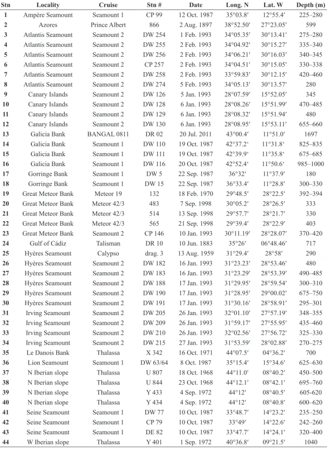
Stn Locality Cruise Stn # Date Long. N Lat. W Depth (m) 1 Ampère Seamount Seamount 1 CP 99 12 Oct. 1987 35°03.8′ 12°55.4′ 225–280
2 Azores Prince Albert 866 2 Aug. 1897 38°52.50′ 27°23.05′ 599
3 Atlantis Seamount Seamount 2 DW 254 1 Feb. 1993 34°05.35′ 30°13.41′ 275–280
4 Atlantis Seamount Seamount 2 DW 255 2 Feb. 1993 34°04.92′ 30°15.27′ 335–340
5 Atlantis Seamount Seamount 2 DW 256 2 Feb. 1993 34°06.21′ 30°16.03′ 340–345
6 Atlantis Seamount Seamount 2 CP 257 2 Feb. 1993 34°04.51′ 30°15.05′ 330–338
7 Atlantis Seamount Seamount 2 DW 258 2 Feb. 1993 33°59.83′ 30°12.15′ 420–460
8 Atlantis Seamount Seamount 2 DW 274 5 Feb. 1993 34°05.13′ 30°13.57′ 280
9 Canary Islands Seamount 2 DW 126 5 Jan. 1993 28°07.59′ 15°52.05′ 345
10 Canary Islands Seamount 2 DW 128 6 Jan. 1993 28°08.26′ 15°51.99′ 470–485
11 Canary Islands Seamount 2 DW 129 6 Jan. 1993 28°08.32′ 15°51.94′ 480
12 Canary Islands Seamount 2 DW 130 6 Jan. 1993 28°08.95′ 15°53.11′ 655–660
13 Galicia Bank BANGAL 0811 DR 02 20 Jul. 2011 43°00.4′ 11°51.0′ 1697
14 Galicia Bank Seamount 1 DW 110 19 Oct. 1987 42°37.2‘ 11°31.8‘ 825–835
15 Galicia Bank Seamount 1 DW 111 19 Oct. 1987 42°39.9‘ 11°35.8‘ 675–685
16 Galicia Bank Seamount 1 DW 116 20 Oct. 1987 42°52.4‘ 11°50.6‘ 985–1000
17 Gorringe Bank Seamount 1 DW 5 22 Sep. 1987 36°32′ 11°37.9′ 180
18 Gorringe Bank Seamount 1 DW 15 22 Sep. 1987 36°33.4′ 11°28.8′ 300–330
19 Great Meteor Bank Meteor 19 132 18 Feb. 1970 29°48.5′ 28°22.5′ 392–394
20 Great Meteor Bank Meteor 42/3 483 7 Sep. 1998 30°05.2′ 28°26.5′ 333
21 Great Meteor Bank Meteor 42/3 514 13 Sep. 1998 29°57.7′ 28°21.7′ 330
22 Great Meteor Bank Meteor 42/3 565 21 Sep. 1998 29°39.4′ 28°22.9′ 403
23 Great Meteor Bank Seamount 2 CP 146 10 Jan. 1993 30°11.19′ 28°28.07′ 370–420
24 Gulf of Cádiz Talisman DR 10 10 Jun. 1883 35°26′ 06°48.46′ 717
25 Hyères Seamount Calypso drag. 3 13 Aug. 1959 31°29.4′ 28°58′ 290
26 Hyères Seamount Seamount 2 DW 182 16 Jan. 1993 31°23.23′ 28°53.46′ 480
27 Hyères Seamount Seamount 2 DW 183 16 Jan. 1993 31°23.29′ 28°53.39′ 490–485
28 Hyères Seamount Seamount 2 DW 188 17 Jan. 1993 31°29.95′ 28°59.54′ 300–310
29 Hyères Seamount Seamount 2 DW 190 17 Jan. 1993 31°28.95′ 29°00.02′ 675–750
30 Hyères Seamount Seamount 2 DW 191 17 Jan. 1993 31°30.16′ 28°58.91′ 295–301
31 Irving Seamount Seamount 2 DW 205 26 Jan. 1993 32°01.10′ 27°57.19′ 348–355
32 Irving Seamount Seamount 2 DW 209 26 Jan. 1993 31°59.17′ 27°55.95′ 435–460
33 Irving Seamount Seamount 2 DW 210 26 Jan. 1993 32°02.56′ 27°56.72′ 325–330
34 Irving Seamount Seamount 2 DW 215 27 Jan. 1993 31°53.59′ 28°02.88′ 270–275
35 Le Danois Bank Thalassa X 342 16 Oct. 1971 44°07.5′ 04°36.2′ 700
36 Lion Seamount Seamount 1 DW 63/64 8 Oct. 1987 35°15.4′ 15°34.6′ 625–630
37 N Iberian slope Thalassa U 807 18 Oct. 1968 44°11.0′ 08°40.2′ 450–500
38 N Iberian slope Thalassa U 844 23 Oct. 1968 44°12.1′ 08°42.1′ 695–760
39 N Iberian slope Thalassa Y 433 4 Sep. 1972 44°12′ 08°40.5′ 605-620
40 N Iberian slope Thalassa Y 434 4 Sep. 1972 44°12′ 08°40.8′ 600–620
41 Seine Seamount Seamount 1 DW 77 10 Oct. 1987 33°48.7′ 14°23.2′ 235–250
42 Seine Seamount Seamount 1 CP 79 10 Oct. 1987 33°49′ 14°22.6′ 242–260
43 Seine Seamount Seamount 1 DE 82 10 Oct. 1987 33°47.7′ 14°24.1′ 320–400
44 W Iberian slope Thalassa Y 401 1 Sep. 1972 40°36.8′ 09°21.5′ 1040
Table 1. List of sampling cruises and the respective stations (Stn #) of the material examined in this
paper. The consecutive station numbers in the first column (Stn) are referenced to in the material section of the respective species.
budded from a distinct pore in the maternal zooid’s distal wall; endooecium fully calcified, ectooecium partially calcified. Ancestrula tatiform with extensive opesia; cryptocyst absent.
Remarks
The taxa that are here combined in the Atlantisinidae fam. nov. are morphologcially somewhat similar to, and share some characters with, the families Escharellidae Levinsen, 1909, Exochellidae Bassler, 1935 and Romancheinidae Jullien, 1888, while the latter is occasionally considered to include the former two (D.P. Gordon, pers. comm. 2014). However, the new taxa described here share distinct traits, which can be regarded as relatively conservative in an evolutionary sense, that are not present in any of the existing families. Assigning the new genera to the Romancheinidae sensu lato would add yet another set of new characters to this large taxon, and would render the family difficult to define precisely, as well as to delimit it from other lepralielloid families, e.g., the Bryocryptellidae Vigneaux, 1949.
Characters common to the species in all three genera presented here comprise the partial calcification of the ectooecium and the position of the distalmost pair of spines in maternal zooids, which are so closely appressed to the peristomial aperture of the ooecium as to leave a furrow on each side when the spine is missing. The species usually have extensive lateral walls composed of smooth gymnocyst, and interzooidal communication takes place via one (occasionally two) large septular pores. Moreover, all species share the same ancestrula-type (tatiform with a large opesia that is somewhat constricted in the distal oral part, while the cryptocyst is practically absent).
In contrast, the representative taxa in the Romancheinidae sensu lato are characterised by ancestrulae with a reduced oval or otherwise shaped opesia that is often bounded by an extensive cryptocyst. The ooecium is also structurally different in that the ectooecium is membranous, and the endooecium fuses with the frontal shield of the distal auto- or kenozooid (Ostrovsky 2013). Both the relatively primitive structure of the ancestrula as well as the morphology of the ooecium in the new taxa may thus suggest that the new family forms a clade basal to the Romancheinidae sensu lato (further discussed below). Genetic studies will be needed to ultimately shed light on the phylogenetic relationships between the new taxa and the Romancheinidae, as well as among the taxa currently placed within the Romancheinidae.
Genus Atlantisina gen. nov.
urn:lsid:zoobank.org:act:B1994BA0-3091-4D78-A5C3-CE8BEE1A19D2
Type species
Atlantisina atlantis gen. et sp. nov.
Diagnosis
Colony encrusting, unilaminar, forming small patches or biserial to multiserial ribbons by intrazooidal budding. Frontal shield umbonuloid, imperforate except for very few minute marginal pores; gymnocystal lateral walls generally extensive, basal pore chambers present, communication via a single large exterior pore per neighbouring zooid that is bounded by a variably distinct cryptocystal rim, a single round and slightly raised septular pore present in the distal vertical wall. Orifice with condyles, proximal margin concave; oral spines present, paired, in two distolateral series with a distal gap. Ovicell hyperstomial, ooecium kenozooidal, budded from the maternal zooid through the distal septular pore; ectooecium partially calcified, proximally usually forming a short tubular apertural peristome wedged in between the distalmost pair of spines; calcified endooecium exposed in central area, surface variably structured, occasionally deeply pitted but imperforate; not closed by operculum (presumably acleithral). No avicularia. Ancestrula tatiform, gymnocyst fairly narrow all around, cryptocyst absent, opesia extensive, slightly constricted in distal (oral) part, surrounded by numerous mural spines; first generation autozooid single, budded distally or (disto)laterally.
Etymology
Named for the occurrence of the type species on Atlantis Smt. Gender feminine.
Remarks
The combined occurrence of the following features distinguishes Atlantisina gen. nov. from all romancheinid genera as well as from the other new genera presented here: (i) a partly calcified ectooecium, (ii) the exclusively kenozooidal origin of the ooecium that is produced from a distinct communication pore in the maternal zooid’s distal wall, (iii) the lack of avicularia, and (iv) the well-developed lateral walls, with a single large communication pore per neighbouring zooid.
The variably developed band of smooth ectooecium around the basal and proximal part of the ooecium is a distinctive character of Atlantisina gen. nov. species (Fig. 1A). Moreover, in the Romancheinidae and most of the remaining atlantisinids the ooecium is either produced by the zooid distal to the maternal zooid, or (much less often) by a distal kenozooid that has an encrusting base, including lateral walls with basal pore chambers from which the distal autozooids are budded. The ooecium in Atlantisina gen. nov. is also a kenozooid (Fig. 1A), but it is budded from a single large communication pore situated in the distal vertical wall of the maternal zooid (Fig. 1B). The pore is slightly raised relative to the remaining lateral communication pores and remains exposed above the frontal shield of the distal zooid throughout ontogeny. Formation of the ovicell may, therefore, not be restricted to the colony margin but may occur opportunistically at any stage during ontogeny whenever breeding conditions are optimal and eggs are fertilised. Owing to the elevated position of this pore the ooecial kenozooid is not in contact with the substratum and the basal ooecium is, if at all, barely touching the frontal shield(s) of subsequently budded distal zooid(s) (Fig. 1A, C). The ooecial kenozooid also lacks communication pores.
Thus, the Atlantisina gen. nov. ovicell differs structurally from the escharelliform ooecium present in the Romancheinidae sensu lato (see Ostrovsky 2013: 138). In the latter, the endooecium fuses with the frontal shield of the distal auto- or kenozooid, and the membranous ectooecium is continuous with the distal zooid’s membranous frontal wall. The ooecium structure in Atlantisina gen. nov., as well as in the other new genera introduced here, can be regarded as evolutionarily more primitive relative to the escharelliform ooecium (A.N. Ostrovsky, pers. comm. 2016). On the other hand, the kenozooidal nature of the ooecium, and its origin from a distinct ooecial pore in the maternal zooid’s distal wall, indicates a derived state within the Atlantisinidae.
The zooecium is divided into a variably sculptured, cryptocystal-type frontal shield and smooth gymnocystal calcification comprising the external lateral and distal vertical walls, including the distal part of the aperture, condyles, and oral spines (Fig. 1A–C). Moreover, the proximal margin of the aperture (i.e., the distal part of the umbonuloid frontal shield) is again composed of smooth gymnocyst, varying in extent from extremely narrow to extremely extensive when forming a suboral mucro (Fig. 1A–C). The interior-walled frontal shield and the gymnocystal lateral walls are separated by distinct sinusoidal sutures lateral to the orifice, leading in a bow to the proximal pair of spines and the condyles (Fig. 1C), where they meet (but do not fuse with) the gymnocystal calcification along the proximal aperture margin (which is again separated by a suture from the cryptocystal-type frontal shield along the proximal side of the mucro).
The usually extensively developed gymnocystal lateral walls are characteristic for Atlantisina gen. nov. species, comprising spacious basal pore chambers (Fig. 1D) with a single, large, external communication pore per neighbouring zooid, which is usually bounded by a distinct cryptocystal rim (Fig. 1B). In contrast, most Romancheinidae (e.g., Hemicyclopora Norman, 1894) have extremely reduced vertical walls with numerous small septula connecting the neighbouring zooids, and gymnocystal calcification is absent (e.g., Hayward & Ryland 1999). The morphology and formation of the frontal shield and
orifice is, nevertheless, vaguely similar between Atlantisina and several romancheinid taxa such as
Hemicyclopora, Escharella Gray, 1848, and Escharoides Milne Edwards, 1836. Moreover, in the latter
genus communication between laterally neighbouring zooids also takes place via a single pore, whereas the distal wall comprises two or three basal pore chambers from which the distal autozooid is budded. The respective number of oral spines in all Atlantisina gen. nov. species is, quite remarkably, extremely constant within and among colonies. Atlantisina meteor gen. et sp. nov. (see below) was the only species
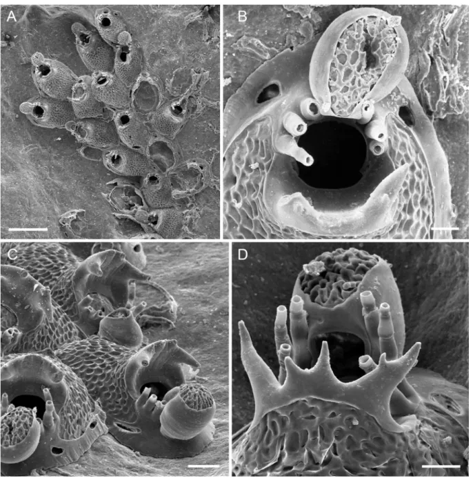
Fig. 1. Morphological characteristics of Atlantisina gen. nov. A. The kenozooidal ooecium of Atlantisina
lionensis gen. et sp. nov. in lateral view (paratype MNHN-IB-2014-67), showing the broad band of
ectooecium and the centrally exposed endooecium; note that the suboral crest is formed by smooth gymnocyst whereas the remaining frontal shield is cryptocystidean. B. Distal view of an autozooid of
Atlantisina meteor gen. et sp. nov. showing two distolateral communication pores and the slightly raised
central pore from which the ooecium is budded (paratype MNHN-IB-2014-50); note the broad band of cryptocyst bounding the septular pores, and that the remaining parts of the distolateral vertical walls and orifice are entirely gymnocystal. C. Oral region of an ovicellate zooid of Atlantisina atlantis gen. et sp. nov. (paratype MNHN-IB-2014-49), showing the contact between the cryptocystidean frontal shield and the gymnocystal distal part of the zooecium; note that the frontal shield is superpositioned on the condyles (white arrow) and meets the distolateral vertical walls in a sinusoidal suture (black arrow).
D. Initial stages of zooid formation with the lateral walls being partly broken, showing the large basal
pore chambers in Atlantisina atlantis gen. et sp. nov. (paratype OLL 2016/123). E. Slightly oblique view of the ancestrula of Atlantisina tricornis gen. et sp. nov. (paratype MNHN-IB-2014-64); note the simple tatiform morphology, the absence of a cryptocyst, and the slightly restricted oral region (top). Scale bars: A–B, D = 100 µm; C, E = 50 µm.
in which a deviation (by one spine) from the specific number of spines occurred among auto- or ovicelled zooids. Even in early astogenetic zooids, which are usually equipped with a higher number of spines than mature zooids in most cheilostomatid species, the specific number of spines is already present. The simple, tatiform ancestrula (Fig. 1E) buds a single distal to lateral autozooid, followed by one to three distolateral zooids that are either situated around the ancestrula or form distal to the first-generation autozooid, apparently depending on microenvironmental clues. While there is also a single first-generation autozooid in Escharella, Hemicyclopora and Neolagenipora Vigneaux, 1949, the ancestrula in Escharoides species usually produces two distolateral zooids (cf. Hayward & Ryland 1999). The ancestrula in Atlantisina gen. nov. also differs from the romancheinid taxa in having a distinctly more extensive opesia, with a constriction in the distal oral part, and in the absence of a crpytocyst.
Species of Atlantisina gen. nov. are presently restricted to bathyal depths along the NE Atlantic continental shelf, islands and seamounts. The northernmost distribution is along the northern Iberian margin (44° N) while Atlantisina gen. nov. was recorded as far west as Atlantis Smt (30° W) and south to the Canary Islands (28° N). No Recent species have been reported from the Mediterranean Sea but there is an early Pleistocene record from Sicily (A. Rosso, pers. comm. 2016).
Atlantisina atlantis gen. et sp. nov.
urn:lsid:zoobank.org:act:D2DA5614-66E4-4014-8FB5-7D931A22B5F2
Figs 1C–D, 2A–F, Table 2
Diagnosis
Frontal shield translucent, surface densely covered by large, flattened, irregularly polygonal nodules; lateral walls well-developed, septular pores large, round to transversely oval. Orifice margin with six oral spines; condyles short and blunt, no suboral mucro. Ovicell hyperstomial, ooecium globular, a little longer than wide; ectooecium relatively narrow, covering (less than) the lower half of ooecium; exposed endooecium relatively large and hemispherical, surface topography irregular, with no distinct pattern. Ancestrula with a pyriform opesia and nine mural spines.
Etymology
Named after its type locality, Atlantis Smt; used as a noun in apposition.
Material examined Holotype
ATLANTIS SMT: a large ovicellate colony marked “H”, together with three smaller colonies of
A. atlantis gen. et sp. nov. and a young colony of Bathycyclopora suroiti gen. et sp. nov. (see below), on
stylasterid skeleton, Stn 8 (MNHN-IB-2014-45).
Paratypes
ATLANTIS SMT: 1 colony on biogenic substrate, Stn 4 (MNHN-IB-2014-46); 3 colonies on a piece of stylasterid skeleton, Stn 7 (MNHN-IB-2014-47); 2 colonies on a piece of stylasterid skeleton, Stn 7 (MNHN-IB-2014-48); 1 coated colony on coral skeleton, Stn 8 (MNHN-IB-2014-49); 3 colonies on rock, Stn 7 (OLL 2016/122); 1 colony on coral, Stn 7 (OLL 2016/123); 2 colonies on coral, Stn 7 (OLL 2016/124).
Other material examined
ATLANTIS SMT: 3 colonies on coral skeleton, Stn 3 (unregistered MNHN material); 4 colonies on coral skeleton, Stn 4 (unregistered MNHN material); ca 33 colonies on coral and stylasterid skeletons, Stn 7
(unregistered MNHN material); 4 colonies on coral skeletons, Stn 8 (unregistered MNHN material); 6 colonies on coral, 1 on bivalve shell, Stn 7 (OLL 2016/125); 3 colonies on coral skeleton, Stn 7 (OLL 2016/126); 3 colonies on coral skeleton, Stn 7 (OLL 2016/127); 1 colony on coral skeleton, Stn 7 (OLL 2016/128); 8 colonies on stylasterid skeleton, Stn 7 (OLL 2016/129).
Description
Colony encrusting, unilaminar, forming small irregular patches or biserial to triserial branching ribbons (Fig. 2A). Zooecium outline oval distally, triangular proximally, wedged in between proximal zooecia
Table 2. Measurements of Atlantisina atlantis gen. et sp. nov.
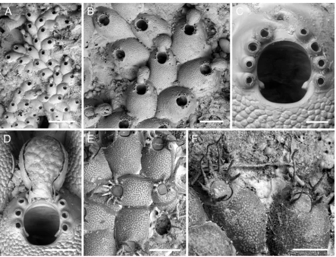
Fig. 2. Atlantisina atlantis gen. et sp. nov., Atlantis Smt. A. Overview of colony growing on a stylasterid
skeleton; note the biserial-branching growth (paratype MNHN-IB-2014-47). B. Several autozooids and ovicellate zooids (paratype MNHN-IB-2014-49). C. Close-up of orifice and the base of a severed ovicell protruding from the distal communication pore (paratype MNHN-IB-2014-49). D. Ooecium (OLL 2016/127). E. Periancestrular region (paratype OLL 2016/123). F. Ancestrula and first-generation autozooid (paratype OLL 2016/123). Scale bars: A = 1 mm; B = 500 µm; C–D = 50 µm; E = 300 µm; F = 100 µm.
ZL ZW OL OW OvL OvW
Mean 521 334 127 112 192 175
SD ± 29 ± 23 ± 7 ± 5 ± 7 ± 16
(Fig. 2B). Frontal shield matted vitreous, convex, densely covered by relatively large, evenly-spaced, irregularly polygonal and flattened nodules (Fig. 2C–F), imperforate except for five or six minute marginal pores, invisible in frontal view or in older zooecia; lateral walls well developed, septular pores in gymnocystal lateral walls large and surrounded by a distinct cryptocystal area, lateral ones usually transversely oval in outline, distal pore suborbicular, very slightly raised relative to lateral ones.
Orifice oval with a fairly straight and narrow proximal margin, slightly longer than wide, broadest in distal third, proximal third delimited by a pair of short and thick, blunt condyles oriented proximomedially; distolateral orifice margins with six short and closely-spaced spines arranged in two groups of three with a distinct distal gap, spine bases thick (Fig. 2C).
Ovicell hyperstomial, ooecium barely touching frontal shield of distal zooid or raised well above substratum when formed at colony margin, globular, with a short tubular proximal peristome wedged in between distalmost pair of spines and terminating at distal orifice margin, in general very little longer than wide; ectooecium smooth, encompassing lower half of ooecium; endooecium accordingly well-exposed, hemispherical, surface structure irregular, with an indistinct reticulate or nodular pattern; ooecial aperture taller than wide, acleithral (Figs 1C, 2B, D).
Ancestrula tatiform, almost oval in outline (ca 300 µm long, 190 µm wide), widest in proximal third, gymnocyst narrow and steeply sloping all around zooid except for proximal part, in which it is slightly better developed and more gently sloping, cryptocyst extremely reduced and only present at proximo-lateral margin, opesia extensive (ca 220 µm long, 150 µm wide), pyriform, distinctly constricted in distal third, surrounded by nine spines arranged in four closely positioned distal spines and five more widely spaced proximal ones; a single first-generation autozooid budded distally or distolaterally (Fig. 2E–F).
Remarks
Atlantisina atlantis gen. et sp. nov. occurs on the central NE Atlantic seamount complex together with A. meteor gen. et sp. nov. (see below), and both are also morphologically similar. The latter differs from
the former in having eight instead of six oral spines, and in that the endooecial surface is more markedly nodular and similar to the zooecial frontal shield. In contrast, the endooecium in A. atlantis gen. et sp. nov. is less conspicuously and variably structured (faint ridges or nodules), and often even lacking any apparent structure (e.g., Fig. 1C), being reminiscent of the early ontogenetic patterning of the frontal shield before the flattened nodules are formed. In some cases, however, a vague pattern of honeycomb depressions is visible, which is similar to the endooecial structure of several species found on or near the continental shelf (see below).
Another species with similar autozooids is Atlantisina inarmata gen. et sp. nov. from the Canary Islands (see below), which differs only in having slightly larger zooids and in that its skeleton is porcelain white, whereas it is translucent in A. atlantis gen. et sp. nov. A further important difference is found in the surface structure of the endooecium, which is densely and deeply pitted in A. inarmata gen. et sp. nov. The ooecium is also larger, and particularly wider, than in A. atlantis gen. et sp. nov.
Ecology
The sampled colonies of Atlantisina atlantis gen. et sp. nov. predominantly encrust coral skeletons, forming small patches or exploiting the surface via bi- or triserial ribbons. They have been found at depths between 275 and 460 m.
Distribution
Atlantisina meteor gen. et sp. nov.
urn:lsid:zoobank.org:act:F9928F6E-D74B-47B5-B985-CAC2BB62ED51
Figs 1B, 3A–F, Table 3
Diagnosis
Frontal shield densely covered by relatively small, irregularly shaped nodules with flattened tips; lateral walls very well developed, septular pores very large, round to elongate transversely oval; distolateral margin of orifice with eight (or rarely nine) slender oral spines, condyles short, blunt and thickened, no suboral mucro. Ectooecium covering more or less the lower half of ooecium; exposed endooecium relatively large and hemispherical, surface topography generally as frontal shield but nodular pattern not as pronounced. Ancestrula presumably with nine spines grouped in five widely spaced proximal ones and four closely spaced distal ones, opesia slightly constricted in distal third, cryptocyst practically absent.
Etymology
Named after its type locality, the Great Meteor Bank; used as a noun in apposition.
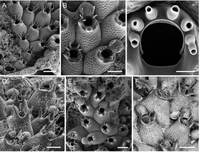
Fig. 3. Atlantisina meteor gen. et sp. nov., Great Meteor Bank A. Overview of holotype (OLL 2016/130a). B. Several autozooids and ovicellate zooids (holotype OLL 2016/130a). C. Orifice (paratype
MNHN-IB-2014-50). D. Ooecium (holotype OLL 2016/130a). E. Periancestrular region (SMF 40.040).
F. Unbleached autozooids with typical whip-like spines (paratype OLL 2016/133a). Scale bars:
Holotype
GREAT METEOR BANK: 2 colonies on limestone, the larger one with eight ovicells is the holotype (OLL 2016/130a), the smaller colony without ovicells is the paratype (OLL 2016/130b), bleached, Stn 20.
Paratypes
GREAT METEOR BANK: 4 colonies on limestone (2 with ovicells, 1 with ancestrula), unbleached, Stn 20 (MNHN-IB-2014-50); 2 colonies on limestone (1 with ancestrula, 1 with ovicells), unbleached, Stn 20 IB-2014-51); 1 colony with ancestrula on limestone, unbleached, Stn 20 (MNHN-IB-2014-52); 1 colony on Cladocora debilis Milne Edwards & Haime, 1849, mounted on stub and sputter-coated, Stn 21 (OLL 2016/131); 1 colony with ancestrula and ovicells on coral base, unbleached, Stn 20 (OLL 2016/132); 2 colonies on limestone, unbleached, Stn 20 (OLL2016/133); 1 colony on bioclast, mounted on stub and sputter-coated, Stn 20 (SMF 40.039); 4 colonies (2 with ancestrula) on limestone, unbleached, Stn 20 (SMF 40.040); 1 colony with ovicells on limestone, bleached, Stn 20 (SMF 40.041).
Other material examined
GREAT METEOR BANK: 10 colonies on Anomocora fecunda (Pourtalès, 1871), Stn 19 (unregistered MNHN material); 1 tiny colony on biogenic debris, sputter-coated, Stn 23 (unregistered MNHN material); 1 colony on C. debilis, mounted on stub and sputter-coated, Stn 21 (OLL 2015/10); 1 colony on C. debilis, mounted on stub and sputter-coated, Stn 19 (OLL 2016/134); 3 zooids (interior frontal shield), mounted on stub and sputter-coated, Stn 20 (OLL 2016/135); several colonies on C. debilis skeletons, unbleached, Stn 21 (OLL 2016/136); numerous colonies on limestone, unbleached, Stn 20 (OLL 2016/137).
IRVING SMT: 2 colonies on small rocks, 3 colonies on Flabellum chunii Marenzeller, 1904, Stn 31 (unregistered MNHN material); 5 colonies on small rocks, Stn 32 (unregistered MNHN material); 12 colonies on small rocks (1 sputter-coated) plus 4 colonies on F. chunii, Stn 33 (unregistered MNHN material); 1 colony on stylasterid skeleton, Stn 34 (unregistered MNHN material).
HYÈRES SMT: 3 colonies on F. chunii, Stn 26 (unregistered MNHN material); 1 small colony on old shell, Stn 27 (unregistered MNHN material); 1 colony on F. chunii and 5 small colonies on shell fragments (one sputter-coated), Stn 28 (unregistered MNHN material); 1 colony on F. chunii, Stn 29 (unregistered MNHN material); 3 colonies on F. chunii, Stn 30 (unregistered MNHN material); 1 colony on rock fragment, Stn 25 (unregistered MNHN material); 1 colony on bioclast, mounted on stub and sputter-coated, Stn 28 (OLL 2016/138); 1 colony on bioclast, mounted on stub and sputter-coated, Stn 28 (OLL 2016/139).
Description
Colony encrusting, unilaminar, forming small patches and bi- to triserial ribbons (Fig. 3A). Zooecia oval to polygonal, with tapering proximal end wedged in between proximal zooecia, separated by deep grooves (Fig. 3B). Frontal shield matted vitreous, convex, surface densely covered with relatively small irregular and flattened nodules, imperforate except for some six to eight very small marginal pores, invisible in frontal view or in older zooecia; lateral walls particularly well developed, septular pores in
Table 3. Measurements of Atlantisina meteor gen. et sp. nov.
ZL ZW OL OW OvL OvW
Mean 561 357 122 115 206 186
SD ± 55 ± 44 ± 9 ± 7 ± 25 ± 8
lateral walls very large and transversely oval, surrounded by a broad area of nodular cryptocyst, distal pore suborbicular, slightly raised relative to lateral ones (Fig. 1B).
Orifice almost as wide as long, broadest in distal third, proximal and lateral margins fairly straight, proximal third delimited by a pair of very short and thick, blunt condyles oriented proximomedially (Fig. 3C); lateral and distolateral margins with eight (very rarely nine) closely-spaced, slender, tapering and slightly curved spines with thick cylindrical bases (Fig. 3C, F), arranged in two groups of four with a distinct distal gap (in case there are nine spines, one group consists of five); all eight spines present in ovicellate zooids, with the distalmost pair thinner and tightly pressed against the ooecial peristome (Fig. 3D).
Ovicell hyperstomial, ooecium barely resting on distal zooid’s frontal shield (Fig. 3B, D), globular, about as long as wide, with a short tubular peristome wedged in between distalmost pair of spines and terminating at distal orifice margin; ectooecium smooth, encompassing approximately lower half of ooecium; exposed endooecium relatively large, hemispherical, surface covered by flattened irregular nodules similar to frontal shield (Fig. 3D); ooecial aperture suborbicular, about as tall as wide, acleithral. Ancestrula oval (ca 280 µm long, 210 µm wide), tatiform, gymnocyst well-developed and gently sloping proximally, becoming steeper and narrower distally; cryptocyst practically absent; opesia oval (ca 185 µm long, 140 µm wide), slightly constricted in distal third; presumably nine mural spines, with five proximal ones widely spaced and four distal ones situated closer together; a single first-generation autozooid budded distally or distolaterally (Fig. 3E).
Remarks
Atlantisina meteor gen. et sp. nov. is the only species in the genus with eight or occasionally even
nine spines surrounding the orifice (all other species have six), and in which the number of spines may occasionally vary. The additional spine is usually thinner and wedged in between the four others on one side of the orifice. Besides this difference in spine number, A. meteor gen. et sp. nov. is very similar to A. atlantis gen. et sp. nov., which occurs on the relatively closely located Atlantis Smt (ca 150 km north of the Great Meteor Bank-Hyères-Irving seamount complex), and also to A. inarmata gen. et sp. nov. from the Canary Islands (see below). However, the nodules on the frontal shield in A. meteor gen. et sp. nov. are slightly smaller and more irregular in outline. Moreover, the surface structure of the endooecium is similar to that of the frontal shield, while it is more indistinctly and variably sculptured in A. atlantis gen. et sp. nov., and deeply pitted in A. inarmata gen. et sp. nov.
Besides the Great Meteor Bank, A. meteor gen. et sp. nov. has also been recorded from the relatively closely located Irving and Hyères seamounts. These three discrete populations differ slightly in the size of their frontal shield nodules, and also in the nodular pattern on the endooecial surface, which may be variably pronounced. However, these differences are very subtle and may also occur within colonies. We thus regard these differences as representing intraspecific variability until genetic analyses can be carried out.
Ecology
The bi- to triserial colonies of this species encrust coral and stylasterid skeletons, shells and pebbles at depths between 270 and 750 m.
Distribution
Atlantisina meteor gen. et sp. nov. occurs on the central Atlantic Great Meteor Bank and probably also
Atlantisina inarmata gen. et sp. nov.
urn:lsid:zoobank.org:act:BDF79949-1519-48E1-81DF-12B922AE7D25
Fig. 4A–F, Table 4
Diagnosis
Frontal shield porcelain white, markedly convex, surface densely covered by large flattened nodules, up to eight tiny marginal pores; lateral walls well developed, septular pores large, round to transversely oval; orifice margin with six oral spines, condyles short and narrow, occasionally slightly thickened distally, operculum yellowish; no suboral mucro. Ooecium as long as wide; ectooecium covering slightly more than the lower half of ooecium; exposed endooecium relatively large and hemispherical, surface densely covered by numerous closely spaced and deep pits separated by thickened ridges. Ancestrula with nine spines separated into two groups of five widely spaced proximal and four closely spaced distal spines, opesia pyriform, cryptocyst a narrow proximal band thinning distally.
Fig. 4. Atlantisina inarmata gen. et sp. nov. Canary Islands. A. Overview of holotype, optical image
(MNHN-IB-2014-53). B. Several autozooids and ovicellate zooids (paratype MNHN-IB-2014-55).
C. Close-up of the orifice and the deeply pitted ooecium (paratype MNHN-IB-2014-55). D.
Peri-ancestrular region (paratype OLL 2016/140). E. An autozooid at the colony growth margin (paratype MNHN-IB-2014-54). F. An autozooid with a borehole in the frontal shield (centre), and one with an intramural bud (at right), indicated by the presence of a secondary orifice rim (paratype MNHN-IB-2014-54). Scale bars: A = 500 µm; B = 300 µm; C = 50 µm; D = 200 µm; E, F = 100 µm.
Etymology
The name refers to the absence of a protective suboral mucro, in contrast to the other species occurring in the nearshore seamounts and the continental slope.
Material examined Holotype
CANARY ISLANDS: 1 large colony (> 50 autozooids), on rock, Stn 10 (MNHN-IB-2014-53).
Paratypes
CANARY ISLANDS: 7 colonies on skeleton, Stn 9 (MNHN-IB-2014-54); 1 ovicellate colony on biogenic substrate, Stn 9 (MNHN-IB-2014-55); 1 young colony with ancestrula, on echinid test, Stn 9 (OLL 2016/140); 1 colony on rock, Stn 11 (MNHN-IB-2014-56).
Other material examined
CANARY ISLANDS: 10 colonies on small rocks, shells and other biogenic substrata, Stn 9 (unregistered MNHN material); 7 colonies on small rocks, Stn 9 (OLL 2016/141); 3 colonies on rock, Stn 11 (OLL 2016/142); 1 small colony with ancestrula on limestone, unbleached, Stn 11 (OLL 2016/143).
Description
Colony encrusting, unilaminar, forming small irregular patches or biserial to triserial branching ribbons (Fig. 4A). Zooecia oval to polygonal, with proximal ends tapering and wedged in between proximal zooecia (Fig. 4B). Frontal shield translucent, very convex, densely covered by relatively large, closely-spaced, flattened nodules (Fig. D, F), imperforate except for up to eight minute marginal pores that may be difficult to detect in frontal view or in older zooecia; lateral walls well developed, septular pores large and surrounded by a distinct cryptocystal area, lateral ones usually transversely oval, slightly raised distal pore suborbicular (Fig. 4E).
Orifice a little longer than wide, with a rounded and broader anter and a fairly straight and narrower proximal margin delimited by a pair of very short, blunt and occasionally distally thickened condyles directing proximomedially (Fig. 4C); distolateral orifice margins with six closely-spaced spines with thick bases, arranged in two groups of three with a distinct distal gap (Fig. 4E).
Ovicell hyperstomial, ooecium barely resting on frontal shield of distal zooid, globular with a short tubular proximal peristome wedged in between distalmost pair of spines and terminating at distal orifice margin, about as long as wide; ectooecium smooth, encompassing slightly more than lower half of ooecium; exposed endooecium relatively large, hemispherical, densely covered by numerous deep pits that give it a perforate appearance (Fig. 4C); ooecial aperture about as tall as wide.
Ancestrula tatiform, broadly oval (ca 320 µm long, 260 µm wide), widest at about mid-distance, gymnocyst relatively well developed all around, gently sloping all around, becoming slightly narrower and steeper distally, cryptocyst forming only a very narrow rim around proximal half of opesia, opesia extensive (ca 215 µm long, 150 µm wide), pyriform, distinctly constricted in distal third, surrounded by
Table 4. Measurements of Atlantisina inarmata gen. et sp. nov.
ZL ZW OL OW OvL OvW
Mean 600 392 137 125 230 232
SD ± 62 ± 47 ± 6 ± 6 – ± 4
nine spines arranged in four closely positioned distal spines and five more widely spaced proximal ones; a single first-generation autozooid budded distally or distolaterally (Fig. 4D).
Remarks
The autozooids of Atlantisina inarmata gen. et sp. nov. are very similar to those of A. atlantis gen. et sp. nov. when observed under the SEM. When observed under a binocular microscope, however, the frontal shield of the former is porcelain-white while that of the latter is rather translucent. Moreover, the ovicells are distinctly different, with A. inarmata gen. et sp. nov. having a deeply and densely pitted endooecial surface structure, while it is rather faint and irregular in A. atlantis gen. et sp. nov. The zooids, orifices and ovicells are also larger in A. inarmata gen. et sp. nov. than in A. atlantis gen. et sp. nov. (see Table 1). Nevertheless, the similarity in autozooidal morphology is remarkable given the distance of ca 1500 km between Atlantis Smt and the Canary Islands.
Ecology
The bi- to triserial colonies of A. inarmata gen. et sp. nov. encrust empty shells, dead skeletons and small rocks at depths between 345 and 485 m. Some zooidal frontal shields show bevelled boreholes (Fig. 4F), which were presumably drilled by predatory microgastropods, while others are damaged around the orifice, and intramural buds occur in damaged or undamaged zooecia (cf. Berning 2008). The relatively high percentage of damaged and repaired zooids may be related to the lack of defensive structures around the orifice apart from oral spines, which characterise all other species from nearshore seamounts and the continental slope described below.
Distribution
Restricted to the island of Gran Canaria (Canary Islands).
Atlantisina seinensis gen. et sp. nov.
urn:lsid:zoobank.org:act:E78F78F3-3E20-4F34-98E9-20441E929E45
Fig. 5A–E, Table 5
Diagnosis
Frontal shield densely covered by relatively large, flattened nodules; lateral walls well developed, septular pores large, round to elongated oval, surrounded by a broad area of cryptocystal calcification; orifice margin with six oral spines; a single, large, pointed mucro with a broad base along the proximal orifice margin is positioned suborally. Ooecium longer than wide; ectooecium covering approximately the lower half of ooecium; exposed endooecium oval, convex, surface densely covered by numerous deep pits bounded by thickened ridges.
Etymology
Named after its type locality, Seine Smt.
Material examined Holotype
SEINE SMT: 1 coated colony on biogenic concretion, Stn 42 (MNHN-IB-2014-57).
Paratypes
SEINE SMT: 1 colony on small rock, Stn 41 (MNHN-IB-2014-58); 3 small colonies (2 ovicellate, 1 immature) on small rock, Stn 42 (MNHN-IB-2014-59); 1 colony on limestone, Stn 41 (OLL 2016/144).
Description
Colony encrusting, unilaminar, forming small patches and/or bi- to triserial ribbons (Fig. 5A). Zooecia oval to polygonal, with tapering proximal end(s) wedged in between proximal zooecia, separated by deep grooves (Fig. 5A). Frontal shield convex, surface densely covered by relatively large, round to polygonal, flattened nodules (Fig. 5B–C), imperforate except for a few very small marginal pores, invisible in frontal view or in older zooecia; suboral frontal shield steeply raised to form a massive mucro with a pointed tip (Fig. 5B–C), lateral and distal part made of gymnocystal calcification, proximal face a continuation of nodular cryptocystal-type calcification of frontal shield, broad base of mucro framing proximal orifice margin and levelling towards proximal pair of spines; lateral walls well developed,
Fig. 5. Atlantisina seinensis gen. et sp. nov., Seine Smt, holotype (MNHN-IB-2014-57). A. Autozooids
and ovicellate zooids. B. Lateral view showing the vertical dimensions of the suboral umbones.
C. Orifice. D. Ooecium. E. Early ontogenetic zooid with a fully formed ooecium. Scale bars: A–B =
200 µm; C–D = 50 µm; E = 100 µm.
Table 5. Measurements of Atlantisina seinensis gen. et sp. nov.
ZL ZW OL OW OvL OvW
Mean 501 329 119 106 203 145
SD ± 49 ± 54 ± 6 ± 6 ± 5 ± 11
septular pores relatively large, round to transversely oval, each pore surrounded by a broad cryptocystal area with an irregular surface (Fig. 5B, D–E).
Orifice suborbicular to oval, slightly longer than wide, broadest at about mid-distance, proximal orifice margin slightly concave, poster comprising about one-third of entire orifice length, delimited from anter by a pair of very short and thick, blunt condyles directing proximomedially (Fig. 5C); lateral and distolateral margins with six closely-spaced spines with thick bases, arranged in two groups of three with a distinct distal gap (Fig. 5B–C); all six spines present in ovicellate zooids with distal pair a little thinner and resting firmly against proximolateral sides of ooecial peristome (Fig. 5D).
Ovicell hyperstomial, ooecium barely resting on frontal shield of distal zooid or free at colony margin, globular, distinctly longer than wide and with a short tubular peristome opening at distal orifice margin, ooecial aperture suborbicular, acleithral; a broad band of smooth ectooecial cover encompassing slightly more than the lower half of ooecium; endooecial surface densely covered by numerous, irregularly shaped, deep pits bounded by thick ridges, giving false appearance of a pseudoporous endooecium (Fig. 5D–E).
An ancestrula was not observed.
Remarks
Atlantisina seinensis gen. et sp. nov. is easily distinguished from all other species of Atlantisina gen. nov.
owing to its single tall, triangular suboral mucro. Concerning frontal shield morphology, this species takes an intermediate position: whereas A. atlantis gen. et sp. nov., A. meteor gen. et sp. nov. and
A. inarmata gen. et sp. nov. have the same type of nodular frontal calcification but lack a suboral mucro,
all remaining Atlantisina gen. nov. species (see below) have complex mucrones but frontal shields with a honeycomb structure. The densely and deeply pitted endooecium in A. seinensis gen. et sp. nov. is, again, shared with A. inarmata gen. et sp. nov. from the Canary Islands.
Ecology
As in the other Atlantisina gen. nov. species, colonies of A. seinensis gen. et sp. nov. are small, combining spot- and runner-type characters (cf. Bishop 1989), and forming small patches with bi- to triserial ribbons. The colonies were found encrusting rocks at depths of 235–260 m.
Distribution
This species is known only from Seine Smt.
Atlantisina tricornis gen. et sp. nov.
urn:lsid:zoobank.org:act:0DB29D39-154D-4C9D-AF4D-F07E525B850B
Figs 1E, 6A–F, Tables 6–7
Hippoporina sp. – d’Hondt 1974: 46, fig. 6.
Romancheinidae gen. et sp. indet. – Souto et al. 2016: 432.
Diagnosis
Frontal shield with a reticulate pattern of raised ridges around polygonal depressions; aperture with six oral spines; orifice suborbicular, proximal edge fairly straight or slightly concave, condyles short, blunt, tip somewhat thickened; suboral area with three tall thick mucrones with pointed tips, occasionally branching from their base or with bifid tips, most often with a central, vertically oriented mucro that is occasionally paired, and two mucrones proximolateral to orifice that are slightly curved and point outwards or are vertically oriented. Ooecium as long as wide or occasionally laterally compressed;
ectooecium well developed, approximately covering two-thirds or more of entire ooecium; exposed endooecium relatively small, occasionally reduced to an elongated central area, surface topography similar to that of frontal shield, but usually with smaller depressions and steeper ridges. Ancestrula with nine spines, the distal four more closely spaced than (and slightly offset from) the proximal five spines, opesia slightly constricted in distal fourth, cryptocyst practically absent.
Etymology
Named for its prominent tridentate suboral mucro.
Material examined Holotype
N IBERIAN SLOPE: 1 large ovicellate colony on biogenic substratum (plus another smaller colony), Stn 39 (MNHN-IB-2014-60).
Fig. 6. Atlantisina tricornis gen. et sp. nov. A. Early colony development; note the presence of the
maternal 5th-generation autozooid at centre right (paratype MNHN-IB-2014-64, N Iberian slope).
B. Ovicellate zooids (paratype MNHN-IB-2014-65, N Iberian slope). C. Close-up of orifice (paratype
MNHN-IB-2014-65, N Iberian slope). D. Lateral view of suboral crests (holotype MNHN-IB-2014-60, N Iberian slope). E. Colony from Galicia Bank forming biserial ribbons; note the relatively broad ooecia (MNHN-IB-2014-279). F. Colony from the W Iberian slope (photo taken by J. Souto); note the bifid tips in some of the mucrones (zooid at lower left) while other suboral crests (zooid at top right) have a simple trident (MNHN-IB-2008-7194). Scale bars: A, E–F = 300 µm; B, D = 200 µm; C = 50 µm.
Paratypes
N IBERIAN SLOPE: 2 colonies on Lophelia pertusa (Linnaeus, 1758), Stn 38 (MNHN-IB-2014-61); 5 colonies on a fragment of coral skeleton, Stn 39 (MNHN-IB-2014-63); 1 coated ovicellate colony with ancestrula, Stn 39 (MNHN-IB-2014-64); 1 coated ovicellate colony, Stn 39 (MNHN-IB-2014-65). LE DANOIS BANK: 1 colony on Balanophyllia thalassae Zibrowius, 1980, Stn 35 (MNHN-IB-2014-62). GALICIA BANK: 1 coated colony, Stn 15 (MNHN-IB-2014-279).
Other material examined
N IBERIAN SLOPE: 2 colonies on old L. pertusa skeleton, Stn 37 (unregistered MNHN material); 2 colonies on coral skeletons, Stn 39 (unregistered MNHN material); 2 colonies on coral skeleton, Stn 40 (unregistered MNHN material); 15 colonies on fragmented coral skeletons, Stn 39 (OLL 2016/145). GALICIA BANK: several coloniesidentified as Romancheinidae gen. et sp. indet. by Souto et al. (2016),
Stn 13 (MNCN 25.03/3955); 1 colony on coral, Stn 14 (unregistered MNHN material); 4 colonies on rock, Stn 15 (unregistered MNHN material); 3 colonies on rock, Stn 16 (unregistered MNHN material). W IBERIAN SLOPE: 2 colonies, identified as Hippoporina sp. by d’Hondt (1974), Stn 44 (MNHN-IB-2008-7194).
Description
Colony encrusting, unilaminar, forming small patches and bi- to triserial ribbons (Fig. 6E). Zooecia polygonal or oval, with tapering proximal end(s) wedged in between proximal zooecia, separated by deep grooves (Fig. 6A–B). Frontal shield convex, distally steeply raised to form a suboral crest, surface with a reticulate pattern of raised ridges around polygonal depressions (Fig. 6B), imperforate except for a few small marginal pores hardly visible in frontal view or in older zooecia; suboral crest usually with three widely spaced and thickly calcified conical mucrones with acuminate tips (Fig. 6D), the mucrones either directed vertically (Fig. 6E) or diverging outwards (Fig. 6F), occasionally one or all mucrones have bifid tips and/or there may be a pair of central mucrones (Fig. 6F); all mucrones rising from a prominent broad area of smooth gymnocystal calcification that slopes towards proximal orifice margin, then narrowing distally and abutting proximal pair of spines; lateral wall moderately well developed, septular pores transversely oval to elongate, area surrounding pores reduced, distal pore large, suborbicular.
Orifice suborbicular, proximal border fairly straight to slightly concave, widest in distal third, proximal third delimited by a pair of very short blunt condyles with slightly thickened tips (Fig. 6C); lateral and distolateral margins with six spines arranged in two series of three, separated by a distinct distal gap (Fig. 6D), spines up to some 350 µm long, comprising a thick tubular base (80–85 µm high) and a thinner whip-like part (200–260 µm long); all 6 spines present in ovicellate zooids, with distal pair abutting proximolateral ooecial wall and wedging in ooecial aperture on both sides.
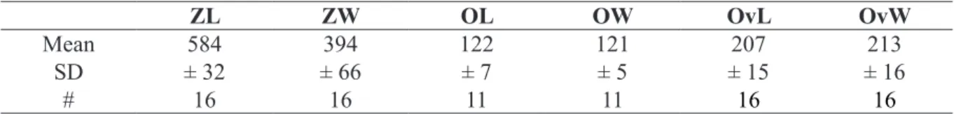
Table 6. Measurements of Atlantisina tricornis gen. et sp. nov. from Galicia Bank population GAL1.
ZL ZW OL OW OvL OvW
Mean 584 394 122 121 207 213
SD ± 32 ± 66 ± 7 ± 5 ± 15 ± 16
Ovicell hyperstomial, ooecium barely resting on frontal shield of distal zooid with its proximal part or entirely free at colony margin, globular, about as long as wide or laterally compressed, with a short tubular peristome terminating at distal apertural margin (Fig. 6B, E–F); ectooecium well developed, covering about two-thirds or more of entire ooecium; exposed endooecium of variable size and shape, either pear-shaped and narrowing on peristome when large, or forming a broad central strip when ooecium is compressed, surface topography similar to that of frontal shield but with smaller and marginally elongate depressions and steeper ridges; ooecial aperture about as tall as wide, acleithral.
Ancestrula oval (ca 350 µm long, 230 µm wide), smooth gymnocyst narrow all around, cryptocyst virtually absent; opesia large, oval to pyriform (ca 235 µm long, 145 µm wide), slightly constricted in distal fourth; nine spines, with five proximal ones widely spaced and four distal ones situated closer together (Figs 1E, 6A).
Remarks
Atlantisina tricornis gen. et sp. nov. is the only species of Atlantisina gen. nov. that was recorded from
the continental slope while all other species occur on seamounts and near islands. Moreover, it is the only species of Atlantisina gen. nov. that has previously been recorded and figured (d’Hondt 1974; Souto et al. 2016).
Atlantisina tricornis gen. et sp. nov. is clearly distinguished from all other congenerics by its thickly
calcified, suboral crest with three pointed mucrones. However, there is some variability in mucro shape and orientation between colonies from the same area, as well as between populations from the three
Table 7. Morphometric comparison between different populations of Atlantisina tricornis gen. et
sp. nov., from the N Iberian slope (NIS), two from Galicia Bank (GAL1, GAL2), and from the W Iberian slope (WIS). All data combined are given under ALL. The measurements are given as the mean ± standard deviation and the number of measurements taken (in parentheses). The subsequent letter (in bold) represents the results of the intergroup comparison of means using a post hoc Tukey’s test; groups not connected by the same letter are significantly different, those with two letters (ab) are not significantly different from a or b. See text for further information and the Material section for the remaining abbreviations.
NIS GAL1 GAL2 WIS ALL
ZL 614 ± 37 584 ± 32 615 ± 50 645 ± 64 616 ± 52 (14) ab (16) b (29) ab (20) a (79) ZW 406 ± 48 394 ± 66 398 ± 38 476 ± 82 419 ± 67 (14) b (16) b (28) b (20) a (79) OL 135 ± 6 122 ± 7 139 ± 9 156 ± 9 138 ± 13 (13) b (11) c (25) b (11) a (60) OW 124 ± 5 121 ± 5 139 ± 7 138 ± 9 132 ± 10 (18) b (11) b (25) a (11) a (65) OvL 226 ± 18 207 ± 15 203 ± 15 233 ± 18 217 ± 20 (16) a (16) b (9) b (8) a (49) OvW 172 ± 16 213 ± 16 177 ± 8 204 ± 17 191 ± 23 (16) b (16) a (9) b (8) a (49) OvL/W 1.32 ± 0.14 0.98 ± 0.08 1.15 ± 0.09 1.15 ± 0.1 1.13 ± 0.17 (16) a (16) c (9) b (8) b (49)
sampled regions, the northern Iberian slope, Galicia Bank and the western Iberian slope. For instance, in the Galicia Bank population the three mucrones are usually single, unbranched, straight, and vertically oriented (Fig. 6E), whereas in some zooids the lateral mucrones may be slightly curved outwards and branched, and the central mucro may be twinned. Branching of mucrones was rather frequent in colonies from the continental shelf off northern Portugal (W Iberian slope; Fig. 6F), while in the colonies from the N Iberian slope the lateral mucrones were constantly unbranched and diverging slightly outwards (Fig. 6A–B, D). However, in some colonies from this region, a secondary acuminate tip may occur laterally on the central mucro. Thus, although the end-members of the mucro-morphotypes are distinctly different, a clear distinction between regions cannot be drawn, as intermediate stages are present. A certain variability was also detected in the development of the ooecium, whose shape ranges from globular with a large suborbicular area of exposed endooecium (Fig. 6E) to laterally compressed ones in which the endooecium is reduced to a narrow central area (Figs 6B, F). Consequently, ooecium width and the length-width ratio (OvL/OvW) may differ between colonies. In order to assess the morphological variability between colonies and regions, one-way ANOVA was were performed on the original length- and width-measurements of zooids, orifices and ooecia. As morphological differences were optically noticeable between colonies occurring in Galicia Bank, the populations were divided into four area-groups, the N Iberian slope (NIS), Galicia Bank 1 (GAL1), Galicia Bank 2 (GAL2), and W Iberian slope (WIS). Statistical analyses of ooecium width data show that, while the mean values are similar within two area-pairs, ooecium width is significantly higher in WIS and GAL1 than in NIS and GAL2 (F = 24.35, p < 0.001; see Table 7). Mean values of ooecium length/width ratio of GAL1 (0.98) are significantly different from NIS (1.32), while similar intermediate values (1.15) are observed in WIS and GAL2 colonies. Although distinct differences in size of autozooids and orifices can be perceived between colonies from the four sampled areas (Table 7), statistical analyses do not show a common hierarchy in the four populations when considering the different dimensions (ZL, ZW, OL, OW). Thus, concerning both morphology and morphometry, there is no clear distinction between the populations of the three geographic areas that would allow separating them at the species level.
With their bifid tips and twinned central mucrones (Fig. 6F), some of the zooids in the Portuguese morphotype of A. tricornis gen. et sp. nov. somewhat resemble Atlantisina lionensis gen. et sp. nov. from Lion and Seine seamounts (see below). However, in the latter the mucrones are not as thickly calcified, are positioned on a distinctly raised and relatively straight crest, and the lateral mucrones point distally. Calcification of the frontal shield’s surface in A. tricornis gen. et sp. nov. and the other new species introduced below is the exact opposite of that of A. atlantis gen. et sp. nov., A. meteor gen. et sp. nov. and A. seinensis gen. et sp. nov. described above. With the reticulate pattern of ridges delimiting round to hexagonal depressions in the former group (Fig. 6B), the precipitation of carbonate seems to be the negative blueprint of the frontally flattened, round to polygonal nodules bounded by grooves in the latter group.
Ecology
Atlantisina tricornis gen. et sp. nov. has been found at depths between 450 and 1040 m on the continental
slope, and between 675 and 1700 m on Galicia Bank (see also Souto et al. 2016: table 22, listed as “Species indet.”). The species forms small patches and bi- to triserial colonies that encrust rocks and biogenic substrata, mainly coral skeletons but also brachiopods, balanid plates and cidarid spines.
Distribution
Atlantisina tricornis gen. et sp. nov. was recovered from the continental slope of northern to western
Atlantisina lionensis gen. et sp. nov.
urn:lsid:zoobank.org:act:54D46F97-6F57-41D2-9417-51DA064B3DF5
Figs 1A, 7A–D, 8, Table 8
Diagnosis
Frontal shield with a reticulate pattern of raised ridges around polygonal depressions; orifice suborbicular, condyles short and without thickened tip; suboral region with a broad band of gymnocystal calcification forming a tall, relatively planar or slightly arched crest carrying three to five pointed mucrones of variable size and shape, the central ones shorter and vertically oriented, the two lateral ones longer and directing distolaterally; lateral walls moderately well developed, septular pores transversely oval to very elongate, area surrounding the pores reduced to absent, distal pore comparatively large, suborbicular, slightly raised relative to lateral ones; orifice margin with six oral spines. Ooecium slightly longer than wide; ectooecium relatively broad, covering about two-thirds of ooecium; exposed endooecium relatively small, imperforate, surface topography similar to that of frontal shield but with elongated depressions and steeper ridges; ooecial peristome relatively short. Ancestrula with 11 spines (four oral, seven mural).
Etymology
Named after its type locality, Lion Smt.
Material examined Holotype
LION SMT: the ovicellate colony marked ‘H’, plus 3 smaller colonies, on a pebble of volcanic rock, Stn 36 (MNHN-IB-2014-66).
Paratypes
LION SMT: 1 coated colony, Stn 36 IB-2014-67); 2 colonies on a pebble, Stn 36 (MNHN-IB-2014-68); 7 colonies and 1 ancestrula on a pebble, Stn 36 (MNHN-IB-2014-69).
Other material examined
LION SMT: ca 20 colonies on rocks, Stn 36 (unregistered MNHN material); 10 colonies on two pebbles, Stn 36 (OLL 2016/146).
SEINE SMT: 1 colony on limestone, Stn 43 (unregistered MNHN material).
Description
Colony encrusting, unilaminar, forming small patches and/or bi- to triserial ribbons (Fig. 7A). Zooecia roughly oval, with tapering proximal end(s) wedged in between proximal zooecia, separated by deep grooves. Frontal shield convex, surface with a reticulate pattern of raised ridges around polygonal depressions, imperforate except for a few small (although comparatively conspicuous) marginal pores faintly visible in frontal view (Fig. 1A); a tall, broad and moderately curved or planar suboral crest is formed predominantly by smooth gymnocystal calcification, sloping distolaterally and abutting proximal pair of spines, generally with two long, pointed lateral mucrones directing distolaterally that may occasionally bear tiny secondary mucrones, and one to three smaller central mucrones pointing vertically (Figs 1A, 7B–D); lateral walls moderately well developed laterally, more extensive in distal part, lateral septular pores transversely oval to extremely elongate, area surrounding pores reduced to absent (Figs 1A, 7B–D); distal pore comparatively large, suborbicular and slightly raised relative to lateral ones.
Orifice orbicular, widest at about mid-distance, proximal border fairly straight to slightly concave, proximal third delimited by a pair of short, blunt condyles, tips usually not thickened (Fig. 7B);
Table 8. Measurements of Atlantisina lionensis gen. et sp. nov.
ZL ZW OL OW OvL OvW
Mean 565 382 114 125 196 172
SD ± 78 ± 57 ± 8 ± 9 ± 20 ± 26
# 30 35 14 14 22 22
Fig. 7. Atlantisina lionensis gen. et sp. nov., Lion Smt, paratype (MNHN-IB-2014-67). A. Colony
overview. B. Orifice and slightly damaged ooecium. C. Ovicellate zooids at the colony growth margin.
distolateral margins with six thick spines arranged in two groups of three with a distinct distal gap; all six spines present in ovicellate zooids with distal pair abutting proximolateral ooecial wall and flanking ooecial aperture on both sides.
Ovicell hyperstomial, ooecium barely resting on frontal shield of distal zooid, globular, slightly longer than wide, with a very short tubular peristome opening at distal orifice margin (Figs 1A, 7B–D); ectooecium relatively broad, covering about two-thirds of ooecium; exposed endooecium relatively small, surface topography similar to that of frontal shield but with steeper ridges delimiting smaller elongated concavities; ooecial aperture slightly taller than wide, acleithral.
Only ancestrula observed has 11 long and thin spines with four oral ones that are oriented vertically and seven mural ones that bend over opesia.
Remarks
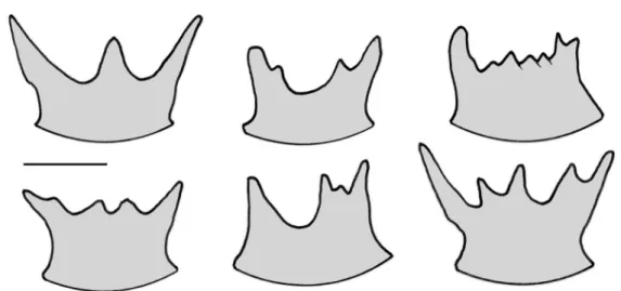
The high morphological plasticity of the suboral crest in Atlantisina lionensis gen. et sp. nov., with a great variability in the number and shape of the mucrones occurring even within the same colony (Fig. 8), is a typical feature of this species. However, with two prominent lateral mucrones pointing in distal directions and shorter intermediate ones, the shape of the suboral crest of A. lionensis gen. et sp. nov. is in general similar to that of A. tricornis gen. et sp. nov., and also to the even larger ones in
A. gorringensis gen. et sp. nov. and A. acantha gen. et sp. nov. (see below).
Ecology
The bi- to triserial colonies of Atlantisina lionensis gen. et sp. nov. were recovered from depths between 320 and 630 m, encrusting small rocks.
Distribution
The species occurs on the Lion and Seine seamounts.
Fig. 8. Atlantisina lionensis gen. et sp. nov. Intraspecific variability in the morphology of the suboral