HAL Id: hal-02667295
https://hal.inrae.fr/hal-02667295
Submitted on 31 May 2020
HAL is a multi-disciplinary open access
archive for the deposit and dissemination of
sci-entific research documents, whether they are
pub-lished or not. The documents may come from
teaching and research institutions in France or
abroad, or from public or private research centers.
L’archive ouverte pluridisciplinaire HAL, est
destinée au dépôt et à la diffusion de documents
scientifiques de niveau recherche, publiés ou non,
émanant des établissements d’enseignement et de
recherche français ou étrangers, des laboratoires
publics ou privés.
Molecular and antigenic evolution and geographical
spread of H5N1 highly pathogenic avian influenza
viruses in western Africa.
Mariette Ducatez, C M Olinger, a A Owoade, Z Tarnagda, M C Tahita, A
Sow, S de Landtsheer, W Ammerlaan, J B Ouedraogo, a D M E Osterhaus, et
al.
To cite this version:
Mariette Ducatez, C M Olinger, a A Owoade, Z Tarnagda, M C Tahita, et al.. Molecular and
antigenic evolution and geographical spread of H5N1 highly pathogenic avian influenza viruses in
western Africa.. Journal of General Virology, Microbiology Society, 2007, 88 (Pt 8), pp.2297-306.
�10.1099/vir.0.82939-0�. �hal-02667295�
Molecular and antigenic evolution and geographical
spread of H5N1 highly pathogenic avian influenza
viruses in western Africa
M. F. Ducatez,
13 C. M. Olinger,
13 A. A. Owoade,
2Z. Tarnagda,
3M. C. Tahita,
3A. Sow,
4S. De Landtsheer,
1W. Ammerlaan,
1J. B. Ouedraogo,
3A. D. M. E. Osterhaus,
5R. A. M. Fouchier
5and C. P. Muller
1Correspondence C. P. Muller
Claude.Muller@LNS.ETAT.LU
1Institute of Immunology, National Public Health Laboratory, 20A rue Auguste Lumie`re, L-1950 Luxembourg, Luxembourg
2Department of Veterinary Medicine, University of Ibadan, Ibadan, Nigeria
3Institut de Recherche en Sciences de la Sante´, 399 Avenue de la liberte´, BP 545 Bobo-Dioulasso, Burkina Faso
4Laboratoire National de l’Elevage, 03 BP 7026 Ouagadougou, Burkina Faso
5Department of Virology, Erasmus Medical Center, Dr Molewaterplein 50, 3015 GE Rotterdam, The Netherlands
Received 21 February 2007 Accepted 21 April 2007
In Africa, highly pathogenic avian influenza H5N1 virus was first detected in northern Nigeria and later also in other regions of the country. Since then, seven other African countries have reported H5N1 infections. This study reports a comparison of full-length genomic sequences of H5N1 isolates from seven chicken farms in Nigeria and chicken and hooded vultures in Burkina Faso with earlier H5N1 outbreaks worldwide. In addition, the antigenicity of Nigerian H5N1 isolates was compared with earlier strains. All African strains clustered within three sublineages denominated A (south-west Nigeria, Niger), B (south-west Nigeria, Egypt, Djibouti) and C (northern Nigeria, Burkina Faso, Sudan, Coˆte d’Ivoire), with distinct nucleotide and amino acid signatures and distinct geographical distributions within Africa. Probable non-African ancestors within the west Asian/Russian/European lineage distinct from the south-east Asian lineages were identified for each sublineage. All reported human cases in Africa were caused by sublineage B. Substitution rates were calculated on the basis of sequences from 11 strains from a single farm in south-west Nigeria. As H5N1 emerged essentially at the same time in the north and south-west of Nigeria, the substitution rates confirmed that the virus probably did not spread from the north to the south, given the observed sequence diversity, but that it entered the country via three independent introductions. The strains from Burkina Faso seemed to originate from northern Nigeria. At least two of the sublineages also circulated in Europe in 2006 as seen in Germany, further suggesting that the sublineages had already emerged outside of Africa and seemed to have followed the east African/west Asian and Black Sea/Mediterranean flyways of migratory birds.
INTRODUCTION
As the avian influenza H5N1 virus swept from Asia through Russia to Europe, Africa seemed to have been spared until January 2006. At that time, commercial poultry farms in
Kaduna State in northern Nigeria (Fig. 1) reported high mortality, which initially was attributed to Newcastle disease. However, on 7 February 2006, the Food and Agricultural Organization reported that the highly patho-genic avian influenza (HPAI) H5N1 virus was the cause of havoc in these farms. Poultry farming is reportedly one of the most important industries in the country, second only to oil production, providing both high-quality animal protein for human consumption and secure jobs for an impoverished population. Many of the large commercial 3These authors contributed equally to this work.
The GenBank/EMBL/DDBJ accession numbers for the sequences reported in this paper are AM262524–AM262578 and AM502998– AM503074.
Journal of General Virology (2007), 88, 2297–2306 DOI 10.1099/vir.0.82939-0
poultry farms maintain a high biosecurity level, but in other large farms and most small farms biosecurity is inadequate. In addition, backyard rearing of chicken and live-poultry markets are common throughout most of Nigeria. The main hub of the poultry industry is located in the small south-western states of the country (Oyo, Osun, Ogun and Lagos).
In a seroepidemiology study that we conducted between 2002 and May 2004 in 65 south-west Nigerian commercial chicken farms, approximately 1000 birds were tested for antibodies against six viruses including avian influenza virus (AIV). Although seroprevalences were very high for most viruses (84 % for infectious bronchitis virus, 41 % for reovirus and 40 % for avian metapneumovirus), no anti-bodies against AIV were confirmed (Owoade et al., 2006). Thus, south-west Nigerian commercial poultry farms seem to have been free of AIV at least until May 2004. Similar studies are not known from other west African countries. Since the first outbreak in Nigeria, seven other African countries have officially reported HPAI H5N1 to the World Organization for Animal Health (OIE): Niger, Egypt, Cameroon, Burkina Faso, Coˆte d’Ivoire, the Sudan and Djibouti (www.oie.int).
In Burkina Faso, poultry is mainly reared in backyards. Poultry imports arrive from all over the world to west Africa, and biosecurity measures such as quarantine are not applied rigorously. Moreover, migratory birds of both the east African/west Asian flyway and the Black Sea/ Mediterranean flyway (Olsen et al., 2006) seek refuge in a number of important bird areas in west Africa, adding to the risk of introduction of HPAI H5N1 in the region.
Therefore, the origin of the outbreaks and the routes of virus transmission remain unclear.
The aim of this study was to investigate and compare the molecular evolution of HPAI H5N1 in Nigeria, Burkina Faso and throughout Africa, utilizing partial and full-length sequences.
METHODS
Samples.Approximately 700 cloacal swabs were collected from more than 100 commercial farms (cockerels, broilers, pullets, layers and breeders) between January and August 2006, in Oyo, Ogun and Lagos states in south-west Nigeria. Flock sizes ranged from 125 to 20 000 birds and ages ranged from 3 weeks to 2 years. Nigeria does not have an official vaccination programme against HPAI H5N1 in poultry. It is highly unlikely that farms from which H5N1 was detected had been vaccinated. In Burkina Faso, paired tracheal and cloacal swabs were collected between 10 March and 25 June 2006, in the eastern, central and western parts of the country (Fada N’Gourouma, Ouagadougou, Bobo Dioulasso and Sokoroni). Five wild birds (three hooded vultures, one little egret and one western grey plantain-eater), as well as birds in 36 backyards or commercial poultry farms (chickens, ducks, geese, pigeons, turkeys and peafowls), were sampled.
RNA isolation, RT-PCR, sequencing and data analysis.RNA was isolated and RT-PCR and sequencing were carried out as described previously (Ducatez et al., 2006a). Phylogenetic and molecular evolu-tionary analyses were conducted using PAUP version 4.0 beta 10 (Swofford, 2003) with the maximum-likelihood method. The model was calculated withMODELTEST(Posada & Crandall, 1998).
Substitution rates were calculated using theBEASTprogram (http:// evolve.zoo.ox.ac.uk/Beast/) (Drummond et al., 2002, 2006). All estimates incorporated the HKR model of DNA substitution as it Fig. 1. Map of HPAI H5N1 outbreaks in Africa. All African countries that reported HPAI H5N1 outbreaks and where sequences are available are shaded. The locations of the studied Nigerian and Burkina Faso farms are indicated.
was the best supported model in MODELTEST (Posada & Crandall, 1998).
Omega (dN/dS) values correspond to nucleotide substitution rate
ratios of non-synonymous and synonymous mutations. These were calculated for each gene usingPAMLversion 3.14 (Yang, 1997; http:// abacus.gene.ucl.ac.uk/software/paml.html). A test was carried out also with PAMLversion 3.14 to detect individual codons under positive
natural selection (Nielsen & Yang, 1998; Yang et al., 2000). The model (M0–M13) best adapted to the dataset was selected with likelihood ratio tests with P,0.01 (Anisimova et al., 2003; Sainudiin et al., 2005; Yang et al., 2000). The Bayes empirical Bayes method was preferred to the naive empirical Bayes method (Yang et al., 2005). Residues with positive natural selection positions were selected only if a,0.05.
Virus isolation and characterization.Virus isolates were obtained by inoculation of embryonated chicken eggs or Madin–Darby canine kidney cells, as described previously (Ducatez et al., 2006a). Antigenic properties of H5N1 virus strains were compared by haemagglutination inhibition using post-infection ferret or duck antisera generated against the following influenza viruses: A/tern/ South Africa/1961 (H5N3) (TE/SA), A/duck/Hong Kong/205/1977 (H5N3) (DK/HK), A/Hong Kong/156/1997 (H5N1) (HK/97), A/ mallard/Netherlands/3/1999 (H5N2) (MA/NL), A/mallard/Sweden/ 49/2002 (H5N9) (MA/SE) and A/turkey/Turkey/1/2005 (H5N1) (TY/ TY) (Table 2). Haemagglutination inhibition assays were performed in duplicate. All serum samples were treated overnight with receptor-destroying enzyme at 37uC and subsequently incubated at 56 uC for 1 h. Twofold serial dilutions of antiserum, starting at 1 : 20, were tested for their ability to inhibit the agglutination of turkey red blood cells using 4 haemagglutinating units of influenza A virus.
RESULTS
H5N1-positive birds
Of the 700 samples collected by the Nigerian–Luxembourg Poultry Virus Network, 35 were positive in a generic real-time influenza A virus RT-PCR assay specific for the matrix (M) gene and an H5-specific real-time RT-PCR assay. In Burkina Faso, out of 206 collected samples, five were positive in both the AIV M gene and H5-specific PCRs. Virus isolates from Nigeria were recovered from animals of 15 flocks and six farms in the south-west of Nigeria (Fig. 1). Increased morbidity was reported in the SO layer farm from mid-January and mortality eventually reached 100 % in the different flocks (mean±SDmortality of 77±33 %). Chickens were 29–79 weeks of age at the time of sample collection and no obvious correlation between flock age and mortality was observed. At necropsy, the chickens showed pinpoint haemorrhages on peritoneal and pericar-dial fat, congested lungs and greenish faecal pasting of the vent, as well as subcapsular hepatic blood clots. In farms in the south-west, layers of 35–45 weeks of age were found to be infected by the H5N1 virus.
In Burkina Faso, H5-positive strains originated from three adult chickens from two backyards with 100 % mortality in Bobo-Dioulasso and in Sokoroni (in the western part of the country, Fig. 1). H5-positive adult hooded vultures (Falconidae, Accipitridae, Aegyptiinae,
Necrosyrtes monachus) collected in Ouagadougou suffered from paralysis, dyspnoea, stiff neck, asthenia and locomo-tion problems (Ducatez et al., 2007).
Viral sequence analyses
Sequencing of haemagglutinin (HA) and neuraminidase (NA) gene segments revealed in all cases that the virus was of the H5N1 subtype. The HA cleavage site sequence PQGERRRKKRG was identical to the cleavage site of the recent HPAI strains in Europe, Russia, central Asia and Africa, but differed by one to three amino acids from south-east Asian HPAI strains. The HA cleavage site of the west African strains contained only one basic amino acid less than the lineages in south-east China, Vietnam and Cambodia (PQRERRRKKRG) and Thailand (PQREKRRKKRG).
Full-length genomes of four strains from the Nigerian SO farm (A/chicken/Nigeria/SO300/2006, -SO452/2006, -SO493/2006 and -SO494/2006), two strains from the Nigerian BA farm (A/chicken/Nigeria/BA210/2006 and -BA211/2006), A/chicken/Nigeria/IF10/2006, A/chicken/ Nigeria/FA7/2006, A/chicken/Nigeria/AB14/2006, A/ chicken/Nigeria/OD9/2006, A/chicken/Burkina Faso/13.1/ 2006 and A/hooded vulture/Burkina Faso/1-2/2006 were compared with full-length sequences available from GenBank. Only sequences from 2005/2006 were closely related and are included in Figs 2 and 3. In addition, partial genomic sequences were obtained from seven birds (seven HA, four M, three NA, five NS, three NP, five PA, four PB1 and two PB2 sequences). These sequences were used in the substitutions analysis, for reassortment evaluation and for substitution rate calculations; but they were not included in the phylogenetic analysis.
Phylogenetic analysis of the HA (Fig. 2) and full genome (Fig. 3) of HPAI H5N1 strains showed that, with the exception of the SO farm, strains from all of the farms in the south-west of Nigeria, A/chicken/Nigeria/1047-8/2006, -10/2006, -30/2006, -34/2006, -54/2006 and A/ostrich/ Nigeria/1047-25/2006, as well as the strains from Niger, were genetically very close to each other. These were collectively referred to as sublineage A strains. The strains from sublineage A shared five specific mutations in HA (168G,237A,1233C,1431A and1587C, all synonymous; num-bering according to Claas et al., 1998 and Ha et al., 2001) and 45 in the complete genome that were not found in the other sublineages. Two additional sublineages emerging from a common node were found in the SO farm (sub-lineage B) and in northern Nigeria (sub(sub-lineage C) (Figs 2 and 3). Sublineage B contained all strains from the SO farm in south-western Nigeria, as well as all Egyptian strains (e.g. A/duck/Egypt/2253-3/2006) and shared six [468A, 633T,
699
T,852G,990T and1658T (SAF), the latter being the only non-synonymous mutation] and 26 specific nucleotides in the HA and full genome, respectively. Sublineage C contained A/chicken/Nigeria/641/2006 and all strains from Burkina Faso, Coˆte d’Ivoire and the Sudan available in
HPAI H5N1 in western Africa
GenBank and was characterized by two (405G and615A) and 13 shared specific nucleotides in the HA and the full genome, respectively.
The phylogenetic analysis also confirmed that the strains from west Africa were more closely related to the central Asian strains than to each other. Sublineages A, B and C were most closely related to strains from Astrakhan, Slovenia and Kurgan, respectively (Figs 2 and 3). For the HA, sequences available from Germany clustered in two of these distinct sublineages: for instance, A/swan/Germany/ R65/2006 in sublineage A and A/common bussard/Bavaria/
2/2006 in sublineage B. Phylogenetic analyses of all gene segments were carried out individually and phylogenetic trees were similar whichever segment was analysed, excluding any reassortment in the newest strains presented here (Fig. 2 for HA; data not shown for other gene segments).
Substitution rates and selection pressure
The 26 available full-length sequences of African HPAI H5N1 were compared with the most likely common
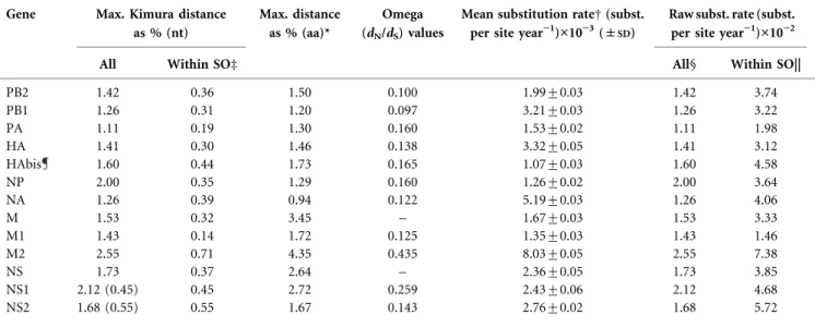
Table 1. Diversity percentages, selection pressure and substitution rates analyses for 26 fully sequenced African 2006 HPAI H5N1 strains
The 26 sequences compared for all the gene segments were as follows: A/chicken/Nigeria/BA210/2006, A/chicken/Nigeria/BA211/2006, A/chicken/ Nigeria/SO300/2006, A/chicken/Nigeria/SO452/2006, A/chicken/Nigeria/SO493/2006, A/chicken/Nigeria/SO494/2006, A/chicken/Burkina Faso/ 13.1/2006, A/hooded vulture/Burkina Faso/2/2006, A/chicken/Nigeria/641/2006, A/chicken/Nigeria/AB14/2006, A/chicken/Nigeria/FA7/2006, A/ chicken/Nigeria/OD9/2006, A/chicken/Nigeria/IF10/2006, A/duck/Niger/914/2006, A/chicken/Coˆte d’Ivoire/1787-34/2006, A/duck/Coˆte d’Ivoire/ 1787-18/2006, A/chicken/Nigeria/1047-34/2006, A/chicken/Nigeria/1047-30/2006, A/chicken/Nigeria/1047-62/2006, A/chicken/Nigeria/1047-54/ 2006, A/ostrich/Nigeria/1047-25/2006, A/chicken/Nigeria/1047-8/2006, A/duck/Egypt/2253-3/2006, A/chicken/Sudan/1784-10/2006, A/chicken/ Nigeria/957-20/2006 and A/chicken/Sudan/1784-7/2006. A/Bar-headed goose/Qinghai/65/2005 was randomly selected as a common ancestor of African HPAI H5N1 strains among 2005 Qinghai Lake strains for substitution rates calculations. It was assumed A/Bar-headed goose/Qinghai/65/ 2005 was 1 year older than any African strain.
Gene Max. Kimura distance as % (nt)
Max. distance as % (aa)*
Omega (dN/dS) values
Mean substitution rateD (subst. per site year”1)¾10”3(±
SD)
Raw subst. rate (subst. per site year”1)¾10”2
All Within SOd All§ Within SO||
PB2 1.42 0.36 1.50 0.100 1.99±0.03 1.42 3.74 PB1 1.26 0.31 1.20 0.097 3.21±0.03 1.26 3.22 PA 1.11 0.19 1.30 0.160 1.53±0.02 1.11 1.98 HA 1.41 0.30 1.46 0.138 3.32±0.05 1.41 3.12 HAbis 1.60 0.44 1.73 0.165 1.07±0.03 1.60 4.58 NP 2.00 0.35 1.29 0.160 1.26±0.02 2.00 3.64 NA 1.26 0.39 0.94 0.122 5.19±0.03 1.26 4.06 M 1.53 0.32 3.45 – 1.67±0.03 1.53 3.33 M1 1.43 0.14 1.72 0.125 1.35±0.03 1.43 1.46 M2 2.55 0.71 4.35 0.435 8.03±0.05 2.55 7.38 NS 1.73 0.37 2.64 – 2.36±0.05 1.73 3.85 NS1 2.12 (0.45) 0.45 2.72 0.259 2.43±0.06 2.12 4.68 NS2 1.68 (0.55) 0.55 1.67 0.143 2.76±0.02 1.68 5.72
*Amino acid distance was calculated with Poisson correction. DCalculated usingBEAST.
dA/chicken/Nigeria/SO300/2006, A/chicken/Nigeria/SO452/2006, A/chicken/Nigeria/SO493/2006 and A/chicken/Nigeria/SO494/2006. §Substitution rates calculated on the basis of Kimura distances with 1 year to ancestor.
||Substitution rates calculated on the basis of Kimura distances with 5 weeks to ancestor (as the start of outbreak within the SO farm was 5 weeks before sampling).
Analysis was made with all 53 African 2006 HPAI H5N1 HA sequences available on GenBank as of 26 October 2006, i.e. with the 26 previously listed strains and the 27 following additional sequences: A/chicken/Egypt/5612NAMRU3-S/2006, A/turkey/Egypt/5613NAMRU3-T/2006, A/hooded vulture/Burkina Faso/1/2006, A/chicken/Burkina Faso/01.03/2006, A/chicken/Egypt/5611NAMRU3-AN/2006, A/chicken/Niger/2130-7/2006, A/ chicken/Egypt/5610NAMRU3-F3/2006, A/chicken/Niger/2130-8/2006, A/chicken/Nigeria/BA209/2006, A/chicken/Nigeria/SO227/2006, A/chicken/ Nigeria/SO294/2006, A/chicken/Nigeria/SO457/2006, A/chicken/Nigeria/SO461/2006, A/chicken/Nigeria/SO470/2006, A/chicken/Nigeria/SO478/ 2006, A/chicken/Nigeria/SO485/2006, A/chicken/Sudan/1784/2006, A/chicken/Egypt/2253-1/2006, A/chicken/Sudan/2115-9/2006, A/chicken/ Sudan/2115-12/2006, A/chicken/Coˆte d’Ivoire/1787/2006, A/chicken/Egypt/960N3-004/2006, A/chicken/Nigeria/FA6/2006, A/chicken/Nigeria/ AB13/2006, A/chicken/Nigeria/FA4/2006, A/chicken/Nigeria/OD8/2006 and A/Egypt/12374/NAMRU3/2006. For calculations within the SO farm, 11 HA sequences were used.
ancestor, A/Bar-headed goose/Qinghai/65/2005, to calcu-late substitution rates (Table 1). African outbreaks from which sequences were available occurred within 7 months and on average 1 year after the sampling of A/Bar-headed goose/Qinghai/65/2005. The mean substitution rates per gene segment ranged from 5.261024 (for NA) to 3.36 1023 (for HA) substitutions per site per year. The exact sequence of the strain that initially entered the SO farm and evolved in this farm during the 5 weeks was unknown. A second method was therefore used to calculate substitu-tion rates without the ancestor sequence and values were compared. The virus substitution rate within 5 weeks in the Nigerian SO farm was slightly higher than the virus substitution rate calculated using the likely ancestor of all African strains (A/Bar-headed goose/Qinghai/65/2005) (Table 1).
In Africa, Europe and Russia, most H5N1 genes were under purifying selection with v values (dN/dS) of 0.097 (for PB1)
to 0.435 (for M2) (Table 1).
Antigenic variation
The antigenic variation of the influenza viruses from sublineage B (SO farm) and sublineage A (BA farm) was in agreement with the genetic data for HA (Table 2). In haemagglutination inhibition assays using post-infection ferret and duck antisera, the two virus isolates from the BA
farm were quite similar to one another and slightly differ-ent from the three virus isolates obtained from the SO farm, in particular when measured with antisera raised against A/turkey/Turkey/1/2005. All five Nigerian virus isolates tested in the haemagglutination inhibition assay were antigenically most similar to influenza virus A/turkey/ Turkey/1/2005, a WHO-recommended vaccine strain. The Nigerian strains were antigenically distinct from A/Hong Kong/156/1997 and revealed antigenic differences to strains isolated previously from Vietnam and Indonesia, as measured with ferret antisera to classical H5 strains and the teal antiserum raised against A/turkey/Turkey/1/2005.
DISCUSSION
Sublineages of HPAI H5N1 in Africa on the basis of full genome sequencing
Detailed phylogenetic analysis of each of the eight gene segments and the full-length genome revealed three African sublineages with a distinct nucleotide and amino acid signature. Although two of the three sublineages, B and C, emerged from a common node, their high Kimura distance and their distinct geographical distribution within Africa suggest three rather than two distinct sublineages. Subline-age A seems to have emerged from Astrakhan 2005 and was originally found in the BA farm in south-western Nigeria
Table 2. Antigenic characterization of influenza A virus isolates using a haemagglutination inhibition assay with ferret and duck post-infection antisera and turkey erythrocytes
Homologous titres are shown in bold. Abbreviated serum names are given in Methods.
Virus Antisera TE/SA* (H5N3) DK/HK* (H5N3) MA/NL* (H5N2) MA/SE* (H5N9) HK/97* (H5N1) TY/TYD (H5N1) TY/TYd (H5N1) Tern/SouthAfrica/63 480 1280 20 80 ,20 320 ,20 Duck/Hongkong/205/77 480 1920 ,20 ,20 ,20 160 ,20 Mallard/Netherl/3/99 640 2560 60 160 ,20 640 ,20 Mallard/Sweden/49/02 320 2560 40 120 ,20 480 ,20 Hong Kong/156/97 1280 2560 160 320 160 1280 20 Vietnam/1194/04 ,20 40 ,20 ,20 ,20 320 ,20 Vietnam/1203/04 ,20 40 ,20 ,20 ,20 320 ,20 Indonesia/5/05 ,20 ,20 ,20 ,20 ,20 320 160 Turkey/Turkey/1/05 20 60 ,20 ,20 ,20 320 240 Nigeria/BA209§ 80 320 ,20 ,20 ,20 1280 640 Nigeria/BA210 80 240 ,20 ,20 ,20 640 480 Nigeria/SO300 40 160 ,20 ,20 ,20 320 160 Nigeria/SO452 20 40 ,20 ,20 ,20 240 160 Nigeria/SO493 160 120 ,20 ,20 ,20 320 320
*Antiserum from a ferret. DAntiserum from a mallard. dAntiserum from a common teal.
§A/chicken/Nigeria/BA209/2006 was not included in the phylogenetic analysis (Figs 2 and 3) because its full genome sequence could not be obtained.
HPAI H5N1 in western Africa
from where it spread more recently to several other farms in the same part of the country and to Niger. This sub-lineage might be even more closely related to swans from Poland and Germany and a buzzard from Denmark (0.3 % diversity in the HA gene; Dr Ian Brown, personal com-munication). In Africa, sublineage B was found in a single farm in the south-west of Nigeria and is the only sublineage reported from Egypt and Djibouti. Its ancestor is less clear, as the different genes have mixed characteristics from European, Russian and Mongolian strains. Interestingly, all reported human cases in Africa (Egypt and Djibouti) have been caused by viruses of this sublineage. The African sublineage C was found in northern Nigeria (A/chicken/ Nigeria/641/2006) and all strains reported from the Sudan and Coˆte d’Ivoire belong to this sublineage. It is closely related to A/chicken/Kurgan/3/2005, but we cannot
exclude a similar or higher homology with unpublished sequences from strains circulating elsewhere. Sublineages B and C seem to have emerged from a more recent common ancestor than the Qinghai Lake 2005 strains and it cannot be excluded that this common ancestor was circulating in Africa.
In the new strains reported here, no evidence of reassort-ment was found, in contrast to A/chicken/Nigeria/1047-62/ 2006 where HA, NP, NS and PB1 gene segments clustered with sublineage C strains and NA, M, PA and PB2 clustered with sublineage A strains (Salzberg et al., 2007). Human cases reported so far in Africa (Egypt and Djibouti) have all been caused by sublineage B, but it would be dangerous to infer a lower infection potential of sublineages A and C strains for humans.
Fig. 2. Phylogeny of the HA gene of Nigerian and Burkina Faso lineages and closest related strains. Nigerian strains are indicated by X and African, non-Nigerian strains by e. The tree was calculated using the maximum-likelihood method implemented in PAUP 4.0. The substitution model was obtained using
MODELTEST. A/duck/Vietnam/01/2005 was used as the outgroup and is indicated by an arrow.
Substitution rates
HPAI H5N1 substitution rates in Africa were high, reaching on average 2.661023 substitutions per site per year. This is well within the range of substitution rates of avian influenza viruses in general and H5N1 in particular, as described by Chen & Holmes (2006) using the same calculation method. During the 1994 H5N2 outbreak in Mexico, higher substitution rates (0.8861022 to 2.861022substitutions per site per year) were reported (Garcia et al., 1997) but flocks were partially vaccinated. To date, estimates of mutation rates from a single farm are rare. Under the assumption of a single introductory event into the SO farm, a direct ancestral relationship between strains isolated subsequently in the farm and a 5 week evolution in the farm (15 January to 20 March 2006), the
substitution rate of the virus for each gene segment including 11 HA sequences was calculated. The rate of 4.5861022substitutions per site per year (for HA, Table 1) was higher than the substitution rate calculated within Africa using the same calculation method (1.6061022 substitutions per site per year; Table 1). This suggests that HPAI H5N1 mutated faster within a farm with a virtually unlimited supply and high density of susceptible (unvacci-nated) animals than on its way from Qinghai to Africa. Based on the HA substitution rate in the SO farm (subline-age B), it would have taken a minimum of 18 weeks for the south-west Nigerian strains (sublineages A and B) to evolve from a common ancestor and 13–14 weeks for each of the south-western strains to develop from the northern strain (A/chicken/Nigeria/641/2006, sublineage C). As the three outbreaks occurred within a maximum of 8 weeks, the
Fig. 3. Phylogeny of the full-length (concat-enated) genomes of HPAI H5N1 from Nigeria and Burkina Faso and their closest related strains. All relevant full-length sequences in GenBank were included. Nigerian strains are indicated by X and African, non-Nigerian strains by e. The tree was calculated using the maximum-likelihood method implemented in PAUP 4.0. The substitution model was obtained using MODELTEST. A/duck/Vietnam/ 01/2005 was used as the outgroup and is indicated by an arrow.
HPAI H5N1 in western Africa
strains probably did not spread from one farm to another. In particular, as H5N1 emerged essentially at the same time in the north (10 January 2006) and in the SO farm (15 January 2006), these two sublineages probably did not develop within Nigeria. Thus, these data and the fact that the closest pre-Nigerian relatives of each of the sublineages were geographically separated further support the inde-pendent introduction of each of the three H5N1 sub-lineages in Nigeria as suggested previously (Ducatez et al., 2006a), even if the B and C sublineages emerged from a common ancestor. The same analysis for the sublineage A strains from the south-west revealed that the 5 months between the outbreaks was in agreement with the genetic distances between strains, as calculated with Kimura para-meters. The HA substitution rate in the SO farm was also compatible with a single introduction of a sublineage C strain into Burkina Faso. Unexpectedly, but as already observed by Chen & Holmes (2006), the change of host (chicken to hooded vulture) did not seem to influence the mutation rate of the virus.
Nucleotide and amino acid substitutions and biological consequences
As previously discussed for Burkina Faso strains (Ducatez et al., 2007), west African H5N1 strains retained the amino acid pattern associated with preferential binding to a2,3-linked sialic acid (mostly present in avian species):
91
Y, 130GVSS134, 149W, 151I, 179H, 186E, 190LY191 and
220
NGQSGR225 (Ha et al., 2001; Shinya et al., 2004). Mutations towards increased binding to human receptors (to a2,6-linked sialic acid) did not seem to have occurred in west African H5N1 strains. In contrast to south-east Asian lineages, PB2 segments of west African strains have a lysine instead of a glutamic acid in position 627, lysine corresponding to a more pathogenic H5N1 phenotype with accelerated viral replication, a reduced host defence and higher mortality in mice (Fouchier et al., 2004; Hatta et al., 2001), as well as increased virulence of H7N7 in human (Chen et al., 2006a). Chen et al. (2006a) studied H5N1 viruses in waterfowl in western China in 2005 and observed lysine residues in position 627 for most but not all of the virus isolates. Among the non-south-east Asian strains, some Russian strains, including A/Cygnus olor/Astrakhan/ Ast05-2-4/2005, A/duck/Kurgan/08/2005 and A/chicken/ Tula/Russia/Oct-5/2005, had a glutamic acid at PB2 position 627, thus also co-existing with the lysine as found in western China. Strains from Russia and western China seem to represent intermediates between the south-east Asian strains with only glutamic acid and the African strains with only lysine.
None of the west African H5N1 strains showed a signatory amino acid pattern associated with resistance to oseltamivir or to amantadine.274H was indeed present in all Nigerian and Burkina Faso NA sequences, whilst 274Y has been observed in oseltamivir-resistant patients in Vietnam (de Jong et al., 2005). Moreover, all west African strains
had a27V/I,30A,31S and34G pattern in M2 and none of the amantadine-resistance markers (27V, 30A, 31S and 34D) (Scholtissek et al., 1998).
Unlike the suggestion by Chen et al. (2006b),99I and268N in HA as well as111R in NA are not always associated with wild birds, as shown by new sequences from domestic poultry in Africa, Europe and Russia. All Nigerian and Burkina Faso H5N1 strains from chickens had these three amino acids, whilst A/hooded vulture/Burkina Faso/1/2006 had268Y instead of the predicted 268N.
In all of the available NS gene sequences from Africa, Europe and Russia, a563GAC substitution was observed, which may correspond to a change in RNA conformation as described by Gultyaev et al. (2007) for the recent H5N1 strains. Thus, the NS gene of the African HPAI H5N1 2006 viruses would be likely to show a hairpin rather than a pseudoknot conformation towards the 39 extremity, which may play a role in mRNA splicing regulation (Gultyaev et al., 2007).
Selection pressure
In Africa, Europe and Russia, most H5N1 genes are under purifying selection with 0.10,v,0.43 (Table 1), suggest-ing that a non-synonymous mutation has only 10–43 % as much chance as a synonymous mutation of being fixed in the population. Only the PB1-F2 gene seems to be under positive natural selection (v58.476, data not shown), but, as discussed by Holmes et al. (2006), this is probably due to the overlap of the PB1 and PB1-F2 ORFs (a shift of 1 nt compared with PB1 ORF) and therefore represents an artefact. In south-east Asian 2002–2005 genotype Z H5N1 strains, M2 was also under positive natural selection, which was not the case in the 26 African strains analysed here or in the available strains from western Asia, Russia and Europe (data not shown). No positively selected codons were detected here in HA, unlike observations in south-eastern strains (Smith et al., 2006).
Our data indicate that the geographical origin of west African HPAI H5N1 was not south-east Asia but the central Asian/European region. The three sublineages A, B and C already existed prior to the arrival of HPAI H5N1 in Africa. They have continued to circulate and to evolve both in Africa and in Europe, e.g. in Germany. Although the relative roles of bird migration and the poultry trade in the spread of recent H5N1 remain a matter for debate, the present study contributes to a better understanding of the evolution and geographical spread of HPAI H5N1 in Africa.
H5N1 virus has been reported from 14 of the 31 Federal Nigerian States and has caused at least five outbreaks in poultry and one outbreak in wild birds in Burkina Faso. It is difficult to see how the virus can be contained without depopulation combined with surveillance and large-scale vaccination. Despite the high mortality caused by H5N1, surveillance is complicated by uncharacteristic organ
lesions at necropsy and multiple viral and bacterial co-infections (Ducatez et al., 2006b; Igbokwe et al., 1996; Owoade et al., 2006, 2004a, b), many of which are associated with significant levels of mortality. Reserva-tions of the developed countries towards vaccination against H5N1 delay crucial decisions and prevent rapid action in Africa. Socio-economic and political hurdles further compromise control measures, whilst the virus continues to spread and threatens to become endemic.
NOTE ADDED IN PROOF
Ghana reported H5N1 for the first time on 28 April 2007. This is not included in Fig. 1.
ACKNOWLEDGEMENTS
The authors gratefully acknowledge the generous financial and moral support of the Ministry of Cooperation, the Ministry of Health and the Ministry of Research of Luxembourg. The support of FAO and WHO is also appreciated. The authors thank the Ministry of Agriculture of Oyo State, Dr Oyewola, the President of Oyo state branch of the Nigerian Veterinary Medical Association, and Dr Oriade, the head of the Poultry unit of Oyo state veterinary Hospital Mokola, Ibadan, Dr Hassan, President of the National Farmers Association, and Dr Bola Oyefolu, Department of Microbiology, Faculty of Science, Lagos State University (LASU), for great support and their enthusiasm in organizing the sample collection. We also acknowledge the hospitality of the University of Ibadan (UI) and the support of the Vice-Chancellor Professor Bamiro and the Deputy Vice-Chancellors Professors Odejide and Ogunkunle, the Dean of the Faculty of Veterinary Medicine, Professor Ogundipe and the Head of the Department of Veterinary Medicine. Technical help and advice from Judith Guldemeester, Pascal Lexmond, Chantal Baas, Theo Bestebroer, Erasmus University Rotterdam; Jacques Kremer and Aure´lie Sausy, National Public Health Laboratory, Luxembourg; and Issaka Yougbare and Ade`le Kam Traore, Institut de Recherche en Sciences de la Sante´, Bobo-Dioulasso, were highly appreciated. C. M. O. and M. F. D. were supported by a BFR fellowship of the Ministry of Research and Higher Education, Luxembourg. The study is part of COST B28 activities. Finally, we thank the participating farmers and individuals in Nigeria and in Burkina Faso for the collection of samples, their patience and their courage in a difficult time. The authors acknowledge (in alphabetical order) A. P. Agafonov, A. Y. Alekseev, T. S. Astakhova, V. M. Blinov, S. I. Braslavskaya, V. V. Drygin, A. G. Durymanov, M. P. Grudinin, K. N. Gruzdev, H. Kida, O. I. Kiselev, T. Y. Kondratieva, N. F. Lomakina, O. V. Lyapina, S. V. Netesov, A. G. Prilipov, Y. N. Rassadkin, G. K. Sadykova, Y. Sakoda, A. N. Sergeev, A. M. Shestopalov, G. A. Shipulin, V. A. Ternovoi, S. S. Yamnikova, S. B. Yatsyshina, A. V. Zaikovskaya and S. I. Zolotykh for the otherwise unpublished sequence data made available on GenBank.
REFERENCES
Anisimova, M., Nielsen, R. & Yang, Z. (2003).Effect of recombination on the accuracy of the likelihood method for detecting positive selection at amino acid sites. Genetics 164, 1229–1236.
Chen, R. & Holmes, E. C. (2006).Avian influenza virus exhibits rapid evolutionary dynamics. Mol Biol Evol 23, 2336–2341.
Chen, H., Li, Y., Li, Z., Shi, J., Shinya, K., Deng, G., Qi, Q., Tian, G., Fan, S. & other authors (2006a).Properties and dissemination of H5N1
viruses isolated during an influenza outbreak in migratory waterfowl in western China. J Virol 80, 5976–5983.
Chen, H., Smith, G. J., Li, K. S., Wang, J., Fan, X. H., Rayner, J. M., Vijaykrishna, D., Zhang, J. X., Zhang, L. J. & other authors (2006b).
Establishment of multiple sublineages of H5N1 influenza virus in Asia: implications for pandemic control. Proc Natl Acad Sci U S A 103, 2845–2850.
Claas, E. C., Osterhaus, A. D., van Beek, R., De Jong, J. C., Rimmelzwaan, G. F., Senne, D. A., Krauss, S., Shortridge, K. F. & Webster, R. G. (1998).Human influenza A H5N1 virus related to a highly pathogenic avian influenza virus. Lancet 351, 472–477.
de Jong, M. D., Tran, T. T., Truong, H. K., Vo, M. H., Smith, G. J., Nguyen, V. C., Bach, V. C., Phan, T. Q., Do, Q. H. & other authors (2005). Oseltamivir resistance during treatment of influenza A (H5N1) infection. N Engl J Med 353, 2667–2672.
Drummond, A. J., Nicholls, G. K., Rodrigo, A. G. & Solomon, W. (2002). Estimating mutation parameters, population history and genealogy simultaneously from temporally spaced sequence data. Genetics 161, 1307–1320.
Drummond, A. J., Ho, S. Y., Phillips, M. J. & Rambaut, A. (2006).
Relaxed phylogenetics and dating with confidence. PLoS Biol 4, e88.
Ducatez, M. F., Olinger, C. M., Owoade, A. A., De Landtsheer, S., Ammerlaan, W., Niesters, H. G., Osterhaus, A. D., Fouchier, R. A. & Muller, C. P. (2006a).Avian flu: multiple introductions of H5N1 in Nigeria. Nature 442, 37.
Ducatez, M. F., Owoade, A. A., Abiola, J. O. & Muller, C. P. (2006b).
Molecular epidemiology of chicken anemia virus in Nigeria. Arch Virol 151, 97–111.
Ducatez, M. F., Tarnagda, Z., Tahita, M. C., Sow, A., De Landtsheer, S., Londt, B. Z., Brown, I. H., Osterhaus, A. D. M. E., Fouchier, R. A. M. & other authors (2007).Genetic characterization of HA1 of HPAI H5N1 viruses from poultry and wild vultures in Burkina Faso. Emerg Infect Dis 13, 611–613.
Fouchier, R. A., Schneeberger, P. M., Rozendaal, F. W., Broekman, J. M., Kemink, S. A., Munster, V., Kuiken, T., Rimmelzwaan, G. F., Schutten, M. & other authors (2004). Avian influenza A virus (H7N7) associated with human conjunctivitis and a fatal case of acute respiratory distress syndrome. Proc Natl Acad Sci U S A 101, 1356–1361.
Garcia, M., Suarez, D. L., Crawford, J. M., Latimer, J. W., Slemons, R. D., Swayne, D. E. & Perdue, M. L. (1997).Evolution of H5 subtype avian influenza A viruses in North America. Virus Res 51, 115–124.
Gultyaev, A. P., Heus, H. A. & Olsthoorn, R. C. (2007). An RNA conformational shift in recent H5N1 influenza A viruses. Bioinformatics 23, 272–276.
Ha, Y., Stevens, D. J., Skehel, J. J. & Wiley, D. C. (2001). X-ray structures of H5 avian and H9 swine influenza virus hemagglutinins bound to avian and human receptor analogs. Proc Natl Acad Sci U S A 98, 11181–11186.
Hatta, M., Gao, P., Halfmann, P. & Kawaoka, Y. (2001).Molecular basis for high virulence of Hong Kong H5N1 influenza A viruses. Science 293, 1840–1842.
Holmes, E. C., Lipman, D. J., Zamarin, D. & Yewdell, J. W. (2006).
Comment on ‘Large-scale sequence analysis of avian influenza isolates’. Science 313, 1573 (author reply 1573).
Igbokwe, I. O., Salako, M. A., Rabo, J. S. & Hassan, S. U. (1996).
Outbreak of infectious bursal disease associated with acute septicae-mic colibacillosis in adult prelayer hens. Rev Elev Med Vet Pays Trop 49, 110–113.
Nielsen, R. & Yang, Z. (1998). Likelihood models for detecting positively selected amino acid sites and applications to the HIV-1 envelope gene. Genetics 148, 929–936.
HPAI H5N1 in western Africa
Olsen, B., Munster, V. J., Wallensten, A., Waldenstrom, J., Osterhaus, A. D. & Fouchier, R. A. (2006).Global patterns of influenza A virus in wild birds. Science 312, 384–388.
Owoade, A. A., Mulders, M. N., Kohnen, J., Ammerlaan, W. & Muller, C. P. (2004a).High sequence diversity in infectious bursal disease virus serotype 1 in poultry and turkey suggests West-African origin of very virulent strains. Arch Virol 149, 653–672.
Owoade, A. A., Oluwayelu, D. O., Fagbohun, O. A., Ammerlaan, W., Mulders, M. N. & Muller, C. P. (2004b).Serologic evidence of chicken infectious anemia in commercial chicken flocks in southwest Nigeria. Avian Dis 48, 202–205.
Owoade, A. A., Ducatez, M. F. & Muller, C. P. (2006).Seroprevalence of avian influenza virus, infectious bronchitis virus, reovirus, avian pneumovirus, infectious laryngotracheitis virus and avian leucosis virus in Nigerian poultry. Avian Dis 50, 222–227.
Posada, D. & Crandall, K. A. (1998).MODELTEST: testing the model of DNA substitution. Bioinformatics 14, 817–818.
Sainudiin, R., Wong, W. S., Yogeeswaran, K., Nasrallah, J. B., Yang, Z. & Nielsen, R. (2005). Detecting site-specific physicochemical selective pressures: applications to the Class I HLA of the human major histocompatibility complex and the SRK of the plant sporophytic self-incompatibility system. J Mol Evol 60, 315–326.
Salzberg, S. L., Kingsford, C., Cattoli, G., Spiro, D. J., Janies, D. A., Aly, M. M., Brown, I. H., Couacy-Hymann, E., De Mia, G. M. & other
authors (2007). Genome analysis linking recent European and African influenza (H5N1) viruses. Emerg Infect Dis 13, 713–718.
Scholtissek, C., Quack, G., Klenk, H. D. & Webster, R. G. (1998).
How to overcome resistance of influenza A viruses against adamantane derivatives. Antiviral Res 37, 83–95.
Shinya, K., Hamm, S., Hatta, M., Ito, H., Ito, T. & Kawaoka, Y. (2004).
PB2 amino acid at position 627 affects replicative efficiency, but not cell tropism, of Hong Kong H5N1 influenza A viruses in mice. Virology 320, 258–266.
Smith, G. J., Naipospos, T. S., Nguyen, T. D., de Jong, M. D., Vijaykrishna, D., Usman, T. B., Hassan, S. S., Nguyen, T. V., Dao, T. V. & other authors (2006).Evolution and adaptation of H5N1 influenza virus in avian and human hosts in Indonesia and Vietnam. Virology 350, 258–268.
Swofford, D. L. (2003).PAUP* – Phylogenetic Analysis Using Parsi-mony (*and other methods). Sunderland, MA: Sinauer Associates.
Yang, Z. (1997).PAML: a program package for phylogenetic analysis by maximum likelihood. Comput Appl Biosci 13, 555–556.
Yang, Z., Nielsen, R., Goldman, N. & Pedersen, A. M. (2000). Codon-substitution models for heterogeneous selection pressure at amino acid sites. Genetics 155, 431–449.
Yang, Z., Wong, W. S. W. & Nielsen, R. (2005).Bayes empirical Bayes interference of amino acid sites under positive selection. Mol Biol Evol 22, 1107–1118.