Linkage mapping of benign familial infantile
convulsions (BFIC) to chromosome 19q
Michel Guipponi
1, Frangois Rivier
12, Federico Vigevano
3, Corinne Beck
1,
Arielle Crespel
1, Bernard Echenne
2, Pierpaolo Lucchini
4, Rosella Sebastianelli
3,
Michel Baldy-Moulinier
1and Alain Malafosse
1-
5*
laboratory of Experimental Medicine, CNRS UPR 9008, INSERM U249, Montpellier, France, 2Department of
Neuropediatrics, Gui-de-Chauliac Hospital, Montpellier, France, 3Section of Neurophysiology, Bambino Gesu
Children's Hospital, Rome, Italy, 4General Hospital, Treviso, Italy and 5Division of Neuropsychiatry, University Hospital of Geneva, Geneva, Switzerland
Received November 4, 1996; Revised and Accepted December 11, 1996
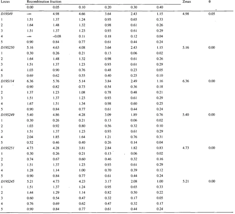
Benign familial infantile convulsions (BFIC) are an autosomal-dominant epileptic syndrome characterized by an age of onset within the first year of life. Although they were first reported in families of Italian descent, BFIC have also been described in non-Italian families. We have mapped the BFIC gene to chromosome 19 by linkage analysis in five Italian families with a maximum two-point lod score of 6.36 at D19S114; maximum multipoint lod scores >8 were obtained for the interval
D19S250-D19S245. BFIC are therefore the third
idiopathic partial epileptic syndrome to be mapped on the human genome.
INTRODUCTION
Epilepsy is one of the most common neurological disorders with a cumulative incidence of - 2 % in the general population by age 40 (1). However, despite intense research, its basic pathogenetic mechanisms remain poorly known. As several lines of evidence suggest an important genetic contribution to the etiology of the epilepsies, gene mapping is potentially an important experimental approach for understanding the molecular basis of these disorders (2). Gene-mapping studies, however, work best when the phenotype can be determined accurately and when the disease gene is highly penetrant (2). Consequently, genetic linkage analysis of epileptic syndromes with a Mendelian mode of inheritance, as a first step in order to clone the responsible genes and define their protein product, is now one of the most promising approaches in identifying the pathogenetic mechanisms underlying epilepsies.
Mendelian epileptic diseases are rare in human. To date only one syndrome with an autosomal-dominant mode of inheritance has been recognized among the idiopathic epilepsies in the International Classification of Epilepsies (3): the benign familial neonatal convulsions (BFNC). Linkage of BFNC to chromosome 20q has been initially reported (EBN1; 4,5) and evidence for
non-allelic genetic heterogeneity has been demonstrated by linkage mapping of a second BFNC locus to chromosome 8q (EBN2; 6). Mapping of the other most common idiopathic epilepsies is difficult because of age-dependent penetrance, spontaneous remission and clinical heterogeneity within the same pedigree. These difficulties have been responsible for the discrep-ancies between the linkage studies of juvenile myoclonic epilepsy (JME) (7).
Benign familial infantile convulsions (BFIC) is a recently recognized idiopathic epileptic syndrome with an autosomal-dominant mode of transmission (8). BFIC was originally described in families of Italian ancestry (8). The disease has also been reported in other Italian (9) and non-Italian families, in France (10,11), Singapore (12), Sweden (Liiovigson, personal communication), Germany (Dr Kurlemann, personal communi-cation) and the United States of America (Dr Ryan, personal communication). BFIC presents with onset between 3.5 and 12 months, seizures of partial type in most cases (according to ictal EEG recordings), no etiological factors, normal psychomotor development, and normal interictal electroencephalograms (EEGs) (8). BFIC is inherited in a manner consistent with an autosomal dominant disorder (8).
To initiate studies that should lead to the identification of the gene defect responsible for BFIC, genetic linkage analysis was performed in five kindreds with this disorder. As a first step, we have used a candidate gene approach in performing linkage analysis using microsatellite polymorphisms flanking the previously mapped epilepsy genes. Due to some similar clinical features between BFNC and BFIC (epileptic syndrome with no etiological factors, normal psychomotor development, normal interictal electroencephalograms, and autosomal dominant mode of inheritance) we first analysed the EBN1 region. This region has been excluded (13) as well as all other candidate regions so far analysed (unpublished data). Consequently, a systematic approach using microsatellite polymorphisms lying in non-excluded regions has been used. We now report that the gene for BFIC maps to the long arm of chromosome 19 in our five pedigrees.
RESULTS
Over 40 microsatellites lying in non-excluded regions were genotyped before linkage was detected. Two-point maximum likelihood calculations were performed after each complete genotyping with one marker. We detected one microsatellite polymorphism giving a maximum two-point lod score of 5.16 at 0 centiMorgan from the marker locus DJ9S250. This prompted us to study chromosome 19q with additional markers. Based on their position reported by Weber et al. (14), five other microsatellite markers (DJ9S49, D19S1J4, D19S249, D19S25I and D19S245) on chromosome 19 were studied. The results of two-point maximum likelihood calculations between the disease
phenotype and each of the marker loci are shown in Table 1, and the pedigree genotyping data for these markers are summarized in Figure 1. Haplotype analysis (Figs 1 and 2), two-point lod scores (Table 1), and multipoint analyses (maximum multipoint lod scores >8 obtained for the interval DI9S250-DJ9S245) suggested that BFIC is linked to chromosome 19 in the five pedigrees. Moreover, haplotype analysis showed that there may be a common haplotype in Families \-A (Fig. 2). Combined with the fact that these 19q-linked families represent a homogeneous group of pedigrees, since they are from Italian descent and seizures recorded in all probands are of partial type (8), this result may indicate the existence of a founder effect.
Table 1. Cumulative and pedigree-specific two-point lod scores for benign familial infantile convulsions versus chromosome 19 markers
Locus D19S49 1 2 3 4 5 DI9S250 1 2 3 4 5 DI9SII4 1 2 3 4 5 DI9S249 1 2 3 4 5 DI9S251 1 2 3 4 5 DI9S245 1 2 3 4 5 Recombination 0.00 - c o 1.51 1.64 1.51 —OO 0.90 5.16 0.30 1.64 1.51 1.03 0.69 6.36 0.90 1.37 1.51 1.67 0.90 5.40 0.30 1.03 1.51 2.04 0.52 4.73 0.30 0.74 1.51 1.28 0.90 5.21 1.51 1.44 0.60 0.76 0.90 fraction 0.05 4.98 1.37 1.48 1.37 -0.08 0.84 4.63 0.26 1.48 1.37 0.90 0.62 5.76 0.82 1.23 1.37 1.51 0.84 4.86 0.26 0.92 1.37 1.85 0.46 4.28 0.26 0.67 1.37 1.14 0.84 4.73 1.37 1.29 0.54 0.69 0.84 0.10 4.66 1.24 1.32 1.23 0.11 0.77 4.08 0.21 1.32 1.23 0.76 0.55 5.14 0.73 1.08 1.23 1.34 0.77 4.28 0.21 0.80 1.23 1.64 0.40 3.81 0.21 0.60 1.23 1.00 0.77 4.23 1.24 1.14 0.47 0.62 0.77 0.20 3.64 0.95 0.98 0.93 0.18 0.61 3.64 0.13 0.98 0.93 0.48 0.40 3.84 0.54 0.78 0.93 0.98 0.61 3.09 0.13 0.56 0.93 1.21 0.26 2.84 0.13 ' 0.46 0.93 0.70 0.61 3.17 0.95 0.82 0.32 0.47 0.61 0.30 2.43 0.65 0.61 0.61 0.12 0.44 2.43 0.06 0.61 0.61 0.23 0.25 2.49 0.36 0.48 0.61 0.60 0.44 1.89 0.06 0.32 0.61 0.76 0.14 1.82 . 0.06 0.32 0.61 0.39 0.44 2.08 0.65 0.50 0.17 0.32 0.44 0.40 1.15 0.33 0.26 0.29 0.04 0.24 1.15 0.02 0.26 0.29 0.05 0.10 1.16 0.18 0.21 0.29 0.25 0.24 0.76 0.02 0.10 0.29 0.31 0.04 0.83 0.02 0.16 0.29 0.12 0.24 1.00 0.33 0.22 0.05 0.17 0.24 Zmax 4.98 0.05 5.16 0.00 6.36 0.00 5.40 0.00 4.73 0.00 5.21 0.00
Italian family D19S49 7 D19S250 3 D19S114 4 D19S249 3 D19S2S1 3 D19S245 3
•
Italian family 20-T-0
O-ri
11:1 D19S49 7 f D19S250 3 D19S1I4 1 D19S249 3 D19S251 4 D19S245 4 L 7 6 2 1 2 J2 7iri
2 4 | • 3l
3 } | 3 3|LJ4•-I-0
•Ir-'•
3 I 4 I 3 I 3 I im 2 3 1 3 3 J4 ' 4 2 3 1 2 3 3 U L D19S49 3 D19S2S0 2 DI9S114 1 D19S249 3 D19S251 DI9S245 41
D19S49 7 D19S25O 3 D19SI14 4 D19S249 3 DI9S2SI 3 D19S24S 5i
2 7 3 3 1 4 2 3 3 3 3 3i
HI I D19S49 7 D19S250 2 D19SU4 4 D19S249 2 D19S2S1 4 D19S245 2u—
j l ns4 2 1 1 Lj2•
111:2I
D19S49 7 I D19S2SO 3 I D19S114 4 I D19S249 3 I D19S251 4 I D19S24S 2 | I"3 -3 ifir2 1 < • 3 4 2 4 4 3U .if
4I
2 22 2 4 4 2 2 2 2 J3 3 r i m JLw m
IV 1 IV D19S49 4 D19S250 1 D19S114 1 D19S249 3 D19S251 5 L D19S245 4 L_r
7 2 1 2 3 J 41
f I " | 4 2 1 5 4 LJi Italian family 4t
DI9S49 4 n f D19S2S0 I D19SI14 1 D19S249 3 D19S2S1 5 DI9S243 < U L•
H
11:2If
7 2 1 2 3 4 r< )6
• i . -Li. 4 3 3 3 4 4 | _ 5 7 3 2 1 1 3 2 3 3 J2 46 i 6
DI9S49 7 D19S250 2 D19S1I4 1 D19S249 2 D19S251 3 D19S245 4 J1U.1
m±O-ri
D19S49 7 D19S2S0 2 D19S1I4 1 D19S249 3 D19S2S1 D19S245 4 « 1 2 I—fl>
2 1 1 2 • 2 2 2 3 5 L J 3 2 4 3 J 3 DI9S49 7 I D19S250 3 I D19S114 4 I D19S249 4 I D19S251 3 I D19S245 4 | Italian family 5 D19S49 7 • D19S250 4 | D19S D19S D19S D19ffi ( D19S49 3 I" D19S250 1 D19SI14 1 D19S249 2 D19S251 4 D19S245 3 L 111:1 D19S49 7 • | ~ | J D19S250 4 • 1 D19SU4 IM 1 D19S249 3 • 2 D19S25I 4 • 4 D19S245 4 | L J 3 14 2 • 49 3 • 51 4 • 45 4 •>T
:1 7 2 4 1 2 LJ 5 17 3 4 3 3 J 3-0
1'2HI
112II
1 3 1 3 6 J 21 i
111:2 III 3s
II
3 1 1 3 4 1 1 3 2 6 5 2 l _ Symbol dcHnllions D O• •
Uaa fretted AfTecled 3 1 1 2 4 J 3Figure 1. The observed alleles with markers D19S49, DI9S250, DI9SI14, D19S249, DI9S25I and DI9S245 in five BFIC families. The phasing of haplotypes was inferred as most likely using all available genotype data for the families. The segment of conserved disease-bearing chromosome inherited by a member is contained within the rectangle. The pedigree designations for all families are consistent with those used previously (13). (A.O.C.O: apparent obligate cross-over).
DISCUSSION
We mapped BFIC to chromosome 19q, with no evidence of heterogeneity within our family sample. The main clinical features of BFIC (afebrile seizures between 3.5 and 12 months of
age, normal neurodevelopmental status, normal interictal electro-encephalogram and no demonstrable underlying pathology) led us to consider this syndrome as an idiopathic epilepsy. Thus BFIC follows autosomal dominant nocturnal frontal epilepsy (15) and a partial epileptic syndrome with auditory features (16) as the
1 2 3 4 5 7 3 4 3 3 3 7 3 4 3 4 ? 3 3 4 3 2 4 7 3 4 4 3 4 7 4 2 3 4 4 Families D19S49 D19S250 D19S114 D19S249 D19S251 D19S245
Figure 2. Haplotypes on benign familial infantile convulsions chromosomes
for D19S49, D19S250, DJ9S114, D19S249, DJ9S25I and D19S245.
third form of inherited idiopathic epilepsy that has had its disease locus genetically mapped. These results confirm the genetic basis, the autosomal inheritance and the accuracy of the clinical diagnostic criteria of BFIC. Moreover, the mapping of the BFIC gene on a different chromosome from BFNC definitively demonstrates that these two idiopathic epilepsies are not allelic, and thus confirm our previous results (13) which indicated that this epileptic syndrome is distinct from 20q-linked BFNC.
The existence of a founder effect in these Italian 19q-linked BFIC families is suggested by the common haplotype found in our pedigrees (Fig. 2). Haplotypes in pedigrees 4 and 5 may indicate that the BFIC gene is located between D19S114 and
D19S249. However, to confirm this hypothesis a larger number
of Italian families and more genealogical information from the pedigrees is required.
Clinical characteristics may indicate that BFIC is a localisation-related epilepsy. The implication of this locus in other inherited IPE remains to be tested. Because of the occipital discharges observed in 19q-l inked BFIC, benign occipital epilepsy, which is characterized by an age of onset between 4 and 8 years and an autosomal dominant mode of inheritance (17), should represent a good candidate for such studies.
It is currently impossible to determine whether the different kinds of seizures in BFNC and BFIC are directly caused by the responsible genes or are in fact the result of interactions between them and different developmental factors. Conversely, the specific ages of onset of BFNC and BFIC may indicate that the corresponding genes play a role in the maturation of regulatory processes of cerebral excitability.
The next step will be to narrow down the BFIC region, and identify candidate genes from these regions. Thus, the identification of the gene defect responsible for BFIC may provide new and unsuspected clues into the mechanisms by which neuronal excitability is regulated.
MATERIALS AND METHODS Clinical description
The five pedigrees included in the present study (Fig. 1) have been previously described (13). Family 7 from Malafosse et al. (13) has not been included because of insufficient data and the impossibility of obtaining additional information. Blood samples were obtained from 16 new individuals, and clinical information was updated for five individuals from Families 1, 3 and 5 (13). The salient distinguishing clinical features of BFIC are the onset of seizures between 3.5 and 12 months of age, and normal psychomotor development (8).
Strategies in the search for linkage and genetic marker typing
EDTA blood samples (20 ml) were collected and DNA was prepared from isolated lymphocytes by SDS lysis, proteinase K digestion, phenol/chloroform extraction, ethanol precipitation and Tris-EDTA resuspension. Using the polymerase chain reaction (PCR), hypervariable microsatellites flanking loci responsible for BFNC (4,6), JME (7) and progressive myoclonus epilepsies (18) were assessed in a first attempt to localize the BIFC gene. Due to the exclusion of these candidate regions, other microsatellites from the Genethon collection (19) and developed by other authors were further investigated. Polymorphism analysis was performed by PCR amplifications in 12 |il containing 50-100 ng of genomic DNA, 1.5 mM MgCl2,100 pmols of each primer, 1.25 mM of each dNTPs, 0.5 u.Ci [oc-32P]dCTP and 0.25 U Taq polymerase (Amersham). Amplification conditions were 95 °C for 10 min followed by 30 cycles at 94 °C for 30 s, specific annealing temperature and duration for each primers, 72°C for 30 s and a final extension of 10 min at 72 °C. Amplified DNA (1 \i\) was mixed with 2 (il formamide and 0.4 ju,l loading buffer (xylene cyanol/bromophenol blue/glycerol), electrophoresed at 1400 V for 2 h in 6% denaturing polyacrylamide gels. The dried gel was exposed to X-ray film for 4-18 h at -70°C.
Linkage analysis
Linkage analysis was performed using the M-LINK and LINKMAP options of the 5.1 version of the LINKAGE program (20). LOD score values were calculated under the assumption of single-gene autosomal dominant inheritance with a complete penetrance and a gene frequency of 0.001 for the BIFC allele, in view of the low prevalence of this condition. Distances between marker loci used in the multipoint linkage analysis were those estimated by Weber et al. (14).
ACKNOWLEDGEMENTS
We thank the families for their kind cooperation, and Genethon for technical assitance. This work was supported by grants from the Association Franchise contre les Myopathies, the Groupement d'Etude et de Recherche sur les Genomes, the Centre National de la Recherche Scientifique, the Institut National de la Recherche Medicale, and the Fond National Suisse pour la Recherche. C.B. received a fellowship from the Association Franc,aise contre les Myopathies. F.R. and A.C. received a fellowship from the Fondation pour la Recherche Medicale.
REFERENCES
1. Hauser,W.A. (1982) Genetics and clinical characteristics of seizures. In Anderson, V.E., Hauser.W.A., PenryJ.K., Sing.C.F. (eds) Genetic Basis of the Epilepsies. Raven, New-York, pp. 3-10.
2. Leppert, M.F., Me Mahon.W.M, Quattlebaum,T., Bjerre, 1., Zonana, J., Shevell,M.I. and Anderman.E. (1993) Searching for human epilepsy genes. A progress report. Brain Pathol., 3, 357—369.
3. Commission on Classification and Terminology of the International League against Epilepsy (1989) A revised proposal for the classification of epilepsy and epileptic syndromes. Epilepsia, 30, 268-278.
4. Leppert, M.F., Anderson,V.E., Quattlebaum,T., Stauffer.D., O'Connell,R, Nakamura,Y., Lalouel,J.M. and White,R. (1989) Benign familial neonatal convulsions linked to genetic markers on chromosome 20. Nature, 337, 647-648.
5. Malafosse.A., Leboyer.M., Dulac.O., Navelet,Y., Plouin.P., Beck,C, Lak-lou.H. and Mallet, J. (1992) Confirmation of linkage of benign neonatal convulsions to D20S19 and D20S20. Hum. Genet., 89, 54-58.
6. Lewis,T.B.,Leach,R.J.,Ward,K.,O'Connell,P. and Ryan,S.G.( 1993) Genetic heterogeneity in benign familial neonatal convulsions: identification of a new locus on chromosome 8q. Am. J. Hum. Genet., 53, 670-675.
7. Delgado-Escueta.A.V., Liu,A., Serratosa,J., Weissbecker.K., Medina.M.T, Gee.M, Treiman.L.J. and Sparkes,R.S. (1994) Juvenile myoclonic epilepsy: is there heterogeneity ? In Malafosse.A., Genton.P., Hirsch,E., Marescaux,C, Broglin.D. and Bernasconi.R. (eds) Idiopathic Generalized Epilepsies: Clinical, Experimental and Genetic aspects. J. Libbey, London, pp. 281 -287. 8. Vigevano.F, Fusco.L., Di Capua,M., Ricci,S., Sebastianelli.R. and
Lucchini,P. (1992) Benign infantile familial convulsions. Eur. J. Pediatr., 151,608-612.
9. Dordi.B., de Marco,P., Biamino.P. and Tiabadon.G. (1992) Convulsioni infantili familiari benigne. Rev. Esp. Epilepsia, 7, 10.
10. Echenne,B-, Humbertclaude.V., Rivier.F, Malafosse.A. and Cheminal,R. (1994) Benign infantile epilepsy with autosomal dominant inheritance. Brain Dev.,16, 108-111.
11. Berquin.P., MacronJ.M., Jacquemart.F, Mathieu,M. and Piussan,C. (1992) Les convulsions partielles b6nignes du nourisson de moins d'un an. Med. Hyg., 50, 425^30.
12. Lee.W.L., Low,P.S. and Rajan.U. (1993) Benign familial infantile epilepsy. J. Pediatr., 123, 588-590.
13. Malafosse,A., Beck,C, Bellet.H., Di Capua,M., DulacO., Echenne.B.. Fusco,L. and Vigevano.F. (1994) Benign infantile familial convulsions are not an allelic form of the benign familial neonatal convulsions gene. Ann. Neuroi, 35,480-482.
14. Weber,J.L., Wang,Z., Hansen,K., Stephenson.M, Kappel.C. Salzman.P. and Keats,B. (1993) Evidence for human meiotic recombination interference obtained through construction of a short tandem repeat-polymorphism linkage map of chromosome 19. Am. J. Hum. Genet., 53. 1079-1095. 15. Phillips,H.A., Scheffer,I.E., Berkovic.S.F., Hollway.G.E., Sutherland.G.R.
and MulleyJ.C. (1995) Localization of a gene for autosomal dominant nocturnal frontal lobe epilepsy to chromosome 20ql3.2. Nature Genet., 10, 117-118.
16. Ottman,R., Risch,N., Hauser.W.A., Pedley.T.A., Lee.J.H.. Barker-Cummings.C, Lustenberger.A., Nagle.K.J., Lee,K.S., Sheuer.M.L.. Neystat,M., Susser.M. and Wilhelmsen,K.C. (1995) Localization of a gene for partial epilepsy to chromosome lOq. Nature Genet., .10, 56-60. 17. Kuzniecky.R. and Rosenblatt,B. (1987) Benign epilepsy with autosomal
dominant inheritance. Epilepsia, 28, 346-350.
18. Genton.P., Labauge,R, Beck.C, Malafosse.A. and Dravet.C. Progressive Myoclonus Epilepsy, (submitted)
19. Dib.C, Faure.S., Fizames.C., Samson.D., Drouot.N.. Vignal.A. and Millasseau,P. (1996) A comprehensive genetic map of the human genome based on 5,264 microsatellites. Nature, 380, 152-154.
20. Lathrop.G.M., Lalouel,J.M. and Ott,J. (1984) Easy calculations of lod scores and genetic risks on small computers. Am. J. Hum. Genet., 36, 460—465.