Robotic lobectomy: tips, pitfalls and troubleshooting
Gregor J. Kocher
a,b,*, Ralph A. Schmid
band Franca M.A. Melfi
aa
Division of Thoracic Surgery, Department of Cardiac, Thoracic and Vascular Surgery, University of Pisa, Pisa, Italy
bDivision of General Thoracic Surgery, University Hospital of Bern, Bern, Switzerland
* Corresponding author. Division of General Thoracic Surgery, University Hospital Bern, 3010 Bern, Switzerland. Tel: +41-31-6322330; fax: +41-31-6322327; e-mail: [email protected], [email protected] (G.J. Kocher).
Received 5 August 2014; accepted 25 August 2014
Abstract
The robotic approach in thoracic surgery has rapidly gained popularity in recent years. As with the introduction of any new technology, this
warrants not only adaptation of the operative technique itself, but also the evolution of appropriate troubleshooting strategies. A selected
number of helpful tips and technical procedural manoeuvres have been compiled to prevent intraoperative problems, as well as to overcome
challenging situations that can arise during robotic lobectomies. In robotic surgery, as opposed to open surgery or video-assisted thoracic
surgery, these tips serve an important purpose for the operating surgeon, as well as the entire surgical team involved in the procedure. All the
assembled recommendations have proved their effectiveness and have been successfully used by the authors in many procedures.
Furthermore, these manoeuvres have been found to be of great importance in the training and proctoring of thoracic surgeons, fellows and
residents (bed-side assistants). This guide of clearly arranged tips and troubleshooting strategies offers surgeons a useful tool to overcome
dif-ficult situations in robotic lobectomy and preferably improve the reproducibility and safety of their procedures.
Keywords:
Minimally invasive surgery
• Robotics • Surgical techniques • Lobectomy • Lung cancer surgery
INTRODUCTION
Since the introduction of the Da Vinci™ robotic surgical system
(Intuitive Surgical, Sunnyvale, CA, USA) in thoracic surgery more
than 10 years ago, the robotic approach for major lung resection
has become increasingly popular worldwide. Besides offering
obvious technical advantages, such as 3D high-de
finition vision and
dedicated endo-wristed instruments, there is increasing evidence
that this technique is associated with reductions in mortality, length
of hospital stay and overall complication rates when compared
with both open and video-assisted thoracic surgery (VATS) [
1
].
Furthermore, in the setting of non-small-cell lung cancer, long-term
oncological results have been shown to be at least equal to open
surgery and VATS [
2
]. Nevertheless, with the introduction of this
new technology, as already experienced in VATS, a shift in
procedural-related problems and challenges can be expected. This prompted
us to compile a list of the most useful tips for prevention of
intra-operative problems, as well as suggestions on how to overcome
challenging situations if and when they occur during robotic
thoracic surgery.
PATIENTS AND METHODS
Our basic robotic approach, which was initially developed and
further re
fined by the senior author (Franca M.A. Melfi), follows
the principle of a totally endoscopic procedure with low CO
2insuf
flation (maximum 6–8 mmHg) using only the four robotic
arms without any assistant port or utility incision (Fig.
1
). One of
the robotic arms is temporarily removed for the purpose of
introducing an endoscopic stapler The trocar position remains the
same for all lobes and both sides, whereas the camera trocar may
be placed slightly more posterior on the left due to the position of
the heart. We generally choose an atraumatic grasper (i.e. Prograsp
or lung grasper) for retraction of the lung for the fourth arm, the
Fenestrated Bipolar Forceps for the left and the Monopolar Cautery
Hook for the right arm. Using this aforementioned approach will
enable the surgeon to stop the bleeding should this occur, either
by monopolar or bipolar cautery, thereby ensuring a clear
field of
vision. The division of the central structures generally begins with
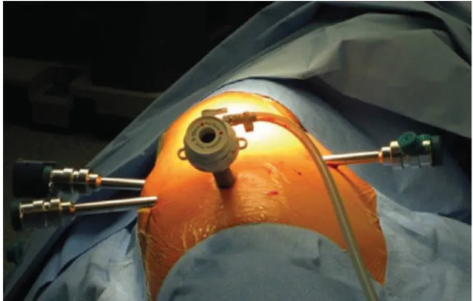
Figure 1:Port mapping for lobectomy on the right. The camera port is placed
in the seventh to eighth intercostal space on the midaxillary line (12 mm, 30°
angled down scope, CO2inlet). The other three 8-mm port incisions are
situ-ated in thefifth to sixth intercostal space on the anterior axillary line, the sixth
to seventh intercostal space on the posterior axillary line and in the auscultatory area (fourth arm).
© The Author 2014. Published by Oxford University Press on behalf of the European Association for Cardio-Thoracic Surgery. All rights reserved.
European Journal of Cardio-Thoracic Surgery 46 (2014) e136–e138
SURGICAL TECHNIQUE
the artery, followed by the vein and
finally the division of the
bron-chus. The
fissures are divided at any suitable point during the
pro-cedure, either by cautery (if only thin) or by stapling (if thick). In
case of fused
fissures, a change of the aforementioned strategy
might be preferable, such that the vein can be divided
first,
fol-lowed by the arterial branches and the bronchus, leaving for the
last the division of the
fissure. Smaller arterial branches can be
ligated (Linen 2.0), while the vein and the bronchus are divided by
means of an endostapler. The specimen is retrieved through the
slightly enlarged ventral porthole (wider rib spaces) and one chest
tube inserted through the camera porthole. We work with a
com-pletely robotic approach; however, in dif
ficult situations it can be
helpful to create an additional ventral assistant port for suction/
irrigation, retraction etc.
RESULTS
Our suggestions have been subdivided into two categories: general
(Table
1
) and problem-speci
fic considerations. These categories
Table 1:
General considerations and common problems and possible solutions
General considerations
Patient positioning • Lateral decubitus position, especially in women, place a pillow under the kidney region to elevate the lower
thorax and prevent the camera from colliding with the hip
Port placement • Keep an adequate distance (≥10 cm) between ports
• Use a needle for testing the optimal position before creating the porthole
Cart placement • Introduce the camera through the port and direct it towards the main target region (central hilum): the robotic
cart (central column) should come from imaginary elongation of the line between the camera trocar and the target. Dock patient cart according to the camera arm sweet spot (the blue arrowhead should be in the middle of the blue line).
Auxiliary material • CO2insufflation helps collapse the lung and evacuates smoke
• Use vessel loops to encircle structures before stapling (easier placement of the anvil)
• Use gauze for oozing instead of suction (time saving, CO2remains)
Temporarily cease CO2insufflation while dissecting/encircling the pulmonary vein, since the vein collapses under intrathoracic pressure levels≥6 mmHg,
which may interfere with safe dissection and encircling of it
Avoid using clips, since they might slip off or get torn off if caught by gauze or passing stapler/instruments Common problems and possible solutions
Pleuropulmonary adhesions • Create space for the camera by digital blunt dissection first, and then introduce VATS endoscopic monopolar
shears through the same incision to create more space for the second port etc.
Exposure problems • Distended lung: let the anaesthetist check the tube and aspirate the airways; simultaneously gently compress the
lung, temporarily use higher CO2pressure
• Cauterize bullae (only in the lobe that needs to be resected) Collision of robotic arms
Collision of the camera with the hip
• Angles of proximal joints of the robotic arms might need to be changed a bit
• Temporarily change the camera angle (from 30° down to up), a pillow under the kidney region elevates the lower chest (especially in women; preoperatively!)
Area of dissection not reachable by the instrument
• Consider switching instruments and their function (dissect with left $ right and retract/expose with
right$left arm)
• Slightly rearrange proximal joints of the robotic arms
Lens repeatedly soiled • Remove the camera trocar, ensure haemostasis, place corner of gauze through the porthole and replace the
trocar (the gauze will compress and absorb bleeding) Trouble passing the stapler around
the vessel
• Try the other access port; test direction with the straight robotic instrument first Full angulation and slight rotation of the anvil while passing the structure Help passing the anvil by gently guiding it from the other side with a grasper Change the direction of traction of the lobe and/or the vessel loop Consider use of a stapling cartridge with a curved tip
Consider use of a short silicon catheter on the stapler anvil tip as a guide [3]
Consider ligation of medium-sized vessels and use of a vessel sealer for small vessels Trouble passing the stapler around
the bronchus
• See above; if bronchus seems too thick, consider use of a conventional open linear stapler (90° angled; the ventral port needs to be slightly enlarged)
Air leak • Avoid air leak by gentle lung tissue handling; avoid injuring fragile lung tissue by using ‘pushing’ instead of
‘grasping’ manoeuvres; if grasping is required, place a gauze on the lung tissue before grabbing it (as with pot cloth)
• Submerge the lung under water for air leak testing • Seal with a stapling device, clips, lung sealant or suture
• In case of fused fissures consider performing the fissure-last technique
Minor bleeding • Diffuse oozing: place thrombostatic material and compress
• From pulmonary artery: compress for several minutes with a sponge, gauze or thrombostatic material (small bleeders often cease spontaneously)
Major bleeding • Compress bleeding with a gauze or lung tissue; use robotic suction (you lose one grasper) or let an assistant
introduce suction through the additional port • Either clip or suture
• Conversion: either let an assistant compress with a sponge or place a clamp, or grasp the bleeding vessel with the robotic instrument and leave the robotic arm in place while performing thoracotomy
VATS: video-assisted thoracic surgery.
SURGIC
AL
T
E
CHNIQUE
have been arranged in a tabular format, providing a clear and
concise overview of the topic as a whole. These suggestions have
evolved over the course of performing many robotic surgical
pro-cedures over the years, and putting them into practice has proved
effective in challenging situations that have arisen. In support of
this, for the last 160 patients, who were operated using the
described four-arm approach, the conversion rate was as low as
5.6% and hospital mortality rate 0% [
4
,
5
]. While most of our
recom-mendations are speci
fic for a robotic approach and therefore have
been applied also by other well-known robotic surgeons [
6
], others
are more non-speci
fic and have, at least in part, also been reported
and successfully applied in VATS [
7
].
COMMENT
The updated, four-arm versions of the da Vinci system
™ [i.e. second
(S) and third (Si) generation systems], along with the development
of new robotic instruments, allow control of any operative step
from the robot
’s console, with the exception of stapling
man-oeuvres. We believe that this development was an important step
enabling the further re
finement of the surgical technique itself,
which has had a favourable impact on the safety and reproducibility
of the operative procedures [
5
].
The purpose of this compilation of tips is two-fold:
first, to
sensitize practitioners to the critical steps of this particular robotic
procedure. We believe that, by considering our helpful tips and
suggestions, one may be able to not only avoid critical situations,
but also to be properly prepared if and when they should arise
during the robotic procedure. Second, and equally as important,
this may not only contribute to improving patient safety, but also
shorten operative time and hopefully have a positive in
fluence on
overall patient outcomes in the end. Although we mainly focused
on the intraoperative part, preoperative planning
—including
proper patient selection
—is of course at least as important, but
would de
finitely go beyond the scope of this contribution.
Con
flict of interest: none declared.
REFERENCES
[1] Kent M, Wang T, Whyte R, Curran T, Flores R, Gangadharan S. Open, video-assisted thoracic surgery, and robotic lobectomy: review of a national database. Ann Thorac Surg 2014;97:236–42.
[2] Park H, Melfi F, Mussi A, Maisonneuve P, Spaggiari L, Da Silca RKC et al.
Robotic lobectomy for non-small cell lung cancer (NSCLC): long-term
oncologic results. J Thorac Cardiovasc Surg 2012;143:383–9.
[3] Ito N, Suda T, Inoue T, Yasui S, Suzuki Y, Taniguchi Yet al. Use of a soft
sili-cone tube guide for an automatic suture device in video-assisted lung lob-ectomy. J Thorac Cardiovasc Surg 2005;130:931–2.
[4] Melfi FM, Menconi GF, Mariani AM, Angeletti CA. Early experience with
robotic technology for thoracoscopic surgery. Eur J Cardiothorac Surg
2002;21:864–8.
[5] Melfi FM, Fanucchi O, Davini F, Romano G, Lucchi M, Dini P et al. Robotic lobectomy for lung cancer: evolution in technique and technology. Eur J Cardiothorac Surg 2014;46:626–31.
[6] Cerfolio RJ, Bryant AS, Minnich DJ. Starting a robotic program in general thoracic surgery: why, how, and lessons learned. Ann Thorac Surg 2011;91:
1729–36.
[7] Demmy T, James T, Swanson S, McKenna R, D’Amico T. Troubleshooting video-assisted thoracic surgery lobectomy. Ann Thorac Surg 2005;79: 1744–53.
G.J. Kocheret al. / European Journal of Cardio-Thoracic Surgery