Embryonic Stem Cells:
New Possible Therapy for Degenerative Diseases
That Affect Elderly People
Qing He, Jian Li, Esther Bettiol, and Marisa E. Jaconi
Biology of Aging Laboratory, Department of Geriatrics, Geneva University Hospitals, Geneva, Switzerland.
The capacity of embryonic stem (ES) cells for virtually unlimited self renewal and differentiation has opened up the prospect of widespread applications in biomedical research and regenerative medicine. The use of these cells would overcome the problems of donor tissue shortage and implant rejection, if the cells are made immunocompatible with the recipient. Since the derivation in 1998 of human ES cell lines from preimplantation embryos, considerable research is centered on their biology, on how differentiation can be encouraged toward particular cell lineages, and also on the means to enrich and purify derivative cell types. In addition, ES cells may be used as an in vitro system not only to study cell differentiation but also to evaluate the effects of new drugs and the identification of genes as potential therapeutic targets. This review will summarize what is known about animal and human ES cells with particular emphasis on their application in four animal models of human diseases. Present studies of mouse ES cell transplantation reveal encouraging results but also technical barriers that have to be overcome before clinical trials can be considered.
C
ELL therapy is an increasingly attractive concept in modern transplantation medicine. For many clinical situations, replacement of lost cells would be the ideal treat-ment. These situations include age-related diseases with progressive cell loss (various types of congestive heart fail-ure, brain degenerative diseases, and sarcopenia), traumatic tissue loss, and iatrogenic destruction of cells (e.g., bone marrow transplantation). In many cases, however, the de-velopment of cell therapeutic treatment approaches is hampered by an increasing lack of donors or by the lack of cells that are suitable for transplantation.A possible solution to this problem lies in xenografts (i.e., transplantation of tissues of animal origin); however, for several reasons (ethical, immunological, infectious dis-eases), this approach has a limited usefulness. A way out of this problem would be the differentiation of embryonic stem (ES) cells into specific cell types and tissues. In fact, recent developments in the field of stem cell biology and, in particular, of human ES cells have generated hope that this lack of suitable cells can be overcome.
Isolated 4 years ago from preimplantation embryos by Thomson et al. (1), human embryonic stem (hES) cells have the capacity to differentiate into virtually all of the cell types building our body. These cells therefore hold the promise of forming any desired tissue in culture that could be used to treat a wide variety of conditions where age, disease, or trauma has led to tissue damage or dysfunction. This radical new approach of disease treatment would overcome the problems of donor tissue shortage and, by making the cells immunocompatible with the recipient, implant rejection.
This review focuses on what is known so far about ES cells, with particular emphasis on the progress made in the characterization of ES cells from mouse and humans, as well as on the present achievements of ES cell-based ther-apies in animal models of human diseases.
THEWORLDoFSTEMCELLS
What are embryonic stem cells and what makes them different from other cells? How do they regulate their self-renewal and how they specialize into a given type? Can we encourage their differentiation towards specific cell line-ages suitable for cell therapies? These are some of the cru-cial questions that scientists all over the world are trying to elucidate.
Stem cells are commonly defined as undifferentiated cells that can proliferate and have the capacity of both self-renewal and differentiation to one or more types of specialized cells. They can be found in the embryo and fetus, and also several organs of the adult human body, with the degree of potentialities commonly decreasing as cells commit to a lineage and specialize. The most promising, and also the most controversial, are embryonic stem cells, which are present in the very early embryo at the stage of the blastocyst, about 1 week after fertilization. They constitute the inner cell mass, a hollow ball of undifferentiated cells (which will form the entire embryo), and are surrounded by a shell of cells (the trophoblast, which will form the placenta). When removed from the blastocyst, ES cells can be cultured and propagated indefinitely in an undifferenti-ated pluripotent state. The isolation of ES cells at the blastocyst stage is imperative, since from this stage onward different populations of cells, when provided with the appropriate signals, begin to specialize and display specific functions [for review see (2)].
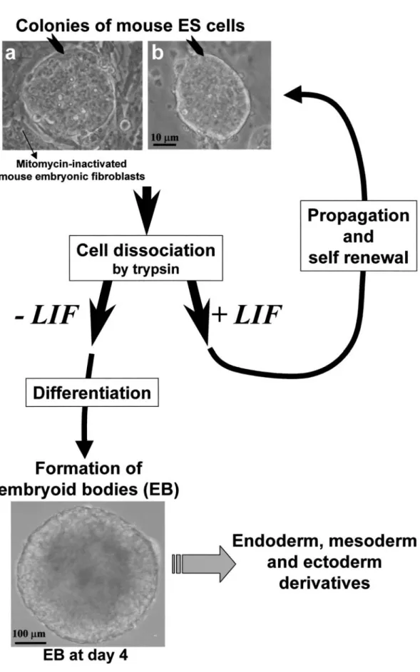
The successful derivation of murine ES cells from the inner cell mass of mouse blastocysts was achieved in 1981, allowing culture conditions to be defined to support their unlimited propagation (3,4). Murine ES cells remain un-differentiated when grown in the presence of leukemia inhibitory factor (LIF) and, for some cell lines, cultured on murine embryonic fibroblasts (MEF) as feeder cells (5,6) 279
(see Figure 1). These cells were soon shown to be pluri-potent, i.e., capable of forming all mature cell phenotypes derived from the three embryonic layers: endoderm, ecto-derm, and mesoderm (2). In fact, when LIF or feeder cells are withdrawn, most types of ES cells differentiate sponta-neously to form aggregates called embryoid bodies. These tridimentional cell–cell contacts allow the formation of heterogeneous cultures of differentiated cell types including cardiomyocytes (7,8), hematopoietic cells (9,10), endo-thelial cells (11–13), neurons (14,15), skeletal muscle (16,17), chondrocytes (18), adipocytes (19), liver (20), and pancreatic islets (21).
HUMAN ES CELLS
The first nonhuman primate embryonic stem cells were described in 1995, maintained in culture for more than a year, while retaining their pluripotency, self-renewing capa-city, and their normal karyotype (22). It was only in 1998 that Thomson et al. reported the first successful derivation and propagation of human ES (hES) cells derived from the inner cell mass of in vitro-fertilized early blastocysts (1). Like the primate ES cells derived earlier, hES are pluri-potent, self-renewing (remaining in the undifferentiated state without losing pluripotency), telomerase positive (an enzyme that confers an unlimited replicative capacity), and have a normal karyotype.
Human pluripotent stem cells, called human embryonic germ (hEG), also could be derived from fetal material ob-tained from medically terminated pregnancies (23). Although obtained from different sources by different laboratory processes, both hES and hEG cells have been demonstrated to be pluripotent (capable of forming all cells and tissues in the body) (24). Several lines of hES cells have been produced, including some that were clonally derived (25).
Human ES cells show several morphological and be-havioral differences from murine ES cells: They grow more slowly and tend to form flat rather than spherical colonies (25,26). Moreover, LIF does not have the same effect on human ES cells as compared to mouse ES cells: To remain undifferentiated, hES require culture on MEF feeder layers in the presence of basic fibroblast growth factor (bFGF) (3,4), on Matrigel, or laminin in MEF-conditioned medium (27).
There is a rapidly increasing number of reports describ-ing hES cell differentiation into neurons (28), endothelium (29,30), hematopoietic cells (31), functionally active pan-creatic cells (32), and beating cardiomyocytes (26,33,34). Current research focuses on how to coax ES cell differ-entiation to a desired lineage, derive highly purified cell populations lacking any carcinogenic potential, and per-form cell implantation in a per-form that will replace or augment the function of diseased or degenerating tissues (26,35).
ROLEoFEXTRACELLULARFACTORS
Studies of gene expression during mammalian embryonic development have led to the identification of some factors that preferentially induce a specific lineage differentiation (36,37). As with murine ES cells, modification of the culture
medium in which human ES cells are grown can encourage the differentiation of certain lineages (36). For example, en-riched populations of proliferating neural progenitors have been obtained by supplementation of the culture medium with specific growth factors (38,39). The temporal interplay between growth factors like FGFs, EGF, Shh, and BMPs can regulate the differentiation of neurons and glia in tissue culture (40,41).
Providing specific local influences through coculture with mature cells can also encourage the formation of a particular lineage. For example, ES cells grown with bone marrow cells or yolk sac endothelium form hematopoietic precursors (31). On the other hand, upon injury, several organs are able to release factors that activate repairing mechanisms and induce resident stem cells, partially committed but not fully differentiated, to further progress and replace damaged or dead cells with new units. Well-known examples are bone marrow, skin, liver, and skeletal muscle. This does not seem to be the case with the brain, which despite possessing a population of neural stem cells, does not seem to be able to activate enough brain stem cells upon major injury or cell loss (e.g., brain stroke, Parkinson’s disease, Alzheimer’s disease) (42). It is therefore conceivable that a healthy organ may not be capable of locally releasing cues to prime the implanted cells, while damaged tissue may be activated to release important molecules for both the recruitment of resident adult stem cells when available or distant stem cells from other compartments (i.e., the bone marrow). The identification of those factors will therefore be fundamental to optimally initiate in vitro a suited differentiation that could be pursued in situ. One could also envisage genetically inducing the implanted cells to transiently secrete factors favoring a neovascularization, for example, via the insertion of vascular endothelial growth factor (VEGF) transgene under the control of an inducible promoter. This would allow optimal integration in the recipient organ and avoid the long-term deleterious effects of uncontrolled long-term secretion (43–45).
TECHNICALOBSTACLES TO THE
CLINICALUSE oFES CELLS
Selection of Suitable Cell Type
For clinical development, it is first necessary to develop methods to purify populations of specific cell types from a complex structure of differentiating stem cells. The removal of undifferentiated stem cells from the cultures prior to clin-ical use is critclin-ical to avoid the risk of teratoma formation.
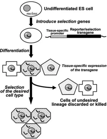
So far, none of the approaches used on murine ES cells can give 100% yield of cells with the required phenotype. Methods such as FACS (fluorescence-activated cell sorting) or MACS (magnetic-activated cell sorting) allow such puri-fication using fluorescence or magnetic microbead-tagged antibodies recognizing a surface marker selective for a desired cell lineage. If this is not available, ES cells can be transduced with a lineage-specific promoter that can drive the expres-sion of a marker, such as green fluorescent protein (46) or an antibiotic resistance gene, as illustrated in Figure 2 (47). This allows for preferential selection of cell subpopulations
Figure 1. Examples of mouse embryonic stem (ES) cell clones, which are propagated in an undifferentiated state by culturing them over mitomycin-inactivated mouse embryonic fibroblasts as feeder cells and in the presence of the cytokine LIF (leukemia inhibitory factor) (panel a). Some cell lines are feeder cell-independent such as the CGR8 line (49) (panel b). Every 2 days, cells are dissociated by trypsinization. Undifferentiated ES cells can be expanded in the presence of LIF, or their differentiation can be initiated by removing LIF and by forming three-dimensional structures called embryoid bodies (EB) containing the three embryonic derivatives.
defined by the cell type specificity of the promoter utilized. This type of approach has been used to select neural and cardiomyocyte phenotypes (48–50).
Immunohistocompatibility
A barrier to overcome is to avoid the rejection of the implanted cells by the recipient. In fact, immunosuppres-sive drugs are associated with many highly unpleasant side effects, and such a treatment would not represent an opti-mally acceptable option. Interestingly, ES cells seem to express less immune-related cell surface proteins (e.g., class I products of the major histocompatibility complex) (51). Drukker et al. (52) addressed the graft rejection issue of cells derived from hES cells by showing that both un-differentiated or un-differentiated hES express no major histo-compatibility complex (MHC)-II proteins or human leukocyte antigen (HLA)-G and very low levels of MHC class I (MHC-I) proteins on their surface. MHC-I molecules, however, may be dramatically and rapidly induced by treating the cells with interferons. If a similar phenomenon occurs after trans-plantation, allogeneic human ES cells might be rejected by cytotoxic T lymphocytes.
It is likely that the problem of rejection of grafted human ES-derived cells could be overcome (or at least minimized) by establishing ‘‘histocompatibility banks’’ of hES with com-pletely HLA-typed ES cell clones derived using good manufacture practice (GMP) protocols. Ideally, if large num-bers of cell lines from genetically diverse populations can be maintained, this would provide isotype-matching cells for virtually any patient.
Other possibilities include means of reducing or abolish-ing cell immunogenicity. ES cells, unlike adult cells, can be easily modified genetically by, for example, inserting immunosuppressive molecules such as Fas ligand, or re-moving immunoactive proteins such as B7 antigens (53). Alternatively, one could delete the foreign MHC genes or insert genes coding for the recipient’s MHC (54).
Ultimately, neoplastic growth or immunopathology could be suppressed by introducing into ES cells before implanta-tion suicide genes that permit their ablaimplanta-tion in case of mis-behavior. For example, herpes thymidine kinase sensitizes mouse ES cells to destruction by the guanosine analog gan-cyclovir (55).
Total immunocompatibility of tissue engineered from human ES cells (56–58) could be theoretically obtained by somatic nuclear transfer (also defined as therapeutical cloning). This procedure uses the transfer of a somatic cell nucleus from an individual into an enucleated oocyte (59,60). Such an oocyte would then undergo embryonic develop-ment to the blastocyst stage prior to isolation from the inner cell mass of hES cells that would be genetically matched to the tissues of the nucleus donor. So far, one group has claimed the nuclear transfer derivation of a human embryo up to only a six-cell stage (61,62), but the success of this result is still questioned. Clearly, this procedure of somatic nuclear transfer is still highly problematic from an ethical and practical point of view.
HOWFAR AREWEFROMCLINICALAPPLICATIONS
USINGES CELLS?
Presently, only allogeneic or matched donor-derived adult stem cells have been used in human cell-grafting therapies. The best known and established example is bone marrow transplantation for the treatment of leukemia and, recently, transplantation of hematopoietic stem cells derived from umbilical cord blood. However, there are still problems with accessibility, low frequency (e.g., in bone marrow there is roughly 1 stem cell per 100,000 cells), restricted differen-tiation potential, and poor growth, which are limiting their applicability to tissue engineering (63). Nevertheless, there are several ongoing phase I trials using bone marrow and skeletal satellite cells for the treatment of human heart fail-ure (64), despite unconvincing or contradictory evidence for a correct in situ transdifferentiation of these adult stem cells implanted in the heart of animal models (65).
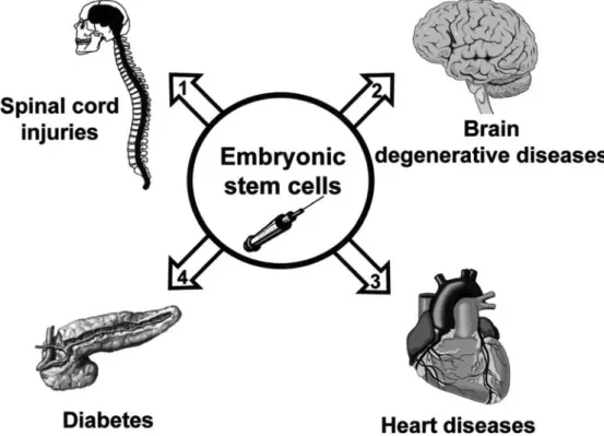
So far, there are few examples of ES cell-based ther-apy using animal models of diseases that have provided encouraging and promising results. As illustrated in Figure 3, these are (a) rat model of spinal cord injury, (b) mouse model of Parkinson’s disease, (c) myocardial infarction (MI) in mice and rats, and (d) diabetes in mouse. We will describe them and discuss the limitations of the present achievements.
Figure 2. Possible methods for the selection of a suited cell lineage include the insertion into embryonic stem (ES) cells of transgenes (i.e., fluorescence reporter genes or antibiotic resistance genes) under the control of tissue-specific promoters. Upon differentiation, the expression of the transgene is dictated by the tissue-specific promoter, allowing the selection of the desired cell type, i.e., using cell sorting or cell recovery after antibiotic treatment. [Adapted from Strom et al. (95).]
Spinal Cord Injury
The absence of spontaneous axonal regeneration in the adult mammalian central nervous system causes devastating functional consequences in patients with spinal cord in-juries. During the past decade, several attempts have been made to find a strategy to repair injured spinal cords in ex-perimental animals, which could provide a novel therapeutic approach in humans. Very interesting results have been achieved recently in a rat model of spinal cord injury (66). When a heterogeneous population of differentiating ES cells (i.e., derived from embryoid bodies cultured 4 days without, then 4 days with, retinoic acid) were transplanted into in-jured spinal cords, they were able to survive, migrate, and dif-ferentiate, allowing a neurological improvement in treated animals, which recovered leg movement, as compared to paralyzed sham-treated controls. More recently, Wichterle et al. showed that developmentally relevant signaling factors can induce mouse ES cells to differentiate into functional motoneurons able to repopulate the embryonic spinal cord, extend axons, and form synapses with target muscles (67). Furthermore, it has been shown that ES cells, when trans-planted into adult rat spinal cord after chemical demyelina-tion or in myelin-deficient mutant mice, differentiated into mature oligodendrocytes, produced myelin, and myelinated host axons (68).
Parkinson’s Disease
Parkinson’s disease (PD) is a common degenerative disorder that affects more than 2% of the population over 65 years of age. PD is characterized by the selective and gradual loss of dopaminergic neurons in the substantia nigra
of the midbrain with a subsequent reduction in striatal dop-amine. The loss of this group of neurons is responsible for most PD symptoms (i.e., tremor, rigidity, and hypokinesia). PD has been treated with grafts of fetal cells, but the limited access of these cells and their poor survival restrict wider application of this approach. ES cells may be particularly valuable for circumventing this problem, as they can proliferate and maintain their developmental po-tential in culture. Dopaminergic neurons have been effi-ciently derived from ES cells in vitro (69). Bjorklund et al. (70) have transplanted a very small number of partially differentiated mouse ES cells derived from embryoid bodies into a rat model of PD, and have shown that at least some of them become dopamine neurons in the striatal regions where the endogenous neurons were previously destroyed. This allowed the Parkinson symptoms to reverse in 50% of the animals, while only 25% of them developed brain tumors (70,71). If a large number of ES cells are implanted into the brain, they grow into every cell type and form teratomas in all cases, eventually killing their host (72). More recently, Kim et al. (73) have characterized the electro-physiological and behavioral properties of highly enriched populations of mouse ES-derived midbrain neural stem cells, able to functionally integrate into host tissue and improve symptoms in a rodent model of PD. The use of neuron-selective media were reported to increase the fraction of neuronal cells, and this type of optimization may be funda-mental for the safe production of selected phenotypes (69). Whether a similar outcome will soon be demonstrated for hES cells will depend on the development of safe strategies that will allow immunotolerance and avoid tumorigenic risks.
Myocardial Infarction and Heart Failure
Chronic congestive heart failure (CHF) is a common consequence of heart muscle or valve damage and represents a major cause of cardiovascular morbidity and mortality in developed countries. When heart muscle is damaged by injury or decreased blood flow (ischemia), functional con-tracting cardiomyocytes are replaced with nonfunctional scar tissue. In fact, cardiomyocyte withdrawal from the cell cycle in the early neonatal period renders the adult heart incapable to regenerate after injury. Therefore, the use of a cell therapy approach to replace lost cardiomyocytes with new engraftable ones would represent an invaluable, low-invasiveness technique for the treatment of heart failure as an alternative to whole heart transplantation.
The way in which hES cells could be used to treat heart disease has already been tested in mice and rats. Mouse ES cells, when cultivated as embryoid bodies, are able to dif-ferentiate in vitro into cardiomyocytes of ventricle-, atrium-, and pacemaker-like cell types characterized by develop-mentally controlled expression of cardiac-specific genes, structural proteins, sarcomeric proteins (74,75), and ion channels (8,76,77). Since only approximately 5% of the cell population within embryoid bodies are cardiomyocytes, the selection of an enriched culture of cardiomyocytes has required genetic manipulation (47,49,50). Clearly, the purity of differentiated ES-derived cardiomyocyte culture is a key issue to avoid the potential formation of teratomas, which would disrupting heart contractility.
Klug et al. were the first to show that ES-derived car-diomyocytes, selected using an antibiotic selection cassette (Figure 2) and injected into the hearts of dystrophin-deficient MDX mice, were able to repopulate the myocardial tissue and integrate with host myocardial tissue (47). Enriched populations of cardiomyocytes were obtained by introducing into ES cells a neomycin resistance gene under the control of the a-cardiac myosin heavy chain promoter. On differentiation, only cardiac-committed cells expressing the antibiotic resistance gene can survive when treated with neomycin. Similarly, cardiomyocytes with a ventricular phenotype have been selected using the ventricular-specific myosin light chain isoform 2v promoter controlling the expression of green fluorescent protein (49,50). So far, how-ever, no study has investigated the engraftment of specific subpopulations of ES-derived cardiomyocytes, such as ventricular, atrial, or pacemaker cells. Whether these ap-proaches can generate sufficient numbers of cardiac cells suited for myocardial repair in vivo remains to be established. Ideally, understanding how to control cardiomyocyte differ-entiation would maximize proliferation of cardiac progenitors cells in culture while impeding their terminal differentiation, which should be undergone after transplantation.
Several animal models of MI using coronary ligation in rats or cryoinjury in mice have been used to test the implantation of fetal-, embryo-, or ES-derived cardiomyo-cytes by direct injection into heart muscle (78–81). Li et al. demonstrated that transplanted fetal cardiomyocytes could integrate cryoinjured cardiac tissue and improve heart func-tion (82,83). Transplantafunc-tion of stage E12 to E15 embryonic cardiomyocytes into MI or cryoinjured hearts attenuated left ventricular dilatation, infarct thinning, and myocardial dysfunction (79,84). Recently, Min et al. engrafted, 30
minutes after MI in rats, ES-derived cardiomyocytes manually dissected from beating embryoid bodies in culture. Although implanted cells survived and integrated the myocardium, which was associated with considerable im-provement of global cardiac function, still many grafts remained isolated and did not differentiate into an adult phenotype (81).
The results published so far indicate that ES cell transplantation is a feasible approach to improve ventricular function in the infarcted failing heart. However, despite encouraging beneficial effects on heart function and remodeling, the mechanism behind these results remains to be demonstrated. Benefits may be associated with en-hanced angiogenesis via the release of angiogenic factors such as VEGF. Several key issues still need to be addres-sed including the extent to which cell engraftment affects cardiac function actively (i.e., by increasing contractility via functional integration or via the secretion of growth factors) or passively (i.e., by limiting infarct expansion and re-modeling). Importantly, the possibility that some ES cells not committed to cardiac lineage could form deleterious teratomas with time has not yet been ruled out.
Diabetes
Diabetes results from abnormal function of pancreatic b-cells, which are responsible for insulin synthesis, storage, and release. Lack or defect of insulin produces diabetes mellitus, a devastating disease suffered by 150 million people in the world. Transplantation of insulin-producing cells could be a cure for type 1 and some cases of type 2 diabetes.
Mouse and human ES cells can produce insulin-secreting cells in culture (32,85). Lumelsky et al. showed that mouse ES cells can coordinately differentiate into multiple types of pancreatic endocrine cells that self-assemble into structures resembling pancreatic islets. When implanted subcutane-ously in diabetic mice, these cells, although able to vascu-larize and remain immunoreactive to insulin, could not reverse high blood sugar levels in mice with symptoms of diabetes (21). This may not be surprising considering the inappropriate site of implantation and the insufficient amount of insulin produced. Nonetheless, grafted animals were able to maintain their body weight and survive for longer periods.
Implantable b-cells are likely to require protection from recurring autoimmunity. This protection might take the form of tolerization, cell encapsulation, or cell engineering with immunoprotective genes. New insights into endocrine pan-creas development using hES are leading to manipulation of progenitor-cell fate towards the b-cell phenotype of insulin production, storage, and regulated secretion, which, in turn, could lead to widespread cell replacement therapy for type 1 diabetes (86).
CONCLUSIONS
The derivation of hES cells is a fundamental discovery that holds promise for three major areas of biomedicine: (a) transplantation medicine, (b) pharmaceutical research and development, and (c) human developmental biology.
Transplantation Medicine
The availability of hES cells opens extraordinary opportunities for tissue transplantation. Examples of cells for transplantation therapies include heart muscle cells for use in repairing the tissue damage inflicted by heart attacks, blood-forming cells for use in bone marrow transplantation procedures for cancer patients, and nerve cells for use in treating patients with spinal cord injury, stroke, Parkinson’s or Alzheimer’s diseases, and diabetes. Frailty is a major problem in geriatric medicine (87–89). Its pathogenesis involves neuronal and muscle cell failure as well as decreased cardiovascular function (90,91). The availability of hES cells represents a tremendous potential to reverse frailty and pre-vent functional decline.
Pharmaceutical Research and Development
Human embryonic stem cells also represent a new technology for pharmaceutical research and development. Until now, the only cell lines available for this work were either animal or abnormal transformed human cells. Per-manent, stable sources for normal human differentiated cells may be developed for drug screening and testing, drug toxicology studies, as well as new drug target identification (92–94). In addition, hES cells may also allow the creation of in vivo models of human disease for drug development as a superior alternative to current mouse models.
Human Developmental Biology
Finally, unraveling the biology of hES cells as they differentiate into functional cell types in vitro offers a unique platform to understand the mechanisms of human embry-onic development, tissue differentiation, and repair. Until now, early genetic events in human embryology have been largely inaccessible to direct observation. Research with hES cells may lead to the discovery of novel genes that fundamentally control tissue differentiation, and may facilitate a molecular understanding of how specific human tissues and organs develop without conducting research on human embryos or fetuses. These gene products could result in the development of therapeutic drugs and proteins with potential applications in wound healing, stroke, heart attack, spinal cord injury, and brain degenerative diseases.
Successful use of any stem cell-based therapy will event-ually depend on our ability to isolate specific cell types in large numbers that will differentiate to a fully functional state, as well as on the challenging demonstration of their in vivo function. To this end, it is crucial to pursue basic research on human ES cell biology to ensure better under-standing of basic principles and what genes and proteins are essential for hES developmental progression. This compre-hension does not only specifically involve the transplantation of ES cell-derived cells but will also help the development of new therapeutic strategies to improve the transdifferentia-tion and expansion of adult stem cells, such as umbilical cord blood cells or bone marrow cells. Many hurdles (not only technical but also ethical) have to be cleared before the research reaches a point where clinical trials can begin.
ACKNOWLEDGMENT
Address correspondence to Marisa E. Jaconi, PhD, Biology of Aging Laboratory, Department of Geriatrics, Geneva University Hospitals, 2 Chemin du Petit Bel-Air, CH-1225 Cheˆne-Bourg, Geneva, Switzerland. E-mail: marisa.jaconi@medecine.unige.ch
REFERENCES
1. Thomson JA, Itskovitz-Eldor J, Shapiro SS, et al. Embryonic stem cell lines derived from human blastocysts.Science. 1998;282:1145– 1147.
2. Smith AG. Embryo-derived stem cells: of mice and men.Annu Rev Cell Dev Biol. 2001;17:435–462.
3. Martin GR. Isolation of a pluripotent cell line from early mouse em-bryos cultured in medium conditioned by teratocarcinoma stem cells. Proc Natl Acad Sci U S A. 1981;78:7634–7638.
4. Evans MJ, Kaufman MH. Establishment in culture of pluripotential cells from mouse embryos.Nature. 1981;292:154–156.
5. Smith AG, Heath JK, Donaldson DD, et al. Inhibition of pluripotential embryonic stem cell differentiation by purified polypeptides.Nature. 1988;336:688–690.
6. Williams RL, Hilton DJ, Pease S, et al. Myeloid leukaemia inhibitory factor maintains the developmental potential of embryonic stem cells. Nature. 1988;336:684–687.
7. Wobus AM, Kaomei G, Shan J, et al. Retinoic acid accelerates embry-onic stem cell-derived cardiac differentiation and enhances develop-ment of ventricular cardiomyocytes. J Mol Cell Cardiol. 1997;29: 1525–1539.
8. Hescheler J, Fleischmann BK, Lentini S, et al. Embryonic stem cells: a model to study structural and functional properties in cardiomyo-genesis.Cardiovasc Res. 1997;36:149–162.
9. Wiles MV, Keller G. Multiple hematopoietic lineages develop from embryonic stem (ES) cells in culture.Development. 1991;111:259–267. 10. Nakano T, Kodama H, Honjo T. In vitro development of primitive and definitive erythrocytes from different precursors. Science. 1996;272: 722–724.
11. Risau W, Sariola H, Zerwes HG, et al. Vasculogenesis and angio-genesis in embryonic-stem-cell-derived embryoid bodies.Development. 1988;102:471–478.
12. Snodgrass HR, Schmitt RM, Bruyns E. Embryonic stem cells and in vitro hematopoiesis.J Cell Biochem. 1992;49:225–230.
13. Yamashita J, Itoh H, Hirashima M, et al. Flk1-positive cells derived from embryonic stem cells serve as vascular progenitors.Nature. 2000; 408:92–96.
14. Bain G, Kitchens D, Yao M, Huettner JE, Gottlieb DI. Embryonic stem cells express neuronal properties in vitro.Dev Biol. 1995;168:342–357. 15. Fraichard A, Chassande O, Bilbaut G, Dehay C, Savatier P, Samarut J. In vitro differentiation of embryonic stem cells into glial cells and func-tional neurons.J Cell Sci. 1995;108:3181–3188.
16. Miller-Hance WC, LaCorbiere M, Fuller SJ, et al. In vitro chamber specification during embryonic stem cell cardiogenesis. Expression of the ventricular myosin light chain-2 gene is independent of heart tube formation.J Biol Chem. 1993;268:25244–25252.
17. Rohwedel J, Maltsev V, Bober E, Arnold HH, Hescheler J, Wobus AM. Muscle cell differentiation of embryonic stem cells reflects myogenesis in vivo: developmentally regulated expression of myogenic determina-tion genes and funcdetermina-tional expression of ionic currents.Dev Biol. 1994; 164:87–101.
18. Kramer J, Hegert C, Guan K, Wobus AM, Muller PK, Rohwedel J. Embryonic stem cell-derived chondrogenic differentiation in vitro: activation by BMP-2 and BMP-4.Mech Dev. 2000;92:193–205. 19. Dani C, Smith AG, Dessolin S, et al. Differentiation of embryonic stem
cells into adipocytes in vitro.J Cell Sci. 1997;110:1279–1285. 20. Hamazaki T, Iiboshi Y, Oka M, et al. Hepatic maturation in
dif-ferentiating embryonic stem cells in vitro.FEBS Lett. 2001;497:15–19. 21. Lumelsky N, Blondel O, Laeng P, Velasco I, Ravin R, McKay R. Differentiation of embryonic stem cells to insulin-secreting structures similar to pancreatic islets.Science. 2001;292:1389–1394.
22. Thomson JA, Kalishman J, Golos TG, et al. Isolation of a primate em-bryonic stem cell line.Proc Natl Acad Sci U S A. 1995;92:7844–848.
23. Shamblott MJ, Axelman J, Wang S, et al. Derivation of pluripotent stem cells from cultured human primordial germ cells.Proc Natl Acad Sci U S A. 1998;95:13726–13731.
24. Itskovitz-Eldor J, Schuldiner M, Karsenti D, et al. Differentiation of human embryonic stem cells into embryoid bodies compromising the three embryonic germ layers.Mol Med. 2000;6:88–95.
25. Amit M, Carpenter MK, Inokuma MS, et al. Clonally derived human embryonic stem cell lines maintain pluripotency and proliferative potential for prolonged periods of culture.Dev Biol. 2000;227:271–278. 26. Odorico JS, Kaufman DS, Thomson JA. Multilineage differentiation from human embryonic stem cell lines.Stem Cells. 2001;19:193–204. 27. Xu C, Inokuma MS, Denham J, et al. Feeder-free growth of undifferen-tiated human embryonic stem cells. Nat Biotechnol. 2001;19: 971–974.
28. Carpenter MK, Inokuma MS, Denham J, Mujtaba T, Chiu CP, Rao MS. Enrichment of neurons and neural precursors from human embryonic stem cells.Exp Neurol. 2001;172:383–397.
29. Amit M, Itskovitz-Eldor J. Derivation and spontaneous differentiation of human embryonic stem cells.J Anat. 2002;200:225–232. 30. Levenberg S, Golub JS, Amit M, Itskovitz-Eldor J, Langer R.
Endothelial cells derived from human embryonic stem cells.Proc Natl Acad Sci U S A. 2002;99:4391–4396.
31. Kaufman DS, Hanson ET, Lewis RL, Auerbach R, Thomson JA. Hematopoietic colony-forming cells derived from human embryonic stem cells.Proc Natl Acad Sci U S A. 2001;98:10716–10721. 32. Assady S, Maor G, Amit M, Itskovitz-Eldor J, Skorecki KL,
Tzukerman M. Insulin production by human embryonic stem cells. Diabetes. 2001;50:1691–1697.
33. Reubinoff BE, Pera MF, Fong CY, Trounson A, Bongso A. Embryonic stem cell lines from human blastocysts: somatic differentiation in vitro. Nat Biotechnol. 2000;18:399–404.
34. Kehat I, Kenyagin-Karsenti D, Snir M, et al. Human embryonic stem cells can differentiate into myocytes with structural and functional properties of cardiomyocytes.J Clin Invest. 2001;108:407–414. 35. Bianco P, Robey PG. Stem cells in tissue engineering.Nature. 2001;
414:118–121.
36. Schuldiner M, Yanuka O, Itskovitz-Eldor J, Melton DA, Benvenisty N. From the cover: effects of eight growth factors on the differentiation of cells derived from human embryonic stem cells.Proc Natl Acad Sci U S A. 2000;97:11307–11312.
37. Schuldiner M, Eiges R, Eden A, et al. Induced neuronal differentiation of human embryonic stem cells.Brain Res. 2001;913:201–205. 38. Reubinoff BE, Itsykson P, Turetsky T, et al. Neural progenitors from
human embryonic stem cells.Nat Biotechnol. 2001;19:1134–1140. 39. Zhang SC, Wernig M, Duncan ID, Brustle O, Thomson JA. In vitro
differentiation of transplantable neural precursors from human em-bryonic stem cells.Nat Biotechnol. 2001;19:1129–1133.
40. Anderson DJ. Stem cells and pattern formation in the nervous system: the possible versus the actual.Neuron. 2001;30:19–35.
41. Lillien L, Raphael H. BMP and FGF regulate the development of EGF-responsive neural progenitor cells. Development. 2000;127: 4993–5005.
42. Cao Q, Benton RL, Whittemore SR. Stem cell repair of central nervous system injury.J Neurosci Res. 2002;68:501–510.
43. Springer ML, Chen AS, Kraft PE, Bednarski M, Blau HM. VEGF gene delivery to muscle: potential role for vasculogenesis in adults.Mol Cell. 1998;2:549–558.
44. Suzuki K, Murtuza B, Smolenski RT, et al. Cell transplantation for the treatment of acute myocardial infarction using vascular endothe-lial growth factor-expressing skeletal myoblasts.Circulation. 2001;104: I207–I212.
45. Yang Y, Min JY, Rana JS, et al. VEGF enhances functional improve-ment of postinfarcted hearts by transplantation of ESC-differentiated cells.J Appl Physiol. 2002;93:1140–1151.
46. Okabe M, Ikawa M, Kominami K, Nakanishi T, Nishimune Y. ‘‘Green mice’’ as a source of ubiquitous green cells. FEBS Lett. 1997;407: 313–319.
47. Klug MG, Soonpaa MH, Koh GY, Field LJ. Genetically selected cardiomyocytes from differentiating embryonic stem cells form stable intracardiac grafts.J Clin Invest. 1996;98:216–224.
48. Nishikawa SI, Nishikawa S, Hirashima M, Matsuyoshi N, Kodama H. Progressive lineage analysis by cell sorting and culture identifies FLK11
VE-cadherin1 cells at a diverging point of endothelial and hemopoietic lineages.Development. 1998;125:1747–1757.
49. Meyer N, Jaconi M, Landopoulou A, Fort P, Puceat M. A fluorescent reporter gene as a marker for ventricular specification in ES-derived cardiac cells.FEBS Lett. 2000;478:151–158.
50. Muller M, Fleischmann BK, Selbert S, et al. Selection of ventricular-like cardiomyocytes from ES cells in vitro. FASEB J. 2000;14: 2540–2548.
51. O’Shea KS. Embryonic stem cell models of development.Anat Rec. 1999;257:32–41.
52. Drukker M, Katz G, Urbach A, et al. Characterization of the expression of MHC proteins in human embryonic stem cells.Proc Natl Acad Sci U S A. 2002;99:9864–9869.
53. Walker PR, Saas P, Dietrich PY. Role of Fas ligand (CD95L) in immune escape: the tumor cell strikes back.J Immunol. 1997;158:4521–4524. 54. Westphal CH, Leder P. Transposon-generated ‘‘knock-out’’ and
‘‘knock-in’’ gene-targeting constructs for use in mice. Curr Biol. 1997; 7:530–533.
55. Fareed MU, Moolten FL. Suicide gene transduction sensitizes murine embryonic and human mesenchymal stem cells to ablation on demand— a fail-safe protection against cellular misbehavior.Gene Ther. 2002;9: 955–962.
56. Lanza RP, Cibelli JB, West MD. Prospects for the use of nuclear transfer in human transplantation.Nat Biotechnol. 1999;17:1171–1174. 57. Lanza RP, Cibelli JB, West MD. Human therapeutic cloning.Nat Med.
1999;5:975–977.
58. Solter D, Gearhart J. Putting stem cells to work.Science. 1999;283: 1468–1470.
59. Wilmut I, Schnieke AE, McWhir J, Kind AJ, Campbell KH. Viable offspring derived from fetal and adult mammalian cells.Nature. 1997; 385:810–813.
60. Wilmut I, Young L, Campbell KH. Embryonic and somatic cell cloning.Reprod Fertil Dev. 1998;10:639–643.
61. Cibelli JB, Kiessling AA, Cunniff K, Richards C, Lanza RP, West MD. Somatic cell nuclear transfer in humans: pronuclear and early embryonic development.J Regener Med. 2001;2:25–31.
62. Cibelli JB, Lanza RP, West MD, Ezzell C. The first human cloned embryo.Sci Am. 2002;286:44–51.
63. Vogel G. Stem cell policy. Can adult stem cells suffice?Science. 2001; 292:1820–1822.
64. Menasche P, Hagege AA, Scorsin M, et al. Myoblast transplantation for heart failure.Lancet. 2001;357:279–280.
65. Reinecke H, Poppa V, Murry CE. Skeletal muscle stem cells do not transdifferentiate into cardiomyocytes after cardiac grafting.J Mol Cell Cardiol. 2002;34:241–249.
66. McDonald JW, Liu XZ, Qu Y, et al. Transplanted embryonic stem cells survive, differentiate and promote recovery in injured rat spinal cord. Nat Med. 1999;5:1410–1412.
67. Wichterle H, Lieberam I, Porter JA, Jessell TM. Directed differen-tiation of embryonic stem cells into motor neurons. Cell. 2002;110: 385–397.
68. Liu S, Qu Y, Stewart TJ, et al. Embryonic stem cells differentiate into oligodendrocytes and myelinate in culture and after spinal cord transplantation.Proc Natl Acad Sci U S A. 2000;97:6126–6131. 69. Lee SH, Lumelsky N, Studer L, Auerbach JM, McKay RD. Efficient
generation of midbrain and hindbrain neurons from mouse embryonic stem cells.Nat Biotechnol. 2000;18:675–679.
70. Bjorklund LM, Sanchez-Pernaute R, Chung S, et al. Embryonic stem cells develop into functional dopaminergic neurons after transplanta-tion in a Parkinson rat model.Proc Natl Acad Sci U S A. 2002;99: 2344–2349.
71. Freed CR. Will embryonic stem cells be a useful source of dopamine neurons for transplant into patients with Parkinson’s disease?Proc Natl Acad Sci U S A. 2002;99:1755–1757.
72. Deacon T, Dinsmore J, Costantini LC, Ratliff J, Isacson O. Blastula-stage stem cells can differentiate into dopaminergic and serotonergic neurons after transplantation. Exp Neurol. 1998;149: 28–41.
73. Kim JH, Auerbach JM, Rodriguez-Gomez JA, et al. Dopamine neurons derived from embryonic stem cells function in an animal model of Parkinson’s disease.Nature. 2002;418:50–56.
74. Sanchez A, Jones WK, Gulick J, Doetschman T, Robbins J. Myosin heavy chain gene expression in mouse embryoid bodies. An in vitro developmental study.J Biol Chem. 1991;266:22419–22426. 75. Muthuchamy M, Pajak L, Howles P, Doetschman T, Wieczorek DF.
Developmental analysis of tropomyosin gene expression in embry-onic stem cells and mouse embryos.Mol Cell Biol. 1993;13:3311– 3323.
76. Boheler KR, Czyz J, Tweedie D, Yang HT, Anisimov SV, Wobus AM. Differentiation of pluripotent embryonic stem cells into cardiomyo-cytes.Circ Res. 2002;91:189–201.
77. Wobus AM, Rohwedel J, Maltsev V, Hescheler J. Development of cardiomyocytes expressing cardiac-specific genes, action potentials, and ionic channels during embryonic stem cell-derived cardiogenesis. Ann N Y Acad Sci. 1995;752:460–469.
78. Koh GY, Soonpaa MH, Klug MG, et al. Stable fetal cardiomyocyte grafts in the hearts of dystrophic mice and dogs.J Clin Invest. 1995; 96:2034–2042.
79. Roell W, Lu ZJ, Bloch W, et al. Cellular cardiomyoplasty improves survival after myocardial injury.Circulation. 2002;105:2435–2441. 80. Roell W, Fan Y, Xia Y, et al. Cellular cardiomyoplasty in a transgenic
mouse model.Transplantation. 2002;73:462–465.
81. Min JY, Yang Y, Converso KL, et al. Transplantation of embryonic stem cells improves cardiac function in postinfarcted rats. J Appl Physiol. 2002;92:288–296.
82. Li RK, Mickle DA, Weisel RD, et al. Natural history of fetal rat cardiomyocytes transplanted into adult rat myocardial scar tissue. Circulation. 1997;96:II-179–186; discussion 186–187.
83. Li RK, Jia ZQ, Weisel RD, et al. Cardiomyocyte transplantation im-proves heart function.Ann Thorac Surg. 1996;62:654–660; discussion 660–661.
84. Etzion S, Battler A, Barbash IM, et al. Influence of embryonic cardiomyocyte transplantation on the progression of heart failure in a rat model of extensive myocardial infarction.J Mol Cell Cardiol. 2001; 33:1321–1330.
85. Soria B. In-vitro differentiation of pancreatic beta-cells.Differentiation. 2001;68:205–219.
86. Efrat S. Cell replacement therapy for type 1 diabetes.Trends Mol Med. 2002;8:334–340.
87. Bortz WM II. A conceptual framework of frailty: a review.J Gerontol Med Sci. 2002;57:M283–M288.
88. Morley JE. Anorexia, sarcopenia, and aging. Nutrition. 2001;17: 660–663.
89. Rockwood K, Hogan DB, MacKnight C. Conceptualisation and measurement of frailty in elderly people. Drugs Aging. 2000;17: 295–302.
90. Roubenoff R. Sarcopenia: a major modifiable cause of frailty in the elderly.J Nutr Health Aging. 2000;4:140–142.
91. Welle S. Cellular and molecular basis of age-related sarcopenia.Can J Appl Physiol. 2002;27:19–41.
92. Rohwedel J, Guan K, Hegert C, Wobus AM. Embryonic stem cells as an in vitro model for mutagenicity, cytotoxicity and embryotoxicity studies: present state and future prospects.Toxicol In Vitro. 2001;15: 741–753.
93. Bremer S, Worth AP, Paparella M, et al. Establishment of an in vitro reporter gene assay for developmental cardiac toxicity.Toxicol In Vitro. 2001;15:215–223.
94. Paparella M, Kolossov E, Fleischmann B, Hescheler J, Bremer S. The use of quantitative image analysis in the assessment of in vitro embryotoxicity endpoints based on a novel embryonic stem cell clone with endoderm-related GFP expression. Toxicol In Vitro. 2002;16: 589–597.
95. Strom TB, Field LJ, Ruediger M. Allogeneic stem cells, clinical trans-plantation, and the origins of regenerative medicine.Transplant Proc. 2001;33:3044–3049.
Received September 30, 2002 Accepted October 9, 2002


