Cerebral Cortex November 2011;21:2589--2598 doi:10.1093/cercor/bhr049
Advance Access publication March 31, 2011
Fake or Fantasy: Rapid Dissociation between Strategic Content Monitoring and Reality
Filtering in Human Memory
Aure´lie Wahlen1, Louis Nahum1, Damien Gabriel1and Armin Schnider1,2 1
Laboratory of Cognitive Neurorehabilitation, Department of Clinical Neurosciences and Dermatology, Medical School, University of Geneva, CH-1211 Geneva 14, Switzerland and2Division of Neurorehabilitation, Department of Clinical Neurosciences, University Hospital, CH-1211 Geneva 14, Switzerland
Address correspondence to Prof. Armin Schnider, Service de Neurore´e´ducation, Hoˆpitaux Universitaires de Gene`ve, Avenue de Beau-Se´jour 26, CH-1211 Geneva 14, Switzerland. Email: armin.schnider@hcuge.ch.
Memory verification is crucial for meaningful behavior. Orbito-frontal damage may impair verification and induce confabulation and inappropriate acts. The strategic retrieval account explains this state by deficient monitoring of memories’ precise content, whereas the reality filter hypothesis explains it by a failure of an orbitofrontal mechanism suppressing the interference of memories that do not pertain to reality. The distinctiveness of these mechanisms has recently been questioned. Here, we juxtaposed these 2 mechanisms using high-resolution evoked potentials in healthy subjects who performed 2 runs of a continuous recognition task which contained pictures that precisely matched or only resembled previous pictures. We found behavioral and electro-physiological dissociation: Strategic content monitoring was maximally challenged by stimuli resembling previous ones, whereas reality filtering was maximally challenged by identical stimuli. Evoked potentials dissociated at 200--300 ms: Strategic monitoring induced a strong frontal negativity and a distinct cortical map configuration, which were particularly weakly expressed in reality filtering. Recognition of real repetitions was expressed at 300--400 ms, associated with ventromedial prefrontal activation. Thus, verification of a memory’s concordance with the past (its content) dissociates from the verification of its concordance with the present. The role of these memory control mechanisms in the generation of confabulations and disorientation is discussed. Keywords: exclusion paradigm, extinction capacity, reward processing, orbitofrontal cortex, feeling of rightness, inverse solution, source monitoring, spatiotemporal analysis
Introduction
Brain damage may induce a state in which patients confabulate experiences of events that never happened, are disoriented regarding time, place, and their current role, and forge plans for the day that disregard their current health status. The study of this state, variably called spontaneous confabulation (Schnider, von Da¨niken, et al. 1996b), confabulation with action (Metcalf et al. 2007), or behaviorally spontaneous confabulation (Schnider 2008), has indicated ways to elucidate the brain processes allowing humans to sense the veracity of memories about the past (‘‘Have I really done this this morning?’’) and their appropriateness as a basis for present action (‘‘Do I really have this appointment today?’’).
Diverse theories postulated the existence of monitoring pro-cesses verifying whether a memory about the past is correct or not (Moscovitch 1989; Johnson et al. 1993; Burgess and Shallice 1996). While functional imaging opened the way to localization of
the hypothesized processes (Mitchell and Johnson 2009), the consequences of their failure, and hence, their clinical impact, has not been experimentally verified or then received negative results (Johnson et al. 1997). The main exception to this rule is the strategic retrieval hypothesis that describes a series of processes, including their anatomical substrate, leading from the evocation of a memory to the verification (‘‘monitoring’’) and the ‘‘felt-rightness’’ about the acceptance or rejection of its content (Moscovitch and Melo 1997; Gilboa et al. 2006). The theory has received empirical support from clinical studies (Moscovitch and Melo 1997; Melo et al. 1999; Gilboa et al. 2006): Confabulating patients produced more false memories when asked to recall personal or historical events (from the past) evoked by a cue word or when recalling bible stories. In a continuous recognition task, they accepted pictures resembling previously presented pictures more often as true repetitions than nonconfabulating patients (Gilboa et al. 2006).
Only a fraction of patients who recount false ideas about the past also have false ideas about their present duties (Schnider 2008). These patients act according to currently inappropriate memories and may, for example, insist on going to appoint-ments that, in reality, have taken place some years ago (Schnider, von Da¨niken, et al. 1996b). Thus, they act on the basis of memories that do not relate to the present. The reality filter hypothesis is meant to explain the capacity to distinguish between memories that relate to current reality and memories that do not, that is, fantasies (Schnider 2003, 2008). It holds that the ability to sort out memories that relate to ongoing reality depends on a filter mechanism mediated by the posterior medial orbitofrontal cortex (area 13) (Schnider, von Da¨niken, et al. 1996a; Schnider and Ptak 1999; Schnider, Treyer, and Buck 2000; Treyer et al. 2003, 2006b), which filters upcoming memories that do not relate to ongoing reality at an early stage of evocation, at 200--300 ms, before their precise content is consciously recognized and re-encoded at 400--600 ms (Schnider et al. 2002). The main experimental paradigm used in these studies—with designs adapted to the used technology—contained repeated runs of a continuous recogni-tion test, whereby each run was composed of the same picture set that was presented in different order each time. Subjects had to indicate for each picture whether it had already been presented within the ongoing run, irrespective of presentation in a previous run. In such tasks, behaviorally spontaneous con-fabulators produced more and more false-positive responses from run to run, believing they had just seen an item in the current run (the ‘‘current reality’’) when, in fact, it had occurred in a previous run (Schnider, von Da¨niken, et al. 1996a, 1996b; Schnider and Ptak 1999; Schnider, Ptak, et al. 2000).
A recent article suggested that reality filtering is a sub-component of strategic retrieval rather than a different process (Gilboa et al. 2006), thus questioning the specificity of reality filtering (Schnider 2008). The proposal is of importance because it suggests a convergence of rather than dissociation between memory control mechanisms. In terms of experimen-tal approach, it was postulated that the ability to sense that an item seen in a previous run has not yet occurred within the ‘‘present reality’’ of the ongoing run (reality filtering) would be related to the ability to sense that an item is not exactly the same as an item previously presented within the same run (strategic monitoring).
The fact that the strategic monitoring hypothesis and the reality filter hypothesis have defined experimental procedures associated with them allows one to directly compare the 2 mechanisms. In the present study, we combined the 2 experimental conditions into one recognition paradigm. Using high-density evoked potentials and spatiotemporal analyses in healthy subjects, we explored whether the monitoring of a memory’s precise content (strategic monitoring) is reflected in the same or different electrocortical activity than the moni-toring of a memory’s relation with the ‘‘now’’ (reality filtering). The task was composed in such a way that it additionally allowed us to test the precision of reality filtering.
Materials and Methods
Participants
Sixteen right-handed subjects (13 women and 3 men, age 25.7±3.6 years) with no history of neurological or psychiatric illness gave written informed consent to participate in this study, which was approved by the Institutional Ethical Committee.
Methods
Subjects made 3 equivalent blocks composed of 2 runs of a continuous recognition task, in which they had to indicate picture recurrences within, and only within, the ongoing run. The 3 blocks were separated by 10-min breaks (to prevent fatigue) and were composed of separate picture sets (to prevent interference between the blocks). The 2 runs of each block were made in immediate succession (60-s break to restart the presentation program). Data from the first runs of the 3 blocks were pooled for the analyses, as were data from the second runs of the 3 blocks.
Strategic content monitoring was tested as proposed by Gilboa et al. (2006): All runs were composed in such a way that some pictures (New items, distracters, Dis) were truly repeated within the run (true repetitions, Rep), while others were followed by an item that was semantically identical but structurally only similar with the previously presented picture (similars, Sim). The distinction between a true repetition and an item that is only similar to a previously presented picture (but is indeed a new picture) has been proposed to rely on strategic monitoring (Gilboa et al. 2006).
Reality filtering was tested by composing the 2 runs within each block according to the same logic as in our previous studies (Schnider, von Da¨niken, et al. 1996b; Schnider and Ptak 1999; Schnider 2003, 2008): The second run of each block was composed in the same manner and of the same item set as the first run of the same block. Thus, the second run demands the ability to sense that an item that appears familiar (from the first run) is indeed new within the ongoing second run (distracters of run 2, Dis2). Behaviorally spontaneous confabulators specifically failed in this capacity (they produced false-positive responses) (Schnider, von Da¨niken, et al. 1996b; Schnider and Ptak 1999). Performance of the second run induced circumscribed activity of orbitofrontal area 13 in healthy subjects (Schnider, Treyer, and Buck 2000). Processing of the first appearance of stimuli in the second run (Dis2), which had already been seen in the first run,
induced a specific electrocortical response at 200--300 ms thought to reflect reality filtering (Schnider et al. 2002).
An additional question in this study was the precision of the orbitofrontal reality filter. To test this aspect, some items of the first runs did not reappear in the identical form in the second runs of the same block but in a similar form (distracters similar with an item from the first run; SimDis2). Thus, these pictures were new pictures that were related to, but not identical with, an item seen in the first run. Would these stimuli be processed in the same way as pictures that had really appeared in the first run (Dis2)? If yes, the reality filter could be assumed to be relatively imprecise and to be challenged by similar stimulus variations as strategic monitoring.
Task Composition
The layout of the task is shown in Figure 1. The whole task was composed of 126 pairs of semantically identical, perceptually related line drawings (126 pictures, 126 similars). In pretests, 9 healthy subjects similarly named 100% of the picture pairs and correctly distinguished 94% of them as different pictures in a continuous recognition task.
In each run, 84 pictures were presented. In the first runs of each block, there were 42 first presentations (Distracters 1, Dis1), 21 true repetitions (Repetitions 1, Rep1—recognition of true repetitions) and 21 presentations of similar pictures (Similars 1, Sim1—test of strategic content monitoring). True repetitions and presentations of similar pictures occurred after 12--18 intervening pictures. Subjects had to indicate as fast as possible by button press whether the picture had been presented before within the ongoing run (right button with right middle finger for Rep1) or not (left button with right index finger for Dis1 and Sim1).
Figure 1. Task design. Both runs of each block were composed of the same items, arranged in different order. Distracters are items that appear for the first time within a run (Dis1, Dis2 5 Distracters of run 1 or 2). Similars resemble a picture previously seen within the same run (Sim1, Sim2 5 Similars of run 1 or 2). Similar distracters (SimDis2) appear for the first time within the second run but resemble a picture seen in the first run. Repetitions are true repetitions within a run (Rep1, Rep2 5 Repetitions within run 1 or 2). The 3 blocks were composed of different pictures and were separated by a break of 10 min.
Participants then immediately performed a second run, which was composed of the same items, mostly represented in the same pictures, as the first run. Again they had to indicate precise picture repetitions within the second run, irrespective of presentations in the first run. Specifically, the second runs contained 21 pictures that had already been presented in the first run and now appeared for the first time within the second run (Distracters 2, Dis2—test of reality filter), 21 pictures that were similar to but not identical with pictures that had been presented and repeated within the first run and now appeared for the very first time in the task (Similar Distracters 2, SimDis2—test of the precision of the reality filter), 21 picture repetitions (Repetitions 2, Rep2), and 21 presentations of pictures similar to but not identical with a picture already presented within the second run (Similars 2, Sim2).
Rep2 and Sim2 stimuli had already been seen within the first run (Fig. 1). Thus, they did not pose a definite cognitive challenge, like the other stimulus types, but rather combined cognitive requirements of strategic content monitoring (within run 2) and reality filtering (across the runs, run 2 vs. run 1). They were included to assure that run 2 had a similar internal structure (same stimulus types, apart from addition of SimDis2) and level of difficulty as run 1 but were not intended to enter analysis.
The meaning of the different stimulus types and expected responses are summarized in Table 1.
As there were 42 Dis1 items per block (126 total), while all other conditions had 21 stimuli per block (63 total), only half of the responses to Dis1 stimuli were randomly selected in each subject to enter analysis.
Stimuli were presented on a computer screen for 2000 ms; inter-stimulus interval was 700 ms.
Behavioral data (reaction times, correct responses) with the factor stimulus type were analyzed using repeated-measures analyses of variance (ANOVAs) and post hoc Fisher’s tests (with a significance level of P <0.05).
ERP Acquisition and Preprocessing
Electroencephalography was continuously recorded with an Active-Two Biosemi EEG system (Biosemi V.O.F.) using 128 scalp electrodes. Signal was sampled at 512 Hz and filtered at bandwidth of 0--104 Hz. Cartool software (http://brainmapping.unige.ch/Cartool.htm) was used to conduct all analyses. Epochs from 200 ms prestimulus to 600 ms poststimulus onset were averaged for each condition and each subject to calculate the event-related potentials (ERPs). Only correct trials were retained. In addition to a±100-lV automated artifact criterion, data were visually inspected during the averaging procedure to reject epochs with eye blinks and movements and other sources of transient noise. ERPs were band-pass filtered to 1--30 Hz and recalculated against the average reference. Artifact electrodes were interpolated using a spherical spline interpolation (Perrin et al. 1987). Baseline correction was applied to the 200-ms prestimulus period before group averaging. The first 600 ms of the ERPs after stimulus onset were retained for analysis.
Spatiotemporal Analysis
In a first step, periods of stable configuration of electrocortical activity (‘‘maps’’) over the whole set of 128 electrodes were determined using a segmentation procedure. To this end, a modified hierarchical cluster analysis as implemented in Cartool software (topographic atomize and agglomerate hierarchical clustering) was run across the 5 experimental conditions Dis1, Sim1, Rep1, Dis 2, and SimDis2 (Michel et al. 2004; Murray et al. 2008). Statistical smoothing was used to eliminate temporally isolated maps with low strength (Pascual-Marqui et al. 1995). As additional constraints, scalp topographies of <20 ms duration were rejected and clusters that correlated >90% were merged. The number of maps explaining the averaged data sets was determined with the cross validation and the Krzanowski--Lai criterion (Pascual-Marqui et al. 1995).
In a second step, the appearance of maps identified in the group-averaged data was statistically verified in the ERPs of the individual subjects. To do this, each map was compared with the moment-by-moment scalp topography of the individual subjects’ ERPs from each condition by strength-independent spatial correlation (Michel et al. 2001, 2009; Murray et al. 2008). That is, for each time point of the individual subjects’ ERPs, the scalp topography was compared with all maps and was labeled according to the one with which it best correlated. It is important to note that this labeling procedure is not exclusive such that a given period of the ERP for a given subject and stimulus condition is often labeled with multiple template maps. Nonetheless, the results of the labeling reveal whether a given ERP is more often described by one map rather than another. Fitting thus allowed us to determine for what period of time (duration) a given topography was observed in a given condition across subjects. The global explained variance (GEV) is the sum of the explained variance weighted by the global field power (GFP, root mean square across the average-referenced electrode values at a given instant in time [Murray et al. 2008; Michel et al. 2009]). The GFP represents the strength of the maps. The GEV describes how well a map configuration explains the individually obtained patterns of activity (Michel et al. 2004; Murray et al. 2008). The GEV and duration of maps were then subjected to repeated-measures ANOVAs with the 2 factors Map and Stimulus type (Dis1, Rep1, Sim1, Dis2, SimDis2) and subjected to Fisher’s post hoc tests.
In order to estimate the sources of the cluster maps, distributed linear inverse solution based on a local auto-regressive average model using a 3D realistic head model with a solution space of 3005 nodes was applied (Grave de Peralta Menendez et al. 2001, 2004; Michel et al. 2004). Current distribution was calculated within the gray matter of the average brain provided by the Montreal Neurological Institute. Source estimation was limited to the time period in which segmentation of scalp ERP demonstrated significantly different map topographies between stimulus types.
Waveform Analysis
For a more traditional view of the evoked potentials, a waveform analysis was also conducted. Three regions of interest (ROIs) were
Table 1
Stimulus types and their meaning
Stimulus type Abbreviation Description Correct response (‘‘Repetition?’’) Measured capacity
Distracter 1 Dis1 Picture appearing for the first time within the first run (new) No Novelty detection Similar 1 Sim1 Picture similar to, but not identical with, a picture previously presented
within the first run
No Strategic content monitoring
Repetition 1 Rep1 Repetition of a picture previously presented within the first run Yes Familiarity judgment (feeling of rightness?) Similar Distracter 2 SimDis2 First appearance of a picture in the second run resembling a picture seen in
the first run
No Precision of reality filter
Distracter 2 Dis2 First appearance within the second run of a picture which has already appeared in the first run
No Reality filtering
Similar 2a
Sim2 Picture similar to, but not identical with, a picture previously presented within the second run; already seen both as Dis1 and Rep1 in the first run
No Strategic monitoring plus reality filtering Repetition 2a
Rep2 Repetition of a picture previously presented within the second run; already seen as Sim1 in the first run
Yes Strategic monitoring (feeling of rightness?) plus reality filtering
Note:a
selected: frontal (16 most frontal electrodes), central (15 most central electrodes), and posterior (16 most posterior electrodes) (Dien and Santuzzi 2005; Fig. 3A). ERPs of the electrodes within each region were averaged for each condition and each subject. Statistical analysis of amplitude differences was centered on the time windows in which the spatial cluster analysis indicated significantly different map configu-rations (200--250 and 300--400 ms). To allow comparison with a previous study using a comparable but simpler paradigm (Schnider et al. 2002), the time window 400--600 ms was also analyzed.
Repeated-measures ANOVAs with the 2 factors ROI (frontal, central, and posterior) and stimulus type (mean of trace amplitudes of Dis1, Rep1, Sim1, Dis2, SimDis2) were run across each time period and subjected to post hoc tests.
Results
Behavioral Results
Table 2 summarizes the behavioral results. There was a signif-icant effect of stimulus type on accuracy (one-factor ANOVA, F4,60 = 9.8, P <0.001) and reaction time (F4,60 = 108.4, P < 0.001). Post hoc tests showed that subjects were significantly less accurate and slower in response to Sim1 items than all other stimulus types. Conversely, they were faster and more accurate in response to SimDis2 than Sim1, Rep1, and Dis2.
The stimuli that did not enter the electrophysiological analysis (Sim2, Rep2) did not differ from their equivalents in the first run (Sim1, Rep1) in terms of accuracy (Table 2). While Sim2 also had similar reaction times as Sim1, reaction times were significantly longer in response to Rep2 than Rep1 (post hoc comparison, P <0.05).
There was no indication of fatigue influencing performance: Reaction times (all stimulus types included) were slowest in the first block, then became faster in the second block and third block (effect of block, F2,30 =24, P <0.0001; post hoc,
block 1 > block 2 = block 3). Reaction times did not vary
between the runs (P=0.7).
Spatiotemporal Analysis
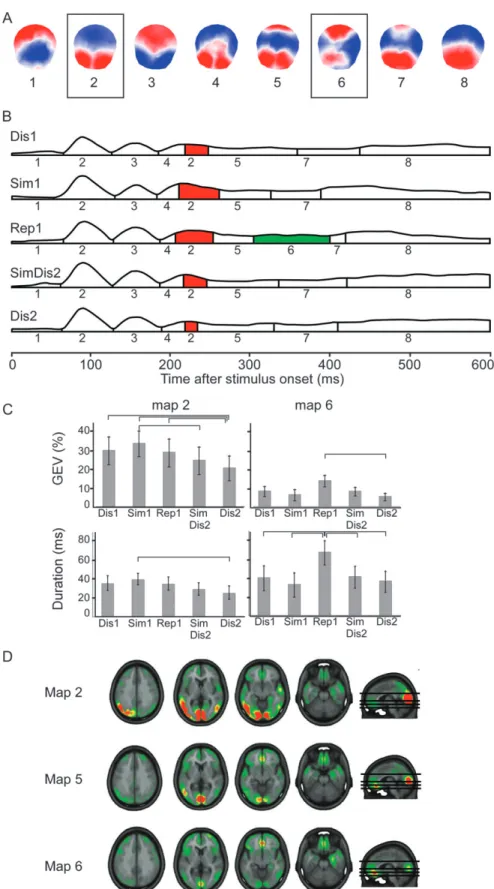
Spatial cluster analysis revealed 8 different cluster maps within the first 600 ms after stimulus onset (Fig. 2A). Temporal succession of the dominant maps is displayed in Figure 2B. Between about 200 and 300 ms, this grand mean analysis suggested that map 2 was less present in response to Dis2 than in the other conditions. Analysis of map appearance in the individual data between 180 and 260 ms showed a significant interaction of GEV between maps 2, 4, and 5 and task conditions (F8,120 = 2.3, P < 0.05). Post hoc tests (Fig. 2C)
confirmed the weaker representation of map 2 in response to Dis2 than all other conditions except SimDis2 and showed a stronger representation of this map in response to Sim1 compared with Dis2 and SimDis2 (statistical differences are showed in Fig. 2C). Fitting of duration of these maps also showed a significant interaction between conditions (F8,120= 2.8, P < 0.05): map 2 was shorter in Dis2 condition than in Sim1 condition (P <0.05). No difference appeared for maps 4 and 5.
Source localization of map 2 (Fig. 2D) indicated that map 2 reflects particularly strong activation of temporo-occipital visual areas and discrete ventromedial prefrontal activation. The ensuing map 5, on which stimuli did not differ significantly, had much less extended activation of these cortical areas but strong ventromedial prefrontal (orbitofrontal) activation (Fig. 2D).
Thus, correct processing of items that only resembled previously presented ones (Sim1), therefore requiring ‘‘strate-gic monitoring,’’ was associated with particularly widespread visual and associative cortical activation. Stimuli known from the previous run but not yet presented within the ongoing second run (Dis2), therefore requiring ‘‘realty filtering,’’ were associated with the weakest activation of these neocortical areas in the 180- to 260-ms period; they essentially left out this neocortical processing stage.
Between 300 and 400 ms after stimulus onset, Figure 2B shows a specific map (map 6) in response to Rep1. Statistical analysis yielded a significant interaction of map3stimulus type between maps 5, 6, 7, and 8 and task conditions regarding GEV (F12,180 = 3.3, P < 0.001) and map duration (F12,180 = 3.5, P<0.001). Figure 2C shows that map 6 was more present (GEV) in response to Rep1 than to Dis2 and longer in Rep1 than in the other conditions (P <0.05). There were no differences across conditions for other maps.
Source localization suggested that map 6 particularly strongly activated prefrontal cortex, in particular the medial orbitofrontal cortex, and the right medial temporal lobe (parahippocampal gyrus).
Waveform Analysis
Figure 3 shows the analysis of waveforms. Repeated ANOVAs yielded significant interactions of ROI3stimulus type in all 3 time widows (4.5 < F8,120< 10.7; P <0.0001). Post hoc tests confirmed the significance of the main apparent findings: At 200--250 ms, Sim1 stimuli (reflecting strategic monitoring) induced the most negative trace over the frontal electrodes, in contrast to Dis2 stimuli (reflecting reality filtering), which induced the least negative trace in comparison to all other stimuli. At 300--400 ms, Rep1 stimuli evoked a markedly more positive wave over frontal electrodes than all other stimuli. At 400--600 ms, Rep1 stimuli appeared to induce a more positive wave than the other stimuli: Post hoc testing confirmed this difference except for the comparison with Dis1 stimuli, which, however, differed from Rep1 stimuli over central and posterior electrodes.
Sim2 and Rep2 stimuli were not included in the EEG analysis for the reasons stated in Methods and are only briefly reported here. Waveform analysis over the frontal electrodes, where the main differences between the responses to Sim1, Rep1, and Dis2 occurred, yielded the following observations: At 200--300 ms, Sim2 stimuli induced the same absence of negative deflection (virtually overlaying trace) as Dis2 stimuli suggesting Table 2
Behavioral results
Run Stimulus type Correct responses Reaction time
(ms) Run1 Dis1 62.6 ± 0.5 776 ± 141 Sim1 50.6 ± 14.3 1001 ± 153 Rep1 57.9 ± 4.1 780 ± 138 Run2 SimDis2 62.6 ± 0.7 752 ± 117 Dis2 61.9 ± 1.5 795 ± 136 Sim2a 49.8 ± 7.5 1025 ± 155 Rep2a 56.1 ± 5.4 926 ± 119
Note: The second column shows mean ± standard deviation (SD) of correct responses for each condition (maximum, 63). The third column shows mean of reaction times ± SD in milliseconds for each condition.
a
Figure 2. Spatiotemporal analysis and source localization. (A) Temporal distribution of the 8 cortical maps obtained from segmentation of the grand mean ERPs within the first 600 ms. Red indicates positivity, and blue indicates negativity. Maps that differed between task conditions are boxed. (B) Distribution of stable map configurations over 600 ms after stimulus onset in the 5 conditions. The numbers below each segment indicate the most representative map of this period. The amplitude of the curves reflects the GFP. Colored segments indicate maps with significant differences between task conditions. (C) Results of post hoc tests of the repeated-measures ANOVAs with the 2 factors Maps and Task condition for maps 2 and 6 regarding GEV and duration of the map. Lines indicate significant differences between 2 conditions. (D) Source localization of maps 2, 5 (which followed map 2), and 6.
that processing at this (preconscious) stage was dominated by reality filtering. Rep2 stimuli evoked a trace similar to Rep1 (overlying trace) with the same differences to other stimulus responses as Rep1 (Fig. 3). At 300--400 ms, Rep2 stimuli induced a similar positive wave as Rep1 but with an amplitude lying between Rep1 and the other stimulus types. According to this intermediate position, their amplitude did not significantly differ from any other stimulus type.
Discussion
This study confirms the dissociation between 2 memory control processes that have been suspected to be related (Gilboa et al. 2006). It demonstrates that the ability to judge whether a stimulus precisely matches a stimulus encountered in the past (strategic monitoring) dissociates from the ability to judge whether the memory of a stimulus relates to the present reality or not (reality filtering). The dissociation appeared both on behavioral and on electrophysiological levels.
On the behavioral level, the stimulus variations challenging the 2 processes dissociated. Stimuli resembling previously presented stimuli within the same run (Sim1) appeared to challenge maximally strategic monitoring, as reflected in slower and less accurate responses to these stimuli than all other stimuli. By contrast, presentation of similar, rather than
equal, distracters between the runs facilitated reality filtering, as reflected in faster responses to SimDis2 than Dis2 stimuli. Thus, the precise matching of memories’ content (strategic monitoring) is particularly challenging when stimuli resemble but are not equal to previously seen stimuli, while realizing that a memory does not apply to present reality (reality filtering) is particularly challenging when stimuli are equal to, rather than only similar to, previously seen stimuli.
Electrophysiologically, the 2 processes dissociated at an early stage: At 200--300 ms, strategic monitoring (processing of Sim1) induced the strongest expression of a specific cortical map configuration (Fig. 2, map 2), while reality filtering (processing of Dis2) was associated with the weakest expres-sion of this configuration. In terms of waveforms, strategic monitoring induced the strongest negative frontal potential, while reality filtering induced the least negative frontal deviation of all stimuli (Fig. 3). This result precisely replicates our previous findings on reality filtering (Schnider et al. 2002). Behavioral measures indicated that these electrophysiolog-ical results truly reflect the cognitive processes of interest rather than unspecific effects such as, for example, fatigue: Reaction times did not differ between the runs, and relative differences of reaction times and error rates among the stimulus types were similar in both runs (Table 2: run 1, Dis1
<Rep1 <Sim1; run 2, Dis2 <Rep 2 <Sim2).
Figure 3. Waveform analysis. (A) Arrangement of all 128 electrodes. The black dots indicate electrodes included in the 3 ROIs (frontal, central, and posterior). (B) Grand average ERPs of electrodes included in each ROI in response to the 5 task conditions (Dis1, Sim1, Rep1, SimDis2 and Dis2). Repeated-measures ANOVAs applied on the mean amplitudes were performed across the 3 time windows boxed in gray: 200--250, 300--400, and 400--600 ms.
The present observations help to refine the understanding of the 2 monitoring mechanisms. Strategic retrieval has been proposed to go through diverse, anatomically distinct stages (Moscovitch and Melo 1997; Moscovitch and Winocur 2002; Gilboa et al. 2006). The present study supports this notion: Strategic retrieval was expressed in 2 time periods with activation of different brain regions.
In the early period, 200--300 ms, stimuli resembling previously presented ones (Sim 1) induced a distinct electrocortical response presumably reflecting the detection of the incongru-ence of the presented stimulus (Sim1) with a previously seen one (Dis1). Within the strategic retrieval account, the capacity to make this distinction is called strategic monitoring. A similar frontal response, also starting at 200 ms, was previously demonstrated in tasks in which subjects had to distinguish between pictures they had truly seen in a previous learning session and items that were perceptually similar to the learned pictures (Ranganath and Paller 1999; Ecker and Zimmer 2009). Source estimation indicated that the capacity to detect that items are only similar to, but not identical with, previously seen items was associated with particularly strong activation of visual and adjacent temporo-occipital association cortex (Fig. 2). This activity may reflect increased attention in response to items resembling true repetitions. More elaborate monitoring of this kind has been shown to activate the lateral prefrontal cortex (Ranganath et al. 2000), as also predicted by the strategic retrieval account (Moscovitch and Melo 1997; Moscovitch and Winocur 2002; Gilboa et al. 2006).
A second period compatible with the strategic retrieval model occurred at 300--400 ms, in which true repetitions (Rep1) induced a distinct positive frontal waveform (Fig. 3) and a specific map configuration that was not present in response to other stimuli (Fig. 2). The primary source of this activity was the bilateral medial orbitofrontal area (Fig. 2). A possible interpretation of this response is that it reflected familiarity, as opposed to the feeling of novelty evoked by Dis1 stimuli. However, in earlier studies using continuous recognition tasks, which did not require stringent content monitoring as the present task (Schnider et al. 2002; James et al. 2009), true repetitions only induced discrete amplitude modulations over posterior electrodes and intensity modulations of electro-cortical maps at 400--600 ms, rather than the distinct, relatively early and frontal response evoked in the present task. These observations indicate that the precise context and difficulty of a task profoundly influence the electrocortical response to seemingly equivalent stimuli, such as true repetitions in differently composed recognition tasks. The specificity of the electrophysiological response to real repetitions in the present study suggests that it reflected a signal of confirmation (‘‘yes, it is a real repetition’’) that is not produced in a simple recog-nition task. The characteristics of this response would be compatible with the so-called ‘‘felt-rightness’’ within the strategic retrieval model, thought to emanate from the ventromedial prefrontal cortex and to signal whether a recovered memory satisfies the goals of the present task (Moscovitch and Melo 1997; Moscovitch and Winocur 2002; Gilboa et al. 2006). In any case, our present data show that, in the context of a difficult recognition task, the recognition of fake repetitions (Sim1) is electrocortically expressed before the recognition of true repetitions.
Reality filtering—as reflected in the response to Dis2 stimuli—had the same electrophysiological signature as in
our previous study (Schnider et al. 2002; Schnider 2003): a markedly weaker, almost absent negative deflection over frontal electrodes at 200--300 ms coinciding with a signifi-cantly weaker expression (virtual absence) of a specific electrocortical map configuration present in response to all other stimulus types. Source estimation indicated that these findings reflected particularly weak activation of posterior association areas. These results support the physiological conclusion already drawn from the previous study (Schnider 2003): Stimuli evoking a memory that does not pertain to ongoing reality (Dis2), that is, a ‘‘fantasy,’’ appear to skip a processing stage at 200--300 ms which is characterized by particularly extensive neocortical activity.
The precise mechanisms of this filtering is not clear, but available studies suggest the following hypothesis: Filtering of memories that do not pertain to reality depends on activity of the posterior medial orbitofrontal cortex (OFC), in particular area 13 (Schnider, von Da¨niken, et al. 1996a; Schnider and Ptak 1999; Schnider, Treyer, and Buck 2000; Treyer et al. 2006b), and seems to be mediated by a frontal--subcortical loop connecting the OFC with the striatum, the substantia nigra, and the medial thalamus (Treyer et al. 2003). Filtering is under dopaminergic modulation: In healthy subjects, a hyperdopaminergic state induced errors with the same pattern as the one observed in patients confusing reality, namely, increased false positives on stimuli equivalent to the Dis2 of the present study (Schnider et al. 2010). Clinical evidence indicated that the OFC mechanism underlying reality filtering is its ability to signal that a previously valid anticipation no longer applies, that is, extinction capacity (Nahum, Ptak, et al. 2009). Extinction in a simple anticipation--outcome paradigm evoked activity of the OFC (Schnider, Treyer, and Buck 2005) and was expressed in the same period (200--300 ms) and by a similar configuration (absence of frontal negativity) (Schnider et al. 2007) as reality filtering in the present and our previous studies. In agreement with this hypothesis, we recently found that medial OFC activity at 200--300 ms was much more dependent on the behavioral relevance of the absence of an expected outcome than the sole absence of reward (Nahum, Gabriel, and Schnider 2011).
How and when precisely the OFC interferes between 200 and 300 ms with the activation of memories is not known. Source estimation as used here is sensitive to activity in midline structures (Nahum, Gabriel, et al. 2011) and, indeed, showed medial OFC activation from 200 ms on (Fig. 2, maps 2 and 5). As in a previous study (Schnider et al. 2002), Dis2 stimuli induced particularly weak expression of a stage characterized by extended temporo-occipital neocortical activation (Fig. 2, map 2), which was followed by a stage with particularly strong medial OFC and weak neocortical activation (Fig. 2, map 5), common to all stimulus types. Our hypothesis is, therefore, that reality filtering—the detection that an upcoming memory does not relate to ongoing reality—is conveyed by transient in-hibition of extended neocortical synchronization between 200 and 300 ms (Schnider 2003, 2008). We suggest that this transient inhibition is induced by a signal from orbitofrontal area 13, produced when true reality contradicts the anticipated outcome (as in extinction trials) (Schnider et al. 2007; Nahum, Gabriel, and Schnider 2011) and transmitted through frontal--subcortical connections (Treyer et al. 2003) that are modulated by dopaminergic neurones (Schnider et al. 2010) known to respond to the appearance or nonappearance of expected
outcomes (rewards in animal experimentation) (Schultz and Dickinson 2000).
The present study indicates that the orbitofrontal reality filter reacts to information’s precise content: It is maximally challenged if a stimulus precisely matches a previously seen stimulus whose pertinence for ongoing reality has to be determined (Dis2). Stimuli only resembling previously seen stimuli (SimDis2) induced a less distinct response than precisely repeated stimuli. Thus, the perception of current reality, the ‘‘now,’’ in human thinking apparently rests on the processing of precise information rather than the gist of memories: the more precisely current reality resembles a past reality, the more the orbitofrontal reality filter will be challenged.
Albeit logical, this result was not necessarily expected. For example, the fact that one performs a task (e.g., a memory task) resembling another one in a similar context already activates orbitofrontal area 13 (Treyer et al. 2006a), a fact that is commonly disregarded but may profoundly influence apparent activations in functional imaging studies. On anatomical grounds, high precision of the reality filter was not necessarily expected either. There is evidence that a neighboring anterior limbic structure, the amygdala, reacts to fast but relatively imprecise (low spatial frequency) information about threaten-ing stimuli (LeDoux 1996) or fearful faces (Vuilleumier et al. 2003). If the OFC processed information according to similar criteria as the amygdala, low resolution of orbitofrontal reality filtering might be expected. However, we recently found that, although potentially harmful archetypic stimuli like spiders induced a very strong ERP response around 200 ms, this response dissociated from the processing of anticipated outcomes by the OFC (Nahum, Morand, et al. 2009). Thus, the OFC seems to process events and outcomes according to different criteria than the neighboring amygdala. In any case, the present study shows that reality filtering is based on a precise comparison of memories with present percepts.
The reality filtering paradigm used in our studies bears superficial resemblance with tasks of exclusion within the dissociation procedure, that is, the ability to indicate that an item (a word or picture) was not present in one series but was rather part of another series (Jacoby 1991). The ability to make this distinction has also been termed reality or source monitoring (Johnson and Raye 1981; Johnson et al. 1993), recency judgment (Mandler 1980; Baddeley and Hitch 1993), or temporal order memory (Squire et al. 1981; Petrides 1989). In contrast to the reality filtering disorder determined with our task, the failure to know explicitly when in the past something happened does not have a clear clinical correlate. Although it may be observed in confabulating patients (Schnider, Gutbrod, et al. 1996), it has no specificity (Johnson et al. 1997; Schnider 2008); it has also been documented in amnesia without confabulations or disorientation (Huppert and Piercy 1976; Squire et al. 1981; Hirst and Volpe 1982; Kopelman et al. 1997) or after prefrontal lobe damage without amnesia (Janowsky et al. 1989; Milner et al. 1991; Shimamura et al. 1991; Kesner et al. 1994; Kopelman et al. 1997). Temporal order tasks activate the lateral prefrontal, rather than orbitofrontal, cortex (Zorrilla et al. 1996). Electrophysiologically, old/new effects in exclusion and temporal order task and context judgments in source memory tasks are not expressed before 300--400 ms, often much later (Wilding and Rugg 1996; Herron and Rugg 2003; Dzulkifli and Wilding 2005). Thus, the explicit
knowl-edge about when in the past something happened (exclusion criterion, recency judgment, temporal order memory) is clinically, anatomically, and electrophysiologically distinct from the ability to sense whether a memory relates to ongoing reality or not (reality filtering).
The present study juxtaposed 2 memory control theories but does not directly relate to the mechanism of the reality confusion characterizing behaviorally spontaneous confabula-tion and disorientaconfabula-tion. The reality filter hypothesis holds that these disorders result from the failure of an orbitofrontal (area 13) mechanism—akin to or identical with extinction capacity--that filters upcoming memories according to their relation with ongoing reality (Schnider 2008; Nahum, Ptak, et al. 2009). The increase of false positives from the second run on in diverse (simpler) versions of the task used in the present experiment (response to Dis2) has proved to be a highly reliable surrogate marker of the memory capacity on which these patients fail. This has been shown in group studies (Schnider, von Da¨niken, et al. 1996b; Schnider and Ptak 1999) and single case studies (Ptak and Schnider 1999; Ptak et al. 2001; Schnider, Bonvallat, et al. 2005; Nahum, Ptak, et al. 2010) on behaviorally spontaneous confabulation and in a group study on disorien-tation (Schnider, von Da¨niken, et al. 1996a). Clinical recovery from behaviorally spontaneous confabulation individually and specifically paralleled recovery of the ability to control false positives in the task (Schnider, Ptak, et al. 2000). In these group studies, patients were recruited irrespective of the cause of brain damage (thus avoiding simple effects of disease severity), were hospitalized at the time of the study (thus allowing us to verify the presence or absence of reality confusion), and were matched with regard to the severity of amnesia (similar degree of delayed free recall deficit); matched in this way, they also did not differ with regard to general executive dysfunction. False-positive and false-negative results occurred but were very rare (discussed in Schnider 2008).
In contradiction to these studies, Gilboa et al. (2006) observed in their study that only patients having an increase of false positives both in our reality filter task and in a task of strategic content monitoring confabulated; thus, deficient reality filtering was considered an insufficient mechanism of behaviorally spontaneous confabulation. Their study included only patients with OFC damage after rupture of an anterior communicating artery aneurysm (thus potentially conflating effects of disease severity with failures specific to confabula-tion) who mostly lived at home (thus rendering verification of reality confusion delicate) and who were not explicitly matched on any cognitive measure, such as amnesia. It is, therefore, possible that their postulate that only the combined failure of content monitoring and reality filtering induces behaviorally spontaneous confabulation essentially compen-sates for the fact that their patients were not matched with respect to the severity of amnesia or other cognitive deficits. Indeed, it is not known whether the failure in reality filtering, as measured with our continuous recognition paradigm, has any specific behavioral correlate in brain-damaged subjects with no amnesia. More intriguingly, severe behaviorally spontaneous confabulation may occur despite intact perfor-mance in the known tasks of strategic monitoring (Nahum, Ptak, et al. 2010). Future studies should include properly matched patient groups and directly contrast strategic content monitoring and reality filtering, as in the present study, to
mechanisms to the reality confusion evident in behaviorally spontaneous confabulation and disorientation.
In conclusion, the present study demonstrates behavioral and electrophysiological dissociation between 2 memory control mechanisms that have been suggested to be related. It shows that the verification of a memory’s content relies on different mechanisms than the ability to sense whether a memory relates to ongoing reality or not.
Funding
Swiss National Science Foundation (320030-132447) to A.S. Notes
We thank Christoph Michel for helpful comments. Cartool software was developed by Denis Brunet, supported by the Center for Biomedical Imaging of Geneva and Lausanne. Conflict of Interest : None declared.
References
Baddeley AD, Hitch G. 1993. The recency effect: implicit learning with explicit retrieval? Mem Cogn. 21:146--155.
Burgess PW, Shallice T. 1996. Confabulation and the control of recollection. Memory. 4:359--411.
Dien J, Santuzzi A. 2005. Application of repeated measures ANOVA to high-density ERP datasets: a review and tutorial. In: Handy T, editor. Event-related potentials. A methods handbook. Cambridge (MA): MIT Press. p. 57--82.
Dzulkifli MA, Wilding EL. 2005. Electrophysiological indices of strategic episodic retrieval processing. Neuropsychologia. 43:1152--1162. Ecker UK, Zimmer HD. 2009. ERP evidence for flexible adjustment of
retrieval orientation and its influence on familiarity. J Cogn Neurosci. 21:1907--1919.
Gilboa A, Alain C, Stuss DT, Melo B, Miller S, Moscovitch M. 2006. Mechanisms of spontaneous confabulations: a strategic retrieval account. Brain. 129:1399--1414.
Grave de Peralta Menendez R, Gonzalez Andino S, Lantz G, Michel CM, Landis T. 2001. Noninvasive localization of electromagnetic epileptic activity. I. Method descriptions and simulations. Brain Topogr. 14:131--137.
Grave de Peralta Menendez R, Murray MM, Michel CM, Martuzzi R, Gonzalez Andino SL. 2004. Electrical neuroimaging based on biophysical constraints. Neuroimage. 21:527--539.
Herron JE, Rugg MD. 2003. Strategic influences on recollection in the exclusion task: electrophysiological evidence. Psychon Bull Rev. 10:703--710.
Hirst W, Volpe BT. 1982. Temporal order judgments with amnesia. Brain Cogn. 1:294--306.
Huppert FA, Piercy M. 1976. Recognition memory in amnesic patients: effect of temporal context and familiarity of material. Cortex. 12:3--20.
Jacoby LL. 1991. A process dissociation framework: separating automatic from intentional uses of memory. J Mem Lang. 30:513--541. James C, Morand S, Barcellona-Lehmann S, Schnider A. 2009. Neural
transition from short to long term memory: an ERP study. Hippocampus. 19:371--378.
Janowsky JS, Shimamura AP, Squire LR. 1989. Source memory impairment in patients with frontal lobe lesions. Neuropsychologia. 27:1043--1056.
Johnson MK, Hashtroudi S, Lindsay DS. 1993. Source monitoring. Psychol Bull. 114:3--28.
Johnson MK, O’Connor M, Cantor J. 1997. Confabulation, memory deficits, and frontal dysfunction. Brain Cogn. 34:189--206.
Johnson MK, Raye CL. 1981. Reality monitoring. Psychol Rev. 88:67--85. Kesner RP, Hopkins RO, Fineman B. 1994. Item and order dissociation in humans with prefrontal damage. Neuropsychologia. 32:881--889. Kopelman MD, Stanhope N, Kingsley D. 1997. Temporal and spatial context memory in patients with focal frontal, temporal lobe, and diencephalic lesions. Neuropsychologia. 35:1533--1545.
LeDoux J. 1996. The emotional brain. The mysterious underpinnings of emotional life.. New York: Simon & Schuster.
Mandler G. 1980. Recognizing: the judgement of previous occurrence. Psychol Rev. 87:252--271.
Melo B, Winocur G, Moscovitch M. 1999. False recall and false recognition: an examination of the effects of selective and combined lesions to the medial temporal lobe/diencephalon and frontal lobe structures. Cogn Neuropsychiatry. 16:343--359. Metcalf K, Langdon R, Coltheart M. 2007. Models of confabulation: a
critical review and a new framework. Cogn Neuropsychiatry. 24:23--47.
Michel C, Koenig T, Brandeis D. 2009. Electrical neuroimaging in the time domain. In: Michel C, Koenig T, Brandeis D, Gianotti L, Wackermann J, editors. Electrical neuroimaging. New York: Cam-bridge University Press. p. 111--143.
Michel CM, Murray MM, Lantz G, Gonzalez S, Spinelli L, Grave de Peralta R. 2004. EEG source imaging. Clin Neurophysiol. 115:2195--2222. Michel CM, Thut G, Morand S, Khateb A, Pegna AJ, Grave de Peralta R,
Gonzalez S, Seeck M, Landis T. 2001. Electric source imaging of human brain functions. Brain Res Brain Res Rev. 36:108--118. Milner B, Corsi P, Leonard G. 1991. Frontal-lobe contribution to recency
judgements. Neuropsychologia. 29:601--618.
Mitchell KJ, Johnson MK. 2009. Source monitoring 15 years later: what have we learned from fMRI about the neural mechanisms of source memory? Psychol Bull. 135:638--677.
Moscovitch M. 1989. Confabulation and the frontal systems: strategic versus associative retrieval in neuropsychological theories of memory. In: Roediger HLI, Craik FIM, editors. Varieties of memory and consciousness. Essays in the honour of Endel Tulving. Hillsdale (NJ): Lawrence Erlbaum Associates. p. 133--160.
Moscovitch M, Melo B. 1997. Strategic retrieval and the frontal lobes: evidence from confabulation and amnesia. Neuropsychologia. 35: 1017--1034.
Moscovitch M, Winocur G. 2002. The frontal cortex and working with memory. In: Stuss DT, Knight RT, editors. Principles of frontal lobe function. New York: Oxford University Press. p. 188--209. Murray MM, Brunet D, Michel CM. 2008. Topographic ERP analyses:
a step-by-step tutorial review. Brain Topogr. 20:249--264.
Nahum L, Gabriel D, Schnider A. 2011. Human processing of behaviorally relevant and irrelevant absence of expected rewards: a high-resolution ERP study. PLoS One. 2011 Jan 27; 6(1):e16173. Nahum L, Gabriel D, Spinelli L, Momjian S, Seeck M, Michel C,
Schnider A. 2010. Rapid consolidation and the human hippocampus: intracranial recordings confirm surface EEG. Hippocampus. doi: 10.1002/hipo.20819.
Nahum L, Morand S, Barcellona-Lehmann S, Schnider A. 2009. Instinctive modulation of cognitive behavior: a human evoked potential study. Hum Brain Mapp. 30:2120--2131.
Nahum L, Ptak R, Leemann B, Lalive P, Schnider A. 2010. Behaviorally spontaneous confabulation in limbic encephalitis: the roles of strategic monitoring and reality filtering. J Int Neuropsychol Soc. 16:995--1005. Nahum L, Ptak R, Leemann B, Schnider A. 2009. Disorientation,
confabulation, and extinction capacity. Clues on how the brain creates reality. Biol Psychiatry. 65:966--972.
Pascual-Marqui RD, Michel CM, Lehmann D. 1995. Segmentation of brain electrical activity into microstates: model estimation and validation. IEEE Trans Biomed Eng. 42:658--665.
Perrin F, Pernier J, Bertrand O, Giard MH, Echallier JF. 1987. Mapping of scalp potentials by surface spline interpolation. Electroencephalogr Clin Neurophysiol. 66:75--81.
Petrides M. 1989. Frontal lobes and memory. In: Boller F, Grafman J, editors. Handbook of neuropsychology. vol. 3. New York: Elsevier Science Publishers. p. 75--90.
Ptak R, Birtoli B, Imboden H, Hauser C, Weis J, Schnider A. 2001. Hypothalamic amnesia with spontaneous confabulations: a clinico-pathologic study. Neurology. 56:1597--1600.
Ptak R, Schnider A. 1999. Spontaneous confabulations after orbito-frontal damage: the role of temporal context confusion and self-monitoring. Neurocase. 5:243--250.
Ranganath C, Johnson MK, D’Esposito M. 2000. Left anterior prefrontal activation increases with demands to recall specific perceptual information. J Neurosci. 20:RC108 (101--105).
Ranganath C, Paller KA. 1999. Frontal brain potentials during recognition are modulated by requirements to retrieve perceptual detail. Neuron. 22:605--613.
Schnider A. 2003. Spontaneous confabulation and the adaptation of thought to ongoing reality. Nat Rev Neurosci. 4:662--671.
Schnider A. 2008. The confabulating mind. How the brain creates reality. Oxford: Oxford University Press.
Schnider A, Bonvallat J, Emond H, Leemann B. 2005. Reality confusion in spontaneous confabulation. Neurology. 65:1117--1119.
Schnider A, Guggisberg A, Nahum L, Gabriel D, Morand S. 2010. Dopaminergic modulation of rapid reality adaptation in thinking. Neuroscience. 167:583--587.
Schnider A, Gutbrod K, Hess CW, Schroth G. 1996. Memory without context. Amnesia with confabulations following right capsular genu infarction. J Neurol Neurosurg Psychiatry. 61:186--193.
Schnider A, Mohr C, Morand S, Michel CM. 2007. Early cortical response to behaviorally relevant absence of anticipated outcomes: a human event-related potential study. NeuroImage. 35:1348--1355. Schnider A, Ptak R. 1999. Spontaneous confabulators fail to suppress
currently irrelevant memory traces. Nat Neurosci. 2:677--681. Schnider A, Ptak R, von Da¨niken C, Remonda L. 2000. Recovery from
spontaneous confabulations parallels recovery of temporal confu-sion in memory. Neurology. 55:74--83.
Schnider A, Treyer V, Buck A. 2000. Selection of currently relevant memories by the human posterior medial orbitofrontal cortex. J Neurosci. 20:5880--5884.
Schnider A, Treyer V, Buck A. 2005. The human orbitofrontal cortex monitors outcomes even when no reward is at stake. Neuro-psychologia. 43:316--323.
Schnider A, Valenza N, Morand S, Michel CM. 2002. Early cortical distinction between memories that pertain to ongoing reality and memories that don’t. Cereb Cortex. 12:54--61.
Schnider A, von Da¨niken C, Gutbrod K. 1996a. Disorientation in amnesia: a confusion of memory traces. Brain. 119:1627--1632. Schnider A, von Da¨niken C, Gutbrod K. 1996b. The mechanisms
of spontaneous and provoked confabulations. Brain. 119: 1365--1375.
Schultz W, Dickinson A. 2000. Neuronal coding of prediction errors. Annu Rev Neurosci. 23:473--500.
Shimamura AP, Janowsky JS, Squire LR. 1991. What is the role of frontal lobe damage in memory disorders?. In: Levin HS, Eisenberg HM, Benton AL, editors. Frontal lobe function and dysfunction. New York: Oxford University Press. p. 173--195.
Squire LR, Nadel L, Slater PC. 1981. Anterograde amnesia and memory for temporal order. Neuropsychologia. 19:141--145.
Treyer V, Buck A, Schnider A. 2003. Orbitofrontal-subcortical loop activation during suppression of memories that do not pertain to ongoing reality. J Cogn Neurosci. 15:610--618.
Treyer V, Buck A, Schnider A. 2006a. Effects of baseline task position on apparent activation in functional imaging of memory. Neuro-psychologia. 44:462--468.
Treyer V, Buck A, Schnider A. 2006b. Selection of currently relevant words: an auditory verbal memory study using PET. Neuroreport. 17:323--327.
Vuilleumier P, Armony JL, Driver J, Dolan RJ. 2003. Distinct spatial frequency sensitivities for processing faces and emotional expres-sions. Nat Neurosci. 6:624--631.
Wilding EL, Rugg MD. 1996. An event-related potential study of recognition memory with and without retrieval of source. Brain. 119:889--905.
Zorrilla LT, Aguirre GK, Zarahn E, Cannon TD, D’Esposito M. 1996. Activation of the prefrontal cortex during judgments of recency: a functional MRI study. Neuroreport. 7:2803--2806.