Case report
Left ventricular assist device (LVAD) enables survival during 7 h
of sustained ventricular fibrillation
Sacha P. Salzberg*, Mario L. Lachat, Gregor Zu¨nd, Marko I. Turina
Department of Cardiovascular Surgery, University Hospital Zurich, Zurich, Switzerland
Received 12 March 2004; received in revised form 30 April 2004; accepted 3 May 2004; Available online 7 June 2004
Abstract
We describe the case of a patient implanted with a DeBakey left ventricular assist device (LVAD) as bridge to transplant who survived 7 h of ventricular fibrillation. He was successfully converted into a stable sinus rhythm.
q2004 Elsevier B.V. All rights reserved.
Keywords: LVAD; Arrhythmia; Ventricular fibrillation; Survival
1. Background
Ventricular fibrillation (VF) is a pulseless arrhythmia in which chaotic electrical activity causes absence of ven-tricular contraction with immediate loss of cardiac output. The subsequent hypo-perfusion creates global tissue ischaemia; brain and myocardium are most susceptible organs and it may lead to irreparable damage and death if not treated immediately [1]. VF is the primary cause of sudden cardiac death in the world. Regular cardiac activity can be restored with mechanical resuscitation or external DC defibrillation. The most important determinant of survival in VF is rapid defibrillation [2]. American Heart Association Guidelines 2000 conference recommends that early defibrillation be available throughout all hospital and outpatient medical facilities[3].
With the rising incidence of congestive heart failure (CHF) due to the aging population, mechanical circulatory support pose themselves as interesting adjuvant therapeutic option. According to the REMATCH study[4], the use of left ventricular assist devices (LVAD) resulted in over twice the survival rate and an improved quality of life, in comparison to optimal medical management. Since 1988 researchers from the Baylor College of Medicine (Houston, TX) with engineers from NASA have developed the DeBakey LVAD (MicroMed Inc.) [5]. This implantable
continuous flow blood pump creates a maximal flow of . 10 l/min, providing relief to the sick heart by taking over part of the pumping action [5]. As this LVAD has been available in our centre since October 1999, we have evaluated this new device in 17 patients.
We report the experience of one patient with a continuous flow LVAD surviving sustained and documen-ted VF for nearly 7 h.
2. Case report
A 61-year-old male suffering from a terminal heart failure (NYHA class III – IV) and a left ventricular ejection fraction of , 25% with two previous cardiac surgeries, namely mechanical aortic valve replacement in 1987 and implantation of a biventricular pacemaker in late 2001 is presented. For a few months cardiac function has been getting worse, and the patient felt a significant decrease of quality of life. As ultimate treatment this patient was selected for heart transplantation (HTX).
Based on our experience with mechanical support systems, and the actual trend in the treatment of chronic heart failure, an LVAD was indicated; this patient was enrolled in our bridge to heart transplantation study and received an LVAD[5,6]as bridge to heart transplantation. This LVAD is a fully implantable miniaturised blood pump used in patients suffering from terminal heart failure
[7]. It creates a constant flow (may exceed 10 l/min) and provides supportive pump action for the sick heart.
European Journal of Cardio-thoracic Surgery 26 (2004) 444–446
www.elsevier.com/locate/ejcts
1010-7940/$ - see front matter q 2004 Elsevier B.V. All rights reserved. doi:10.1016/j.ejcts.2004.05.010
* Corresponding author. Address: Department of Cardiothoracic Surgery, 1190 Fifth Avenue, Box 1028, New York, NY 10029, USA. Tel.: þ 1-212-659-1360; fax: þ 1-212-659-6818.
In the course of a multicentre study, we have successfully implanted 17 patients. Eight have been successfully bridged to HTX, 8 deaths occurred, 6 among 8 emergent implants and 2 among 7 elective procedures. As mortality of transplan-tation candidates on waiting lists is high and with the increased survival after HTX, LVAD are considered a safe and efficient bridging tool in the setting of terminal heart failure[4].
The patient was implanted successfully, successfully weaned from the respirator on the second post-operative day and transferred to the regular ward after 4 days. The rest of his stay in the hospital was uneventful. He was released into a specialised cardiac rehabilitation facility after 4 weeks.
Patients on LVAD support are discharged as soon as possible into a cardiac rehabilitation centre. They come to our centre once a week for a thorough check-up by our HTX team. In this case all visits were satisfactory. Pulmonary vascular resistance (PVR) decreases; increased exercise and regression of plasma b-type natriuretic peptide (BNP) were all stigmata of regressing heart failure. The patient himself described his physical state as a rebirth and was good spirited toward his upcoming HTX. The anticoagulation regimen of all our LVAD patients consists of only acetylsalicylic acid 100 mg/day and warfarin with an aim INR of 1.5 – 2.5.
Eighteen days after discharge (73 days of LVAD support), we received a call from the rehabilitation centre, informing us that the patient was taken ill while on a long walk during a hot summer day in the mountains. He had suffered a sudden onset of light-headedness, nausea and cold sweats, no collapse or loss of consciousness was noted. After a short rest on a bench and a cold beverage, the symptoms improved and he was bought back to the centre. An ECG was immediately done showing what initially was interpreted as atrial flutter activity with irregular pacemaker activity. Immediate treatment was initiated (intravenous saline and bolus of 300 mg Amiodarone) while the transfer to our centre was organised.
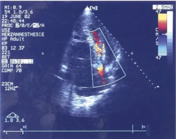
On arrival at our hospital this oligosymptomatic patient was walking about the hospital ward as if nothing happened. Physical examination showed turgescent jugular veins and absence of heart sounds, the pump making its normal turbine like noise. Precordial ECG, however, showed VF with irregular pacemaker activity. The patient was rapidly transferred to the ICU. Pacemaker memory function (MedTronic InSync III, with automatic diagnosis system) yielded very surprising information, the onset of VF happened 6 h prior the onset of symptoms. Pressure curves displayed on the clinical data acquiring system (CDAS) of the LVAD and on the ICU monitoring screens were completely flat, attesting to the absence of left ventricular ejection (Fig. 1). Absence of flow through the aortic valve and impactment of the right ventricle on the left were documented by echography (Fig. 2). The INR was 2.7 and potassium was found at 4 mmol/l. Heart enzymes had risen significantly (Troponin T 5.88 mg/l, CK tot. 712 U/l,
CK-Mb act. 71 U/l, Myoglobin 278 mg/l). After intravenous sedation (propofol and fentanyl) and bolus of 5000 IE heparin we proceeded to electroconvert the patient. Three attempts with A/P external defibrillation (360 J) and intravenous lidocaine were necessary to restore regular sequential paced heart rhythm. Restored left ventricular ejection and flow through the aortic valve were observed on echocardiography, cardiovascular monitoring and on the display of the CDAS. The patient regained consciousness a few minutes later with a stable cardiac rhythm requiring no pharmacological support. Laboratory values showed a normalisation of cardiac enzymes after 5 days (Troponin T 1.61 mg/l, CK tot. 73 U/l, CK-Mb act. 11 U/l, Myoglobin 88 mg/l).
Twelve hours after defibrillation the patient developed severe neurological symptoms. A cerebral CT-scan showed a massive frontal intra-cerebral haemorrhage. Upon this all anticoagulation was stopped. The patient never recuperated from this neurological event and the LVAD was turned of 14 of ICU treatment.
3. Comment
Very few reports describing survival after sustained VF exist in the literature, VF being lethal if not treated immediately[8,9].
Fig. 1. ICU monitoring before electro conversion.
Fig. 2. Trans thoracic echocardiography (during VF) shows accelerated flow through compressed left ventricle with dilated right ventricle. S.P. Salzberg et al. / European Journal of Cardio-thoracic Surgery 26 (2004) 444–446 445
This LVAD provides assistance for the failing heart. In this report we illustrate the role of this small device creating sufficient blood flow to allow viable end-organ function even with absence of left ventricular ejection.
The mechanisms causing VF are well known. Generally the aetiology is cardiac (dilatative or ischaemic cardiomyo-pathy) or metabolic. We believe that hypo-kaliemia induced through the diuretic action of furosemide combined with an excess of physical exercise under extreme conditions (altitude, heat, sun) caused this arrhythmia. The patient having an underlying cardiomyophathy known to cause arrhythmia, this VF was probably precipitated by the hypokaliemia.
By survival and recuperation of efficient cardiac activity after such a long period of VF, the normalisation of cardiac enzymes (CK, Myoglobin, Troponin T) provide insight into the physiological consequences of VF. The flow (2.5 – 3.4 l/min) provided by the LVAD, was sufficient to avoid irreversible ischaemic cardiac damage. In contrast to the maximum flow capacity of this device (. 10 l/min) the turbine was functioning in a low speed mode (only 8500 rpm—maximum 15,000 rpm). This shows the inten-sity of necessary flow to effectively replace cardiac output. The physiological mechanism allowing a sustained effective pulmonary circulation in the absence of left ventricular ejection is due to two factors: the increase in central venous pressure and the negative pressure at the inflow-tract generated by the turbine in this micro-axial pump. The blood is passively moved through the lungs, suctioned by the turbine and propelled into the aorta. In this case this was enough to allow sufficient oxygenation and end organ perfusion for survival.
This report demonstrates the efficacy of the LVAD not only as circulatory support but also as effective substitute in absence of any cardiac activity.
Bleeding is the most frequent complication of mechan-ical support systems (MCS). But the medmechan-ical management of terminal heart failure very often includes oral antic-oagulation and platelet inhibition of any kind. In this case, retrospectively one could consider that the IV bolus of heparin (standard protocol for electro conversion) may have lead to the neurological outcome. We also believe that it is further necessary to study the precise effects of heparin, continuous flow and the brain blood barrier, all this in
the setting of long-term support periods which destination therapy patients will undergo.
Furthermore the question now may arise as to the artificial heart. If left ventricular assistance is sufficient for life support, additional fully implantable devices replacing the heart will not be necessary, adding implantable automatic defibrillators would be enough for long-term assistance. Further studies are being conducted in this field, and we hope that our experience will be a valuable brick in the fight against heart failure.
References
[1] Al-Khatib SM, Granger CB, Huang Y, Lee KL, Califf RM, Simoons ML, Armstrong PW, Van de Werf F, White HD, Simes RJ, Moliterno DJ, Topol EJ, Harrington RA. Sustained ventricular arrhythmias among patients with acute coronary syndromes with no ST-segment elevation: incidence, predictors, and outcomes. Circulation 2002;106:309– 12. [2] Angelos MG, Menegazzi JJ, Callaway CW. Bench to bedside:
resuscitation from prolonged ventricular fibrillation. Acad Emerg Med 2001;8:909– 24.
[3] Shuster M, Tang A. Advanced cardiovascular life support Guidelines 2000: pharmacological changes to the treatment of ventricular fibrillation/pulseless ventricular tachycardia. Can J Cardiol 2001;17: 1022 – 5.
[4] Rose EA, Gelijns AC, Moskowitz AJ, Heitjan DF, Stevenson LW, Dembitsky W, Long JW, Ascheim DD, Tierney AR, Levitan RG, Watson JT, Meier P, Ronan NS, Shapiro PA, Lazar RM, Miller LW, Gupta L, Frazier OH, Desvigne-Nickens P, Oz MC, Poirier VL. Long-term mechanical left ventricular assistance for end-stage heart failure. N Engl J Med 2001;345:1435 – 43.
[5] Noon GP, Morley DL, Irwin S, Abdelsayed SV, Benkowski RJ, Lynch BE. Clinical experience with the MicroMed DeBakey ventricular assist device. Ann Thorac Surg 2001;71:S133 – 8. Discussion S144 – 6. [6] Frazier OH. The development of an implantable, portable, electrically
powered left ventricular assist device. Semin Thorac Cardiovasc Surg 1994;6:181– 7.
[7] Cohn JN. Mortality as an endpoint for cardiac failure therapy. J Card Fail 1995;1:191– 4.
[8] Lewis CT, Graham TR, Marrinan MT, Chalmers JA, Colvin MP, Withington PS, Coumbe A. The use of an implantable left ventricular assist device following irreversible ventricular fibrillation secondary to massive myocardial infarction. Eur J Cardiothorac Surg 1990;4:54 – 6. [9] McGinley AM, Matthews AS, Graham TR. Use of an implantable left ventricular assist device for irreversible ventricular fibrillation secondary to massive myocardial infarction—a case study. Intensive Care Nurs 1990;6:138 – 49.
S.P. Salzberg et al. / European Journal of Cardio-thoracic Surgery 26 (2004) 444–446 446