Review article
Less invasive aortic valve surgery: rationale and technique
q
Ludwig K. von Segesser
a,*, Stephen Westaby
b, Jose Pomar
c, Daniel Loisance
d,
Peter Groscurth
e, Marko Turina
faDepartment for Cardio-vascular Surgery, University Hospital Vaudois, CHUV, CH-1011 Lausanne, Switzerland bOxford Heart Center, Oxford, UK
cDepartment for Cardio-vascular Surgery, Hospital Clinico, Barcelona, Spain
dService de chirurgie thoracique et cardio-vasculaire, Henri Mondor Hospital, CreÂteil/Paris, France eInstitute of Anatomy, University of Zurich, Zurich, Switzerland
fDepartment of Cardio-vascular Surgery, University Hospital, Zurich, Switzerland
Received 20 January 1999; received in revised form 10 March 1999; accepted 16 March 1999
Abstract
The unquestionable aims for a less invasive operations are less morbidity, less discomfort, and a reduced hospital stay through an operation which proves equally durable to the conventional approach. Such an operation must be carried out without further risk to the patient or increased dif®culty for the surgeon. Whilst most de®nitions of less invasive coronary surgery include the phrase without cardiopulmonary bypass, this is clearly not yet possible in valve surgery. In valve surgery, the de®nition of less invasive relates only to the size of incision and rate of recovery. As a result of the discussions during the Heart Lab International Workshop on video-assisted heart surgery in ZuÈrich, October 22±25, 1998, the following conclusions emerged. The partial upper sternotomy with J- or L- shaped extension to the right is the preferred approach for minimally invasive aortic valve surgery. Other methods which sacrify the internal thoracic arteries, open pleural cavities or predispose to long hernia are less satisfactory. A detailed description of the technique proposed is given and its indications and contraindications are discussed. q 1999 Elsevier Science B.V. All rights reserved.
Keywords: Aortic valve surgery; Less invasive surgery; Minimally invasive surgery; Limited access; Partial sternotomy
1. Introduction
The two great advances that promoted widespread adop-tion of cardiac surgery were cardiopulmonary bypass and median sternotomy. Cardiopulmonary bypass [1] allowed open operations without time constraint on an open quiet heart. Median sternotomy [2] provided direct access for central cannulation without the complications of the femoral route or pain of the thoracotomy incision. Many of the bewildering so called less invasive cardiac approaches now deviate from these sound principles by increasing surgical dif®culty (and risk to the patients) for the sake of a fractionally shorter set of incisions. Most require expen-sive new equipment presently unfamiliar to experienced surgeons. Particularly questionable is the role of incremen-tal cosmesis in the surgery of elderly patients with life threa-tening illness. The unquestionable aims for a less invasive
operation are less morbidity, less discomfort, and a reduced hospital stay through an operation which proves equally durable to the conventional approach. Such an operation must be carried out without further risk to the patient or increased dif®culty for the surgeon. Whilst most de®nitions of less invasive coronary surgery include the phrase without cardiopulmonary bypass [3], this is clearly not yet possible in valve surgery. In valve surgery the de®nition of less invasive relates only to the size of incision and rate of recovery [4]. However, nothing that signi®cantly prolongs cardiopulmonary bypass can be described as less invasive [5]. There can be no trade off between length of skin inci-sion and increased cerebral morbidity [6]. When suf®cient neurons are lost, the patient does not notice the size of an incision. Within these constraints we believe that one parti-cular approach conveys substantial bene®ts and warrants adoption by all cardiac surgical centers. The upper partial sternotomy with unilateral L- or J-shaped extension to the right through the third or forth intercostal space [7] provides a window through which the aortic root is freely accessible. Many mitral operations can also be performed through the roof of the left atrium [7] using this approach. All cannulas
1010-7940/99/$ - see front matter q 1999 Elsevier Science B.V. All rights reserved. PII: S1010-7940(99)00119-0
qSummary of the discussions during the Heart Lab International
Work-shop on Video-assisted Heart Surgery, Zurich, October 22±25, 1998. * Corresponding author. Tel.: 1 3142280; fax: 1 41-21-3142279.
are introduced through this incision and no new instruments, retractors, and ports are necessary. With only modest increase in dif®culty and without additional risk to the patients the surgeon employs familiar techniques and the patient bene®ts from expedite recovery provided the patients are properly selected.
2. Patient selection
Aortic valve or root replacement [8] can only be performed by partial upper sternotomy if the patient does not require concomitant coronary artery bypass (see also Table 1). Aortic valve reoperations are feasible by this method but certain anatomical factors such as the length of the ascending aorta and adverse pathology including extensive calci®cation may mitigate against a less invasive approach. The technique is particularly applicable to the elderly and those with impaired respiratory function since both pleural cavities can be kept intact.
The level of the sternal division necessary to provide access to the aortic annulus varies greatly with body habitus, the presence or absence of chronic obstructive airway disease, and whether the heart lies transversely or longitud-inally within the chest. The standard preoperative chest radiography provides valuable information about the rela-tive positions of the ascending aorta, its root and the ster-num. In some cases one can even recognise the exact level of the aortic valve because of its calci®cations. Transeso-phageal echocardiography as suggested by Sardari et al. [9] can be used to locate more precisely the aortic annulus and the depth of the echoprobe from the teeth used to predict whether the third or the fourth interspace is suitable for the length of the incision. During the learning curve use of the fourth interspace is suggested. Given that the principle aim
of the method is stability of the thoracic cage, ease of access to the aortic root and right atrial appendage is far more important than the length of the skin incision. Young female patients with an eye on cosmesis may prefer a submammary incision with lower partial sternotomy and cannulation of the femoral artery. Such patients are unlikely to suffer retro-grade embolism from a diseased throaco-abdominal aorta and division of the sternum as far as the manubrium provides suf®cient access for aortic crossclamping. Partial upper sternotomy has also been applied in infants and chil-dren for the arterial switch operation, tetralogy of Fallot and both aortic and mitral valve repair. Further reported proce-dures include aortoplasty for reduction of ascending aortic dilatation, repair of limited aortic dissection and resection of hypertrophic subaortic stenoses (septal myectomy) [10].
Table 1
Contraindications for less invasive aortic valve or root replacement through a partial upper sternotomy
Contraindications Signi®cant coronary artery disease
Very short ascending aorta Very long ascending aorta Ascending aorta with extensive calci®cation
Ascending aorta with severe atheromatosis
Porcelain aorta
Small aortic annulus in the elderly requiring patch enlargement of the aortic root Low left ventricular ejection fraction
Transesophageal
echocardiography not available Thin friable atrial wall
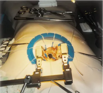
Fig. 1. Upper partial midline sternotomy (L- or J- type). The position of the surgeon is slightly different in that a view over the patients right shoulder gives direct vision of the aortic root (MT, SW, PG, LS, from the left).
Fig. 2. Upper partial midline sterotomy L- or J- type (mini-chest spreader by Ulrich AG, St. Gallen, Switzerland). Excellent view (SW in Fig. 1) of the opened aortic root during minimally invasive implantation of a state of the art mechanical prosthesis.
3. The surgical technique
Access to the aortic valve and root by partial upper ster-notomy allows the use of standard retractors, familiar cannulation, perfusion and myocardial protection techni-ques, and a straightforward valve operation without special instruments or videoscopy. The only difference between limited sternotomy and the conventional approach is that the surgeon has access only to the relevant part of the heart rather than the whole organ. The ascending aorta and right atrial appendage are upper midline structures within easy reach after upper midline sternal division. The position of the surgeon is slightly different in that a view over the patients right shoulder (Fig. 1) gives direct vision of the aortic root (Fig. 2). The patient is prepared and draped accordingly to provide access to the whole length of the sternum with the groins and upper legs prepared as for all valve operations. A single lumen endotracheal tube is used. The skin incision should be as long as the sternal incision since tension on the extremities of the soft tissues causes ischemia, an in¯ammatory reaction and the risk of hyper-trophic scarring. The skin incision is made in the mid line from just below the suprasternal notch to the third or fourth interspace according to the length of the ascending aorta and its position. An oscillating saw is then used to divide the sternum as far as the third or fourth interspace when a second narrow blade is used to transversely divide the right half of the sternum only (L-shaped sternotomy). Alter-natively the sternotomy can be started at the third or fourth intercostal space in an oblique fashion in order to join the
midline and turn there with the technique well established for a jigsaw (J-shaped incision). The right internal thoracic artery is usually 1 cm away from the sternal edge and can be protected by undercutting the edge of the skin incision and placing a forceps around the sternal edge to push the right internal thoracic artery laterally away from the saw. The L-or J-incision provides better sternal stability than the T approach [11] since the whole of the left side of the sternum remains intact. Usually the right pleural cavity can also be preserved. Thymic tissue is dissected and excised if neces-sary providing the usual access to the upper anterior peri-cardium. Keeping to the mid line the pericardium is then opened from the innominate vein to beneath the lower intact
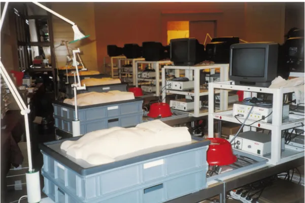
Fig. 3. Realistic chest phantoms with preserved porcine thoracic organs (Heart Lab International, Stans, Switzerland) ready for hands-on less invasive aortic valve replacement (video equipment by Treier/Wolf, BeromuÈnster, Switzerland).
Fig. 4. Delegates were able to practice the less invasive approach (Heart Lab in Zurich, October 22±25, 1998).
sternal table. Pericardial stay sutures are used to elevate the heart into the incision. The aorta is then cannulated just proximal to the innominate artery and a two stage venous cannula inserted into the right atrial appendage. After cardi-opulmonary bypass empties the right atrium gentle traction on the purse string of the two stage venous cannula helps to expose the aortic sinuses. The mode of cardioplegic arrest depends on surgeon's preference. Direct anterograde deliv-ery of cold crystalloid or blood cardioplegia is simple but a retrograde cannula can also be placed lower in the right atrium either blindly or with transesophageal echocardio-graphic guidance. A standard angled or curved aortic cross clamp is applied and sits out of the surgical ®eld if well placed. From this point the aortic procedure does not deviate from normal until the de-airing stages. Valve or full root replacement or repair is accomplished according to surgeon's preference. For stentless aortic valve replacement a transverse incision 0.5 cm above the aortic sinuses and at least 1 cm above the right coronary ostium is preferable. Appropriately positioned stay sutures eliminate the need for retractors in the operating ®eld and further deliver the aortic root into the limited incision. For mechanical valve implan-tation and use of stented bioprostheses some may prefer the standard oblique incision extending down to the annulus in the non-coronary sinus. Again stay sutures are preferable to retractors. The surgical ®eld is kept dry by a suction vent in either the left superior pulmonary vein, main pulmonary artery or the bottom of the left ventricle (trans-aortic) all of which are easily accessible with this method.
With the new valve reliably implanted it is important to secure closure of the aortotomy since bleeding from the root is less easily accessible via this approach when the heart is full. The de-airing process must also be thorough since this is achieved predominantly through the highest point of the aorta. Vent suction is discontinued as the aortotomy is closed so that the heart ®lls. The patient is tipped head down and rhythmic in¯ation of the lungs helps to expel air into the left ventricular out¯ow tract. A suction vent on the highest point of the aorta helps to evacuate bubbles. Partial aortic cross-clamping distally to the suction vent can improve the de-airing process. If the heart does not sponta-neously de®brillate then internal paediatric sized paddles are applied to the epicardium. Alternatively, sterilised exter-nal paddles are used within the surgical ®eld or soft patches positioned on the chest prior to the draping are activated. Transesophageal echocardiography is used continuously to check de-airing and to detect right ventricular disfunction due to air embolism which may require a period of contin-ued support. It is useful to place the right ventricular pacing wires and a pericardial drain before the heart is ®lled.
After discontinuing cardio-pulmonary bypass the cannu-las are clamped and removed. Protamine is administered. The wounds are checked for bleeding and then the pericar-dial stay sutures are released. The sternal edges are then accurately opposed with wire sutures taking care not to damage the internal thoracic arteries. With the L- or
J-incision three or four wires between the two halves of the sternal table are suf®cient. There is no need to add wires between the upper and lower sternal portions. The great advantage of this approach is the simplicity with which full sternotomy can be performed in the event of dif®culty. Appropriate anaesthesia allows early (less than 2 h) extuba-tion in the recovery area and the patient in sinus rhythm with a stentless biological valve may leave hospital with antipla-telet therapy only on the third to fourth postoperative day (SW: personal experience).
4. Comment
Early experience has shown that many surgeons are unwilling to accept procedures complicated by dif®cult access, limited control, femoral cannulation and video-scopic techniques [12] with which they are unfamiliar. Many dispute the cosmetic advantage of multiple stab wounds, additional groin incisions, the risk of endoaortic occluders [13], and the alleged less painful thoracotomy incision. However none of these disadvantages apply to the technique of aortic valve replacement described. The upper partial sternotomy offers the comfort factor of ster-notomy over thoracotomy but prevents complications of other distentions at the costovertebral joint or brachial plexus traction at the thoracic inlet. The integrity of the thoracic cage is better preserved and the pleural cavities are usually kept intact thereby reducing the risk of pleural effusion. The risks of sternal instability are reduced as long as partial sternotomy is performed with care. Aside from a marginally better cosmetic result the role of the small inci-sion on patient psychology should not be underestimated. These patients mobilise earlier and cough more effectively. In relation to cost the operation takes only a little longer in the operating room and does not require new equipment. This is in contrast to the robot assisted techniques with innumerable new instruments and devices which greatly prolong operating room time and duration of cardiopulmon-ary bypass.
What then are the disadvantages of this technique? Most surgeons instinctively rely on seeing the whole heart and manual cardiac manipulation contributes to the de-airing process. Without being able to see the left ventricle dif®cul-ties in weaning from cardio-pulmonary bypass may be unexpected and right ventricular dysfunction through air embolism is less apparent. However the routine use of trans-esophageal echocardiography is advised for less invasive aortic valve replacement and provides excellent information on air in the heart and ventricular dysfunction on either side. Though the surgical sites are readily accessible during the course of the procedure, access to the aortic root is more dif®cult when the heart is full. Videoscopic inspection of the operative ®eld and especially behind the aorta as well as behind the sternum can be useful under these circumstances in order to complete the hemostasis. Unexpected bleeding or
cardiac tamponade in the recovery area is correspondingly less accessible without rapid access to full sternotomy. In the event of profound haemodynamic deterioration immedi-ate availability of a scalpel and a sternal saw in the recovery area is therefore of prime importance. As usual, the success of a surgical procedure is initiated well before the skin incision, namely with the indication. Therefore the contra-indications listed in Table 1 have to be stressed here once more.
All new techniques have a learning curve. This is parti-cularly true for new surgical approaches. The Food and Drug Administration (USA) now recommends that stentless aortic valve replacement with the newer bioprostheses should be taught in surgical workshops. Similar wisdom should be applied to the teaching of less invasive operations. This report follows such a workshop convened by the authors in Zurich (Fig. 3). The relative merits and disadvan-tages of various less invasive approaches to the aortic valve were discussed with the ®rm conclusion that the partial upper sternotomy with L- or J- shape extension to the right was preferable. Other methods which sacri®ced the internal thoracic arteries, open the pleural cavities or pre-disposed to long hernia are less satisfactory. Delegates were able to practice the less invasive approach (Fig. 4) by using a realistic anatomical model of the chest which covered the preserved thoracic organs of a 70±80 kg pig. The biological component was directly positioned to simulate the human mediastinum, heart and great vessels. With this model work-shop participants were able to work through the steps of less invasive aortic valve replacement using intra-annular, supra-annular, and mini-root replacements before applying these techniques to their own patients.
References
[1] Gibbon JH. Application of mechanical heart and lung apparatus to cardiac surgery. Minnesota Med 1954;37:171.
[2] Westaby SM, Benetti FJ. Less invasive coronary surgery: consensus from Oxford Meeting. Ann Thorac Surg 1996;62:924±931. [3] Cala®ore AM, Di Giammarco G, Teodori G. Left anterior descending
coronary artery grafting via left anterior small thoracotomy without cardiopulmonary bypass. Ann Thorac Surg 1996;61:658±665. [4] Cooley DA. Minimally invasive valve surgery versus the
conven-tional approach. Ann Thorac Surg 1998;66:1101±1105.
[5] Kirklin JK, Westaby S, Blackstone EH, Kirklin JW, Chenoweth DE, Paci®co AD. Complement and the damaging effect of cardiopulmon-ary bypass. J Thorac Cardiovasc Surg 1983;86:845±857.
[6] Taylor KM. Brain damage during cardiopulmonary bypass. Ann Thorac Surg 1998;65:S20±S26.
[7] Svensson LG, D'Agostino RS. `J' incision minimal access valve operations. Ann Thorac Surg 1998;66:1110±1112.
[8] Westaby S, Katsumata T. Less invasive aortic root replacement. Ann Thorac Surg 1998;66:1400±1401.
[9] Sardari FF, Schlunt ML, Applegate RI, Gundry SR. The use of trans-esophageal echocardiography to guide sternal division for cardiac operations via mini-sternotomy. J Cardiac Surg 1997;12:67±70. [10] von Segesser, L.K. Die allgegenwaÈrtige Zukunft/Le futur preÂsent
(Editorial). Swiss Surgery 1998;(Suppl. 2):2-3.
[11] Cosgrove III DM, Sabik JF. Minimally invasive approach for aortic valve operations. Ann Thorac Surg 1996;62:596±597.
[12] Tevaearai H, Mueller X, Stumpe F, Ruchat P, von Segesser LK. Remote control of video assistance during minimally invasive coron-ary bypass procedures. Ann Thorac Surg 1999; in press.
[13] Falk V, Walther T, Diegeler A, Wendler R, Autschbach R, van Son JAM, Siegel LC, Pompilli MF, Mohr FW. Echocardiographic moni-toring of minimally invasive mitral valve surgery using an endoaortic clamp. J Heart Valve Dis 1996;5:630±671.