Clinical picture
An early sign of superior vena cava syndrome
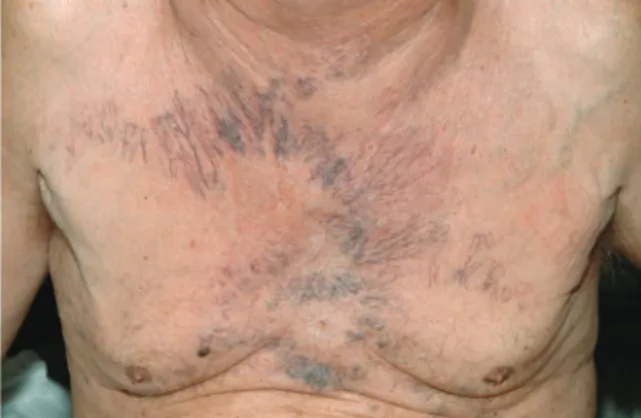
A 68-year-old man noticed progressive enlargement of the superficial veins on his anterior chest. One month later he developed dyspnea, headache and swelling of the neck and face. On examination, tele-angiectatic distension of the superficial veins of the anterior chest wall was found to be prominent (Figure 1). A clinical diagnosis of superior vena cava syndrome was made.
Further investigation by computer tomography of the chest showed extensive mediastinal lymph node enlargement with obstruction of the superior cava vein. Histological work up revealed small cell lung cancer.
Tortuous dilatation of the subcutaneous veins of the chest wall in the shape of a garland may be an early sign of impaired venous return from the upper extremity and often precedes other signs of the superior vena cava syndrome for weeks. Its’ appear-ance should lead to a thorough investigation of the etiology. In most cases, it is caused by malignant disorders, but thrombosis or inflammatory disease is also possible.
Photographs and text from: Stephan Wieser, MD, Malcolm Kohler, MD, Division of Pulmonary Medicine, University Hospital of Zurich, Raemistrasse 100, CH-8091 Zurich, Switzerland. email: stephan.wieser@sunrise.ch
Figure 1. Dilated tortuous superficial veins of the chest wall in the shape of a garland.
! The Author 2009. Published by Oxford University Press on behalf of the Association of Physicians. All rights reserved. For Permissions, please email: journals.permissions@oxfordjournals.org
Q J Med 2010;103:707