A Consideration of Biomarkers to be
used for Evaluation of Inflammation
in Human Nutritional Studies
P.C. Calder
1, N. Ahluwalia
2, R. Albers
3,4, N. Bosco
5, R. Bourdet-Sicard
6,
D. Haller
7, S.T. Holgate
1, L.S. Jo¨nsson
8, M.E. Latulippe
8, A. Marcos
9,
J. Moreines
10, C. M’Rini
11, M. Mu¨ller
12, G. Pawelec
13, R.J.J. van Neerven
14,
B. Watzl
15and J. Zhao
161. University of Southampton, Southampton SO16 6YD, UK 2. Inserm U557, University of Paris 13, Bobigny, France 3. Unilever R&D, 3130 AC, Vlaardingen, The Netherlands 4. NutriLeads, 3235 KT, Rockanje, The Netherlands 5. Nestle´ Research Center, 1000 Lausanne, Switzerland 6. Danone Research, 91767 Palaiseau, France
7. Technical University of Munich, 85354 Freising, Germany 8. ILSI Europe, 1200 Brussels, Belgium
9. ICTAN-CSIC Spanish National Research Council, Madrid 28040, Spain 10. Pfizer, Madison, NJ 07940, USA
11. Institut Me´rieux, 69002 Lyon, France
12. Wageningen University, 6703 HD, Wageningen, The Netherlands 13. University of Tu¨bingen, D-72072 Tu¨bingen, Germany
14. FrieslandCampina, 3818 LE, Amersfoort, The Netherlands 15. Max Rubner-Institut, 76131 Karlsruhe, Germany
16. Yakult Europe, 1332 EN, Almere, The Netherlands
Commissioned by the
ILSI Europe Nutrition and Immunity Task Force
Correspondence: ILSI Europe a.i.s.b.l. - Avenue E. Mounier 83, Box 6 - 1200 Brussels - Belgium
Email: [email protected] - Fax þ 32 2 762 00 44
https:/www.cambridge.org/core/terms. https://doi.org/10.1017/S0007114512005119
Vol. 109
Supplement No. S1
January 2013
British Journal of Nutrition
Table of Contents
Biomarkers of inflammation S3 – S9
Modifiers of inflammatory markers S9 – S16
Challenge models S16 – S20
Emerging markers of inflammation S20 – S22
Emerging technologies S22 – S24
Summary, conclusions, identification of key gaps, overall recommendations
and overall key messages S24 – S25
Acknowledgements S25
Key words:Inflammation: Leucocytes: Cytokines: Chemokines: Acute phase
Correspondence: ILSI Europe a.i.s.b.l., Av. E. Mounier 83, Box 6, 1200 Brussels, Belgium, fax: þ 32 2 762 00 44, email: [email protected]
Abbreviations: ASCA, anti-Saccharomyces cerevisiae antibodies; CPN, chaperone; CRP, C-reactive protein; ER, endoplasmic reticulum; IBD, inflammatory bowel disease; ICAM, intercellular adhesion molecule; ILSI, International Life Sciences Institute; LPS, lipopolysaccharide; MCP, monocyte chemoattractant protein; miRNA, microRNA; OGTT, oral glucose tolerance test; P-ANCA, perinuclear anti-neutrophil cytoplasmic antibodies; RA, rheumatoid arthritis; UPR, unfolded protein response; VCAM, vascular cell adhesion molecule; Xbp-1, X-box binding protein 1.
qILSI Europe [2013]
https:/www.cambridge.org/core/terms. https://doi.org/10.1017/S0007114512005119
To monitor inflammation in a meaningful way, the markers used must be valid: they must reflect the inflammatory process under study and they must be predictive of future health status. In 2009, the Nutrition and Immunity Task Force of the International Life Sciences Institute, European Branch, organized an expert group to attempt to identify robust and predictive markers, or patterns or clusters of markers, which can be used to assess inflammation in human nutrition studies in the general population. Inflammation is a normal process and there are a number of cells and mediators involved. These markers are involved in, or are produced as a result of, the inflammatory process irrespec-tive of its trigger and its location and are common to all inflammatory situations. Currently, there is no consensus as to which markers of inflammation best represent low-grade inflammation or differentiate between acute and chronic inflammation or between the various phases of inflammatory responses. There are a number of modifying factors that affect the concentration of an inflammatory marker at a given time, including age, diet and body fatness, among others. Measuring the concentration of inflammatory markers in the bloodstream under basal conditions is probably less informative compared with data related to the concentration change in response to a challenge. A number of inflammatory challenges have been described. However, many of these challenges are poorly standardised. Patterns and clus-ters may be important as robust biomarkers of inflammation. Therefore, it is likely that a combination of multiple inflammatory markers and integrated readouts based upon kinetic analysis following defined challenges will be the most informative biomarker of inflammation.
The overall aim of the present study is to attempt to identify robust and predictive markers, or patterns or clusters of mar-kers, which can be used to assess inflammation in human nutrition studies in the general population. Inflammation is a normal component of host defence, initiating the mechanisms involved in pathogen killing and protection against other insults. Thus, in a physiological context, inflammation is pro-tective. Typically, the inflammatory response is activated rapidly in response to infection or another trigger and then follows a temporal pattern of cellular activation and chemical mediator release. Once the infection or the other insult is eliminated, or at least controlled, mechanisms come into play to terminate inflammation in order to limit further damage to the host and to initiate tissue repair. This process is termed resolution of inflammation, and it is now recognised to be an active process involving specific mediators that act to down-regulate the processes that were earlier activated. Fail-ure to resolve inflammation may permit the normally acute inflammatory processes to become chronic. Chronic inflam-mation may also be driven by continuous exposure to the triggering agent. Chronic inflammation is a well-recognised component of many pathologies and is a target for many phar-macologic interventions. In order to examine the role of diet, foods or specific nutrients on inflammation, it is necessary to identify candidate biomarkers. This review describes a sys-tematic approach to identify generic biomarkers of inflam-mation and to consider the factors that may influence the status of those biomarkers.
Biomarkers of inflammation Inflammation: a general overview
Inflammation is a normal host defence mechanism that pro-tects the host from infection and other insults; it initiates pathogen killing, as well as tissue repair processes, and
helps to restore homeostasis at infected or damaged sites(1).
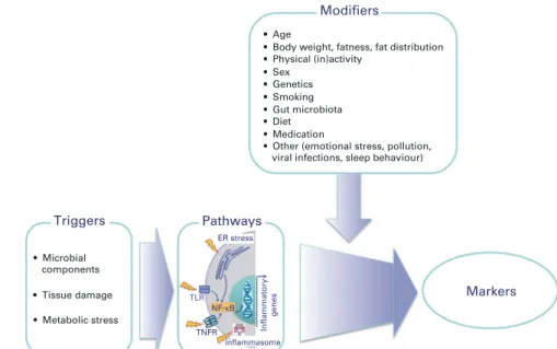
A number of different triggers can initiate inflammation. These triggers include the presence of microbial products, tissue damage and metabolic stress (Fig. 1). Exposure to the
trigger may involve a breakdown in barrier function or a loss of normal immune tolerance. Irrespective of the nature
of the trigger, a common set of cellular pathways is initiated(2).
These pathways include activation of signalling via Toll-like receptors and nuclear factor (NF)-kB, formation of a multi-protein complex termed the inflammasome that is responsible for the release of several inflammatory cytokines, and
endo-plasmic reticulum (ER) stress (Fig. 1)(2). The resulting
inflam-matory response is typified by redness, swelling, heat, pain and loss of function, and involves interactions among many cell types and the production of, and responses to, a vast number of chemical mediators. The precise timing and vigour of the inflammatory response, including the exact cells and mediators involved, is determined by the nature, extent and location of the trigger. These factors act to shape and maintain the inflammatory response. Once the infection or the other trigger is eliminated, or at least controlled, mech-anisms come into play to terminate inflammation in order to limit further damage to the host and to initiate tissue repair. This self-regulating process is termed resolution of inflam-mation and involves the activation of negative feedback mech-anisms such as the secretion of anti-inflammatory cytokines, the inhibition of pro-inflammatory signalling cascades, the shedding of receptors for inflammatory mediators and the acti-vation of regulatory cells. Properly controlled inflammation is essential to remain healthy and maintain homeostasis, but inflammation that involves a loss of tolerance or regulatory
processes may become pathological(1).
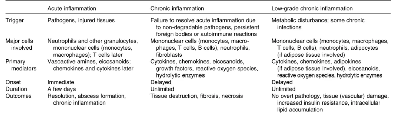
Inflammation may be classified as acute or chronic (Table 1). Acute inflammation is the initial response of the body to an infectious agent or another inflammatory trigger (e.g. tissue damage by wounding or irradiation) and is achieved by the increased movement of plasma and leucocytes (especially granulocytes) from the blood into the site of infection or injury. A cascade of biochemical events matures and propa-gates the inflammatory response, involving the local vascular system, the immune system and various cells within the injured tissue. Acute inflammation is typically self-limiting with resolution being activated once the cause of the initial
Biomarkers of inflammation S3
British
Journal
of
Nutrition
https:/www.cambridge.org/core/terms. https://doi.org/10.1017/S0007114512005119response is contained or eliminated. Resolution is itself an active process involving specific cell types and specific
anti-inflammatory cytokines(3) and pro-resolving lipid
mediators(4,5). Prolonged, or chronic, inflammation involves
a progressive shift in the type of cells present at the site of inflammation and simultaneous destruction and healing of the tissue due to the ongoing inflammatory process. Inflam-mation may become pathological due to the loss of tolerance and/or of regulatory processes such as resolution. Where this becomes excessive, irreparable damage to host tissues and
disease can occur(1). Such diseases are characterised by
mark-edly elevated concentrations of inflammatory markers and of activated inflammatory cells at the site of tissue damage and in the systemic circulation; this state of inflammation may be regarded as ‘high grade’. These diseases include rheumatoid arthritis (RA), inflammatory bowel diseases (IBD), atopic der-matitis, psoriasis and asthma. Chronic inflammation can also be of a ‘low grade’ and under such circumstances, overt clini-cal manifestations can be minimal or absent; the elevation in the concentrations of inflammatory markers and of inflamma-tory cells in the systemic circulation is not as great as that seen in the frank chronic inflammatory conditions described above. Low-grade asymptomatic inflammation can occur in adipose
tissue as a feature of obesity(6). Under these conditions,
the adipocyte itself becomes the source of inflammation-associated adipokines, although there is also infiltration of adipose tissue by macrophages and T cells, which make important contributions to the inflammatory output from
adi-pose tissue(7)
. The cytokines and adipokines released
prob-ably contribute to the insulin resistance seen in obesity(8).
Older people also commonly exhibit low-grade chronic inflammation that may contribute to frailty, depending on the plasma levels of factors such as tumor necrosis factor (TNF) and interleukin (IL)-6.
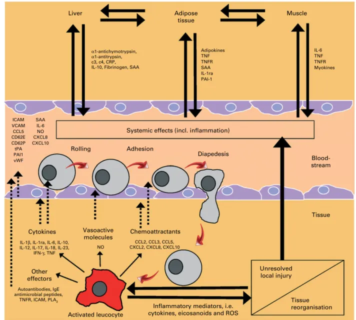
Common to both acute and chronic inflammation is that they have an afferent phase, in which the presence of a trigger is sensed by some types of cells, and an efferent phase, in which an inflammatory response is generated to eliminate the perceived hostile intruder (i.e. the source of the trigger). Irrespective of the type of inflammation, the response involves four major events. The first event is increased blood supply to the site of inflammation. The second event is increased capil-lary permeability caused by retraction of endothelial cells. This permits larger molecules, not normally capable of traversing the endothelium, to do so and thus delivers some soluble mediators to the site of inflammation. The third event is leucocyte migration from the capillaries into the surrounding tissue (Fig. 2). This is promoted by the release of chemo-attractants from the site of inflammation and by the up-regulation of adhesion molecules on the endothelium. Once in the tissue, the leucocytes move to the site of inflammation. The fourth event is the release of mediators from leucocytes at the site of inflammation. These may include lipid mediators (e.g. prostaglandins (PG), leukotrienes; see Table 2), peptide mediators (e.g. cytokines, chemokines; see Table 3), reactive oxygen species (e.g. superoxide),
Triggers Pathways ER stress TLR TNFR Inflammasome Inflammatory genes NF-κB Inflammatory response • Microbial components • Tissue damage • Metabolic stress
Fig. 1. Schematic overview of the common inflammatory response initiated by different triggers. Triggers are those factors that can directly initiate an inflammatory response. ER, endoplasmic reticulum; TLR, Toll-like receptor; TNFR, TNF receptor.
Table 1. Features of acute, chronic and low-grade chronic inflammation
Acute inflammation Chronic inflammation Low-grade chronic inflammation Trigger Pathogens, injured tissues Failure to resolve acute inflammation due
to non-degradable pathogens, persistent foreign bodies or autoimmune reactions
Metabolic disturbance; some chronic infections
Major cells involved
Neutrophils and other granulocytes, mononuclear cells (monocytes, macrophages); T cells later
Mononuclear cells (monocytes, macro-phages, T cells, B cells), neutrophils, fibroblasts
Mononuclear cells (monocytes, macrophages, T cells, B cells), neutrophils, adipocytes (if adipose tissue involved)
Primary mediators
Vasoactive amines, eicosanoids; chemokines and cytokines later
Cytokines, chemokines, eicosanoids, growth factors, reactive oxygen species, hydrolytic enzymes
Cytokines, chemokines, adipokines (if adipose tissue involved), eicosanoids, reactive oxygen species, hydrolytic enzymes
Onset Immediate Delayed Delayed
Duration A few days Unlimited Unlimited
Outcomes Resolution, abscess formation, chronic inflammation
Tissue destruction, fibrosis, necrosis No overt pathology, tissue (vascular) damage, increased insulin resistance, intracellular lipid accumulation P. C. Calder et al. S4
British
Journal
of
Nutrition
https:/www.cambridge.org/core/terms. https://doi.org/10.1017/S0007114512005119amino acid derivatives (e.g. histamine) and enzymes (e.g. matrix proteases), depending upon the cell type involved, the nature of the inflammatory trigger, the anatomical site involved and the stage during the inflammatory response. Several of the mediators involved act to amplify the inflam-matory process by acting, for example, as chemoattractants. Some of the inflammatory mediators escape the inflammatory site into the circulation and from there, they can exert systemic effects (Fig. 2). For example, the cytokine IL-6 released into the bloodstream from an inflammatory locus induces hepatic synthesis of the acute-phase protein C-reactive protein (CRP), while the cytokine TNF elicits metabolic effects within skeletal muscle, adipose tissue and bone. Thus, inflam-mation at one site, such as within the intestinal mucosa, can cause inflammation-driven changes at distal sites, such as in bone.
The need to identify biomarkers of inflammation
Inflammation has long been recognised to play a key role in the pathology of a number of human diseases, including RA, psoriasis, Crohn’s disease, allergic asthma and atopic dermati-tis, being centrally involved in the tissue damage that is seen. These diseases with a well-recognised inflammatory component are usually treated with general or specific anti-inflammatory pharmaceuticals with the aim of inducing a clini-cal benefit. More recently, it has become clear that metabolic diseases such as atherosclerosis, type 2 diabetes and obesity
have an inflammatory component(6,9 – 11), albeit low-grade;
however, the extent to which this is causal in the pathology is not yet clear. These diseases are not usually treated with anti-inflammatory pharmaceuticals. It is considered that many dietary factors can influence various aspects of
inflam-mation(1,6); thus, nutrition may play a role in predisposing to
ICAM VCAM CCL5 CD62E CD62P SAA Rolling Cytokines Chemoattractants Tissue Unresolved local injury Tissue reorganisation Blood-stream Liver Adipose tissue Adipokines TNF TNFR SAA IL-1ra IL-6 TNF TNFR Myokines PAI-1 Muscle Vasoactive molecules NO CCL2, CCL3, CCL5, CXCL2, CXCL8, CXCL10 Other effectors Activated leucocyte
Inflammatory mediators, i.e. cytokines, eicosanoids and ROS
Autoantibodies, IgE antimicrobial peptides,
TNFR, ICAM, PLA2 IL-1β, IL-1ra, IL-6, IL-10, IL-12, IL-17, IL-18, IL-23,
IFN-γ, TNF
Adhesion
Diapedesis Systemic effects (incl. inflammation)
α1-antichymotrypsin, α1-antitrypsin, c3, c4, CRP, IL-10, Fibrinogen, SAA
IL-6 NO CXCL8 CXCL10 tPA PAI1 vWF
Fig. 2. An overview of inflammation. C, complement; CRP, C-reactive protein; SAA, serum amyloid A; TNFR, TNF receptor; ra, receptor antagonist; PAI, plasmi-nogen activator inhibitor; ICAM, intercellular adhesion molecule; VCAM, vascular cell adhesion molecule; CCL, chemokine (C – C motif) ligand; CD62E, E-selectin endothelial leukocyte adhesion molecule 1; CD62P, P-selectin endothelial leukocyte adhesion molecule 1; CXCL, chemokine (C – X – C motif) ligand; tPA, tissue plasminogen activator; vWF, von Willebrand factor; IFN, interferon; PLA, phospholipase A; ROS, reactive oxygen species.
Biomarkers of inflammation S5
British
Journal
of
Nutrition
https:/www.cambridge.org/core/terms. https://doi.org/10.1017/S0007114512005119conditions that have an inflammatory component and altered nutrition may be useful in the prevention or treatment of such conditions(1,6).
In order to elucidate the role of diet or its component foods, nutrients and non-nutrients in inflammation, either as a predis-posing, preventative or therapeutic factor, there is a need to be able to monitor the inflammatory process in the subjects being studied. This requires the identification of biomarkers of inflammation. A biomarker is defined as ‘a characteristic that is objectively measured and evaluated as an indicator of normal biological processes, pathogenic processes, or
phar-macologic responses to an intervention’(12). However, clear
recommendations on which biomarkers to use and how to interpret patterns of biomarkers and changes in biomarkers in the context of inflammation are lacking. The Institute of Medicine recently presented a general concept for identifi-cation of biomarkers and surrogate endpoints in chronic
diseases(13). The Institute of Medicine recommended
basing the evaluation process for biomarkers on three steps:
(1) analytical validation of the biomarker (reference range, accuracy, limits of detection, reproducibility, etc.); (2) qualifi-cation of the available evidence on an association between the biomarker and disease states including data showing the effects of an intervention on both the biomarker and clini-cal outcomes; (3) use of a utilisation step that primarily includes the determination of whether analytical validation and qualification conducted provide sufficient support for
the proposed use of the biomarker(13). The overall aim of
this review is to identify robust and predictive markers, or patterns or clusters of markers, that can be used to assess inflammation in human nutrition studies in the general popu-lation and to create a consensus for grading these biomarkers.
How were the biomarkers chosen?
In order to select likely biomarkers of relevance to nutrition studies, it was decided to first identify general and disease-specific markers of inflammation within the circulation Table 2. Lipid mediators associated with inflammation*
Class Mediator Substrate Receptor(s)
Prostanoids PGD2 Arachidonic acid via COX DP1, DP2
PGE2 Arachidonic acid via COX EP1, EP2, EP3, EP4
PGF2a Arachidonic acid via COX FP
PGI2 Arachidonic acid via COX IP
TXA2 Arachidonic acid via COX TP
PGE1 Dihomo-g-linolenic acid via COX EP1, EP2, EP3, EP4
PGD3 EPA via COX DP1, DP2
PGE3 EPA via COX EP1, EP2, EP3, EP4
Leukotrienes 5-HETE Arachidonic acid via 5-LOX BLT2
5-HPETE Arachidonic acid via 5-LOX OXE
LTB4 Arachidonic acid via 5-LOX BLT1, BLT2
LTC4, D4, E4(termed cys-LT) Arachidonic acid via 5-LOX CysLT1, CysLT2
15-HETE Arachidonic acid via 15-LOX BLT2
15-HPETE Arachidonic acid via 15-LOX BLT2
12-HETE Arachidonic acid via 12-LOX BLT2
LTB5 EPA via 5-LOX BLT1, BLT2
Lipoxins LXA4 Arachidonic acid via 15-LOX and 5-LOX or 5-LOX
and 12-LOX (transcellular)
FPR2/ALX Endocannabinoids 2-Arachidonoylglycerol 1,2-Diacylglycerol with arachidonic acid at the sn-2
position
CB1, CB2 Anandamide N-arachidonoylphosphatidylethanolamide via
phos-pholipase D; in turn, N-arachidonoylphosphatidy-lethanolamide is formed from phosphatidylcholine with arachidonic acid at the sn-1 position and phosphatidylethanolamine
CB1, CB2
Resolvins, protectins and maresins
RvE1 EPA via acetylated COX-2 and 5-LOX (transcellular) RvE1 (ChemR23), BLT1 RvD1 DHA via acetylated COX-2 and 5-LOX or via 15-LOX
and 5-LOX (transcellular)
RvD1 (GPR32), ALX/FPR2 PD1 (NPD1) DHA via 15-LOX and LOX (transcellular) Not yet known
MaR1 DHA via 15-LOX and 12-LOX (transcellular) Not yet known Lysolipids PAF Phosphatidylcholine with diethyl ether link at the sn-1
position
PAF-R Lyso-PA Phosphatidic acid, which in turn is synthesised
from phosphatidylcholine
LPA1, LPA2, LPA3, LPA4, LPA5, LPA6
Sphingosine-1-phosphate Sphingosine, which in turn is synthesised from ceramide
S1P1, S1P2, S1P3, S1P4, S1P5
COX, cyclo-oxygenase; TX, thromboxane; HETE, hydroxyeicosatetraenoic acid; LOX, lipoxygenase; HPETE, hydroperoxyeicosatetraenoic acid; cys, cysteinyl; LT, leukotriene; LX, lipoxin; Rv, resolvin; P, protectin; NP, neuroprotectin; MaR, maresin; PAF, platelet-activating factor; Lyso-PA, lysophosphatidic acid; DP, EP, FP, IP, TP, prostanoid receptors; BLT, leukotriene B4 receptors; OXE, oxoeicosanoid receptor; D4, E4, Leukotriene receptors; FPR2, formyl peptide receptor 2; ALX, oncogenic receptor tyrosine kinase; CB, cannabinoid receptor; ChemR23, chemokine receptor-like 1 (also known as Chemerin receptor 23); RvD, resolving D; GPR32, G protein-coupled receptor 32; LP, lysophospholipid; S1P, sphingosine-1-phosphate.
* Note that the listing is not exhaustive.
P. C. Calder et al. S6
British
Journal
of
Nutrition
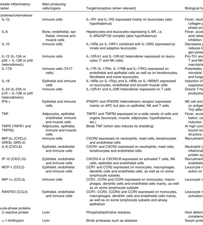
https:/www.cambridge.org/core/terms. https://doi.org/10.1017/S0007114512005119Table 3. Soluble peptides associated with inflammation* Soluble inflammatory
marker
Main producing
cells/organs Target/receptors (when relevant) Biological function(s)
Cytokines/chemokines
IL-1b Immune cells IL-1R1 and IL-1R2 expressed mainly on leucocytes (also
hypothalamus)
Fever, neutrophil attraction and activation, amino acid release by muscle, collagen production by fibroblasts, B and T lymphocyte activation, acute-phase protein synthesis by liver
IL-6 Bone, endothelial,
epi-thelial, immune and muscle cells
Hepatocytes and leucocytes expressing IL-6R, i.e. IL-6Ra/GP130 complex (also hypothalamus)
Fever, acute-phase protein synthesis, amino acid release by muscle, fatty acid release from adipose tissue, neutrophil and lymphocyte activation, inhibition of TNF and IL-1 and activation of IL-1ra and IL-10 secretion
IL-10 Immune cells IL-10Ra (or IL-10R1) combined with IL-10R2 expressed by
innate and adaptive leucocytes
Decreases pro-inflammatory cytokine expression, reduces Th17 generation, reduces MHC class II and co-stimulatory molecule expression by anti-gen-presenting cells, improves B-cell survival and antibody production IL-12 (IL-12A or
p35 þ IL-12B or p40 heterodimeric)
Immune cells IL-12R-b1 and IL-12R-b2 heterodimer expressed on
leuco-cytes (T and NK cells)
Pro-Th1 and anti-Th2 function, Th1 and NK cell proliferation, stimulation of T and NK cell cytotoxic activity, up-regulation of IL-18R, activation of macrophages, anti-angiogenic
IL-17A Immune cells (Th17
cells)
IL-17R (IL-17RA, IL-17RB and IL-17RC) expressed on endothelial and epithelial cells as well as on keratinocytes, fibroblasts and some leucocytes
Potentiates chemokine and pro-inflammatory cytokine release, triggers anti-microbial peptide secretion, clear role in skin protection against bacterial and fungal infections, involved in allergic responses
IL-18 Epithelial and immune
cells
IL-18Ra (or IL-1Rrp) and IL-18Rb (or IL-18RAP) expressed on leucocytes, endothelial and smooth muscle cells
Induction of IL-1b, IFN-g and TNF production. In combination with IL-12 suppresses Th2-mediated B-cell responses (IgE and IgG1 secretion) IL-23 (IL-23A or
p19 þ IL-12B or p40 heterodimeric)
Immune cells IL-12R-b1 and IL-23R heterodimer expressed on T cells Directs T-helper cell differentiation (Th17), induces IFN-g and IL-12
production
IFN-g Epithelial and immune
cells
IFNGR1 and IFNGR2 heterodimeric receptor expressed mainly on APC but also on epithelial, NK and T cells
NK cell and macrophage activation, stimulates expression of MHC I and II on antigen-presenting cells, promotes Th1 differentiation, suppresses Th2 differentiation, viral replication inhibition
TNF Adipocytes, epithelial,
endothelial, immune and muscle cells
TNFR1 and TNFR2 expressed on a wide variety of cells and organs (leucocyte, muscle, adipocytes, hypothalamus, etc.)
Fever, acute-phase protein synthesis, procoagulation, Th-cell differen-tiation, cachexia, apoptosis, inhibits viral replication, antitumour activity, induction of TNFR shedding as sTNFR
TNFR (TNFR1 and TNFR2)
Adipocytes, epithelial, immune and muscle cells
Binds TNF (which also induces its shedding) At high concentration inhibits TNF function (competition with
membrane-bound receptors). At low concentration, can stabilise TNF trimeric structure and improve half-life of TNF
MIP-2a (CXCL2; GROb; GRO-2)
Immune cells CXCR2 expressed on neutrophils, mast cells, keratinocytes
and endothelial cells
Recruitment of leucocytes and haematopoietic stem cells
IL-8 (CXCL8) Epithelial, endothelial
and immune cells
CXCR1 and CXCR2 expressed on neutrophils, mast cells, keratinocytes and endothelial cells
Neutrophil (and basophil) attraction and activation to the site of inflammation, angiogenic properties, stimulation of acute-phase protein synthesis
IP-10 (CXCL10) Epithelial, endothelial
and immune cells
CXCR3-A or CXCR3-B expressed on activated T cells, NK cells, epithelial and endothelial cells
Recruitment/trafficking of leucocytes and promotion of adhesion to endothelial cells. Expression largely controlled by IFN-g
MCP-1 (CCL2) Epithelial, endothelial
and immune cells
CCR1 and CCR2 expressed on monocytes, macrophages, dendritic cells and endothelial cells, as well as on some lymphocyte subsets
Leucocyte recruitment (monocytes, dendritic cells and memory cells); some action on mast cell degranulation
MIP-1a (CCL3) Immune cells CCR1, CCR4 and CCR5 expressed on monocytes,
macro-phages, dendritic cells and endothelial cells mainly, as well as on some lymphocyte subsets
Leucocyte recruitment (monocytes, B and T cells, as well as eosinophils)
RANTES (CCL5) Epithelial, endothelial
and immune cells
CCR1, CCR3, (CCR4) and CCR5 expressed on monocytes, macrophages, dendritic cells and endothelial cells mainly, as well as on some lymphocyte subsets and airway epithelium
Leucocyte recruitment (T cells, basophils and eosinophils), NK cell activation. Highly expressed upon pro-inflammatory cytokine exposure
Acute-phase proteins
C-reactive protein Liver Phosphatidylcholine residues Host defence (clearance of pathogens and dead cells) by opsonisation and
complement activation
a-1-Antitrypsin Liver Binds proteases such as elastase Serpin protease inhibitor (tissue protection)
Biomarkers
of
inflammation
S7
British Journal of Nutrition
https:/www.cambridge.org/core/terms
.
https://doi.org/10.1017/S0007114512005119
Downloaded from
https:/www.cambridge.org/core
. University of Basel Library
, on
10 Jul 2017 at 15:46:22
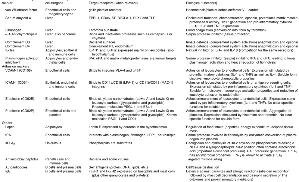
Table 3. Continued Soluble inflammatory marker
Main producing
cells/organs Target/receptors (when relevant) Biological function(s)
von Willebrand factor Endothelial cells and
megakaryocytes
gp1b platelet receptor Haemostasis/platelet adhesion/factor VIII carrier
Serum amyloid A Liver FPRL1, CD36, SR-BI/CLA-1, P2X7 and TLR Cholesterol transport, chemoattraction, opsonin, potentiates matrix
metallo-proteinase 9 activity, Th17 generation and pro-inflammatory cytokine (IL-1b, IL-6 and TNF) expression
Fibrinogen Liver Thrombin substrate Blood coagulation (conversion into fibrin by thrombin)
a-1-Antichymotrypsin Liver, also pancreas Binds and inactivates proteases such as cathepsin G or
chymase
Serpin protease inhibitor (tissue protection)
Complement C3 Liver Bacterial surfaces Innate defence (complement system activation) anaphylatoxin and opsonin
Complement C4 Liver Complement R1, endothelium Innate defence (complement system activation) anaphylatoxin and opsonin
IL-1ra Adipocytes, epithelial
and immune cells
IL-1R1 and IL-1R2 expressed mainly on leucocytes (also hypothalamus)
Natural inhibitor of IL-1a and IL-1b (competition for the same receptors) Plasminogen activator
inhibitor-1
Adipocytes and endo-thelial cells
tPA, uPA and matrix metalloproteinases are known targets Serine protease inhibitor (serpin) inhibiting tPA and uPA, leading to lower plasminogen activation and hence reduction of fibrinolysis
Adhesion markers
VCAM-1 (CD106) Endothelial cells Binds to integrins VLA-4 and a4b7 Adhesion of leucocytes to endothelial cells. Expression stimulated by
pro-inflammatory cytokines (IL-1 and TNF) as well as IL-4. Soluble form displays lymphocyte chemotactic properties
ICAM-1 (CD54) Epithelial, endothelial
and immune cells
Binds to CD11a/CD18 (LFA-1) or CD11b/CD18 (MAC-1) integrins
Adhesion of leucocytes to endothelial cells or antigen-presenting cells. Expression stimulated by pro-inflammatory cytokines (IL-1 and TNF). Soluble form displays macrophage activation properties and reduction of leucocyte adhesion to endothelium
E-selectin (CD62E) Endothelial cells Binds sialylated carbohydrates (Lewis A and Lewis X) on
leucocyte surface (glycoproteins and glycolipids). Proposed molecules PSGL-1 and ESL-1
Adhesion/recruitment of leucocytes to endothelial cells. Expression stimu-lated by pro-inflammatory cytokines (IL-1 and TNF). No clear specific functions for soluble form
P-selectin (CD62P) Endothelial cells and
platelets
Binds sialylated carbohydrates (Lewis A and Lewis X) on leucocyte surface (glycoproteins and glycolipids). Known molecules PSGL-1 and CD24
Adhesion/recruitment of leucocytes to endothelial cells. Aggregation of platelets. Expression stimulated by histamine and thrombin. No clear specific functions for soluble form
Others
Leptin Adipocytes Leptin R expressed by neurons in the hypothalamus Regulation of food intake (appetite), energy expenditure, adipose tissue
mass
tPA Endothelial cells Interacts with plasminogen, fibrinogen, LRP1, neuroserpin Serine protease involved in fibrinolysis by enzymatic conversion of
plasmi-nogen into plasmin
sPLA2 Ubiquitous Phospholipids are substrates Recognition and hydrolysis of sn-2 acyl-bound phospholipids releasing a
NEFA and a lysophospholipid. Sn-2 position often contains arachidonic acid (important eicosanoid precursor). PAF precursor generation. sPLA2 has antimicrobial properties. IFN-g is known to activate sPLA2
Antimicrobial peptides Paneth cells and
immune cells
Bacteria and some viruses Targeted microbe killing
Autoantibodies B cells and plasma cells Self antigens (protein, DNA, lipids, etc.) Cell/tissue destruction
IgE B cells and plasma cells Fc1R1 and Fc1R2 expressed on basophils and mast cells
(plus other granulocytes and platelets)
Defence against parasites and allergic reactions (allergen recognition followed by mast cell degranulation and basophil secretion of Th2 cytokines and pro-inflammatory mediators)
ra, receptor antagonist; Th, helper T; NK, natural killer; IL-18Ra, IL-18 receptor alpha; IL-1Rrp, IL1 receptor-related protein; IL-18Rb, IL-18 receptor beta; IL-18RAP, IL-18 receptor accessory protein; IFN, interferon; APC, antigen-presenting cells; R, receptor; MIP, macrophage inhibitory protein; CXCL, chemokine (C – X – C motif) ligand; GRO, growth-regulated protein; CXCR, chemokine (C – X – C motif) receptor; IP-10, interferon g-induced protein 10; MCP, monocyte chemoattractant peptide; CCL, chemokine (C – C motif) ligand; CCR, chemokine (C – C motif) receptor; RANTES, regulated on activation, normal T expressed and secreted; TLR, Toll-like receptor; tPA, tissue plasminogen activator; VCAM, vascular cell adhesion molecule; ICAM, intercellular adhesion molecule; sPLA2, soluble phospholipase A2; PAF, platelet-activating factor; GP130, glycoprotein 130; FPR, formyl peptide receptor; CD, cluster of differentiation; SR-BI, scavenger receptor class B member 1; P2X7, P2X purinoceptor 7; uPA, urokinase receptor; LFA-1, lymphocyte function-associated antigen 1; MAC-1, macrophage-1 antigen; PSGL-1, P-selectin glycoprotein ligand 1; ESL-1, E-selectin ligand 1; LRP1, Low density lipoprotein receptor-related protein 1; Fc1R1, high-affinity IgE receptor 1; Fc1R2, high-affinity IgE receptor 2.
* Note that the listing is not exhaustive.
P . C. Calder et al. S8
British Journal of Nutrition
https:/www.cambridge.org/core/terms
.
https://doi.org/10.1017/S0007114512005119
Downloaded from
https:/www.cambridge.org/core
. University of Basel Library
, on
10 Jul 2017 at 15:46:22
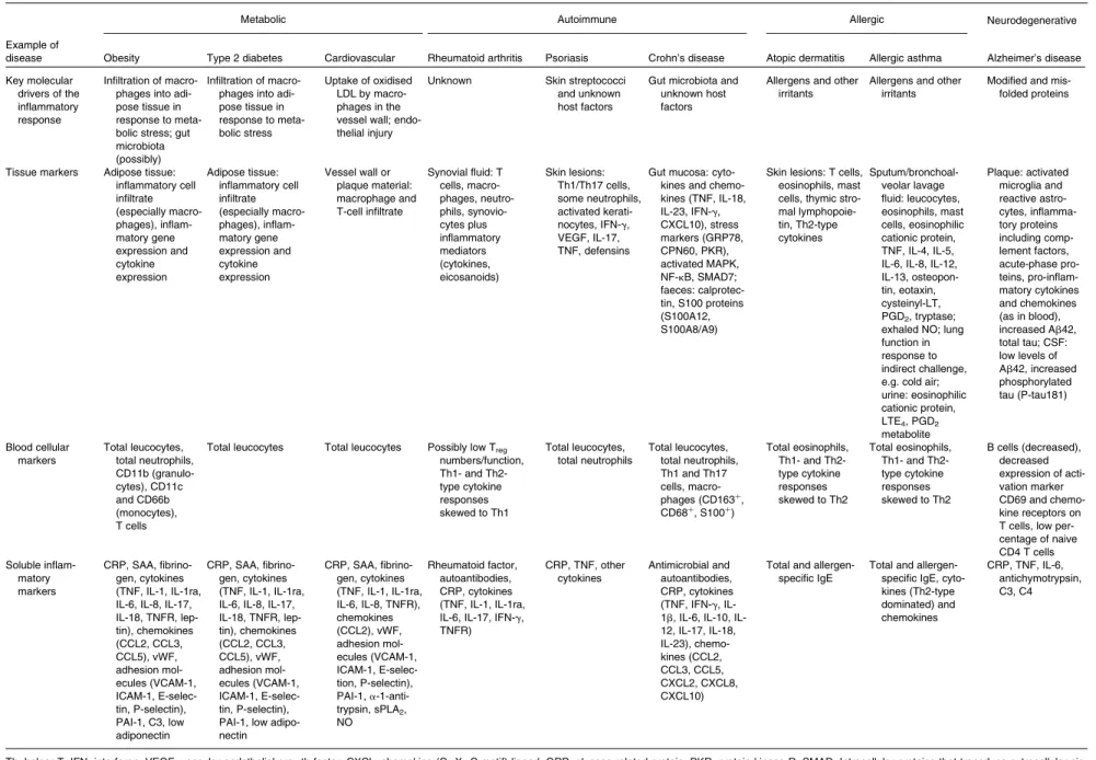
(i.e. soluble within plasma/serum or as circulating cells) and within tissue sites. These markers were identified by considering a range of conditions with a recognised inflammatory component elicited within different body com-partments (i.e. adipose tissue, brain, gastrointestinal tract, vas-cular wall, joints, airways or skin). The objective of considering such a wide range of pathological conditions was to identify those markers that are common to inflam-mation, irrespective of its causative trigger. The following four general families of inflammatory disease are discussed herein: metabolic conditions (obesity, type 2 diabetes and cardiovascular disease (CVD)), immune-mediated inflamma-tory diseases (RA, Crohn’s disease and psoriasis), allergic diseases (asthma and atopic dermatitis) and a prototypical neurodegenerative disease (Alzheimer’s disease). These dis-eases each have different molecular triggers, which are mostly exogenous to the host individual but are endogenous in some cases. For each disease, Table 4 lists the plasma/ serum (hereafter termed soluble), blood cell and tissue markers of inflammation commonly reported in the literature related to that disease; the listing is not exhaustive.
Although inflammation-induced tissue damage occurs in an organ-specific manner (adipose tissue, vascular wall, joints, skin, gut, airways and brain) in the different diseases or conditions, there is some commonality among the responses and markers seen in the different organs (summar-ised in Tables 5 and 6 for soluble markers and blood cellular markers, respectively). Indeed, although triggers, localisation and resulting clinical symptoms are different, many of the pro-cesses, cells and molecules involved in the actual inflamma-tory response are remarkably similar. Most, if not all, of the chronic inflammatory diseases considered here are character-ised by an increase in the number of leucocytes in the blood-stream and by overproduction and appearance in the bloodstream of increased concentrations of inflammatory cytokines (TNF, IL-1b, IL-6 and interferon-g) and chemokines (IL-8 and monocyte chemoattractant protein (MCP)-1). Elev-ated levels of these mediators act to amplify the inflammatory process (e.g. by attracting further inflammatory cells to the site), contribute to tissue destruction, elicit systemic effects (e.g. hepatic CRP synthesis) and, in many cases, actually cause the clinical symptoms observed (Fig. 2; Table 4). Thus, a consideration of a wide range of inflammatory dis-eases affecting distinct sites within the body has enabled the identification of a set of ‘common’ or ‘general’ markers of inflammation that can be measured in the bloodstream (Tables 5 and 6). The selection of soluble markers of inflam-mation has focused on peptides and proteins; lipid mediators have not been considered further because of their instability and relative difficulty of measurement. For most of the soluble markers identified (Table 5), there is no commonly accepted normal range of concentrations. In contrast, for the blood cel-lular markers of inflammation (Table 6), there is a generally accepted normal range and some of these markers are used clinically.
In addition to the general, non-specific markers of inflam-mation identified above, there are a number of inflammatory markers that are more disease specific (Table 4). Sometimes
these markers are present at the site of the inflammatory locus, such as thymic stromal lymphopoietin in skin lesions in atopic dermatitis. On other occasions, they may appear in the circulation (e.g. rheumatoid factor in RA). Because the aim of this evaluation of inflammatory markers is to identify those to be assessed in nutrition studies in the general popu-lation, disease-specific markers are not considered to be of rel-evance to this aim. Likewise, assessment of general, non-specific inflammatory markers at sites of disease activity (e.g. cytokines in synovial fluid in RA, in bronchoalveolar lavage fluid in asthma, in skin lesions in psoriasis or in gut mucosa in Crohn’s disease) was not considered relevant to the non-disease setting. For these reasons, inflammatory markers that are disease specific and assessment of inflammatory markers in tissues or lesions are not further considered here. Thus, this review is restricted to the consideration of inflammatory markers in the blood, either soluble markers in plasma/ serum (Table 5) or circulating cells (Table 6).
Key messages
(1) Acute inflammation is a normal physiological response critical to maintain homeostatic control, but when it becomes chronic, inflammation contributes to the patho-physiology of a range of diseases.
(2) Common soluble markers of inflammation include cytokines, chemokines, lipids, acute-phase proteins and adhesion molecules.
(3) Common blood cellular markers of inflammation include various subclasses of leucocytes. The same markers are involved in both acute and chronic inflammatory processes, meaning that the markers themselves, in iso-lation, cannot be used to distinguish the two processes.
Modifiers of inflammatory markers
There is a considerable between-individual variation in the numbers of leucocytes and of leucocyte subclasses and in the concentrations of the soluble markers of inflammation. There is no generally accepted ‘normal range’ for most of these markers, although there are clinically accepted normal ranges for total leucocytes and the leucocyte subclasses (Table 6). A number of modifiers, other than the presence of an inflammatory trigger, have been shown to influence the concentrations of these markers; these modifiers include age, body fatness, physical (in)activity, sex, genetics, smoking, gut microbiota composition, diet, use of medications and other factors such as emotional stress, pollution, viral infection and sleep behaviour (Fig. 3; Tables 5 and 6). These modifiers should all be considered when designing a study aimed at measuring inflammatory biomarkers.
Influence of age
A certain degree of inflammation may be part of adaptability and homeostasis in older people, and it has been demon-strated that their ability to mount a vigorous inflammatory
Biomarkers of inflammation S9
British
Journal
of
Nutrition
https:/www.cambridge.org/core/terms. https://doi.org/10.1017/S0007114512005119Table 4. Markers associated with inflammation in specific diseases
Metabolic Autoimmune Allergic Neurodegenerative
Example of
disease Obesity Type 2 diabetes Cardiovascular Rheumatoid arthritis Psoriasis Crohn’s disease Atopic dermatitis Allergic asthma Alzheimer’s disease Key molecular
drivers of the inflammatory response
Infiltration of macro-phages into adi-pose tissue in response to meta-bolic stress; gut microbiota (possibly)
Infiltration of macro-phages into adi-pose tissue in response to meta-bolic stress Uptake of oxidised LDL by macro-phages in the vessel wall; endo-thelial injury
Unknown Skin streptococci and unknown host factors
Gut microbiota and unknown host factors
Allergens and other irritants
Allergens and other irritants
Modified and mis-folded proteins
Tissue markers Adipose tissue: inflammatory cell infiltrate (especially macro-phages), inflam-matory gene expression and cytokine expression Adipose tissue: inflammatory cell infiltrate (especially macro-phages), inflam-matory gene expression and cytokine expression Vessel wall or plaque material: macrophage and T-cell infiltrate Synovial fluid: T cells, macro-phages, neutro-phils, synovio-cytes plus inflammatory mediators (cytokines, eicosanoids) Skin lesions: Th1/Th17 cells, some neutrophils, activated kerati-nocytes, IFN-g, VEGF, IL-17, TNF, defensins
Gut mucosa: cyto-kines and chemo-kines (TNF, IL-18, IL-23, IFN-g, CXCL10), stress markers (GRP78, CPN60, PKR), activated MAPK, NF-kB, SMAD7; faeces: calprotec-tin, S100 proteins (S100A12, S100A8/A9)
Skin lesions: T cells, eosinophils, mast cells, thymic stro-mal lymphopoie-tin, Th2-type cytokines Sputum/bronchoal-veolar lavage fluid: leucocytes, eosinophils, mast cells, eosinophilic cationic protein, TNF, IL-4, IL-5, IL-6, IL-8, IL-12, IL-13, osteopon-tin, eotaxin, cysteinyl-LT, PGD2, tryptase; exhaled NO; lung function in response to indirect challenge, e.g. cold air; urine: eosinophilic cationic protein, LTE4, PGD2 metabolite Plaque: activated microglia and reactive astro-cytes, inflamma-tory proteins including comp-lement factors, acute-phase pro-teins, pro-inflam-matory cytokines and chemokines (as in blood), increased Ab42, total tau; CSF: low levels of Ab42, increased phosphorylated tau (P-tau181) Blood cellular markers Total leucocytes, total neutrophils, CD11b (granulo-cytes), CD11c and CD66b (monocytes), T cells
Total leucocytes Total leucocytes Possibly low Treg numbers/function, Th1- and Th2-type cytokine responses skewed to Th1 Total leucocytes, total neutrophils Total leucocytes, total neutrophils, Th1 and Th17 cells, macro-phages (CD163þ, CD68þ, S100þ) Total eosinophils, Th1- and Th2-type cytokine responses skewed to Th2 Total eosinophils, Th1- and Th2-type cytokine responses skewed to Th2 B cells (decreased), decreased expression of acti-vation marker CD69 and chemo-kine receptors on T cells, low per-centage of naive CD4 T cells Soluble inflam-matory markers CRP, SAA, fibrino-gen, cytokines (TNF, IL-1, IL-1ra, IL-6, IL-8, IL-17, IL-18, TNFR, lep-tin), chemokines (CCL2, CCL3, CCL5), vWF, adhesion mol-ecules (VCAM-1, ICAM-1, E-selec-tin, P-selectin), PAI-1, C3, low adiponectin CRP, SAA, fibrino-gen, cytokines (TNF, IL-1, IL-1ra, IL-6, IL-8, IL-17, IL-18, TNFR, lep-tin), chemokines (CCL2, CCL3, CCL5), vWF, adhesion mol-ecules (VCAM-1, ICAM-1, E-selec-tin, P-selectin), PAI-1, low adipo-nectin CRP, SAA, fibrino-gen, cytokines (TNF, IL-1, IL-1ra, IL-6, IL-8, TNFR), chemokines (CCL2), vWF, adhesion mol-ecules (VCAM-1, ICAM-1, E-selec-tion, P-selectin), PAI-1, a-1-anti-trypsin, sPLA2, NO Rheumatoid factor, autoantibodies, CRP, cytokines (TNF, IL-1, IL-1ra, IL-6, IL-17, IFN-g, TNFR) CRP, TNF, other cytokines Antimicrobial and autoantibodies, CRP, cytokines (TNF, IFN-g, 1b, 6, 10, IL-12, IL-17, IL-18, IL-23), chemo-kines (CCL2, CCL3, CCL5, CXCL2, CXCL8, CXCL10)
Total and allergen-specific IgE
Total and allergen-specific IgE, cyto-kines (Th2-type dominated) and chemokines CRP, TNF, IL-6, antichymotrypsin, C3, C4
Th, helper T; IFN, interferon; VEGF, vascular endothelial growth factor; CXCL, chemokine (C – X – C motif) ligand; GRP, glucose-related protein; PKR, protein kinase R; SMAD, Intracellular proteins that transduce extracellular sig-nals from TGF beta ligands; CPN, chaperone; MAPK, mitogen-activated protein kinase; LT, leukotriene; CRP, C-reactive protein; SAA, serum amyloid A; ra, receptor antagonist; R, receptor; CCL, chemokine (C – C motif) ligand; vWF, von Willebrand factor; VCAM, vascular cell adhesion molecule; ICAM, intercellular adhesion molecule; PAI, plasminogen activator inhibitor; sPLA2, soluble phospholipase A2; C, complement.
P . C. Calder et al. S10
British Journal of Nutrition
https:/www.cambridge.org/core/terms
.
https://doi.org/10.1017/S0007114512005119
Downloaded from
https:/www.cambridge.org/core
. University of Basel Library
, on
10 Jul 2017 at 15:46:22
Table 5. Soluble markers of inflammation and their associated modifying factors
Marker
Typical range reported in healthy subjects* Association with increasing age Association with increasing body fatness Association with habitual physical activity Association
with smoking References
Cytokines/chemokines
TNF 0·75 – 5 ng/l $ / " " # / $ " 18, 19, 22, 55 – 57, 77, 110, 133, 301
TNFRI and TNFRII 400 – 700 ng/l " " # 24, 25, 55, 64, 302, 303, 304
IL-6 0·4 – 1·4 ng/l $ / " " # " 14, 18, 20, 21, 24, 57, 61, 62, 77, 110, 130 – 132, 305 – 307 IL-1b 200 – 500 ng/l $ / " / # " $ $ 18, 19, 21, 60, 110, 301 IL-8 2 – 10 ng/l $ / " $ / " " $ 24, 63, 64, 77, 308, 309, 310 IFN-g 1 – 5 ng/l " 24 CCL2 200 – 400 ng/l $ / " " 24, 31, 63, 65, 66, 77 CCL3 0·1 – 11·3 ng/l " 308, 309 CCL5 20 – 50 ng/ml " $ $ $ 308, 309 Acute-phase proteins CRP 0·1 – 10 mg/l† " " # " 14, 26, 42, 62, 67, 68, 77, 135, 136, 138, 139, 311, 312 SAA 15 – 35 mg/l " " $ / " 42, 141, 256, 313 Fibrinogen 1500 – 4000 mg/l " " # " 14, 20, 28, 135 – 137, 139, 140, 314, 315
Von Willebrand factor 5 – 15 mg/l " " 316 – 320
Antichymotrypsin 300 – 460 mg/l "
C3 1 – 3 mg/l " # " 321 – 325
IL-1ra 350 – 700 ng/l " " $ 14, 21, 25, 58 – 60, 326
Soluble adhesion markers
VCAM-1 500 ng/ml " " # 29, 30, 57, 70 ICAM-1 175 – 200 ng/ml " " # " 58, 70 – 73, 130, 327 E-selectin 43 – 80 ng/ml $ " # " 30, 70, 72 – 74, 130, 328 P-selectin 50 – 60 ng/ml " " # 57, 71 Adipokines Leptin 10 ng/ml $ " # $ / # 55, 61, 65, 75, 327, 329 Adiponectin 5 – 15 mg/l $ / " # " # 33, 34, 76 – 78, 330
R, receptor; IFN, interferon; CCL, chemokine (C – C motif) ligand; CRP, C-reactive protein; SAA, serum amyloid A; C, complement; ra, receptor antagonist; VCAM, vascular cell adhesion molecule; ICAM, intercellular adhesion molecule; ", positive association; #, negative association; $, no clear association.
* These values are based on groups of healthy subjects in studies published in scientific literature. These are therefore indicative of what could be expected in such populations. † Values . 10 mg/l considered to be indicative of inflammation beyond ‘low grade’.
Biomarkers
of
inflammation
S11
British Journal of Nutrition
https:/www.cambridge.org/core/terms
.
https://doi.org/10.1017/S0007114512005119
Downloaded from
https:/www.cambridge.org/core
. University of Basel Library
, on
10 Jul 2017 at 15:46:22
response is associated with enhanced survival(14). On the other hand, inflammation is considered to play an important role in the loss of lean body mass and in progression towards frailty in older persons. Studies investigating a possible effect of age on inflammatory markers have generally adopted one of two approaches. The first has been to look for an associ-ation between age and the concentrassoci-ations of markers of inter-est, whereas the second has been to compare marker concentrations between two or more groups of individuals of different ages. Studies have generally stratified populations and set arbitrary thresholds varying from ages . 60, 65, 70 to
$75 years to identify aged individuals. These studies, for the
most part, have suggested that ageing is accompanied by a low-grade chronic inflammatory state, clearly shown by two-to fourfold higher concentrations of serum levels of several inflammatory markers such as cytokines and acute-phase
pro-teins in older persons(15). Several studies have reported
age-associated increases in total leucocyte numbers(16,17) and in
soluble concentrations of soluble pro-inflammatory cytokines
such as IL-1(18,19), IL-6(14,18,20,21)and TNF(22,23), although this
has not been seen in all studies(21,24). Others have reported
higher concentrations of IL-1 receptor antagonist(14,21,25) and
of TNF-receptors(24,25)with increased age. In line with these
findings, many studies have reported higher concentrations of downstream markers such as acute-phase proteins like CRP(14,26), a1-acid glycoprotein(27) and fibrinogen(14,20,28). Fewer studies have addressed the relationship of ageing with chemokines and adhesion molecules, although in some
studies, vascular cell adhesion molecule (VCAM)-1(29,30) and
intercellular adhesion molecule (ICAM)-1(30) have been
shown to have higher concentrations in older persons while
E-selectin was not(30). Similarly, there is some evidence that
con-centrations of MCP-1(31), plasminogen activation inhibitor-1(32)
and adiponectin(33,34)are increased with ageing.
Overall, these changes support a generally increased state of low-grade chronic inflammation in older people compared with younger adults. The more limited literature on blood cel-lular markers would support this contention, but indicates that numbers and proportions of leucocytes in the older people differ considerably depending on the state of health of the
individual(35). Although older individuals have been reported
to have increased total leucocytes, the increase in neutrophils, in particular, has been related to reduced survival in longitudi-nal studies, whereas total lymphocyte numbers are not
signifi-cantly associated with survival(36). However, within the total
lymphocyte population, there may be decreased numbers of B cells and increased numbers of CD8 cells in older persons, which can also have an impact on survival and which to a
great extent depend on microbiological exposures(37).
It is recognised that ageing represents cumulative effects of exposure to various triggers such as diet, physical (in)activity and stress, as well as micro-organisms, over the lifetime, and it is hypothesised that failure of anti-inflammatory mechanisms
to neutralise such inflammatory processes triggered
continuously lifelong plays a role in low-grade chronic
inflam-mation in older people(38). Inflammatory markers in older
per-sons may also be influenced by co-morbidity, medication use
or malnutrition(39 – 43). However, there are some indications that
once co-morbidities are accounted for, inflammation is still
associated with ageing(14,15). A number of other factors that
may affect and modulate circulating levels of inflammatory mediators alter with ageing, including body composition, physi-cal (in)activity, sex hormone concentrations, gut microbiota,
emotional stress and a poor social environment(44 – 48). It is
likely that in some studies, these confounders have not been considered when the effect of age on inflammation has been
evaluated(44 – 48). Thus, it is challenging to dissect out the
effect of ageing per se on inflammatory markers in humans and essentially impossible to exclude all residual confounding concomitant modifying factors and lifelong exposures. One possibility is to examine inflammatory markers in centenarians as a model of successful ageing. Low levels of the pro-inflamma-tory cytokine IL-6 and high levels of anti-inflammapro-inflamma-tory cyto-kines such as IL-10 and transforming growth factor (TGF)
have been noted in centenarians(49,50), suggesting that these
extremely old persons do not suffer the ‘inflammaging’ noted in many older persons who do not survive to 100 years of age or beyond.
Influence of body fatness and fat distribution
As stated earlier, white adipose tissue secretes cytokines and chemokines, and macrophage and T-cell infiltration is
dra-matically increased in the adipose tissue of obese
humans(51). Obesity is associated with a state of low-grade
chronic inflammation. Total blood leucocyte numbers are positively associated with obesity and the metabolic
syn-drome(16,17,52 – 54). Differential leucocyte counts indicate that
neutrophils, but not monocytes or lymphocytes, account for
this predictive potential(17). There are higher concentrations
Table 6. Blood cellular markers of inflammation and their modifying factors
Marker Normal range*
Association with increasing age Association with increasing body fatness Association with habitual physical activity Association
with smoking References
Total leucocytes 4 – 11 £ 109/l " " # " 16, 17, 53, 89, 128, 129 Total neutrophils 2 – 7·5 £ 109/l " " # " 17, 54, 245
Total T lymphocytes 0·6 – 2·5 £ 109/l $ " # " 35, 89, 331 Total eosinophils 0 – 0·4 £ 109/l # " " " 89, 332, 333
Total monocytes 0·2 – 0·8 £ 109/l " " " # 89, 335
Up arrow, Positive association; Down arrow, Negative association; Double arrow, No clear association. * Based on data reported in MacLennon & Drayson(336).
P. C. Calder et al. S12
British
Journal
of
Nutrition
https:/www.cambridge.org/core/terms. https://doi.org/10.1017/S0007114512005119of circulating cytokines and chemokines such as TNF(55 – 57), IL-1(58 – 60), IL-6(57,61,62), IL-8(63,64)and MCP-1(63,65,66)in obese individuals compared with normal-weight subjects. Several acute-phase proteins are elevated in overweight and obese subjects. For example, the circulating concentration of CRP is higher in obese persons, and obese subjects who lost
weight showed decreased levels of CRP(67,68). A meta-analysis
showed a strong association between Body Mass Index (BMI)
and serum amyloid A concentration(69). Elevated levels of
sol-uble adhesion molecules, such as VCAM-1, ICAM-1, E-selectin and P-selectin, are found in obese individuals compared with
normal-weight subjects(57,70 – 74). Leptin concentrations are
higher in obese than in normal-weight subjects(55,61,65,75),
while adiponectin concentrations are lower in obese individ-uals(76 – 78). Although these studies have focused on obesity
per se, the distribution of adipose tissue appears to be
import-ant in determining inflammatory burden. Visceral adipose tissue has a greater production of inflammatory mediators such as cytokines than subcutaneous adipose tissue, although the precise contribution of each to the circulating pool is not
clear(79). Furthermore, other adipose tissue depots in so-called
‘ectopic sites’, such as within the liver, heart or skeletal muscle, may contribute to the production of inflammatory mediators
even in the absence of obesity(80). In this regard, the local
pro-duction of inflammatory molecules by adipose tissue within the heart may be important; the amount of this tissue and its proximity to the coronary vessels could contribute to the
development of coronary pathologies(81,82). Although there
has been considerable focus on the role of low-grade chronic inflammation in overweight, obesity and the metabolic
syn-drome(6), it is important to recognise that underweight also
appears to be associated with low-grade chronic inflam-mation. For example, adolescent anorexia is associated with elevated levels of some inflammatory cytokines including
TNF-a(83), and low-grade chronic inflammation is associated
with low body weight in older subjects(84). Several
pro-inflammatory cytokines, most notably TNF-a, have a causal
role in cachexia(85).
Influence of physical (in)activity
Physical (in)activity resulting in inadequate energy expendi-ture relative to intake may lead to the accumulation of visceral fat and consequently to the activation of inflammatory
processes associated with this(86). In contrast, physical activity
promotes health and this may relate to the anti-inflammatory effect of regular exercise, which reduces visceral fat mass
and induces an anti-inflammatory environment(87,88). Subjects
engaged in regular physical activity have lower blood
leucocyte numbers(89). Plasma concentrations of IL-6 are
lower in individuals who undertake regular physical activity
than in those who do not(90). The lower concentrations of
IL-6 in the circulation will subsequently result in lower CRP levels. Several studies of large population cohorts have provided evidence for an inverse, independent dose – response relationship between the level of habitual physical activity and plasma CRP concentration in both men and
women(91 – 96). Moreover, CRP levels in 2120 Finnish
partici-pants were associated with obesity indices (positively) and
physical activity (inversely) among both sexes(97).
Cross-sec-tional studies have demonstrated that cardiorespiratory fitness levels are inversely associated with CRP concentrations
and also the prevalence of elevated CRP concentrations(98).
However, conflicting findings exist and several training inter-ventions have not produced changes in basal IL-6 or CRP
levels(99 – 106). There seems to be more consistency in patients
with metabolic or coronary disease and in older individuals than in healthy or young people. In a relatively large interven-tion study of exercise training in cardiac rehabilitainterven-tion patients, CRP concentration was reduced by 41 %, whereas
Triggers Pathways ER stress TLR TNFR Inflammasome Inflammatory genes NF-κB Markers Modifiers • Microbial components • Age
• Body weight, fatness, fat distribution • Physical (in)activity • Sex • Genetics • Smoking • Gut microbiota • Diet • Medication
• Other (emotional stress, pollution, viral infections, sleep behaviour)
• Tissue damage • Metabolic stress
Fig. 3. Factors that influence the status of soluble and cellular markers of inflammation in the bloodstream. Triggers are those factors that can directly initiate an inflammatory response. Modifiers are endogenous and exogenous factors that may increase or decrease the response induced by a trigger. ER, endoplasmic reticulum; TLR, Toll-like receptor; TNFR, TNF receptor.
Biomarkers of inflammation S13
British
Journal
of
Nutrition
https:/www.cambridge.org/core/terms. https://doi.org/10.1017/S0007114512005119CRP concentrations did not change in subjects who did not
exercise(107). Rankovic et al.(108) showed that 6 weeks of
aerobic exercise can significantly reduce the inflammatory state by decreasing CRP and VCAM-1 levels in patients with stable coronary heart disease. One possible explanation could be that patients and older people have higher basal levels of inflammation before becoming physically active and physical activity is more effective in lowering the CRP
levels in those with the highest initial CRP concentrations(107).
Given that physical activity and obesity are often inversely related, it is not clear as to whether the anti-inflammatory health benefits of a physically active lifestyle are due to exer-cise per se or result from favourable changes in body compo-sition. A systematic review addressed whether fitness or
fatness has the greatest impact on inflammatory factors(90)
and concluded that both fatness and the lack of fitness are associated with systemic inflammatory status, although the relative contributions of both may be dependent on age, dis-ease status and sex. Different types of exercise to attain and maintain a state of physical fitness may have different effects
on inflammation(109), although this is little studied.
Influence of sex
Few studies have examined the effect of sex on inflammatory markers and the literature presents mixed findings. Some studies have reported no differences between males and females for most inflammatory markers examined, including
TNF, TNF-receptor, IL-1, MCP-1(24) or IL-6 and ICAM-1(29).
However, in a study of adolescents (aged 13 to 17 years), although CRP, C3 and C4 concentrations did not differ between girls and boys, adolescent boys had lower levels of
adiponectin, leptin and L-selectin than girls, whereas girls
had lower levels of ICAM-1, VCAM-1 and plasminogen
acti-vation inhibitor-1 than boys(86). Some investigators have
reported lower IL-1b levels in men(110), whereas others(111)
have found that men have higher levels of IL-1b-secreting monocytes than women. Importantly, few studies have reported on sex effects after adjusting for variables that can affect inflammatory markers, such as body fatness, fat
distri-bution and physical (in)activity. Marques-Vidal(110) showed
in a Swiss population-based sample that males have higher levels of IL-6 and TNF and slightly but significantly lower levels of CRP when compared with females in crude analysis and after adjusting for age, BMI, smoking status and leisure-time physical activity in multivariate analysis. There is a need for further population-based studies in healthy adults to examine sex differences in inflammatory markers taking into account potential important confounders.
Influence of genetics
Both epigenetic and genomic variations influence the extent of the response of an inflammatory marker to a given trigger. Epigenetics refers to chemical modifications to chromatin or DNA that influence gene expression but are not encoded in the DNA sequence itself. The two major epigenetic mechan-isms are the post-translational modification, often acetylation,
of histone proteins in chromatin and the methylation of DNA itself. DNA methylation occurs at CpG sites of the gene sequence. Epigenetic variations appear to play an important
role in determining the risk of inflammatory disorders(112 – 114).
Differences in the methylation status of CpG sites have been observed in key inflammatory response genes and influence
the expression of those genes(115). This is an area in which
there are likely to be major advances in the next few years. It is important to note that epigenetic modifications are not fixed but may be modified by a number of different exposures, including stress hormones, oxidative stress, pol-lution and diet. This may partly explain the ability of those fac-tors to act as modifiers of the inflammatory response.
SNP are variations in the DNA sequence at a single nucleo-tide that occur in at least 1 % of the population. They can occur in both coding and non-coding regions of the genome. It is estimated that the human genome contains 3 million SNP. Genotype is a description of the allele pair pre-sent at a given site in the genome, and typically there will be three possible genotypes (e.g. AA, AG and GG). There are variations in allele frequencies and genotypes among different human populations because certain alleles and genotypes may have offered an evolutionary advantage in one geographical region and not in another. SNP within a coding sequence do not necessarily change the amino acid sequence of the protein that is produced, because of the degeneracy of the genetic code. SNP that are not in protein-coding regions may affect transcription factor binding or gene splicing or the sequence of non-coding RNA and, as a result, the gene expression, and hence the level of the encoded protein may differ among individuals with different genotypes. It is likely that SNP occur in regions of the genome associated with each of the inflammatory proteins being considered here (e.g. cytokines, chemokines, adhesion molecules, acute-phase proteins), encoding enzymes responsible for the syn-thesis of non-peptide inflammatory mediators (e.g. in cyclo-oxygenase and lipcyclo-oxygenase enzymes, NADPH oxidase and inducible NO synthase) and encoding regulatory proteins such as transcription factors. In addition, such genetic vari-ations underlie the huge number of possible variants in the human leucocyte antigens involved in antigen presentation. This variation in possible human leucocyte antigens allows an individual to have the potential to deal with an enormous number of possible antigens that may be encountered. How-ever, some human leucocyte antigens variants have strong associations with inflammatory diseases, such as human leuco-cyte antigens-DR4 with RA. A number of SNP are of functional significance, meaning that individuals with different genotypes will produce different levels of a particular inflammatory mediator in response to a given stimulus or trigger than
other individuals(116 – 119). Thus, genotype can influence the
plasma concentration of inflammatory markers(116,120,121). For
example, the G to C polymorphism at the 2 174 position of the IL-6 gene has been reported to significantly influence plasma IL-6 concentrations: mean concentrations (pg/ml) after adjusting for age, BMI, sex and smoking were 2·74 for GG homozygotes, 2·64 for heterozygotes and 1·63 for CC
homozygotes(116). Similarly, the polymorphism at the þ 252
P. C. Calder et al. S14