Dendritic Plasticity in the Adult Rat
Following Middle Cerebral Artery
Occlusion and Nogo-A Neutralization
Catherine M. Papadopoulos
1, Shih-Yen Tsai
1, Joseph L.
Cheatwood
1,3, Melanie R. Bollnow
1,3, Bryan E. Kolb
4,
Martin E. Schwab
5and Gwendolyn L. Kartje
1,2,3,61
Research, Hines VA Hospital, Hines, IL 60141, USA,
2
Neurology Service, Hines VA Hospital, Hines, IL 60141, USA,
3
Department of Neurology, Loyola University, Maywood, IL
60153, USA,
4Department of Psychology, University of
Lethbridge, Lethbridge, AB, Canada,
5Brain Research Institute,
University of Zurich and Swiss Federal Institute of
Technology, Zurich, Switzerland and
6Department of Cell
Biology, Neurobiology and Anatomy, Loyola University,
Maywood, IL 60153, USA
Our work has shown that following focal ischemic lesion in adult
rats, neutralization of the axon growth inhibitor Nogo-A with the
monoclonal antibody (mAb) IN-1 results in functional recovery.
Furthermore, new axonal connections were formed from the
contralesional cortex to subcortical areas corresponding to the
observed functional recovery. The present study investigated
whether dendritic changes, also known to subserve functional
recovery, paralleled the axonal plasticity shown after ischemic
lesion and treatment with mAb IN-1. Golgi--Cox-stained layer V
pyramidal neurons in the contralesional sensorimotor cortex were
examined for evidence of dendritic sprouting. Results demonstrated
increased dendritic arborization and spine density in the mAb
1-treated animals with lesion. Interestingly, administration of mAb
IN-1 without lesion resulted in transient dendritic outgrowth with no
change in spine density. These results suggest a novel role for
Nogo-A in limiting dendritic plasticity after stroke.
Keywords: dendritic arbors and spines, Golgi--Cox, layer V,
motor cortex, rat, stroke
Introduction
Ischemic stroke, the lack of blood flow to the brain, is the third
most common cause of death in developed countries. One-third
of the 4.6 million stroke survivors in the USA are left
perma-nently disabled, thus representing the leading cause of serious
long-term neurological disability (American Heart Association,
2004). Neuronal loss due to stroke may result in chronic
impairment of sensory and motor function that is resistant to
therapeutic strategies.
Incomplete functional recovery after injury to the adult
central nervous system (CNS) is a result of the inability of
central neurons to form correct axonal and dendritic
connec-tions through regenerative or plastic responses. The failure of
adult neurons to establish new connections after CNS lesions is
most likely not an intrinsic deficit in the growth potential of the
central neuron (Richardson et al., 1980; David and Aguayo,
1981), but rather an effect of the non-permissive adult
environ-ment. A major contributor to the presence of growth-inhibitory
proteins that creates a non-permissive environment and limits
the structural plasticity of injured mature neurons is CNS myelin
(Schwab and Caroni, 1988; Qui et al., 2000). Several
myelin-associated proteins and chondroitin-sulfate proteoglycans
(CSPGs) have been characterized as inhibitors of axonal growth:
oligodendrocyte myelin glycoprotein (OMgp) (Wang et al.,
2002), myelin-associated glycoprotein (MAG) (McKerracher
et al., 1994; Mukhopadhyay et al., 1994), Nogo-A (Spillman
et al., 1997; Chen et al., 2000; GrandPre et al., 2000; Prinjha
et al., 2000), and the CSPGs Versican V2 (Schweigreiter et al.,
2004) and Brevican (Schmalfeldt et al., 2000). Nogo-A is
expressed in oligodendrocytes and myelin, and in certain
neuronal populations (Caroni and Schwab, 1988a; Chen et al.,
2000; Huber et al., 2002; Buss et al., 2004; Dodd et al., 2005; this
study). Based on several regeneration and plasticity studies,
Nogo-A has proven to be a powerful inhibitor of axonal growth
(for review, see Schwab, 2004). Regeneration studies have
supported this inhibitory role by neutralization of Nogo-A
with the specific antigen, monoclonal antibody (mAb) IN-1
(Fiedler et al., 2002) with in vitro growth assays (Caroni and
Schwab, 1988b) and in vivo spinal cord injury studies (Schnell
and Schwab, 1990; Bregman et al., 1995). Important to the
present study, intraventricular administration of mAb IN-1
resulted in axonal plasticity of intact fiber tracts after cortical
aspiration lesion (Kartje et al., 1999; Wenk et al., 1999) and
improved functional recovery of skilled movements after
unilateral pyramidotomy (Thallmair et al., 1998; Z’Graggen
et al., 1998), cortical aspiration lesion (Emerick and Kartje,
2004) and permanent middle cerebral artery occlusion (MCAO)
(Papadopoulos et al., 2002; Seymour et al., 2005) in adult rats.
Clinical studies have implicated the contralesional cortex as
an important contributor to motor recovery after stroke
(Cramer et al., 1997; Cuadrado et al., 1999). Functional imaging
studies demonstrated increased cerebral blood flow (Weiller
et al., 1993) as well as altered patterns of activation (Feydy et al.,
2002; Johansen-Berg et al., 2002) in the intact cerebral
hemi-sphere of human patients recovering from unilateral stroke that
paralleled functional recovery. Our past work has demonstrated
the critical role in functional recovery for neuronal (axonal)
plasticity originating from the contralesional hemisphere to
re-innervate deafferented areas after cortical lesions and
treat-ment with mAb IN-1 (Wenk et al., 1999; Kartje et al., 1999;
Papadopoulos et al., 2002). Also, using intracortical
micro-stimulation mapping techniques, our laboratory has shown
that reorganization in the contralesional forelimb motor cortex
parallels functional recovery after aspiration lesion and
treat-ment with mAb IN-1 (Emerick et al., 2003). Since dendritic
plasticity is also associated with functional recovery after
cortical lesions (Jones and Schallert, 1994; Kawamata et al.,
1997b; Rowntree and Kolb, 1997; Biernaskie et al., 2004), we
questioned whether neuronal (dendritic) changes paralleled
the axonal plasticity shown after stroke and treatment with
mAb IN-1. Therefore, we analyzed the dendritic arbors of layer V
pyramidal neurons within the forelimb motor cortex of the
contralesional hemisphere using a modified Golgi--Cox staining
procedure. Our results demonstrate a dramatic increase in both
Cerebral Cortex April 2006;16:529--536 doi:10.1093/cercor/bhi132
the apical and basilar dendritic arborization and spine density of
pyramidal neurons in the stroke/mAb IN-1-treated group
com-pared with controls. These results point to a role for Nogo-A in
restricting dendritic plasticity after stroke.
Materials and Methods
Animals and In Vivo Manipulations
Adult male Long Evans, black-hooded rats (250--360 g) were divided into
eight experimental groups: (i) MCAO only, six-week survival (n=6); (ii)
MCAO plus mAb IN-1, six-week survival (n=6); (iii) MCAO plus control
antibody treatment, six-week survival (n=5); (iv) mAb IN-1 only,
two-week survival (n=5); (v) mAb IN-1 only, six-week survival (n=3); (vi)
control Ab only, two-week survival (n=4); (vii) control Ab only,
six-week survival (n=3); and (viii) normal animals (n=5). Animals were
number-coded and investigators were blinded to the treatment groups. Experiments were approved by the Institutional Animal Care and Use Committees of Loyola University/Hines VA.
MCAO was performed as described in Chen et al. (1986). Animals were anesthetized with sodium pentobarbital (50 mg/kg, i.P.). Bilateral common carotid arteries (CCA) were isolated, a vertical 2 cm long incision was made between the eye and ear, and the temporalis muscle was retracted. A burr hole was made to expose the left MCA and it was permanently occluded with a 10-0 suture. The CCA ipsilateral to the MCAO was permanently occluded with a 4-0 suture and the contralat-eral CCA was temporarily occluded for 45 min. Body temperature was maintained with a heating pad and monitored with a rectal probe. The wounds were then closed and animals warmed under a heating lamp until awake.
Antibody application was performed immediately after MCAO as in our previous study (Papadopoulos et al., 2002). A cell suspension (6 ll)
containing a total of 13105of either mAb IN-1 (Caroni and Schwab,
1988b) or the control antibody (anti-HRP) (Schnell and Schwab, 1990) secreting mouse hybridoma cells was injected posterior to the lesion (4 mm caudal, 5 mm lateral and 5 mm ventral to bregma) on the side of the lesion. MCAO only animals received vehicle only injections in the same coordinates. All animals, including those in the MCAO only group, received cyclosporin A (10 mg/kg, i.P.) daily starting from the day before surgery to 7 days after surgery to prevent rejection of the hybridoma xenograft.
Histology
Either 2 or 6 weeks after MCAO and/or antibody treatments, animals were overdosed with sodium pentobarbital (100 mg/kg, i.P.) and transcardially perfused with 0.9% saline. Brains were removed and immersed whole in Golgi--Cox solution (Glaser and van der Loos, 1981) for 14 days and then placed in 30% sucrose solution for 2 days. Brains were cut into 200 lm coronal sections using a vibratome, slide mounted, and reacted according to the procedure described by Gibb and Kolb (1998).
Stroke Volume Analysis
The stroke volume was quantitatively analyzed on Golgi--Cox stained
sections (+4.7 to –5.2 mm from bregma according to Paxinos and
Watson, 1998) using a computer-interfaced imaging system (NIH Image 1.51) by the method as described in Kawamata et al. (1997a) (total area of the intact contralateral hemisphere -- total area of the intact ipsilateral hemisphere multiplied by the total distance between the sections). Stroke size was expressed as a percentage of the intact contralateral hemispheric volume.
Nogo-A Immunostaining
A rat was euthanized and perfused intracardially with 0.9% saline and 4% paraformaldehyde. The brain was removed and flash frozen in
isopen-tane, then stored in a–80°C freezer until cutting. Sections were cut at 50
lm on a freezing microtome and placed into tris-buffered saline (TBS). For immunohistochemistry, sections were first treated with a blocking solution of 10% normal goat serum (NGS)/TBS/0.5% Triton-X for 60 min. Sections were then incubated in TBS containing 0.5% Triton-X, 5% NGS, a rabbit polyclonal anti-Neurofilament 200 (1:250; Sigma, St Louis, MO)
and the mouse IgG 11C7, a monoclonal antibody specific for Nogo-A (1:500; Novartis, Basel, Switzerland), overnight at 4°C. Sections were rinsed thoroughly and incubated in TBS containing 0.5% Triton-X, 5% NGS, a tetramethylrhodamine-conjugated goat anti-rabbit IgG (1:1000; Molecular Probes, Eugene, OR) and an AlexaFluor 488-conjugated goat anti-mouse IgG (1:1000; Molecular Probes) for 60 min at room temperature. After rinsing, sections were incubated in a 300 nM solution of 49-6-diamidino-2-phenylindole (DAPI) in 0.1% Triton-X/TBS for 30 min. After a final rinse, sections were mounted onto slides from dilute TBS solution and coverslipped with the aqueous mounting medium Fluoromount-G (Electron Microscopy Sciences, Hatfield, PA).
Neuroanatomical Analysis
Seven layer V pyramidal neurons per animal located within the forelimb motor cortex and five layers II/III pyramidal neurons per animal located within the occipital cortex of the contralesional hemisphere were identified with an atlas (Zilles, 1985) and according to previous electrophysiological studies (Neafsey et al., 1986). Criteria for inclusion in the analyses were that the neuron had to be well impregnated, unobstructed by other dendrites, blood vessels or glial cells, and the dendritic arborizations intact and visible in the plane of section. For all analyses the slides were coded and investigators were blind to the treatment group.
Apical and basilar dendrites of layers II/III and V pyramidal neurons in
the intact hemisphere opposite the lesion were traced at 325
magnification with the aid of a camera lucida drawing tube. Both apical and basilar dendritic trees were analyzed for total dendritic length (Sholl, 1956) and total number of branch segments within a branch order (Coleman and Riesen, 1968). Branch order was determined by considering that branches emanating directly from the cell body (basilar) or the primary apical dendrite were first order; once bifurcation occurred, two secondary dendrites were formed and so on.
Dendritic spine densities were analyzed from layer V pyramidal neurons within the forelimb motor cortex of the intact hemisphere.
Using a3100 oil immersion lens, a 50 lm length of one terminal basilar
branch segment of third or higher order and one terminal apical segment from the midpoint of the apical tree were drawn from seven neurons per animal. Spine density was expressed as the number of spines per lm. All the data presented in this study are expressed as
means
±
SEM. Statistical comparison for layer V and layers II/III neuronswas assessed by one-way analysis of variance (ANOVA) followed by Newman--Keuls test for post hoc analyses.
Results
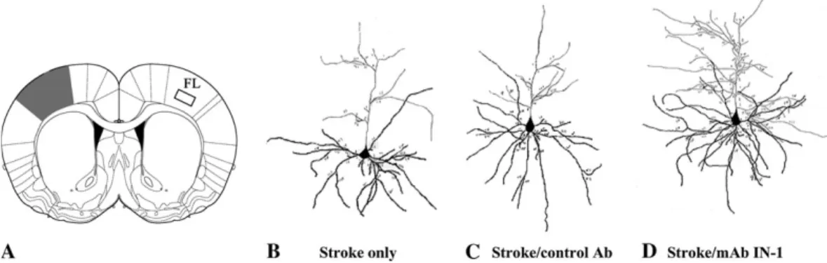
Lesions are Localized in the Sensorimotor Cortex
The topographical location of all lesions included the
sensori-motor cortex ipsilateral to the occluded MCA (Fig. 2A, shaded
area) with minimal subcortical damage. Measurement of the
infarct volumes showed no difference between the stroke only
(16.46
±
5.01), stroke/control antibody (13.82
±
3.98) and
stroke/mAb IN-1 groups (11.70
±
1.91) (P
=0.58, one-way
ANOVA).
Forelimb Cortex Layer V Neurons are
Strongly Nogo-A Positive
Immunohistochemical staining with the Nogo-A specific mouse
monocloncal antibody 11C7 (labeled with a green fluorophore)
and a rabbit polyclonal against the neuronal structural protein
Neurofilament 200 (labeled with a red fluorophore) revealed
that the inhibitory protein Nogo-A is present in many neurons
across cortical layers II through VI of rat forelimb cortex (FL), as
evidenced by the overlap of green and red fluorescence in the
processed tissue (Fig. 1A). The cell body and dendritic processes
of the large layer V pyramidal neurons stain very robustly for
Nogo-A, but there is no visible staining in the nucleus (Fig. 1B).
Dendritic Complexity is Enhanced after Stroke and
mAb IN-1 Treatment
The significant functional recovery and increased axonal
plasticity observed previously in rats after stroke and immediate
or delayed mAb IN-1 treatment (Papadopoulos et al., 2002) led
us to investigate whether dendritic changes paralleled these
events. Both apical and basilar dendritic arborizaton and spine
densities of Golgi--Cox stained layer V pyramidal neurons within
the intact, contralesional forelimb motor cortex were examined
(Fig. 2A, boxed area). Representative camera lucida drawings of
pyramidal neurons from stroke only, stroke/control antibody
and stroke/mAb IN-1 treatment groups are shown in Figure
2B--D. Apical dendrite patterns in animals with stroke lesion only, in
stroke animals treated with control antibody and in normal
animals showed no statistical difference between groups.
However, stroke animals with mAb IN-1 treatment were
significantly different from all control groups (Fig. 3A,C,D).
mAb IN-1-treated animals had significantly greater apical
den-drite arborization when measured for the number of branch
segments within branch orders (Fig. 3A), total number of
branches (P
<0.001, Fig. 3C) and dendritic length (P
<0.01,
Fig. 3D).
As with apical dendritic arborization, a similar pattern in
basilar dendrites was observed. While the control groups
showed no significant difference from each other, the stroke/
mAb IN-1-treated animals showed an increased basilar arbor
complexity when compared with controls. Analyses
demon-strated that the stroke/mAb IN-1-treated group had significantly
greater number of branch segments per branch order (Fig. 3B),
total number of branches (P
<0.001, Fig. 3C) and dendritic
length (P
<0.001, Fig. 3D).
Quantitative analysis of spine densities in layer V pyramidal
neurons of the contralesional forelimb motor cortex showed
a significant increase in the number of spines of both apical and
basilar terminal branch segments in stroke animals treated with
mAb IN-1 when compared with stroke only, stroke/control Ab
and normal groups (P
<0.001, Fig. 4).
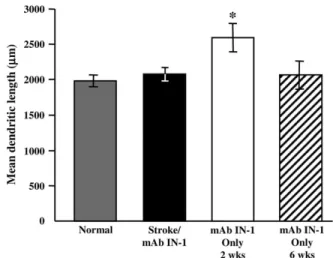
Lasting Enhanced Dendritic Complexity is Specific to
the Motor Cortex after Stroke and mAb IN-1 Treatment
The significant dendritic plasticity observed in the
contrale-sional forelimb motor cortex of stroke/mAb IN-1 animals raises
the question whether the effect on plasticity is specific to an
area or is found globally throughout the cortex. To determine
this, we assessed the dendritic structures in the lateral area of
the secondary visual cortex (Oc2L), a distant region from the
motor cortex. Apical and basilar dendritic arbors of layers II/III
neurons within the contralesional occipital cortex were
exam-ined. Quantitative analysis demonstrated no difference in
occipital neuron total dendritic length between the stroke/
mAb IN-1 group, normal animals, and normal animals treated
with mAb IN-1 at 6 weeks (P
>0.05, Fig. 5). However, total
dendritic length was enhanced after 2 weeks of mAb IN-1
treatment in normal animals, but this difference resolved at
6 weeks (P
<0.05).
Dendritic Complexity is Transient after
mAb IN-1 Treatment in Normal Animals
Treatment with mAb IN-1 in the absence of CNS injury induces
transient axonal outgrowth in adult cerebellar Purkinje neurons
(Buffo et al., 2000) and corticospinal neurons (Bareyre et al.,
2002). We therefore investigated whether mAb IN-1
adminis-tration without stroke had the same effect on dendritic
out-growth. Apical and basilar dendritic arbors and spine densities
of layer V pyramidal neurons within the forelimb motor cortex
were examined.
At 2 weeks after antibody administration, control
antibody-treated animals showed no increase in dendritic complexity
when compared with stroke/mAb IN-1 animals in all parameters
measured (P
<0.01) (Fig. 6A,B). Conversely, the dendritic
arbors of mAb IN-1-treated animals were similar to those of
stroke/mAb IN-1 animals in both total number of branches
(Fig. 6A) and length (Fig. 6B), showing an overall increase in
dendritic outgrowth. Despite this effect on dendrite growth,
spine density in mAb IN-1-treated animals without stroke
remained unaffected by antibody treatment. This was
signifi-cantly different to stroke/mAb IN-1-treated animals that
showed more spines per dendritic length (P
<0.001) (Fig. 6C).
At 6 weeks after antibody administration, animals treated with
mAb IN-1 were similar to control antibody-treated animals in
dendritic length and total branch number when compared with
the increased dendritic complexity of stroke/mAb IN-1-treated
animals (P
<0.01) (Fig. 6A,B). This indicated that the dendritic
growth triggered by mAb IN-1 alone without stroke at 2 weeks
was transient over time. In addition, spine density remained
unaffected in mAb IN-1-treated animals when compared with
the increased density in the stroke/mAb IN-1 animals (P
<0.001) (Fig. 6C).
Figure 1. Nogo-A is expressed on layer V pyramidal neurons in cortical area FL in the rat. (A) Low magnification (332) view of Nogo-A (green), Neurofilament 200 (NF200; red) and DAPI (blue) in the cortical layers of area FL. Nogo-A staining is most robust on the large pyramidal neurons in layer V. Box represents the approximate field shown in (B). Scale bar 5 500 lm. (B) Layer V pyramidal neurons staining positively for Nogo-A (green) and NF200 (red). Scale bar 5 30 lm.
Discussion
The present study demonstrates that treatment with the mAb
IN-1 to neutralize Nogo-A after stroke in adult rats results in
a significant increase in the dendritic arbors and spine density of
layer V pyramidal neurons within the forelimb motor cortex of
the opposite, intact hemisphere. Additionally, analysis of mAb
IN-1-treated animals without lesion revealed a transient
in-crease in dendritic arborization of layer V pyramidal neurons
with no increase in spine density. An increase in neuronal
complexity as seen here has been correlated with improved
motor function in animals after cortical lesions (Jones and
Schallert, 1994; Kawamata et al., 1997b; Rowntree and Kolb,
1997; Biernaskie et al., 2004). Importantly, strategies such as
enriched environment and drug intervention have been shown
to enhance dendritic plasticity and lead to better functional
outcome (for review, see Kolb and Metz, 2003).
In recent years, Nogo-A has been identified as a primary
inhibitor of axonal growth after injury in the adult CNS. Indeed,
our earlier work has shown that neutralizion of Nogo-A with the
mAb IN-1 resulted in axonal plasticity from cortical neurons
Figure 2. Qualitative assessment of increased apical and basilar dendritic arbor complexity in stroke/mAb IN-1-treated rats. (A) Schematic diagram illustrating a typical lesion (shaded area) after permanent MCAO. The location of the affected area includes the sensorimotor cortex. Boxed area indicates the contralesional forelimb motor cortex from which layer V pyramidal neurons were analyzed. (B--D) Representative camera lucida drawings of layer V pyramidal neurons respective of treatment condition, i.e. stroke only (B), stroke/ control Ab (C) and stroke/mAb IN-1 (D). All data shown (A--D) are from 6 weeks after stroke. Panel A modified and reprinted from The Rat Brain in Stereotaxic Coordinates(CD-ROM), 5thEdition, George Paxinos and Charles Watson, p. 65, Copyright 2004, with permission from Elsevier.
Figure 3. Increased dendritic arbor complexity of layer V contralesional motor cortex neurons in stroke/mAb IN-1-treated rats. (A) Quantification of the total number of apical branch segments within a particular branch order showing the mean of seven neurons per animal for each treatment group: stroke only (n 56), stroke/control Ab (n 5 5), stroke/ mAb IN-1 (n 5 6), and normal (n 5 5). Br, branch order. (B) Quantification of the total number of basilar branch segments within a particular branch order showing the mean of seven neurons per animal for each treatment group: stroke only (n 56), stroke/control Ab (n 5 5), stroke/mAb IN-1 (n 5 6) and normal (n 5 5). Br, branch order. (C) Quantification of the total number of branches within the apical and basilar arbors showing the mean of seven neurons per animal for each treatment group: stroke only (n 56), stroke/control Ab (n 5 5), stroke/mAb IN-1 (n 5 6) and normal (n 5 5). (D) Quantification of total dendritic length showing the mean of seven neurons per animal for each treatment group: stroke only (n 56) (clear bars), stroke/control Ab (n 5 5) (hatched bars), stroke/mAb IN-1 (n 5 6) (black bars) and normal (n 5 5) (gray bars). All data are represented as mean ± SEM. *P \ 0.05; **P \ 0.01; ***P \ 0.001 (one-way ANOVA, Newman--Keuls test for post hoc comparison). All data shown (A--D) are from 6 weeks after stroke.
in the opposite cerebral hemisphere after unilateral stroke
(Papadopoulos et al., 2002; Seymour et al., 2005) or aspiration
lesion (Emerick and Kartje, 2004). This axonal plasticity
paralleled the improved behavioral recovery on skilled motor
tests as observed in these animals. Using intracortical
micro-stimulation, we have recently shown that, following cortical
lesion and treatment with anti-Nogo-A antibody, cortical
re-organization occurred in the opposite, intact hemisphere
(Emerick et al., 2003). We now show an important role for
Nogo-A in regulating dendritic growth after ischemic stroke in
this contralesional hemisphere.
The dendritic plasticity observed in the present study may be
the result of a complexity of factors. Early events such as
physiological and molecular alterations may facilitate a growth
permissive environment after stroke. Alterations in
physiolog-ical properties have been shown to promote excitability by
disinhibition of the contralesional hemisphere after focal brain
injury in human subjects (Liepert et al., 2000; Manganotti et al.,
2002) and animal studies (Rouiller et al., 1994; Fabri and
Manzoni, 1996; Buchkremer-Ratzmann and Witte, 1997), and
facilitate the induction of synaptic plasticity (Carmichael
and Chesselet, 2002). In addition, the cortex opposite the
lesion has an increased expression of factors implicated in
plasticity such as the immediate early genes c-fos and
nerve-growth factor inducible-B (Johansson et al., 2000; Keyvani
et al., 2002), phosphorylated cAMP-response element (Tanaka
et al., 1999, 2001; Keyvani et al., 2002), and activity-regulated
cytoskeleton-associated protein, a protein enriched in dendrites
(Keyvani et al., 2002). Several neurotrophins are also
up-regulated in the hemisphere contralateral to the stroke lesion,
including nerve growth factor (Lee et al., 1996), insulin-like
growth factor (Garcia-Estrada et al., 1992) and basic fibroblast
growth factor (Schallert et al., 2000). Moreover, the
growth-associated protein GAP-43 is up-regulated in the contralesional
hemisphere with a subsequent increase in synaptophysin levels,
suggesting the development of mature synapses (Stroemer
et al., 1995).
Dendritic structural events have also been reported in the
hemisphere opposite the lesion. After unilateral cortical
elec-trolytic lesions, dendritic sprouting in the contralesional cortex
was shown by an increase in the apical and basilar dendritic
complexity of layer V pyramidal neurons up to 18 days following
the lesion with a subsequent decrease by day 30 (Schallert and
Jones, 1993; Jones et al., 1996). However, others using
electro-lytic or aspiration lesions found no evidence of transient
Figure 4. Increased spine density of layer V contralesional motor cortex neurons instroke/mAb IN-1-treated rats. (A--D) Representative photomicrographs of dendritic spines from layer V pyramidal neurons in the forelimb motor cortex respective of treatment condition, i.e. normal (A), stroke only (B), stroke/control Ab (C), and stroke/ mAb IN-1 (D). Note the increased spine density in stroke/mAb IN-1 animals and their apparently different morphology, i.e. many multi-headed spines when compared with controls. Scale bar equals 10 lm. (E) Quantification of basilar and apical spine density from a terminal branch segment showing the mean of seven branch segments per animal for each treatment group: stroke only (n 56), stroke/control Ab (n 5 5), stroke/ mAb IN-1 (n 5 6) and normal (n 5 4). All data are represented as mean ± SEM. ***P \ 0.001 (one-way ANOVA, Newman--Keuls test for post hoc comparison). All data shown (A--E) are from 6 weeks after stroke.
Figure 5. Lasting increased dendritic plasticity is specific to the contralesional motor cortex after stroke/mAb IN-1. Quantification of total dendritic length from layers II/III neurons in the occipital cortex showing the mean of five neurons per animal for each treatment group: stroke/mAb 1 (six-week survival; n 5 6), normal (n 5 5), mAb IN-1 only (two-week survival; n 5 5) and mAb IN-IN-1 only (six-week survival; n 5 3). No significant difference is shown in occipital neuron total dendritic length between the stroke/mAb IN-1 group, normal animals and animals treated with mAb IN-1only at 6 weeks survival (P [ 0.05, Fig. 5). However, total dendritic length was significantly enhanced in animals treated with mAb IN-1 only at two-week survival, indicating a transient response of occipital neurons to treatment with mAb IN-1(P \ 0.05). All data are represented as mean ± SEM. *P \ 0.05 (one-way ANOVA, Newman--Keuls test for post hoc comparison).
dendritic growth in the contralesional hemisphere (Forgie
et al., 1996; Prusky and Whishaw, 1996). In our study we
examined dendritic changes at 6 weeks post-stroke in order to
investigate a time point when any possible transient growth
would have returned to normal. Interestingly, in our study
normal animals treated with mAb IN-1 without stroke showed
a transient increase of dendritic arbors at 2 weeks that returned
to normal by 6 weeks post-treatment, indicating that Nogo-A
neutralization has an effect on dendritic plasticity that is not
dependent upon a coexisting cortical lesion.
We previously reported that treatment with mAb IN-1 or
other antibodies against Nogo-A increases cortico-efferent
plasticity from the opposite, forelimb motor cortex into
deaf-ferented motor areas with concomitant improved functional
outcome after pyramidotomy (Thallmair et al., 1998; Z’Graggen
et al., 1998), cortical aspiration lesions (Emerick and Kartje,
2004) and ischemic stroke (Papadopoulos et al., 2002; Wiessner
et al., 2003). Here we report that after unilateral stroke, animals
treated with mAb IN-1 showed a significant increase in apical
and basilar dendritic growth and spine density in the same
population of layer V pyramidal neurons of the opposite,
forelimb motor cortex. These results implicate Nogo-A as
a ‘stop’ signal for both dendritic and axonal growth, since
neutralization with an anti-Nogo-A antibody leads to increased
dendritic arborization and spine formation. The physiological
mechanism responsible for the dendritic sprouting of these
same cortical neurons shown here is speculative. However, an
important hypothesis to note is that dendritic growth is
regulated by afferent innervation and synaptic contact in an
activity-dependent manner (Kossel et al., 1997; Engert and
Bonhoeffer, 1999; Maletic-Savatic et al., 1999; McAllister, 2000;
Whitford et al., 2002). Consequently, the robust axonal
sprout-ing observed after treatment with mAb IN-1 in previous studies
may provide the afferent innervation for the present dendritic
growth. Whether the activity originates from local axonal
collaterals in the homotopic cortex, from distant transcallosal
connections or from the combination of both has yet to be
determined.
Previous studies have shown that treatment with mAb IN-1 to
block Nogo-A in normal non-injured adult animals promoted
transient axonal sprouting of the CST (Bareyre et al., 2002) and
cerebellar Purkinje cells (Buffo et al., 2000). However, after
corticospinal tract injury and treatment with mAb IN-1, axonal
sprouting was target specific and stable, and was paralleled by
an increased expression of guidance molecules and
growth-related proteins (Bareyre et al., 2002). In the present study we
show that after stroke/mAb IN-1 treatment the increase in
dendritic complexity is stable and possibly responding to the
same mechanistic cues as axons. Similar to axonal studies in
non-injured animals, administration of mAb IN-1 in normal
animals without stroke resulted in transient growth of dendritic
length and branching which returned to normal by 6 weeks, and
showed no increase in the number of spines at any time.
Furthermore, our results show that the effect on dendrites is
not specific to motor neurons, i.e. the occipital neurons also
showed a transient effect from anti-Nogo treatment and
returned to normal at 6 weeks. Therefore, our effect of
sustained dendritic plasticity in the contralesional motor cortex
Figure 6. Transient increase in dendritic arbor complexity of layer V contralesional motor cortex neurons after mAb IN-1 treatment in normal rats. (A) Quantification of the total number of branches within the apical and basilar arbors showing the mean of seven neurons per animal for each treatment group: stroke/mAb IN-1 (n 5 6), mAb IN-1 only (two-week survival; n 5 4), mAb IN-1 only (six-(two-week survival; n 5 3), control Ab only (two-(two-week survival; n 5 4) and control Ab only (six-(two-week survival; n 5 3). (B) Quantification of total dendritic length showing the mean of seven neurons per animal for each treatment group: stroke/mAb IN-1 (n 5 6), mAb IN-1 only (two-week survival; n 5 4), mAb IN-1 only (six-week survival; n 5 3), control Ab only (two-(six-week survival; n 5 4) and control Ab only (six-(six-week survival; n 5 3). (C) Quantification of basilar and apical spine density from a terminal branch segment showing the mean of seven branch segments per animal for each treatment group: stroke/mAb IN-1 (n 5 6), mAb IN-1 only (two-week survival; n 5 4), mAb IN-1 only (six-week survival; n 5 3). All data are represented as mean ± SEM. **P \ 0.01; ***P \ 0.001 (one-way ANOVA, Newman--Keuls test for post hoc comparison).is not due to the intrinsic properties of motor neurons, but most
likely has some relationship to the location in the contralesional
motor cortex. The increased plasticity is specific to the
contralesional forelimb motor cortex, a region directly affected
by injury-related factors needed for guidance, attraction and
stabilization. These results support the notion of lesion-induced
events playing an important role in the stable remodeling of
dendrites after mAb IN-1 treatment.
The role of myelin-associated Nogo-A in axonal growth
inhibition is well established. We now show a role for Nogo-A,
which is present in CNS myelin and certain neuronal subtypes
(Dodd et al., 2005), including layer V and layers II/III pyramidal
neurons (Huber et al., 2002; this study), in dendritic growth
restriction as well. Treatment with mAb IN-1 to neutralize
Nogo-A may contribute to the plasticity of the system through
the orchestration of the same growth promoting mechanisms in
axons and dendrites and their subsequent stabilization. This
combination of axonal sprouting to deafferented targets and
increased dendritic arborization in the contralesional cortex
following ischemic stroke and mAb IN-1 treatment most likely
contributes to the dramatic functional recovery as reported
after anti-Nogo-A therapy.
Notes
We thank Robin Gibb and Grazna Gorny for expert technical assistance in Golgi--Cox staining techniques. We gratefully acknowledge Novartis for providing us with the 11C7 antibody. This work was supported by the Department of Veterans Affairs, NIH Grant NS 40960, a Schmidt Fellowship to C.M.P. and the Swiss NSF.
Address correspondence to Catherine Papadopoulos, Edward Hines VA Hospital, Research Service 151, Hines, IL 60141, USA. Email: [email protected].
References
American Heart Association (2004) Heart disease and stroke statistics — 2004 update. Dallas, TX: American Heart Association.
Bareyre FM, Haudenschild B, Schwab ME (2002) Long-lasting sprouting and gene expression changes induced by the monoclonal antibody IN-1 in the adult spinal cord. J Neurosci 22:7097--7110.
Biernaskie J, Chernenko G, Corbett D (2004) Efficacy of rehabilitative experience declines with time after focal ischemic brain injury. J Neurosci 24:1245--1254.
Bregman BS, Kunkel-Bagden E, Schnell L Dai HN, Gao D, Schwab ME (1995) Recovery from spinal cord injury mediated by antibodies to neurite growth inhibitors. Nature 378:498--501.
Buchkremer-Ratzmann I, Witte OW (1997) Extended brain disinhibition following small photothrombotic lesions in rat frontal cortex. Neuroreport 8:519--522.
Buffo A, Zagrebelsky M, Huber AB, Skerra A, Schwab ME, Strata P, Rossi F (2000) Application of neutralizing antibodies against NI-35/250 myelin-associated neurite outgrowth inhibitory proteins to the adult rat cerebellum induces sprouting of uninjured Purkinje cell axons. J Neurosci 20:2275--2286.
Buss A, Sellhaus B, Wolmsley A, Noth J, Schwab ME, Brook GA (2005) Expression pattern of Nogo-A protein in the human nervous system. Brain 128:356--364.
Carmichael ST, Chesselet M-F (2002) Synchronous neuronal activity is a signal for axonal sprouting after cortical lesions in the adult. J Neurosci 22:6042--6070.
Caroni P, Schwab ME (1988a) Two membrane protein fractions from rat central myelin with inhibitory properties for neurite growth and fibroblast spreading. J Cell Biol 106:1281--1288.
Caroni P, Schwab ME (1988b) Antibody against myelin-associated inhibitor of neurite growth neutralizes nonpermissive substrate properties of CNS white matter. Neuron 1:85--96.
Chen MS, Huber AB, Van der Haar ME, Frank M, Schnell L, Spillmann AA, Christ F, Schwab ME (2000) Nogo-A is a myelin-associated neurite outgrowth inhibitor and an antigen for monoclonal antibody IN-1. Nature 403:434--439.
Chen ST, Hsu CY, Hogan EL, Maricq H, Balentine JD (1986) A model of focal ischemic stroke in the rat: reproducible extensive cortical infarction. Stroke 17:738--743.
Coleman PD, Riesen AH (1968) Environmental effects on cortical dendritic fields. I. Rearing in the dark. J Anat 102:363--374. Cramer SC, Nelles G, Benson RR, Kaplan JD, Parker RA, Kwong KK,
Kennedy DN, Finklestein SP, Rosen BR (1997) A functional MRI study of subjects recovered from hemiparetic stroke. Stoke 28:2518--2527. Cuadrado ML, Egido JA, Gonzalez-Gutierrez JL, Varela-de-Seijas E (1999) Bihemispheric contribution to motor recovery after stroke; a longitudinal study with transcranial Doppler ultrasonography. Cerebrovasc Dis 9:337--344.
David S, Aguayo AJ (1981) Axonal elongation in peripheral nervous system bridges after central nervous system injury in adult rats. Science 214:391--393.
Dodd DA, Niederoest B, Bloechlinger S, Dupuis L, Loeffler JP, Schwab ME (2005) Nogo-A, -B, and -C are found on the cell surface and interact together in many different cell types. J Biol Chem 280:12494--12502. Emerick A, Kartje GL (2004) Behavioral recovery and anatomical plasticity after cortical aspiration lesion and treatment with mono-clonal antibody IN-1. Beh Brain Res. 152:315--325.
Emerick AJ, Neafsey EJ, Schwab ME, Kartje GL (2003) Functional reorganization of the motor cortex in adult rats after cortical lesion
and treatment with monoclonal antibody IN-1. J Neurosci
23:4826--4830.
Engert F, Bonhoeffer T (1999) Dendritic spine changes associated with hippocampal long-term plasticity. Nature 399:66--70.
Fabri M, Manzoni T (1996) Glutamate dexarboxylase immunoreactivity in corticocortical projecting neurons of rat somatic sensory cortex. Neuroscience 72:435--448.
Feydy A, Carlier R, Roby-Brami A, Busssel B, Cazalis F, Pierot L, Burnod Y, Maier MA (2002) Longitudinal study of motor recovery after stroke recruitment and focusing of brain activation. Stroke 33:1610--1617. Fiedler M, Horn C, Bandtlow C, Schwab ME, Skerra A (2002) An
engineered IN-1 F(ab) fragment with improved affinity for the nogo-A axonal growth inhibitor permits immunochemical detection and shows enhanced neutralizing activity. Protein Eng 11:931--941. Forgie ML, Gibb R, Kolb B (1996) Unilateral lesions of the forelimb area
of rat motor cortex: lack of evidence for use-dependent neural growth in the undamaged hemisphere. Brain Res 710:249--259. Garcia-Estrada J, Garcia-Segura LM, Torres-Aleman I (1992) Expression
of insulin-like growth factor I by astrocytes in response to injury. Brain Res 592:343--347.
Gibb R, Kolb B (1998) A method for vibratome sectioning of Golgi--Cox stained whole rat brain. J Neurosci Methods 79:1--4.
Glaser EM, van der Loos H (1981) Analysis of thick brain sections by obverse-reverse computer microscopy: application of a new, high clarity Golgi--Nissl stain. J Neurosci Methods 4:117--125.
GrandPre T, Nakamura F, Vartanian T, Strittmatter SM (2000) Identifi-cation of the Nogo inhibitor of axon regeneration as a reticulon protein. Nature 403:439--443.
Huber AB, Weinmann O, Bro¨samle C, Oertle T, Schwab ME (2002) Patterns of Nogo mRNA protein expression in the developing and adult rat and after CNS lesions. J Neurosci 22:3553--3567.
Johansen-Berg H, Rushworth MFS, Bogdanovic MD, Kischka U, Wimalaratna S, Matthews PM (2002) The role of the ipsilateral premotor cortex in hand movement after stroke. Proc Natl Acad Sci USA 99:14518--14523.
Johansson IM, Wester P, Hakova M, Gu W, Seckl JR, Olsson T (2000) Early and delayed induction of immediate gene expression in a novel focal cerebral ischemia model in the rat. Eur J Neurosci 12:3615--625. Jones TA, Schallert T (1994) Use-dependent growth of pyramidal
neurons after neocortical damage. J Neurosci 7:119--126.
Jones TA, Kleim JA, Greenough WT (1996) Synaptogeneisis and dendritic growth in the cortex opposite unilateral sensorimotor cortex damage in adult rats: a quantitative electron microscopic examination. Brain Res 733:142--148.
Kartje GL, Schulz, MK, Lopez A, Schnell L, Schwab ME (1999) Cortico-striatal plasticity is restricted by myelin-associated neurite growth inhibitors in the adult rat. Ann Neurol 45:778--786.
Kawamata T, Speliotes EK, Finklestein SP (1997a) The role of poly-peptide growth factors in recovery from stroke. In: Brain plasticity, advances in neurology (Freund HJ, Sabel BA, Witte OW, eds), pp. 377--382. Philadelphia, PA: Lippincott-Raven.
Kawamata T, Dietrich WD, Schallert T, Gotts JE, Cocke RR, Benowitz LI, Finklestein SP (1997b) Intracisternal basic fibroblast growth factor enhances functional recovery and up-regulates the expression of a molecular marker of neuronal sprouting following focal cerebral infarction. Proc Natl Acad Sci USA 94:8179--8184.
Keyvani K, Witte OW, Paulus W (2002) Gene expression profiling in perilesional and contralateral areas after ischemia in rat brain. 22:153--60.
Kolb B, Metz G (2003) Handbook of neuropsychology, vol. 9, 2nd edn. Amsterdam: Elsevier Science.
Kossel AH, Williams CV, Schweizer M, Kater SB (1997) Afferent innervation influences the development of dendritic branches and spines via both activity-dependent and non-activity-dependent mechanisms. J Neurosci 17:6314--6324.
Lee TH, Kato H, Kogure K, Itoyama Y (1996) Temporal profile of nerve growth factor-like immunoreactivy after focal cerebral ischemia in rats. Brain Res 713:199--210.
Liepert J, Hamzei F, Weiller C (2000) Motor cortex disinhibition of the
unaffected hemisphere after acute stroke. Muscle Nerve
23:1761--1763.
Manganotti P, Patuzzo S, Cortese F, Palermo A, Smania N, Fiaschi A (2002) Motor disinhibition in affected and unaffected hemisphere in the early period of recovery after stroke. Clin Neurophysiol 113:936--943.
Maletic-Savatic M, Malinow R, Svoboda K (1999) Rapid dendritic morphogenesis in CA1 hippocampal dendrites induced by synaptic activity. Science 283:1923--1927.
McAllister AK (2000) Cellular and molecular mechanisms of dendritic growth. Cereb Cortex 10:963--973.
McKerracher L, David S, Jackson DL, Kottis V, Dunn RJ Braun PE (1994) Identification of myelin-associated glycoprotein as a major melin-derived inhibitor of neurite outgrowth. Neuron 13:805--811. Mukhopadhyay G, Doherty P, Walsh FS, Crocker PR, Filbin MT (1994)
A novel role for myelin-associated glycoprotein as an inhibitor of axonal regeneration. Neuron 13:757--767.
Neafsey EJ, Bold EL, Haas G, Hurley-Gius KB, Quirk G, Sievert CF, Terreberry RR (1986) The organization of the rat motor cortex: a microstimulator mapping study. Brain Res 396:77--96.
Papadopoulos CM, Tsai S-Y, Alsbiei T, O’Brien TE, Schwab ME, Kartje GL (2002) Functional recovery and neuroanatomical plasticity following middle cerebral artery occlusion and IN-1 antibody treatment in the adult rat. Ann Neurol 51:433--441.
Paxinos G, Watson C (1998) The rat brain in stereotaxic coordinates, 4th edn. New York: Academic Press.
Prinjha R, Moore SE, Vinson M, Blake S, Morrow R, Christie G, Michalovich D, Simmons DL, Walsh FS (2000) Inhibitor of neurite outgrowth in humans. Nature 403:383--384.
Prusky G, Whishaw IQ (1996) Morphology of identified corticospinal cells in the rat following motor cortex injury: absence of use-dependent change. Brain Res 714:1--8.
Qui JQ, Cai D, Filbin MT (2000) Glial inhibition of nerve regeneration in the mature mammalian CNS. Glia 29:166--174.
Richardson PM, McGuinness UM, Aguayo AJ (1980) Axons regenerate into PNS grafts. Nature 284:264--265.
Rouiller EM, Babablian A, Kazennikov O, Moret V, Yu XH, Wiesendanger M (1994) Transcallosal connections of the distal forelimb represen-tations of the primary and supplementary motor cortical areas in macaque monkeys. Exp Brain Res 102:227--243.
Rowntree R, Kolb B (1997) Blockade of basic fibroblast growth factor retards recovery from motor cortex injury in rats. Eur J Neurosci 9:2432--2442.
Schallert T, Jones TA (1993) ‘Exuberent’ neuronal growth after brain damage in adult rats: the essential role of behavioral experience. J Neural Transplant Plast 4:193--198.
Schallert T, Leasure JL, Kolb B (2000) Experience-associated structural events, subependymal cellular proliferative activity, and functional recovery after injury to the central nervous system. J Cereb Blood Flow Metab 20:1513--1528.
Schnell L, Schwab ME (1990) Axonal regeneration in the rat spinal cord produced by an antibody against myelin-associated neurite growth inhibitors. Nature 343:269--272.
Schmalfeldt M, Bandtlow CE, Dours-Zimmermann MT, Winterhalter KH, Zimmermann DR (2000) Brain derived versican V2 is a potent inhibitor of axonal growth. J Cell Sci 113:807--816.
Schwab ME, Caroni P (1988) Oligodendrocytes and CNS myelin are nonpermissive substrates for neurite growth inhibitors. J Neurosci 8:2381--2393.
Schwab ME (2004) Nogo and axon regeneration. Curr Opin Neurobiol 14:118--124.
Schweigreiter R, Walmsley AR, Niederost B, Zimmermann DR, Oertle T, Casademunt E, Frentzel S, Dechant G, Mir A, Bandtlow CE (2004) Versican V2 and the central inhibitory domain of Nogo-A inhibit neurite growth via p75NTR/NgR-independent pathways that con-verge at RhoA. Mol Cell Neurosci 27:163--174.
Seymour AB, Andrews EM, Tsai S-Y, Markus TM, Bollnow MR, Brenneman MM, O’Brien TE, Castro AJ, Schwab ME, Kartje GL (May 11, 2005) Delayed treatment with monoclonal antibody IN-1 1 week after stroke results in recovery of function and corticorubral plasticity in adult rats. J Cereb Blood Flow Metab doi: 10.1038/sj.jcbfm.9600134. Sholl DA (1956) The organization of the cerebral cortex. London:
Methuen.
Spillmann AA, Amberger VR, Schwab ME (1997) High molecular weight protein of human central nervous system myelin inhibits neurite outgrowth: an effect which can be neutralized by the monoclonal antibody IN-1. Eur J Neurosci 9:549--555.
Stroemer RP, Kent TA, Hulsebosch CE (1995) Neocortical neural sprouting, synaptogenisis, and behavioral recovery after neocortical infarction in rats. Stroke 26:2135--2144.
Tanaka K, Nagata E, Suzuki S et al. (1999) Immunohistochemical analysis of cyclic AMP response element binding protein phosphorylation in focal cerebral ischemia in rats. Brain Res 818:520--526.
Tanaka K, Nogawa S, Ito D, et al. (2001) Phosphorylation of cyclic adenosine monophosphate response element binding protein in oliodendrocytes in the corpus callosum after focal cerebral ischemia in the rat.. J Cereb Blood Flow Metab 21:1177--1188.
Thallmair M, Metz GAS, Z’Graggen WJ, Raineteau O, Kartje GL, Schwab ME (1998) Neurite growth inhibitors restrict plasticity and func-tional recovery following corticospinal tract lesions. Nat Neurosci 1:124--131.
Wang KC, Koprivica V, Kim JA, Sivasankaran R, Guo Y, Neve RL, He Z (2002) Oligodendrocyte-myelin glycoprotein is a Nogo receptor ligand that inhibits neurite outgrowth. Nature 471:941--944. Weiller C, Ramsey SC, Wise RJS, Friston KJ Frackowiak RSJ (1993)
Individual patterns of functional reorganization in the human cerebral cortex after capsular infarction. Ann Neurol 33:181--189. Wenk CA, Thallmair M, Kartje GL, Schwab ME (1999) Increased
corticofugal plasticity after unilateral cortical lesions combined with neutralization of the IN-1 antigen in adult rats. J Comp Neurol 410:143--157.
Whitford KL, Dijkhuizen P, Polleux F, Ghosh A (2002) Molecular control of cortical dendrite development Annu Rev Neurosci 25:127--149. Wiessner C, Bareyre FM, Ballegrini PR, Mir AK, Fretzel S, Zurini M,
Schnell L, Oertle T, Schwab ME (2003) Anti Nogo-A antibody infusion 24 hours after experimental stroke improved behavioral outcome and corticospinal plasticity in normotensive and spontaneously hypertensive rats. J Cereb Blood Flow 2:154--165.
Z’Graggen WJ, Metz GAS, Kartje GL, Thallmair M, Schwab ME (1998) Functional recovery and enhanced cortico-fugal plasticity after unilateral pyramidal tract lesion and blockade of myelin-associated neurite growth inhibitors in adult rats. J Neurosci 18:4744--4757. Zilles K (1985) The cortex of the rat: a stereotaxic atlas. Berlin: Springer