Normalization of high pulmonary vascular resistance with LVAD support
in heart transplantation candidates
*Sacha P. Salzberg*, Mario L. Lachat, Kai von Harbou, Gregor Zu
¨nd, Marko I. Turina
Department of Cardiovascular Surgery, University Hospital Zurich, Zurich, SwitzerlandReceived 27 July 2004; received in revised form 29 October 2004; accepted 1 November 2004; Available online 16 December 2004
Abstract
Objective: Pulmonary hypertension (PH) and elevated pulmonary vascular resistance (PVR) lead to poor outcome after heart transplantation due to postoperative failure of the non-conditioned right ventricle. The role of continuous flow left ventricular assist device (LVAD) support in the reduction of elevated PVR was evaluated in a series of clinical implants. Methods: Among 17 patients with terminal heart failure receiving a MicroMed DeBakey LVAD as bridge to transplant, there were six patients with pulmonary hypertension (mean systolic PAP 47 mmHg) and high PVR (398 dyne s/cm5), previously not considered suitable for heart transplantation, who underwent serial right heart catheters during their
LVAD support period. Results: In these patients mean systolic pulmonary pressure dropped to 29 mmHg and PVR decreased to a mean 167 dyne s/cm5under LVAD support. Clinical improvement was significant in all patients. Four patients were successfully transplanted without
major postoperative difficulties (mean duration 130 days support) and all are doing well to date. Post-transplant-PVR remained in the normal range in all transplanted patients. Conclusions: Elevated PVR and severe PH were both previously considered as contraindication for heart transplantation. A period of LVAD pumping leads to a progressive decrease of PVR and normalization of pulmonary pressures, making these patients amenable for heart transplantation. LVAD as bridge to heart transplantation is safe and highly beneficial for terminal heart failure patients with severe PH.
q2004 Elsevier B.V. All rights reserved.
Keywords: LVAD; Heart failure; Pulmonary vascular resistance; Pulmonary hypertension; Right ventricular failure
1. Introduction
Terminal heart failure brings with it a high morbidity and mortality rate, with therapeutic options remaining limited for this ever-increasing population. Orthotopic heart transplantation is the gold standard for these patients [1]. It renders reasonable quality of life while significantly increasing life expectancy [1,2]. Due to the shortage of donor organs and medical contraindications to heart transplantation, mechanical circulatory support (MCS) such as left ventricular assist devices (LVAD) have shown to be very beneficial as a bridge to transplantation
[3,4]. Furthermore as an alternative to heart transplan-tation in terminal heart failure, the REMATCH trial has established LVADs as such[5–7]. Disease progression brings backward failure, which in turn alters the pulmonary circulation ultimately leading to decreased right
ventricular function. The remodeling occurs quickly, leading to changes of the pulmonary vasculature, which at first seems to be reversible, but over time becomes irreversible. On a capillary level, increased vascular remodeling, in situ thrombosis and vasoconstriction are all perpetuating mechanisms, leading to fixed pulmonary hypertension (PH) [8]. Fixed PH and high PVR are both tributaries of advancing heart failure and lead to poor outcome after heart transplantation due to the post-operative failure of the non-conditioned right ventricle[9]
increasing early morbidity and mortality. However after heart transplantation, 80% of survivors will normalize their PVR at 1 year [10–12], highlighting the fact that even with fixed PH, reversal of pulmonary disease is achieved with through normalized blood flow, as seen in patients under LVAD support.
The role of LVAD support, especially with the new continuous flow devices, as provided by new generation LVADs, was evaluated in a series of clinical implants. We studied the role of LVAD support as bridge to heart transplantation, but more specifically we identified a subset of patients with pulmonary hypertension (PH) and high pulmonary vascular resistance (PVR).
European Journal of Cardio-thoracic Surgery 27 (2005) 222–225
www.elsevier.com/locate/ejcts
1010-7940/$ - see front matter q 2004 Elsevier B.V. All rights reserved. doi:10.1016/j.ejcts.2004.11.001
*
Read at the 23rd Meeting of the Society of Cardiac Surgeons/Sociedad de Cardiocirujanos, Magog, Canada 2003 (J Card Surg 2004;19:83).
*Corresponding author. Address: Department of Cardiothoracic Surgery, Mount Sinai Medical Center, 1190 Fifth Avenue, Box 1028, New York, NY 10029, USA. Tel.: C1 212 659 1360; fax: C1 212 659 6818.
2. Methods
2.1. Study population and protocol
Over a period of 45 months from October 1999 until June 2003 a total of 17 patients were enrolled in a multicenter phase III trial evaluating the DeBakey LVAD (MicroMed Inc, Houston, TX) as a bridge to heart transplantation. All 17 patients (mean age 42 years (18–64)), received a DeBakey LVAD as a bridge to transplantation. Inclusion criteria for this study are resumed in Table 1. Over the course of LVAD support the following parameters were followed in all patients: plasma BNP, hepatic and renal function, hemolysis, and pump-parameters such as revolutions per minute, flow and power consumption.
A subset of six patients with elevated PVR and pulmonary hypertension (mean PA pressure O25 mmHg) where ident-ified by preoperative pulmonary artery catheterization. Within this group, four patients were seen to have fixed pulmonary hypertension as defined by the lack of significant decrease of pulmonary artery pressures following vasodila-tor testing. Table 2 shows patient’s demographics, and
Table 3 the hemodynamics in this patient subset prior to LVAD implantation.
All patients were operated on electively under cardio pulmonary bypass with cannulation of femoral artery and vein and on the beating heart, as previously reported[4].
This subset of patients was then prospectively followed during the bridge to transplant support period especially in regard to PA pressures. The patients underwent measure-ment of their PA pressures by serial postoperative right heart catheters. During the entire LVAD support period, these patients were removed from the heart transplantation list. Once clinical status improved, right heart catheters showed
a significant decrease in PA pressures and PVR, and a positive metabolic status was achieved, the decision to put these patients on the list with a ‘super-urgent’ status was taken.
This research was conducted with approval by the institutional review board of the University Hospital of Zurich. All patients signed an informed consent prior to enrollment.
A precise description of the DeBakey LVAD was done previously by Noon et al.[13]. Details of this series, such as outcome, anticoagulation and clinical follow-up have recently been published by our team[4].
2.2. Statistical analysis
Hemodynamic and clinical data are reported as meanG range. Paired variables were analysed by paired Student’s t-test and unpaired variables were compared using the Wilcoxon non-parametric test. A P value less than 0.05 was deemed significant.
3. Results
3.1. LVAD implantation and support period
LVAD implantation was successful in all six patients. Operative mortality for this subset was 0 vs. 21.5% in the entire bridge to transplant group (P!0.05). Duration of surgery was 156 (110–200) min. The mean ICU stay was 16 days (5–58). Of the six patients only one patient required use of inhaled nitric oxide (NO) for early postoperative RV failure after LVAD implantation, during a period of 12 h. The same patient required prolonged ventilation and subsequent tracheostomy due to iatrogenic lesion with a TEE probe to the soft palate. The patient recovered and was weaned from the respirator and discharged from the ICU after 58 days.
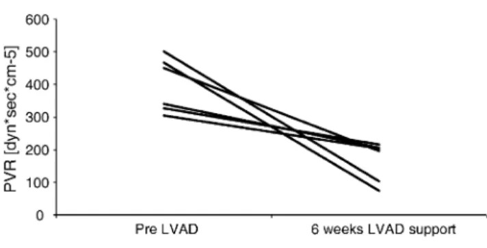
Serial right heart catheters were done on these six study patients. After a period of 6 weeks, the mean PVR decreased from 398 (501–305) to 167 (74–216) dyne s cmK5 (Fig. 1), systolic pulmonary artery pressure from a mean of 47 (29–67) to 29 (12–49) mmHg (Fig. 2), P!0.05 in all.
Patients increased their exercise tolerance with maxi-mum achieved power increasing from a mean of 35 to 71 W (P!0.05). Plasma BNP dosages were also done in this patient population, values dropped significantly during support period from 950 to 162 ng/l (P!0.05). Functional status measured by NYHA classification decreased from 4 to 1.5 (P!0.05). One patient suffered from a 7-h episode of
Table 1 Inclusion criteria
Left atrial or pulmonary capillary wedge pressure O188 mmHg Mean arterial pressure !90 mmHg
Cardiac index !2.0 l/min per m2
LVEF !25%
Incomplete response to intravenous therapy IABP
High risk of sudden cardiac death, and
Accepted by multidisciplinary institutional transplantation committee
Table 2
Preoperative patient demographics
Patient 1 2 3 4 5 6 Age (years) 48 41 37 52 64 61 Sex F M M M M M Height (cm) 172 176 186 182 185 170 Weight (kg) 67 72 92 92 64 68 HR (bpm) 100 80 90 95 88 84 AOP (mmHg) 105/75 100/54 90/60 85/50 80/50 85/65
NYHA III IV III III III III
Etiology RCMP CAD CAD CAD VALV DIL
Redo-surgery No No Yesa No Yesb No
AOP, aortic pressure; NYHA, New York Heart Association; RCMP, restrictive cardiomyopathy; CAD, coronary artery disease; VALV, valvular heart disease; M, male; F, female.
a
Previous coronary artery bypass grafting.
b Previous aortic valve replacement.
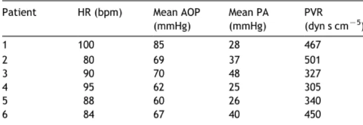
Table 3
Preoperative patients hemodynamics Patient HR (bpm) Mean AOP
(mmHg) Mean PA (mmHg) PVR (dyn s cmK5) 1 100 85 28 467 2 80 69 37 501 3 90 70 48 327 4 95 62 25 305 5 88 60 26 340 6 84 67 40 450
HR, heart rate; AOP, aortic pressure; PA, pulmonary artery pressure; PVR, pulmonary vasculature resistance.
ventricular fibrillation (recorded by his pace-maker). Fol-lowing electro conversion he remained in sinus rhythm for over 3 weeks (see case report)[14]. Two patients died while on support in this subgroup at days 81 (unknown cause) and 76 (thrombo-embolic event, see case report[14]).
3.2. Heart transplantation procedure and follow-up Four patients underwent successful heart transplantation after a mean duration of 130 (25–355) days. Only one patient required the use of inhaled nitric oxide in the early post-operative period after transplantation. Operative mortality was 0% and all patients were discharged. Mortality after a mean of 24-month follow-up remains 0%. Follow-up right heart catheters have shown all patients to have normal PA pressures and PVR.
4. Conclusions
4.1. LVAD as bridge to transplant
The DeBakey LVAD was the first new generation of LVAD, with continuous flow to offer a new alternative for the treatment of terminal heart failure [15]. As bridge to transplant, LVAD’s now seem to be established. The benefit of increased end-organ perfusion with very small, fully implantable devices seems substantial in ensuring good outcome of LVAD support and heart transplantation, especially with the acute donor organ shortage present around the world. This is the first report of a homogenous series of patients with high PVR and pHTN receiving the same type of LVAD. Martin et al.[16]just recently reported their experience in similar patients undergoing LVAD implantation with intent to treat pulmonary hypertension in
transplantation candidates. In our series we obtained similar results with patients normalizing their PVR within 6 weeks and significantly bettering their clinical status. Furthermore, we experienced very few adverse events, and no device related events occurred in our series.
4.2. LVAD and pulmonary circulation
Some authors advocate use of right ventricular support in patients with high PVR and pHTN. It seems however that with the advent of these new micro-axial continuous flow pumps, the mechanisms leading of right heart failure may be altered. The left and right ventricles are closely interdependent, with the use of pulsatile LVADs the unloading of the ventricle, especially the left, is not continuous during the cardiac cycle. These new continuous flow pumps unload the ventricle during the entire cardiac cycle, leading to a geometric restoration of ventricular geometry (left and right). Further-more the lateral shift of the septum which occurs when the left ventricle is not sufficiently unloaded, is decreased in these scenarios. In addition to a continuous negative pressure at the inflow cannula of the LVAD in the left ventricle, both these effects lead to an increased transpul-monary flow. Increased LV compliance during systole, decreased RV dilatation and less tricuspid insufficiency, all may contribute to the clinical improvement in these patients
[17]. The role of possible associated resynchronization therapy, i.e. biventricular pacing and continuous flow LVAD support seems like a very interesting combination in this patient population and warrants further investigation.
The early postoperative period has been identified by many authors as a critical period in patients with high PVR and pHTN leading to a low threshold for additional right ventricular assist device placement[18]. However there has been a lack of evidence in regard to PVR being a risk factor for postoperative right ventricular failure [19]. It is our belief that both pharmacological management (inotropic support), volume loading and fine tuning of pump par-ameters (rpm and flow adjustments, but also cardiac pacing) can avoid the necessity of right ventricular support. We use a low rpm/flow setting in the early phases of LVAD implan-tation to allow the aortic valve to open, but also to prevent septal shift in hypovolemic states leading to decreased right ventricular function and hemodynamically significant arrhythmias. All these measures are intended to ensure efficacious synchrony between the actively unloaded left ventricle and the passively assisted right ventricle. Once the early postoperative phase is overcome the flow can gradually be increased to match the clinical needs of these patients. 4.3. Outcome after heart transplantation
Elevated PVR and PH, medically unresponsive or fixed PH were previously considered contraindications for heart transplantation[20]. This study suggests that LVAD support allows continuous reduction of post capillary load inducing normalization of PVR in patients otherwise not considered as transplantation candidates. As reports have previously shown, normalization of high PVR and pulmonary hyperten-sion does occur after heart transplantation[10,12]. LVAD as bridge to transplant is safe and beneficial for terminal heart
Fig. 1. PVR during LVAD support (nZ6).
Fig. 2. Pulmonary systolic pressures during LVAD support (nZ6).
S.P. Salzberg et al. / European Journal of Cardio-thoracic Surgery 27 (2005) 222–225 224
failure patients with pHTN and high PVR and may allow some patients with pulmonary hypertension to convert from ineligible to eligible in regard to heart transplantation status
[21]. In our opinion, emergency heart transplantation is obsolete with the advent of LVAD therapy, especially in regard to the high mortality associated with emergency procedures [22]. Offering patients with terminal heart failure the option of mechanical circulatory support is a big therapeutic opening, which will save lives.
On the other hand, elevated PVR and fixed PH, notions incompatible with successful heart transplantation in the past, need to be revisited. Furthermore, this hemodynamic preconditioning, with LVAD and/or new pharmacologic agents prior to cardiac surgery seem to lead to excellent functionality of the pulmonary vasculature and come with increased postoperative survival.
References
[1] Massad MG. Surgical options for the management of congestive heart failure. Cardiology 2004;101:5–6.
[2] Massad MG. Current trends in heart transplantation. Cardiology 2004; 101:79–92.
[3] Wheeldon DR. Mechanical circulatory support: state of the art and future perspectives. Perfusion 2003;18:233–43.
[4] Salzberg S, Lachat M, Zund G, Oechslin E, Schmid ER, DeBakey M, Turina M. Left ventricular assist device as bridge to heart transplan-tation—lessons learned with the MicroMed DeBakey axial blood flow pump. Eur J Cardiothorac Surg 2003;24:113–8.
[5] Kukuy EL, Oz MC, Rose EA, Naka Y. Devices as destination therapy. Cardiol Clin 2003;21:67–73.
[6] Frazier OH, Myers TJ, Westaby S, Gregoric ID. Clinical experience with an implantable, intracardiac, continuous flow circulatory support device: physiologic implications and their relationship to patient selection. Ann Thorac Surg 2004;77:133–42.
[7] Entwistle III JW, Bolno PB, Holmes E, Samuels LE. Improved survival with ventricular assist device support in cardiogenic shock after myocardial infarction. Heart Surg Forum 2003;6:316–9.
[8] Nauser TD, Stites SW. Pulmonary hypertension: new perspectives. Congest Heart Fail 2003;9:155–62.
[9] Stobierska-Dzierzek B, Awad H, Michler RE. The evolving management of acute right-sided heart failure in cardiac transplant recipients. J Am Coll Cardiol 2001;38:923–31.
[10] Klotz S, Deng MC, Hanafy D, Schmid C, Stypmann J, Schmidt C, Hammel D, Scheld HH. Reversible pulmonary hypertension in heart transplant candidates—pretransplant evaluation and outcome after orthotopic heart transplantation. Eur J Heart Fail 2003;5:645–53.
[11] Lindelow B, Andersson B, Waagstein F, Bergh CH. High and low pulmonary vascular resistance in heart transplant candidates. A 5-year follow-up after heart transplantation shows continuous reduction in resistance and no difference in complication rate. Eur Heart J 1999;20: 148–56.
[12] Bhatia SJ, Kirshenbaum JM, Shemin RJ, Cohn LH, Collins JJ, Di Sesa VJ, Young PJ, Mudge Jr GH, Sutton MG. Time course of resolution of pulmonary hypertension and right ventricular remodeling after ortho-topic cardiac transplantation. Circulation 1987;76:819–26.
[13] Noon GP, Morley DL, Irwin S, Abdelsayed SV, Benkowski RJ, Lynch BE. Clinical experience with the MicroMed DeBakey ventricular assist device. Ann Thorac Surg 2001;71:S133–S8 [discussion S144–6].
[14] Salzberg S, Lachat M, Zund G, Turina M. Left ventricular assist device (LVAD) enables survival during 7 h of sustained ventricular fibrillation. Eur J Cardiothorac Surg 2005.
[15] Goldstein DJ. Worldwide experience with the MicroMed DeBakey ventricular assist device as a bridge to transplantation. Circulation 2003;108(Suppl 1):II272–II277.
[16] Martin J, Siegenthaler MP, Friesewinkel O, Fader T, van de Loo A, Trummer G, Berchtold-Herz M, Beyersdorf F. Implantable left ventri-cular assist device for treatment of pulmonary hypertension in candidates for orthotopic heart transplantation—a preliminary study. Eur J Cardiothorac Surg 2004;25:971–7.
[17] Kucuker SA, Stetson SJ, Becker KA, Akgul A, Loebe M, Lafuente JA, Noon GP, Koerner MM, Entman ML, Torre-Amione G. Evidence of improved right ventricular structure after LVAD support in patients with end-stage cardiomyopathy. J Heart Lung Transplant 2004;23: 28–35.
[18] Kavarana MN, Pessin-Minsley MS, Urtecho J, Catanese KA, Flannery M, Oz MC, Naka Y. Right ventricular dysfunction and organ failure in left ventricular assist device recipients: a continuing problem. Ann Thorac Surg 2002;73:745–50.
[19] Ochiai Y, McCarthy PM, Smedira NG, Banbury MK, Navia JL, Feng J, Hsu AP, Yeager ML, Buda T, Hoercher KJ, Howard MW, Takagaki M, Doi K, Fukamachi K. Predictors of severe right ventricular failure after implantable left ventricular assist device insertion: analysis of 245 patients. Circulation 2002;106:I198–I202.
[20] Rose EA, Gelijns AC, Moskowitz AJ, Heitjan DF, Stevenson LW, Dembitsky W, Long JW, Ascheim DD, Tierney AR, Levitan RG, Watson JT, Meier P, Ronan NS, Shapiro PA, Lazar RM, Miller LW, Gupta L, Frazier OH, Desvigne-Nickens P, Oz MC, Poirier VL. Long-term mechanical left ventricular assistance for end-stage heart failure. N Engl J Med 2001;345:1435–43.
[21] Al-Khaldi A, Ergina P, DeVarennes B, Lachappelle K, Cecere R. Left ventricular unloading in a patient with end-stage cardiomyopathy and medically unresponsive pulmonary hypertension. Artif Organs 2004;28: 158–60.
[22] Espinoza C, Manito N, Castells E, Rodriguez R, Octavio de Toledo MC, Calbet JM, Fontanillas C, Saura E, Miralles A, Granados J, Benito M, Roca J, Mauri F, Ramon JM, Obi C, Quiles C, Claret G. Perioperative mortality risk factors after orthotopic heart transplantation. Transplant Proc 1999;31:2509–10.