Chemistry meets biology in colitis-associated carcinogenesis
The MIT Faculty has made this article openly available. Please share
how this access benefits you. Your story matters.
Citation Mangerich, A., P. C. Dedon, J. G. Fox, S. R. Tannenbaum, and G. N. Wogan. “Chemistry Meets Biology in Colitis-Associated Carcinogenesis.” Free Radic Res 47, no. 11 (November 2013): 958– 986.
As Published http://dx.doi.org/10.3109/10715762.2013.832239
Publisher Informa UK (Informa Healthcare)
Version Author's final manuscript
Citable link http://hdl.handle.net/1721.1/91231
Terms of Use Creative Commons Attribution-Noncommercial-Share Alike
1
Chemistry meets biology in colitis-associated carcinogenesis
Aswin Mangerich1,5*, Peter C. Dedon1,4, James G. Fox1,3,4, Steven R. Tannenbaum1,2,4, and Gerald N. Wogan1,2,4
Departments of Biological Engineering1 and Chemistry2, 3Division of Comparative Medicine, and 4Center for Environmental Health Science, Massachusetts Institute of Technology, 77 Massachusetts Avenue, Cambridge, MA 02139; 5Department of Biology, Molecular Toxicology Group, University of Konstanz, D-78457 Konstanz, Germany
KEY WORDS: Inflammation, cancer, inflammatory bowel disease, innate immunity, nitric oxide, peroxynitrite, hypochlorous acid, iNOS, myeloperoxidase, DNA damage, DNA repair, biomarkers
ABBREVIATIONS:
AOM, azoxymethane; CAC, colitis-associated carcinogenesis; GPx, gluthathione peroxidases; GRx, glutaredoxins; GSR: glutathione reductase; IBD, inflammatory bowel disease; iNOS, inducible nitric oxide synthase; MPO, myeloperoxidase; NOX, NADPH oxidase; NOx: NO
oxidation products; RNS, reactive nitrogen species; ROS, reactive oxygen species; TNBS, 2,4,6-trinitro benzene sulfonic acid; TxnRD, thioredoxin reductases; XOR, xanthine oxidoreductase
*CORRESPONDENCE: Aswin Mangerich; Department of Biological Engineering,
Massachusetts Institute of Technology; and Department of Biology, University of Konstanz; Email: amang@mit.edu or aswin.mangerich@uni-konstanz.de
2
Abstract
The intestine comprises an exceptional venue for a dynamic and complex interplay of numerous chemical and biological processes. Here, multiple chemical and biological systems, including the intestinal tissue itself, its associated immune system, the gut microbiota, xenobiotics, and metabolites meet and interact to form a sophisticated and tightly regulated state of tissue homoeostasis. Disturbance of this homeostasis can cause inflammatory bowel disease (IBD) – a chronic disease of multifactorial etiology that is strongly associated with increased risk for cancer development. This review addresses recent developments in research into chemical and biological mechanisms underlying the etiology of inflammation-induced colon cancer. Beginning with a general overview of reactive chemical species generated during colonic inflammation, the mechanistic interplay between chemical and biological mediators of inflammation, the role of genetic toxicology and microbial pathogenesis in disease development are discussed. When possible, we systematically compare evidence from studies utilizing human IBD patients with experimental investigations in mice. The comparison reveals that many strong pathological and mechanistic correlates exist between mouse models of colitis-associated cancer, and the clinically relevant situation in humans. We also summarize several emerging issues in the field, such as the carcinogenic potential of novel inflammation-related DNA adducts and genotoxic microbial factors, the systemic dimension of inflammation-induced genotoxicity, and the complex role of genome maintenance mechanisms during these processes. Taken together, current evidence points to the induction of genetic and epigenetic alterations by chemical and biological inflammatory stimuli ultimately leading to cancer formation.
3
1 Introduction: inflammatory bowel disease and colon cancer
With respect to a functional interplay between chemistry and biology, the intestinal tract is one of the most complex organs in the body. In the intestine, local epithelial cells closely interact with the body’s immune system and meet with a complex bacterial microflora and a plethora of endogenous metabolites and xenobiotic substances. All of these factors need to be tightly balanced to ensure intestinal homeostasis and to guarantee efficient uptake of nutrients and excretion of metabolites. If this balance is disturbed, pathological states can appear, such as inflammatory bowel disease (IBD). As a major risk factor for colon cancer, IBD entails chronic, relapsing inflammation of the gastrointestinal tract that affects millions of people worldwide, with >1 million new patients each year in the US alone [1]. The paucity of safe and effective therapies for IBD amplifies its public health impact and motivates the hunt for the molecular etiology of the disease and the mechanisms linking colitis with colon carcinogenesis. This review addresses recent developments in chemical and biological mechanisms underlying inflammation-induced colon cancer, starting with a general overview of the reactive chemical species generated during colonic inflammation followed by a discussion of the mechanistic interplay between chemical and biological mediators of inflammation and the role of microbial pathogenesis and genetic toxicology in the etiology of inflammation-induced colon cancer. The reader is referred to several recent reviews of molecular aspects of IBD and cancer development not covered here, in particular immunological and genetic studies [1-7].
1.1 Inflammatory bowel disease: Crohn’s disease and ulcerative colitis
Inflammatory bowel disease comes in two distinct forms: Crohn’s disease and ulcerative colitis. In Crohn’s disease, inflammation often affects the entire thickness of the bowel wall and affects mainly the ileum and colon, but can discontinuously spread throughout any part of the intestine. In contrast, the inflammation in ulcerative colitis is usually confined to the mucosal surface of the intestines and limited to colon and rectum, spreading in a continuous fashion (reviewed in [1]). The pathophysiology of IBD is multifactorial, with contributions from genetic, epigenetic, environmental, and endogenous microbial factors (reviewed in [3,8]). Genetic background is an important determinant for IBD, evidenced by the fact that family history is a risk factor for disease development with a concordance rate in monozygotic twins of 10-15% in ulcerative colitis and 30-35% in Crohn’s disease. At present, more than 150 genetic loci have been associated with disease development [9]. IBD-associated genes are involved in several
4 disease-relevant pathways crucial for intestinal homeostasis, including epithelial barrier function, microbial defense mechanisms, regulation of immunity, autophagy, generation of reactive oxygen and nitrogen species (ROS, RNS) and signaling functions. About 30% of these loci are shared between ulcerative colitis and Crohn’s disease, so the two pathological states share some similar molecular mechanisms, but also exhibit many disease-specific features [3,9]. The contribution of environmental factors is not well understood, but evidence indicates that they play a significant role, as suggested by the high prevalence of IBD in industrialized countries. This pattern of high prevalence appears not to be related to genetic factors, since migrant studies have demonstrated that individuals relocating from a region with low prevalence to one of high prevalence are at increased risk of developing IBD [10]. Environmental influences include life-style factors such as smoking and dietary composition among others, but elevated IBD prevalence may also be related to improved sanitation and hygiene. The latter is based on the hypothesis that lack of childhood exposure to enteric pathogens may lead to inappropriate immune responses to new antigens in adulthood and an imbalance in host-microbe interaction, which play an important role in the maintenance of mucosal homeostasis [10]. An imbalance of host-microbe homeostasis is of paramount importance in the etiology of IBD, supported by the fact that some IBD patients benefit from antibiotic treatment and most mouse models of IBD require the presence of intestinal bacteria to develop colitis [1]. Furthermore, an altered immune response to the intestinal flora plays a pivotal role in the etiology of IBD [8]. Notably, the intestine and its associated tissues represent the largest immunological organ of the body. In particular, the intestinal lamina propria contains a complex mixture of immune cells that maintain immune tolerance and pathogen defense in a tightly controlled balance (reviewed in [1]). Active IBD is characterized by a massive infiltration of innate immune cells, including neutrophils, macrophages, dendritic cells, and natural killer cells, which fulfill complex roles under physiological as well as pathophysiological conditions. Importantly in the context of this review, activated neutrophils and macrophages are major sources of ROS and RNS that are among the main factors against invading pathogens, but can also cause collateral damage to the host tissue, thereby potentially initiating and promoting carcinogenesis (see below). Apart from innate immune cells, those of the adaptive immune system (e.g., B and T cells), also control intestinal homeostasis. Perturbation of the balance of Th17 cells, which produce the pro-inflammatory signature cytokines IL-17, IL-21, and IL-23, and regulatory T-cells, which produce the anti-inflammatory cytokines IL-10 and TGF-β appear to be of particular importance. An imbalance in
5 these systems seems to play a crucial role in the etiology of IBD (reviewed in [2]). For an in-depth discussion on the intestinal immunology in the pathology of IBD, the reader is referred to several recent reviews [2,4,6,11-17]. In summary, IBD is a multifactorial disease that is thought to result from a perturbation in host-microbe interactions leading to an immunological imbalance and chronic inflammation in genetically susceptible individuals [3].
Strong epidemiological evidence indicates that inflammation existing in Crohn’s disease and ulcerative colitis is associated with increased risk of colon cancer, but the responsible molecular mechanisms remain largely undefined. More than 20% of patients with IBD develop colitis-associated cancers within 30 years of disease onset, and >50% of these patients die from them [7]. Notably, patients who develop IBD at a young age (< 30 yrs) have a much greater risk of cancer development [18]. In general, the risk for colon cancer increases with duration and severity of disease, whereas it decreases when patients are treated with anti-inflammatory drugs such as mesalamine and corticosteroids, consistent with a causative role for inflammation in colon carcinogenesis (reviewed in [19]).
Spontaneous colorectal cancer shares many common pathophysiological mechanisms with colitis-associated cancer, but differs in some distinct ways (reviewed in [7]). For example, as with spontaneous colorectal cancer, colitis-associated cancers may progress through a sequence of aberrant crypt foci, polyps, adenomas, and carcinomas. However, dysplasia in spontaneous colorectal cancer is often focal, whereas colitis-associated cancers often develop through a sequence of chronic inflammation, tissue injury, and multifocal dysplasia leading to the formation of poorly-defined carcinomas. The genetic alterations such as chromosomal instability, microsatellite instability, and DNA hypermethylation found in spontaneous colon cancers also occur in colitis-associated cancers; these characteristics arise in inflamed tissue before the appearance of histological evidence of dysplasia or cancer [19]. Common tumor-related signaling pathways, including factors such as Wnt, β-catenin, K-ras, p53, TGF-β, and DNA repair, are affected in development of spontaneous as well as colitis-associated cancers, but at different stages of tumor formation [7]. It is very likely that crypt stem cells represent the cells-of-origin for cancer development, in both spontaneous colorectal and colitis-associated cancers [20]. However, recent studies demonstrate that NF-κB signaling can lead to dedifferentiation of intestinal epithelial cells into tumor-initiating cells with stem cell-like properties [21]. This is consistent with the fact that colitis-associated carcinogenesis (CAC) is
6 characterized by production of NF-κB and Stat3-dependent pro-inflammatory cytokines, and activation or inactivation of oncogenes and tumor suppressors, respectively. Reactive chemical species are likely to play an important role in these processes and there is increasing experimental evidence supporting the hypothesis that during inflammation, cells of the innate immune system generate large amounts of ROS and RNS inducing genetic and epigenetic changes, which may lead to mutations and tumor initiation and promotion [22].
1.2 Mouse models of colitis-associated carcinogenesis
Several mouse models of colitis have been developed to study molecular disease mechanisms and to define targets for translational approaches. Colitis is induced by chemical agents, by genetic intervention, by infection or by combinations of these factors (Table 1). In light of the recent criticism of mouse models of human disease [23,24], which in our opinion has been overstated and inaccurate, we demonstrate that many compelling pathological and mechanistic correlates exist between mouse models of colitis-associated cancer and the clinically relevant disease in humans. Indeed, as we review here, a plethora of studies in mouse models of colitis have revealed that many chemical and biological mechanisms of pathology share strong similarities across species boundaries (Table 2), which is most evident by the fact that human IBD and various forms of colitis in mice lead to an increased risk of colon carcinogenesis. Only through the combination of animal models and studies utilizing human IBD patients can a deeper understanding be obtained of the chemistry and biology in CAC, through the use of multidisciplinary approaches from genetics to microbiology, chemistry, systems biology and mathematical modeling, combined with thoughtful data interpretation.
We recently conducted a systematic analysis of mechanisms and biomarkers of Helicobacter
hepaticus (H. hepaticus)-induced colitis in immunodeficient 129 Rag2-/- mice with respect to various aspects of disease development [25-29]. The gram-negative spiral bacterium H.
hepaticus colonizes the liver and intestinal crypts of the cecum and the colon, establishing a
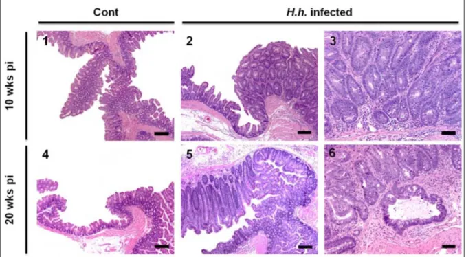
life-long infection [25,26,30,31]. H. hepaticus does not typically cause disease in immunocompetent mice, but infection in susceptible mouse strains or in immunodeficient mice, such as those lacking the recombinase-activating gene-2 (Rag2), results in chronic colitis and colon cancer [25] (Figure 1). H. hepaticus infection in Rag2-/- mice emulates many aspects of human IBD and as discussed here, this mouse model has proved to be a valuable tool specifically to study features of innate immunity during CAC.
7
2 Immunological chemistry of inflammatory bowel disease
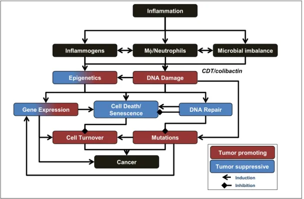
Colonic inflammation results in a local release of cytokines and other chemotactic factors that cause infiltration and activation of innate immune cells to produce large quantities of other cytokines and chemokines resulting in the activation of various enzymes that generate a wide spectrum of reactive chemical species as summarized in Figure 2 and discussed in the following sections in detail. In particular, we summarize evidence that these chemical mediators of inflammation can react with all types of macromolecules, including proteins, RNA, and DNA, to cause mutagenic and cytotoxic damage, thereby contributing to colon carcinogenesis.
2.1 Cell types involved in the generation of reactive chemical species during IBD
In addition to the endogenous production of NO by colonic epithelial cells [32-35], macrophages and neutrophils are thought to be responsible for the bulk of ROS and RNS generation during acute and chronic inflammation (Figure 2). Both cell types are closely related and cooperate during the onset, progression and resolution of inflammation. Usually, tissue-resident macrophages and dendritic cells sense inflammatory stimuli, which causes chemokine-dependent recruitment of neutrophils followed by infiltration of blood monocytes, both of which then differentiate and become activated (reviewed in [14]).
Initial support of the hypothesis that innate immune cells can act as carcinogens was obtained by Weitzman and colleagues, who exposed mouse fibroblasts to activated human neutrophils or a cell-free ROS-producing system. Following injection of the fibroblasts into nude mice, tumors developed, but only in mice injected with exposed cells. Mice injected with control cells remained tumor-free, which indicated that activated neutrophils and ROS are able to mediate malignant cell transformation [36]. It is well established that neutrophils and macrophages play a pivotal role in the induction and maintenance of inflammation during colitis in mice and humans [26,37-39]. In addition, several studies support the functional relevance of these cell types during colon carcinogenesis. For example, in a rat model of dextran sodium sulfate (DSS)-induced acute colitis, selective depletion of neutrophils by intraperitoneal injection of a neutrophil-specific monoclonal antibody significantly reduced pathology [40]. A similar antibody-based approach was used to selectively deplete mature neutrophils in H. hepaticus-infected Rag2-/- mice, a model in which there is massive infiltration of macrophages and neutrophils during disease development [26,27,29]. Interestingly, depletion of neutrophils in
8 these mice significantly decreased the severity of colon pathology, including cancer rates, and led to lower expression of pro-inflammatory cytokines and ROS/RNS-generating enzymes (see below) [26]. Macrophages can also contribute to disease progression and severity, as shown in a mouse model of H. bilis-induced typhlocolitis, in which macrophages were selectively depleted by treatment with clodronate-containing liposomes. Clodronate-treated mice exhibited significantly lower histopathology scores and suppressed expression of macrophage-related factors, such as TNF-α, Il-1β, and iNOS, suggesting that macrophages are important mediators of
H. bilis-induced colitis. However, the finding that clodronate treatment concomitantly reduced
the number of MPO-positive neutrophils suggests that both macrophages and neutrophils closely interact in disease development on a functional level [41]. This view is supported by the H.
hepaticus Rag2-/- mouse model that revealed cytokine signatures suggestive of both neutrophil
and macrophage activity [29]. Interestingly, this finding correlated well with results from patients with Crohn’s disease. In contrast, cytokine signatures of patients with ulcerative colitis were more suggestive of neutrophil activity only, indicating specific differences between the two types of IBD on a molecular level [29].
2.2 NADPH oxidases, xanthine oxidoreductase, and the generation of ROS
Superoxide (O2•-), is the primary source of ROS in humans. Under physiological conditions,
mitochondrial respiration is the major source, whereas NADPH oxidase (official gene symbol:
Nox, not to be confused with NOx which refers to NO oxidation products) and xanthine
oxidoreductase (XOR) significantly contribute to the generation of O2•- under pathophysiological
conditions (reviewed in [42-44]) (Figure 2).
NADPH oxidases represent a widely distributed family of enzymes, with each species consisting of a flavoprotein, cytochrome b and several regulatory subunits. When activated, the enzymes assemble in the cell membrane and reduce oxygen to O2•-. Phagocytic cells express
NADPH oxidase 2 in the phagosomal membrane, where it generates high fluxes of O2•- (reviewed
in [42,43,45]). In addition to ‘phagocyte NADPH oxidases 2’, NADPH oxidases 1 is highly expressed in the gastrointestinal tract. Levels of the mRNA of NADPH oxidase 1, also known as the ‘colon NADPH oxidase’, increase from proximal to distal colon [45]. Even under physiological conditions, NADPH oxidase 1 is highly expressed in colon epithelial cells, both within the crypts and on the luminal surface [46,47]. Maturation-dependent factors seem to regulate the expression of NADPH oxidase 1 in colonic epithelial cells, which leads to the
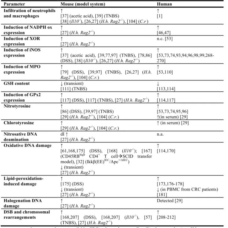
9 highest expression levels in surface mucous cells where NADPH oxidases are thought to contribute to host defense. However, under pathophysiological conditions, they may mediate tissue damage by generating ROS. This view is consistent with results from H. hepaticus-induced colitis in Rag2-/- mice, in which gene expression of NADPH oxidases 1 and 2 (Nox 1,
Nox2) was significantly up-regulated in inflamed tissue. This was accompanied by higher
expression levels of several regulatory subunits of the NADPH oxidase system, such as Ncf1 (p47phox) and Ncf2 (p67phox) [27], presumably a result of tissue infiltration of innate immune cells. In addition, studies in mice and human cells have shown that pro-inflammatory cytokines induced during colitis activate expression of NADPH oxidases [48,49]. The role of NADPH oxidase in human IBD and colon carcinogenesis is not well characterized. Some evidence comes from patients with chronic granulomatous disease, many of whom have genetic defects in the NADPH oxidase system that results in an immunodeficiency phenotype characterized by an inability of phagocytes to kill invading pathogens. These patients exhibit an increased risk for IBD, suggesting that NADPH oxidases have a direct role in controlling intestinal homeostasis [45,50,51]. Szanto et al. reported strong expression of NADPH oxidase 1 in active lesions from patients with Crohn’s disease and ulcerative colitis [46] and Fukuyama et al. reported significant up-regulation of NADPH oxidase 1 in colonic tumor tissue [47], suggesting a functional role of NADPH oxidases in disease progression. Despite these findings, comprehensive understanding of the functional role of NADPH oxidases in CAC is incomplete, and conclusions rely mainly on correlative studies, while mechanistic studies in mice and humans are limited. Genetic studies analyzing mechanisms of CAC in mice carrying mutations or deletions in the NADPH oxidase system would be of interest with respect to pharmacological interventions, since NADPH oxidases are already being considered as potential drug targets for treatment of other inflammatory disorders, such as vascular disease [43].
Xanthine oxidoreductase (XOR) is a multifunctional enzyme that is expressed and secreted by many epithelial cells including those in the colonic mucosa (reviewed in [44]) (Figure 2). The enzyme possesses two activities, as both a reductase and an oxidase. The reductase catalyzes the oxidative hydroxylation of hypoxanthine to xanthine and subsequently of xanthine to uric acid. In this functional state, xanthine reductase acts as a housekeeping enzyme with a role in purine catabolism, detoxification and the regulation of the cellular redox potential. Various stimuli induce the conversion into an oxidase, either reversibly by thiol oxidation or irreversibly
10 by limited proteolysis. In contrast to the reductase, xanthine oxidase is associated with production of large amounts of ROS including O2•- [44]. The enzyme has also been directly
implicated in the activation of macrophages through effects on chemokine expression and other factors, thereby participating in macrophage-mediated neutrophil recruitment [52].
In the human colon, XOR immunostaining follows a gradient along the villus-crypt axis in a pattern similar to that observed for NOX1, with luminal epithelial cells showing the strongest staining. XOR staining was also observed in cells at the base of the crypt, as well as in neutrophils and endothelial cells in the lamina propria [53]. Data on the role of XOR in colitis and associated cancer development is limited and controversial. H. hepaticus-infected Rag2 -/-mice showed higher expression levels of XOR mRNA in inflamed tissue, but the origin of this increase is currently unknown [27]. Up-regulation of XOR is consistent with the finding that XOR is stimulated by IFN-γ, TNF-α, and IL-1, and some of these factors also trigger the reductase-oxidase conversion [44]. Furthermore, Siems et al. reported that the XOR inhibitor oxypurinol protects mice from morphological and molecular changes occurring in TNBS-induced colitis. However, as is it the case with every pharmacological inhibitor, specificity of the compound is an issue. Although these results indicate a role of XOR in murine colitis and XOR has been associated with other inflammatory diseases [44], its role in human IBD awaits further clarification. Kruidenier et al. reported that epithelial apoptosis in patients with Crohn’s disease was strongly associated with XOR expression, suggesting a role for XOR in the regulation of epithelial homeostasis. However, in this study, overall levels of XOR were unaffected during intestinal inflammation, indicating that XOR contributes to disease development more through local and functional changes in its activity [53]. Mouse genetics could shed some light on the role of XOR in colitis, which could be instrumental to test potential treatment options by targeting this enzyme, In this respect, generation of colon-specific knock-out mice or genetically engineered mice that specifically lack the enzymes’s oxidase function may represent one possible line of investigation, since Xor-/- mice die within the first six weeks after birth due to renal failure [54,55].
Superoxide can be transformed by superoxide dismutases (SOD) into hydrogen peroxide (H2O2), which can be either detoxified by catalases and peroxidases into H2O and O2, or it can be
reduced by divalent metal ions in Fenton chemistry that gives rise to highly reactive hydroxyl radicals (•OH) [42] (Figure 2). Since H2O2 reacts poorly with most biological molecules and is
11 highly diffusible, it acts as an important signal transducer (reviewed in [42,56]). For example, H2O2 activates phosphorylation cascades resulting in downstream effects such as enhanced stress
resistance, cell proliferation, cytokine release, cell adhesion, growth arrest and apoptosis. Its damaging effects are mainly caused by its reaction with seleno-, thiol or heme peroxidases to produce radical and non-radical species as well as through •OH formed by Fenton chemistry [42]. In mice with TNBS-induced colitis, H2O2 levels immediately increased in the muscularis externa
within the first week after treatment, with peak levels achieved after the first day [57]. That the generation of H2O2 acts as a detoxifying pathway is suggested by studies of SOD, the major
source of H2O2. For example, in DSS-induced colitis, Cu/Zn-SOD-overexpressing transgenic
mice showed a survival benefit that was accompanied by an enhanced clearance of neutrophils [58]. Moreover, studies in which SOD and SOD mimetics were administered during TNBS and DSS-induced colitis revealed beneficial effects of SOD with regards to disease severity and molecular damage [59-62]. In studies of human colitis, MnSOD was strongly up-regulated in inflamed epithelium, whereas the Cu/ZnSOD content decreased with inflammation. On a cellular level, Cu/Zn and MnSOD were prominently present in neutrophils and macrophages, while extracellular (EC)-SOD was mainly localized in small vessels, stromal cells, and neutrophils. This differential regulation of the SOD isoforms indicates that ROS generation and its detoxification are important factors in the etiology of IBD, whose specific role needs to be clarified in future studies [63].
2.3 Nitric oxide synthases and the generation of reactive nitrogen species (RNS)
Nitric oxide (NO) is the key molecule during the generation of RNS (Figure 2). Since NO has a low molecular weight, is uncharged, and is soluble in both aqueous and hydrophobic environments, it is highly diffusible across cell membranes and through tissues. The diffusion coefficient for NO is 3300 µm2s-1, which is comparable to that for O2 (2800 µm2s-1) and implies
that NO is able to diffuse as far as 5-10 cell diameters in 1 sec [56]. NO production in cell culture studies coincides with the appearance of NO2- and NO3-, which are formed at
approximately equal and constant rates [64]. These metabolites arise from NO auto-oxidation by reacting with O2 to form nitrogen dioxide radical (NO2•) that can react further with NO2• or NO to
form N2O4 or N2O3, respectively. These higher N-oxides can subsequently form NO2-, NO3- and
nitroso products, which are closely related with enzymatic as well as non-enzymatic conversions with complex (patho-)physiological consequences [65-67]. Furthermore, NO reacts with O2•- at
12 diffusion controlled rates to form peroxynitrite (ONOO-) [68] (Figure 2). ONOO- represents an important source of a complex spectrum of highly reactive secondary radicals [42]. Its protonated form, ONOOH, undergoes rapid (t1/2 ~ 1 sec) homolysis to form the strongly
oxidizing •OH and the weaker oxidant NO2•. In addition, ONOO- reacts with CO2, which is
present in millimolar concentrations in tissues, to form nitrosoperoxycarbonate (ONOOCO2-),
which decomposes with a half life of ~50 msec into NO2• and highly reactive carbonate radical
anions (CO3•-) [69]. When the concentration of NO• exceeds that of O2•-, which predominantly
occurs within cellular membranes, autoxidation leads to the formation of NO2• and N2O3. N2O3
itself is an S- and N-nitrosating species, but readily decomposes with a half-life of 0.2 msec [56,69].
Three isoforms of NO synthases exist that can generate NO over a wide range of concentrations in an NADPH and O2-dependent process by converting L-arginine to citrulline
and NO [70]. Neuronal and endothelial NOS produce NO at low levels in nerve and endothelial cells, respectively, while activation of inducible NOS (iNOS) leads to high NO concentrations in macrophages during inflamation (reviewed in [71]). In general, the chemical biology of NO can be divided into two categories: (i) direct effects of NO mediated by chemical reactions that occur fast enough to allow NO to react directly with biological target molecules, and (ii) indirect effects that require that NO reacts with other ROS to generate RNS, which then react with biological targets [56]. Biological functions of NO are highly concentration dependent, with relevant concentrations varying over three orders-of-magnitude. One of the major challenges in the study of colitis has been to define the concentrations of NO arising in the inflamed colon and the biological consequences of the different concentrations of NO and its derivatives in the inflamed tissue. In this regard, significant insights have been obtained from computational modeling. By describing rates of reaction and diffusion in realistic geometries, mathematical modeling has estimated that maximal NO concentrations reached at the base of a crypt are in the sub-micromolar range (~0.3 µM) and the cumulative NO dose an epithelial cell is experiencing during migration from the base to the top of the crypt is ~560 µM min [72] (Figure 3). Taking into account findings that epithelial cells contribute to the NO production in the colon [32,73,74] and that macrophages are able to infiltrate into the lamina propria, these models predict maximal NO concentrations of ~ 0.4 µM and maximal cumulative doses of ~1 µM min [72] (Figure 3). These calculations are in accordance with cell culture studies demonstrating that upon stimulation
13 macrophages produce levels of NO up to 1 µM [64]. As a rule of thumb, at low (nM) concentrations NO promotes cell survival and proliferation and acts as an important signaling molecule in the cardiovascular, nervous and immune systems, while, at higher concentrations (≤ 1 µM), such as may occur at sites of inflammation, it can be cytotoxic and cytostatic, inducing cell cycle arrest, apoptosis and senescence [56,69,75,76].
During colitis, iNOS is considered the main source of NO. In healthy mice, iNOS mRNA is expressed in the ileum, but not in the jejunum or colon. iNOS protein is also detected in normal ileal epithelial cells, suggesting that it may also fulfill a role in maintaining intestinal homeostasis [33]. In the inflamed gut, iNOS is present throughout the intestinal tract, including the jejunum and colon [26,27,33,37,77,78]. Induction of iNOS is regulated by neutrophils and macrophages in combination with cytokine stimuli, as suggested by the fact that depletion of Gr-1+ neutrophils,
clodronate-induced depletion of macrophages, and blocking of TNF-α in the colon, respectively, prevented iNOS induction [26,41]. Various mouse studies demonstrated that colitis-induced activation of iNOS is accompanied by increased levels of NO-derived species in plasma and urine [26,38,39,79]. Saijo et al. studied the dynamics of biomarkers of NO production, such as NO2-, NO3-, and nitroso and nitrosyl species during DSS-induced chronic and acute colitis [80],
and showed that blood levels of nitroso products correlated well with disease development. This correlation was also true for plasma levels of NO2- and NO3-, but only in acute colitis during the
onset of the inflammatory response. A more complex response of these ions was observed in tissues as well as in the chronic colitis model. Interestingly, for all markers analyzed, similar changes were observed in organs other than colon, such as liver, brain, aorta, and heart, highlighting the systemic effects of this disease. In general, the study by Saijo et al. suggests that NO2- serves as a suitable indicator of colitis per se, although the rise of plasma NO2-may not
correlate well with the progression and resolution of inflammation [80].
Supporting the notion that NO generation during colitis can be attributed to the activation of iNOS, no increase in NO-derived species was observed during colitis in Il-10-/- mice in an iNos -/-background [38]. Similarly, in H. hepaticus-infected Rag2-/- mice concurrent administration of an iNOS inhibitor prevented NO3- production and reduced epithelial pathology and cancer
incidence [26]. Similarly, an iNOS inhibitor significantly reduced colonic adenocarcinoma formation in a dose-dependent manner in a CAC model using DSS-treated ApcMin/+ mice [81]. These effects can be directly attributed to iNOS inhibition, since during H. hepaticus-induced
14 colitis, iNOS inhibitor treatment had no influence on the numbers of neutrophils and macrophages [26]. Interestingly, administration of iNOS inhibitors also decreased the levels of pro-inflammatory cytokines such as TNF-α and IL-1β in DSS-induced colitis, indicating a positive-feedback regulation of these components under inflammatory conditions [79,81]. The notion that ONOO- can not only act as a downstream effector molecule, but also as an active driving force of disease development is supported by the finding that intrarectal administration of ONOO- itself was sufficient to induce the generation of NO-derived species, iNOS expression, and colitis in rats [82].
Ambiguous results were obtained from genetic studies of colitis in iNos-/- mice. Several groups reported a protective effect of iNOS depletion on tissue inflammation, damage, and cancer rates [39,78,83-85], whereas others found no effect [38,86] or even more severe tissue damage and pathology in iNos-/- mice [37,87]. Krieglstein et al. performed bone marrow transplantation
experiments using iNOS-proficient or -deficient cells transplanted into wild-type or iNos-/- mice. In mice deficient in either tissue or blood cell iNOS, or both, DSS-induced colitis was attenuated. Interestingly, MPO activity was also lower in iNos-/- mice and occurred at the lowest levels in
iNos-/- mice transplanted with blood cells carrying functional iNOS. These results suggest that both blood-cell and derived iNOS plays a role in DSS-induced colitis, with tissue-associated iNOS making a larger contribution to the recruitment of MPO-positive inflammatory cells [84]. Similarly, Beck et al. showed that chimeric mouse lines with iNOS-deficient blood cells were more resistant to DSS and TNBS-induced colitis; in the DSS model neutrophils were the main source of iNOS [88]. This is in agreement with our finding that depletion of Gr-1+ neutrophils of H. hepaticus-infected mice prevented iNOS induction in the colon [26]. Another study analyzed mouse lines deficient in iNOS, nNOS, eNOS or e/nNOS and revealed that loss of iNOS or eNOS was protective on DSS-induced colitis, whereas loss of nNOS resulted in more severe disease and higher mortality rates. These results indicate that NO produced by individual isoforms plays different roles in the modulation of intestinal inflammation [85]. In terms of signature biomarkers of iNOS activity, H. hepaticus-induced colitis in Rag2-/- mice led to significantly higher levels of nitrotyrosine in colon tissue as well as in serum [29]. Similarly, TNBS-induced colitis led to increased levels of nitrotyrosine [39]. In this model, ablation of iNOS resulted in reduced levels of nitrotyrosine indicating that iNOS is required for the generation of nitrosative damage [39]. On the other hand, in iNos+/+ and iNos-/- mice with
15 chronic DSS-induced colitis showed no differences in nitrotyrosine levels, pointing to a role of MPO activity (see below) [86]. These apparently contradictory results may be reconciled by a mechanism in which depletion of iNOS has a protective effect mostly in models of acute, severe colitis, whereas, in models of chronic colitis, depletion of iNOS has either no effect or results in more severe pathology [37,38,86,87]. The different effects of iNOS depletion in different colitis models may therefore be dependent upon the severity of disease at the time point analyzed and may represent the double-edged role of NO as a signaling molecule on the one hand and a tissue-damaging factor on the other. Moreover, there is evidence for endogenous production of NO by iNOS in epithelial cells [87,88[73,74]. Recently, Shaked et al. demonstrated that mice with a deletion in one allele of the Apc gene specifically in intestinal epithelial cells (IEC) in combination with constitutively active NF-κB signaling in these cells (Ikkβ(EE)IEC/Apc+/ΔIEC
mice) show increased levels of iNOS expression, DNA damage induction and elevated colon carcinogenesis [32]. Interestingly, these mice developed intestinal tumors despite the absence of signs of pathology and hyperplasia typical of colonic inflammation. Treatment of these mice with an iNOS inhibitor reduced cancer rates and molecular damage markers highlighting the iNOS-dependency of the observed effects (see below for further discussion of this mouse model) [32].
Although there is a controversy about the factors that trigger human macrophages to produce NO and the quantity of NO generated [89-92], the results obtained with rodent models of IBD have largely been confirmed in humans. Thus, increased iNOS expression and activity were also identified in patients with Crohn’s disease and ulcerative colitis [73,74,93,94]. Interestingly, as observed in mouse models, iNOS and nitrotyrosine were not only detected in mononuclear cells, but also in neutrophils, which suggests a significant role for this cell type in NO production during human disease [74]. Several studies have found a correlation between increased iNOS expression during IBD and nitrotyrosine levels and NO production itself [73,74,94-97], but correlation of NOS activity with severity of disease was not always evident, and profound differences in molecular fingerprints existing between Crohn’s disease and ulcerative colitis [93,98,99]. In terms of biomarker development, a recent study of 38 patients with ulcerative colitis and 42 patients with Crohn’s disease revealed only slightly elevated plasma levels of nitrotyrosine in active compared to inactive IBD, and no significant differences were found compared to non-IBD control subjects. These results suggest that NO-derived damage is less
16 pronounced in humans compared to rodent colitis models and that nitrotyrosine may not be the most suitable biomarker of human disease [29].
2.4 MPO and the generation of HOCl
Hypochlorous acid (HOCl,) is one of the most potent antimicrobial compounds released by activated immune cells as a first-line defense mechanism against invading pathogens. During inflammation, HOCl could lead to injurious reactions in surrounding host tissue. HOCl is generated from Cl- and H2O2 by heme peroxidases, such as neutrophil MPO or eosinophil
peroxidase (EPO) (Figure 2). Analogous reactions generate hypobromous acid (HOBr) and hypothiocyanous acids (HOSCN). All of these molecules are particularly reactive with thiols, methionine, and tyrosine, and are also capable of damaging nucleobases (see below, reviewed in [42,100,101]). For a more in-depth discussion of the various and complex reactions mediated by MPO, the reader is referred to recent reviews [102,103].
Although there are reports that MPO is expressed in specific monocytes and macrophages, its major site of expression is the neutrophil, and MPO immunostaining is often used as a marker for the in situ detection of neutrophils. Within neutrophils, MPO is stored mainly in azurophilic granules, which are released into the extracellular space during neutrophil activation [103]. It is therefore not surprising that MPO protein levels and activity are elevated during colitis, since neutrophils are recruited to sites of inflammation [27,79,104]. In mouse models of C. rodentium-induced acute colitis and H. hepaticus-rodentium-induced chronic colitis, infiltration of MPO-positive cells was associated with increased tissue and serum levels of protein damage markers of HOCl (e.g., chlorotyrosine) [27,29,104]. Interestingly, in both mouse models, increases in chlorotyrosine were much more pronounced than increases in nitrotyrosine, indicating that MPO-mediated halogenation chemistry is of central importance during infection-induced colitis in mice. Increases in nitrotyrosine in these mouse models may be also attributable to MPO activity, since it was shown that MPO (and EPO) can use H2O2 to convert NO2- to NO2• via a nitryl chloride
(NO2Cl) intermediate [105]. During inflammation this process may also contribute to
cytotoxicity mediated by tyrosine nitration and chlorination, and other macromolecular damage. Using MPO- and EPO-deficient mice, Brennan et al. demonstrated that MPO and EPO participate in nitrotyrosine formation in vivo, possibly by generation of NO2• and, to a lesser
extent, ONOO-; in some instances, MPO and EPO accounted for the majority of the nitrotyrosine formed [106]. Similar results were obtained in a model in which peritonitis was induced either
17 by cecal ligation and puncture or by infection with K. pneumoniae; only the latter led to an increase of total NO2- and NO3- levels of about 20-fold and to increased levels of nitrotyrosine
[107]. In contrast, nitrotyrosine levels remained unchanged in K. pneumoniae-infected mice deficient in MPO, supporting the view that MPO is capable of generating RNS in vivo [107]. Also noteworthy was the finding in a human study that increases in nitrotyrosine in ulcerative colitis correlated only with MPO expression and not with that of iNOS [53].
In contrast to reports of studies in iNos-/- mice, none have been reported in Mpo-/- -deficient mice to assess the role of MPO during colitis and CAC. However, some information is available from other pathological states. While Mpo-/- mice were more susceptible to several types of infections, such as infections with Candida albicans and Klebsiella pneumoniae, they paradoxically showed more severe atherosclerosis [108]. Additionally, deficiency in MPO was associated with increased infarct volumes and nitrotyrosine formation in a mouse model of ischemic brain injury [109]. These studies thus suggest a protective, anti-inflammatory role of MPO, in at least select inflammation-related pathologies [103,108,109]. Thus, the possibility cannot be excluded that MPO expression and activity during colitis might serve as in part a mechanism of tissue protection rather than tissue destruction. Studies in Mpo-/- mice with respect to colitis and colon cancer development would help to clarify this interesting question.
In the context of the fact that MPO expression is well correlated with human IBD, a recent study revealed that MPO immunostaining may serve as marker to assess disease activity in patients with ulcerative colitis [110]. This is consistent with our own study in patients with ulcerative colitis and Crohn’s disease where MPO-derived chlorotyrosine was significantly elevated in serum from patients with ulcerative colitis and Crohn’s disease, potentially contributing to a set of biomarkers for IBD activity in human serum [29].
2.5 Antioxidant defense
Efficient antioxidant defense systems exist to keep the cellular redox state in balance. The most prominent ones include the glutathione/glutathione disulfide (GSH/GSSG), cysteine/cystine (Cys/CySS), and thioredoxin/thioredoxin disulfide (TRx/TRxSS) redox couples. These systems maintain mucosal integrity and intestinal homeostasis, which is evident by the fact that, for example, GSH is present in millimolar concentrations in the intestinal epithelium and its de novo synthesis can be efficiently induced upon pathological stimuli. Nevertheless, conditions of
18 oxidative and nitrosatative stress can occur if ROS and RNS levels exceed the cellular antioxidative capacity.
Evidence supporting an active role of antioxidant defense mechanisms in the control of intestinal homeostasis comes from both mouse and human studies. Ardite et al. suggested that TNBS-induced colitis is triggered by ROS generation followed by GSH depletion. Following treatment with TNBS, GSH pools in colon mucosa were found to be depleted, an effect that was inhibited by administration of the GSH precursor and antioxidant N-acetylcysteine (NAC), which resulted in significantly less histological injury. Underpining the dynamic and tightly regulated control of antioxidant systems, GSH levels recovered after ~1 week of TNBS treatment due to induction of γ-glutamylcysteine synthase gene expression [111]. NAC-protective effects were also observed in a long-term DSS-induced colitis model, in which NAC treatment reduced histopathology accompanied by reduced numbers of nitrotyrosine and iNOS-containing cells in the mucosa. Mechanistically, the finding that NAC administration reduced tumor incidence may be attributed to sensitization of epithelial cells to apoptosis [112]. Redox regulation also plays an important role in human IBD, which is evident by findings that intestinal subepithelial myofibroblasts in patients with Crohn’s disease exhibited an increased oxidative state due to a decrease in the GSH/GSSG ratio [113] and GSH content in tissue from colorectal tumors was much lower than in non-malignant tissue [114].
Several enzymes are involved in the intra-intestinal antioxidant defense. For example, glutaredoxins (GRx) and glutathione reductase (GSR) catalyze the reduction of GSSG through thiol-disulfide exchanges. Similarly, thioredoxin reductases (TxnRD) are involved in the regeneration of thioredoxin. Gluthathione peroxidases (GPx) are the major H2O2-reducing
intestinal selenoproteins and GSH S-transferases catalyze GSH-detoxification reactions of luminal electrophiles and carcinogens (reviewed in [115]). These antioxidant defense systems are dynamically regulated in the course of colitis development as recently revealed by sensitive qPCR-array-based gene expression profiling in H. hepaticus-infected Rag2-/- mice [27]. Unexpectedly, several antioxidant enzymes, such as MnSOD and catalase, showed modestly decreased levels in inflamed tissue, whereas others, including glutathione and thioredoxin reductases (Gsr and Txnrd1) as well as Gpx2, displayed higher expression levels, which points to complex regulatory networks that counteract oxidative stress during colitis.
19 In particular, GPx2 is an interesting factor in the etiology of colitis. GPx2 is a gastrointestinal-specific form of GPx that is highly expressed at crypt bases. Similar to other glutathione peroxidases, GPx2 reduces hydroperoxides using GSH. However, GPx2 seems to play a double-edged role during the etiology of IBD [116]. GPx2 is up regulated during colitis, and possibly could protect colon tissue by reducing concentrations of inflammation-derived hydroperoxides, which may otherwise give rise to highly reactive •OH [117]. In accordance with this view, Gpx2-/- mice were susceptible to DSS-induced colitis and Gpx2-/- mice treated with a combination of DSS and azoxymethane (AOM) tended to develop higher incidence of tumors than wild-type animals [118]. Of note, tumor incidence in this model showed a strong positive correlation with inflammation scores [116]. Some defects caused by GPx2 deficiency are thought to be compensated by up-regulation of GPx1 [119]. This becomes more evident by the finding that in GPx1/2 double deficiency spontaneous development of colitis was accompanied by elevated levels of MPO activity, increased mutation frequencies, and spontaneous cancer development [120]. In spite of the evidence for a protective role, tumor sizes were smaller in
Gpx2-/- mice. This has been associated with anti-apoptotic and pro-proliferative properties of
GPx2 and highlights the double-edged role of this enzyme in CAC [116,118,119]. This view is consistent with human data showing that expression levels of GPx1 and GPx3 were significantly decreased in colorectal tumor samples, whereas those of GPx2 were increased [114].
Gpx2 expression is regulated by the transcription factor NRF2, which acts as a cellular
master switch in controlling expression of a plethora of oxidative stress response genes. Its activity is controlled by KEAP1, which binds to NRF2 and retains it in the cytosol, inducing its degradation via ubiquitylation. Upon oxidation or nitrosylation of specific thiol residues, KEAP1 releases NRF2 to the nucleus, where it induces expression of antioxidant and detoxifying genes, such as heme oxygenase, GSTs, and GPx2 [121]. It is important to note that in addition to their direct role in redox regulation, GSH and TRx serve as trans-nitrosylation agents with functional consequences on several important biochemical signaling pathways [122]. Because GSH is the predominant pool of thiol groups in cells, it can serve as the initial pool of nitrosothiols (RSNO), which are then available for transnitrosylation to many other proteins and peptides [123]. For example, it was shown that GSNO is able to site-specifically transnitrosate TRx in a redox-controlled manner which is then able to catalyze caspase-3 nitrosation leading to inhibition of cell death [124-126]. With regard to NRF2 regulation, studies in the human colon cancer cell line
20 HCT116 revealed that S-nitrosylation can have direct functional consequences to cellular mechanisms in response to oxidative stress by regulating NRF2 activity [34]. Thus, direct S-nitrosylation of KEAP1 may trigger nuclear translocation of NRF2, where it induces protective responses to counteract NO-induced damage to prevent CAC. (N.B.: As discussed below, this may contribute to the relative resistance of HCT116 cells to NO-induced cytotoxicity [34]). Support for such a hypothesis comes from several animal studies demonstrating that Nrf2-/- mice have an increased susceptibility to DSS-induced colitis and associated colon cancer. Specifically, increases in the severity of colitis and cancer incidence in Nrf2-/- mice were associated with repression of antioxidant/detoxifying genes and increased expression of mediators of inflammation such as iNOS, MPO, IL-1β, and TNFα as well as increased levels of nitrotyrosine staining [127-129]. Interestingly, Choi et al. showed that Nrf2 expression is modestly suppressed by oxidative stress during TNBS-induced colitis. NRF2 stimulation by sulforaphane significantly reduced DNA instability during inflammation, indicating that suppression of NRF2 by unknown mechanisms has functional consequences in CAC [57]. The finding that Nrf2 is suppressed during colitis may also account for the down-regulation of some antioxidant and detoxifying genes in H. hepaticus-induced murine colitis [27]. Significantly, an epidemiological study suggested that a promoter polymorphism in the human Nrf2 gene is associated with ulcerative colitis in a Japanese population [130].
In summary, these results provide a potential foundation for development of pharmacological intervention to treat IBD. Several mouse studies with antioxidant compounds have shown promising results, while clinical trials with these compounds largely proved to be negative because of adverse effects, non-specific drug distribution, and low retention in the colon [131-133]. A recent report addressed the pharmacokinetic and pharmacodynamic problems encountered in previous studies by using NO-containing nanoparticles that exhibit antioxidant properties. These particles showed improved accumulation and retention in the colonic mucosa and less up-take into the blood circulation compared to the low-molecular weight analogue TEMPOL. Subsequently, therapeutic effects were significantly improved in a mouse model of DSS-induced colitis, suggesting that this type of treatment could offer a useful perspective for human therapy [134].
21
3 Genotoxicology of ROS and RNS during inflammatory bowel disease
Many of the chemical mediators of inflammation are thought to act as anti-microbial factors or signaling molecules, depending on their source, location and concentration. In particular oxidative and nitrative modification of proteins and changes in the cellular oxidative state are thought to fulfill various signaling functions that lead to diverse biological outcomes, such as cell proliferation, cell cycle regulation, and induction of senescence and apoptosis. On the other hand, these reactive chemical species can damage all classes of cellular biomolecules, including DNA, by both direct and indirect mechanisms, which elicit mutagenic and cytotoxic effects. In the following section, we provide an overview of the genetic toxicology of inflammation-related chemical species during colitis. We begin with an overview on RNS-related cytotoxicity studies, and then examine more closely genotoxicological mechanisms involved in murine colitis and human IBD. Finally, we discuss the regulation of DNA repair mechanisms during colitis and summarize work providing evidence that non-repaired, persistent damage products contribute to genomic mutations that initiate and promote CAC.
CytotoxicityApplication of precise and biologically relevant concentrations of NO and its derivatives is essential to mimic the biological environment and to study the physiological and cytotoxic functions of these chemical species. In general, NO can be delivered to cells via three approaches: NO donor compounds, such as NONOates, NO gas, or NO produced by activated macrophages [135] (Figure 4 A-C). The most rigorous conditions are achieved by using of NO and O2-permeable polydimethylsiloxande (Silastic) tubing to deliver gases at constant and
predictable rates. These delivery systems are specifically designed and optimized to provide controlled, steady state concentrations of NO, O2, and NO2, thereby mimicking the chemical
environment thought to exist in inflamed tissues [136-140].The range of estimated concentrations of NO in inflamed tissue, as obtained by mathematical modeling (Figure 3), are in reasonable agreement with those found to be cytotoxic in studies applying NO and O2 via Silastic tubing to
cell culture media (Figure 4 D). For example, the minimum NO dose that was associated with cytotoxicity in TK6 human lymphoblastoid cells was ~150 µM min [141]. Interestingly, TK6-isogenic cells that lack p53 (NH32 cells) [141] and cells derived from colonic origin (HCT116 cells) [34,139] were more resistant, displaying threshold doses of 300 µM min and 1000-1600 µM min, respectively (Figure 4 D). These findings may be related to the importance of p53-dependent apoptosis in cytotoxic mechanisms (see below) and to the activation of the
NRF2-22 KEAP1 signaling pathway to mitigate NO-mediated damage (see above), respectively. NO production in macrophages stimulated by LPS and IFN-γ resulting in growth inhibition and impaired cell viability, which could be attenuated by addition of the NOS inhibitor, NMA [142]. Moreover, co-culture of TK6 cells with stimulated macrophages also resulted in a substantial decrease in cell viability, both in the macrophages and the TK6 cells. Again, application of NMA reduced cytotoxicity in both cell types, which verifies the role of NO in the induction of these responses [143].
These results provide strong evidence that local NO concentrations, as they exist in the vicinity of activated immune cells, can reach pathophysiological and cytotoxic levels in vivo. Mechanistic studies revealed that NO-mediated cytotoxicity is an extremely complex process involving several cell death pathways, including apoptosis and necrosis, depending on the cell type studied. Potential mechanisms include energy depletion upon acotinase inhibition, lipid peroxidation and protein modification through nitrotyrosine formation [144]. However, the earliest cellular effects observed include the inhibition of DNA synthesis, most likely as a result of DNA damage, leading to cell cycle arrest and apoptosis induction [145]. Most of the cells, however, will experience sub-toxic doses of ROS and RNS, causing macromolecular damage. As discussed in the following sections if such damage occurs in DNA, this may lead to mutational events driving tumor initiation and progression.
3.1 DNA damage during inflammatory bowel disease
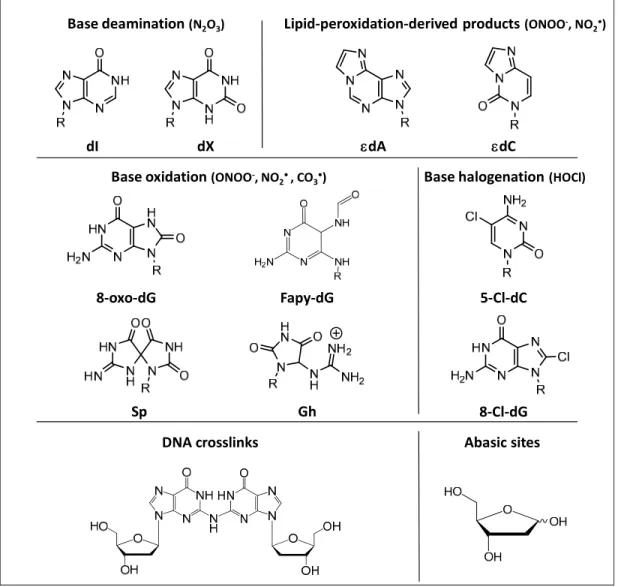
ROS and RNS-mediated DNA damage products span a wide range of reactions, including nitrosation, nitration, oxidation, and halogenation reactions (Figure 5), which are discussed in the following with a special emphasis on studies analyzing murine and human colitis. For a more general overview of the chemical biology of oxidative and nitrosative DNA damage the reader is referred to more specific reviews on this topic [146,147].
3.1.1 Nitrosative deamination
N2O3 which is thought to be the primary agent causing nitrosative deamination of DNA.
Other mechanisms include spontaneous hydrolysis and enzymatic processes by deaminases. DNA damage products formed by nitrosative deamination include 2’-deoxy-uridine (dU) derived from cytosine, deoxy-inosine (dI) derived from adenine, deoxy-xanthosine (dX) or 2’-deoxyoxanosine (dO) derived from guanine, and 2’-deoxythymidine (dT) derived from
23 methylcytosine [69,148] (Figure 5). Due to their mutagenic potential, formation of these damage products is of high functional relevance. For example, during replication formation of deamination products mainly cause single-base transition mutations such as A:T to G:C (dI), C:G to T:A (dU), and G:C to A:T transitions (dX). In addition to deamination of exocyclic amines, nitrosation of heterocyclic nitrogens such as N7 or N3 in dA and N7 in dG can induce depurination creating abasic sites [149]. In an attempt to define the spectrum of nitrosative DNA lesions induced by RNS, purified DNA was exposed to NO (1.3 µM) and O2 (190 µM) [149].
Subsequent quantification of several DNA adducts by stable isotope-dilution mass spectrometry revealed that dX, dI and dU were formed at nearly identical rates to the extent of ~80 lesions per 106 nt after 12 h exposure to NO. Furthermore, abasic sites were generated at a rate of ~10 per 106 nt after 12 h of exposure to NO. In combination with other studies, which demonstrated that
nitrosative guanine deamination can lead to the formation of interstrand cross-links (see below) [150], it can be estimated that under these conditions the following spectrum of nitrosative DNA damage occurs: 25-35% each of dX, dI, and dU, 4-6% abasic sites and ~2% dG-dG cross-links [149]. In cell culture, a ~4-fold protective effect by the cellular environment was observed against the formation of dX, dI and dU. Notably, significant elevations of these products of ~4-fold were only observed when cells were exposed to cytotoxic doses of NO and N2O3 (i.e., 1.75
µM NO and 186 µM O2 for >12 h) [135,151]. An earlier study detected a similar increase in dX
levels of about ~6-fold using stimulated macrophages [152]. The slightly higher values obtained in the latter study may be related to the possibility that intracellular NO levels in stimulated macrophages as used in the study by Dong and Dedon may exceed local concentrations of 1-2 µM [135,152]. In vivo, recent results obtained from H. hepaticus-infected Rag2-/- mice revealed a small increase in levels of dI in inflamed colon tissue by 26%, whereas similar levels of dX were observed in colitis-affected and control tissue [27].
Taken together, these results indicate that nitrosative deamination products arise in cells and tissues under conditions of inflammation, but the relatively small increases suggest either limited nitrosative chemistry in nuclear DNA or effective protection and repair mechanisms, both in cell culture models and in the mouse [27,135]. That this holds true for the human situation awaits further study.
24
3.1.2 DNA oxidation
DNA can also be directly damaged by nucleobase oxidation by ONOO− and ONOOCO2−derived oxidants such as OH•, NO2•,and CO3•- [148]. While ONOO- itself, mainly
causes 2-deoxyribose oxidation (i.e., strand breaks) and some base oxidation, ONOOCO2- causes
mainly base oxidation [153]. Due to its low reduction potential, guanine represents the most vulnerable nucleobase, which shows DNA sequence- and agent-specific variations in damage chemistry [147,154-156]. Consequently, in vitro experiments demonstrated significant reactivity of ONOO- only with dG, leading to the formation of two major oxidation products, i.e., 8-oxo-7,8-dihydro-2’-deoxy-guanosine (8-oxo-dG) and 8-nitro-dG [147,157] (Figure 5). Apart from iNOS- and NADPH oxidase-dependent generation of ROS and RNS, which is thought to occur mainly in macrophages, nitration and oxidation of dG can be also be induced by neutrophils via MPO-dependent reactions that generate NO2• [158,159]. 8-oxo-dG and 8-nitro-dG are
significantly more reactive with ONOO- than dG itself, however 8-nitro-dG is not stable and depurinates to 8-nitro-G generating an abasic site in the DNA backbone (t1/2 ~ 1 h at 37°C),
which can cause base substitutions such as G à T transversions [147,157]. In contrast, 8-oxo-dG is more stable than 8-nitro-8-oxo-dG, but exhibits a ~1,000-fold higher reactivity than 8-oxo-dG and gives rise to more than 10 different secondary oxidation products [153]. It is important nonetheless to keep in mind that even a 1000-fold difference in the reactivity of dG and 8-oxo-dG means that dG is still the major target in cellular DNA when 8-oxo-dG is present at steady state levels of 1-10 per 1-106 nucleotides [151,154]. Secondary dG oxidation products include spiroiminodiydantoin (Sp), guanidinohydantoin (Gh), and oxazalone (Ox), all of which have been extensively described in vitro [154,160-163]. In contrast to nitrosative DNA damage that mainly causes transition mutations, oxidative lesions tend to cause transversion mutations. While 8-oxo-dG itself can lead to C:G to A:T transversion mutations [164], secondary oxidation products such as Sp cause G:C to T:A and G:C to C:G transversions and can act as a strong inhibitor of DNA polymerase extension leading to replicative stress. Similarly, Gh lesions are also highly mutagenic causing G:C to C:G transversions, though they can be bypassed nearly as efficiently as 8-oxo-dG. For both secondary damage products, mutation frequencies are at least an order of magnitude higher than for 8-oxo-dG [165].
Because of their promutagenic potential, significant biological consequences are expected if such primary and secondary oxidation products are formed in mammalian DNA in vivo. While
25 8-oxo-dG is one of the most widely studied DNA damage lesions in vivo [166], data on the existence of guanine secondary oxidation products is not as abundant. Our recent LC-MS/MS-based study on a comprehensive set of inflammation-related DNA damage products in H.
hepaticus-infected Rag2-/- mice aimed to detect guanine secondary oxidation products in mammalian tissues. In fact, Sp and Gh were detected in mouse liver and colon at absolute levels of ~1-5 lesions per 108 nucleotides, which are ~100 times lower than those of 8-oxo-dG. Interestingly, levels of Sp and Gh displayed tissue-specific differences and correlated strongly with their parent molecule 8-oxo-dG, indicating that these molecules indeed arise from 8-oxo-dG
in vivo. In this study, no strong influence of colitis induction on whole tissue levels of 8-oxo-dG
and its secondary oxidation products was observed. Thus, while 8-oxo-dG levels showed a slight and transient decrease during colitis development, levels of Sp tended to correlate positively with corresponding colonic histopathology indices, suggesting that these lesions may have biological significance during inflammation [27]. Similarly, Shaked et al. found modestly elevated levels of oxidative DNA damage lesions, such as deoxyadenosine (cdA) and 8,5′-cyclo-2′-deoxyguanosine (cdG), in intestinal epithelium in a mouse model with constitutive induction of iNOS expression measured by LC-MS/MS analysis (see above) [32]. It is important to stress that these studies analyzed lesion frequencies in tissue samples. Therefore, even small changes as detected in these studies may be related to more drastic effects on a single cell level. Although immuno-chemical methods of DNA damage detection based on DNA-damage-specific antibody binding have their limitations such as lack of chemical specificity and high background signals, these methods allow assessment of DNA damage on a single cell level. In this respect an analysis of an immunological colitis model detected elevated immuno-reactivity towards 8-oxo-dG and 8-nitro-G in nuclei of epithelial cells. These levels correlated with iNOS expression, suggesting a functional interplay between those factors [167]. Furthermore, stronger 8-oxo-dG immuno-reactivity was also detected in colonic epithelia of DSS-treated mice - an effect that could be almost completely suppressed by ectopic expression of extracellular SOD, which also considerably improved disease severity [61]. Interestingly, systemic effects of oxidative DNA damage were identified in mouse models of DSS-induced and immune mediated colitis using
Il-10-/- mice: circulating blood leukocytes isolated from diseased mice showed significantly increased 8-oxo-dG immuno-reactivity, which correlated to oxidative damage detected in inflamed tissue [168]. Such systemic effecs may be related to activation of circulating innate
![Table 1: Common mouse models of colitis-associated carcinogenesis. For more detailed reviews on mouse models of IBD see [261-263]](https://thumb-eu.123doks.com/thumbv2/123doknet/14285148.492036/53.918.100.841.741.1095/table-common-models-colitis-associated-carcinogenesis-detailed-reviews.webp)
![Figure 2. Chemical and biological mechanisms of inflammation [Illustration by Jeff Dixon]](https://thumb-eu.123doks.com/thumbv2/123doknet/14285148.492036/57.918.145.820.159.634/figure-chemical-biological-mechanisms-inflammation-illustration-jeff-dixon.webp)