Cognition in healthy aging and Parkinson’s disease:
Structural and functional integrity of neural circuits
by
David A. Ziegler
B.S., Pyschobiology Denison University, 1999
Submitted to the Department of Brain and Cognitive Sciences in partial fulfillment of the requirements for the degree of
Doctor of Philosophy in Neuroscience at the
Massachusetts Institute of Technology September, 2011
© Massachusetts Institute of Technology. All rights reserved
Signature of Author _____________________________________________________________ David Ziegler Department of Brain and Cognitive Sciences
Certified by____________________________________________________________________ Suzanne Corkin Professor of Behavioral Neuroscience, MIT Thesis Supervisor
Accepted by ___________________________________________________________________ Earl K. Miller Picower Professor of Neuroscience Director, BCS Graduate Program
Cognition in healthy aging and Parkinson’s disease:
Structural and functional integrity of neural circuits
by David A. Ziegler
Submitted to the Department of Brain and Cognitive Sciences
on August 5, 2011 in partial fulfillment of the requirements for the degree of Doctor of Philosophy in Neuroscience
Abstract
This dissertation documents how healthy aging and Parkinson’s disease (PD) affect brain anatomy and physiology and how these neural changes relate to measures of cognition and perception. While healthy aging and PD are both accompanied by a wide-range of cognitive impairments, the neural underpinnings of cognitive decline in each is likely mediated by deterioration of different systems. The four chapters of this dissertation address specific aspects of how healthy aging and PD affect the neural circuits that support sensory processes and high-level cognition.
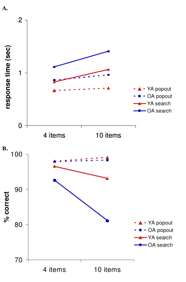
The experiments in Chapters 2 and 3 examine the effects of healthy aging on the integrity of neural circuits that modulate cognitive control processes. In Chapter 2, we test the hypothesis that the patterns of age-related change differ between white matter and gray matter regions, and that changes in the integrity of anterior regions correlate most strongly with performance on cognitive control tasks. In Chapter 3, we build upon the structural findings by examining the hypothesis that age-related changes in white matter integrity are associated with disrupted oscillatory dynamics observed during a visual search task. Chapter 4 investigates healthy age-related changes in somatosensory mu rhythms and evoked responses and uses a computational model of primary somatosensory cortex to predict the underlying cellular and neurophysiolgical bases of these alterations.
In contrast to the widespread cortical changes seen in healthy OA, the cardinal motor symptoms of PD are largely explained by degeneration of the dopaminergic substantia nigra, pars compacta (SNc). Cognitive sequelae of PD, however, likely result from disruptions in multiple neurotransmitter systems, including nondopaminergic nuclei, but research on these aspects of the disease has been hindered by a lack of sensitive MRI biomarkers for the affected structures. Chapter 5 presents new multispectral MRI tools that visualize the SNc and the cholinergic basal forebrain (BF). We applied these methods to test the hypothesis that degenerative processes in PD affect the SNc before the BF. This experiment lays important groundwork for future studies that will examine the relative contribution of the SNc and BF to cognitive impairments in PD.
Thesis Supervisor: Suzanne Corkin
Table of Contents
Chapter 1: Introduction... 7
Structural integrity of cognitive control networks in healthy aging... 7
The role of oscillatory activity in top-down control ... 12
Age-related changes in primary sensory rhythms... 15
Neuropathological and neuroimaging studies of Parkinson’s disease... 17
References ... 19
Chapter 2: Cognition in healthy aging is related to regional white matter integrity, but not cortical thickness... 31 Abstract... 32 Introduction ... 33 Methods... 39 Results... 47 Discussion ... 51 References ... 62
Tables and Figures ... 70
Chapter 3: Age-related changes in white matter integrity disrupt top-down modulation of oscillations during visual search... 77
Abstract... 78
Introduction ... 79
Methods... 84
Results... 92
References ... 106
Tables and Figures ... 114
Chapter 4: Transformations in Oscillatory Activity and Evoked Responses in Primary Somatosensory Cortex in Middle Age: A Combined Computational Neural Modeling and MEG Study... 123 Abstract... 124 Introduction ... 125 Methods... 129 Results... 139 Discussion ... 155 References ... 166
Tables and Figures. ... 170
Tables and Figures. ... 170
Chapter 5: New multispectral MRI tools reveal stage-dependent decreases in basal forebrain and substantia nigra volumes in early Parkinson’s disease... 185
Abstract... 186 Introduction ... 187 Methods... 190 Results... 196 Discussion ... 197 References ... 210
Tables and figures. ... 217
Chapter 1: Introduction
Structural integrity of cognitive control networks in healthy aging.
Healthy aging is characterized by functional declines that cross multiple cognitive domains, including attention, working memory, and long-term declarative memory (Craik & Salthouse, 2000; Hedden & Gabrieli, 2004; Glisky, 2007; Piguet & Corkin, 2007). Common to many of the tasks that reveal the effects of age is a reliance on cognitive control processes. Cognitive control encompasses a collection of top-down processes that enable us to modulate the impact of sensory inputs on our brain. These processes include planning a series of actions, managing goals, coordinating and monitoring automatic processes, inhibiting prepotent responses, focusing attention selectively, and suppressing irrelevant sensory inputs (Salthouse & Meinz, 1995; Spencer & Raz, 1995; Fuster, 2000; Miller, 2000; Braver et al., 2001; Miller & Cohen, 2001). This ability to manage the barrage of sensory information that we encounter in the world allows us to engage in complex, goal-directed behaviors, and it is exactly these processes that are most susceptible to breakdown during the course of healthy aging (Buckner, 2004; Gazzaley & D'Esposito, 2007). Notably, older adults (OA) consistently show reduced performance on tasks that place high demands on cognitive control, including multi-tasking (Jimura & Braver, 2010; Clapp et al., 2011), attention (McDowd, 1986; Hawkins et al., 1992; Milham et al., 2002; West, 2004), episodic and source memory (Craik & McDowd, 1984; Spencer & Raz, 1994), and working memory (Hasher & Zacks, 1988; Salthouse, 1994; Gazzaley et al., 2005; Emery et al., 2008), or on tasks such as free recall, which provide relatively little
environmental support during encoding or retrieval (Craik, 1990). In contrast, OA appear to remain relatively unimpaired on most measures of nondeclarative or implicit memory, which are believed to rely on more automatic and less attention-demanding processes (Light & Singh, 1987; La Voie & Light, 1994; Fleischman & Gabrieli, 1998; Bergerbest et al., 2009). Consequently, an emerging theme is that impaired or inefficient deployment of cognitive control processes may be a common mechanism underlying many of the cognitive deficits in healthy aging (Miyake et al., 2000; Fletcher & Henson, 2001; Braver & Barch, 2002; Salthouse et al., 2003; Braver & Ruge, 2006; Glisky, 2007).
Cognitive control processes are supported by broadly distributed networks within association areas, including prefrontal (PFC) and posterior parietal cortex, that exert top-down control over posterior sensory and motor areas (Goldman-Rakic, 1988; Wise et al., 1996; Miller, 2000; Miller & Cohen, 2001; Stuss & Knight, 2002; Koechlin et al., 2003; Tanji & Hoshi, 2008). Degeneration of any of the nodes or links of these networks could interfere with efficient deployment of cognitive control processes in OA. Evidence for age-related changes in the structural integrity of frontoparietal control regions come from a variety of sources and point toward changes in both cortical gray matter and its underlying white matter. Based on the patterns of neuroanatomical change observed during the course of healthy aging, the frontal aging hypothesis has been proposed as a theoretically parsimonious explanation of deficits in cognitive control processes in OA (West, 1996; Tisserand & Jolles, 2003), although this hypothesis is not universally embraced (Greenwood, 2000; Van Petten et al., 2004; Raz & Rodrigue, 2006).
Magnetic resonance imaging (MRI)-based studies consistently reveal a global age-related decline in gray matter volumes (Pfefferbaum et al., 1994; Raz et al., 1997; Salat et al., 1999; Bartzokis et al., 2001; Allen et al., 2005; Raz et al., 2005; Walhovd et al., 2005), but regional differences exist in terms of the relative rate and magnitude of change. An ever-growing literature documents the most dramatic volume losses in PFC (Jernigan et al., 1991; Raz et al., 1997; Salat et al., 1999; Sowell et al., 2003; Raz et al., 2004; Grieve et al., 2005; Raz & Rodrigue, 2006), while more posterior areas tend to show less shrinkage with age (Raz et al., 1997; Salat et al., 1999; Bartzokis et al., 2001; Resnick et al., 2003; Allen et al., 2005; Raz et al., 2005). Within PFC, lateral PFC and orbital cortices show the greatest degree of volumetric decline (Raz et al., 2004; Raz et al., 2005). Newer methods for examining the thickness of discrete areas across the cerebral cortex provide confirmatory evidence of pronounced thinning of the superior and inferior frontal gyri, but also reveal thinning in occipital and parietal lobes (Salat et al., 2004; Fjell et al., 2009; Lemaitre et al., 2010). A postmortem study confirmed that age-related thinning of the frontal and temporal lobes, with greater atrophy in the frontal lobe (Freeman et al., 2008). Futher, cortical thinning was not associated with decreased neuron number or density, suggesting that changes in neuropil, decreased dendritic arborization, or a loss of ascending projections account for age-related cortical thinning. Thus, while strong evidence shows that the detrimental effects of aging occur earlier and have a greater magnitude of effect on frontal lobe gray matter, compared to posterior cortices, significant degeneration has also been documented in all four lobes of the brain bilaterally (Cowell et al., 1994; Bartzokis et al., 2001; Tisserand et al., 2002; Raz et al., 2004; Van Petten, 2004; Van Petten et al., 2004; Allen et al., 2005),
Postmortem studies indicate that healthy aging is also associated with marked degradation of white matter (Double et al., 1996; Guttmann et al., 1998; Peters, 2002; Hinman & Abraham, 2007; Ikram et al., 2007; Piguet et al., 2009), but the evidence for volumetric decline remains equivocal: Some studies report reduced global and regional white matter volumes (Guttmann et al., 1998; Salat et al., 1999; Courchesne et al., 2000; Bartzokis et al., 2001; Jernigan et al., 2001; Allen et al., 2005; Piguet et al., 2009), whereas others fail to find such decreases (Pfefferbaum et al., 1994; Good et al., 2001; Sullivan et al., 2004). More compelling in vivo evidence of white matter decline comes from other MRI markers of degeneration, including diffusion-tensor imaging (DTI)-based measures of white matter integrity. Like cortical thickness, declines in white matter integrity are widespread (Good et al., 2001; Salat et al., 2004; Lutz et al., 2007; Yoon et al.), but tend to be most striking in anterior areas, such as the genu of the corpus callosum and the white matter underlying the PFC (O'Sullivan et al., 2001; Head et al., 2004; Pfefferbaum et al., 2005; Salat et al., 2005; Sullivan & Pfefferbaum, 2006; Ardekani et al., 2007; Madden et al., 2007; Yoon et al.).
While the patterns of age-related cognitive and neuroanatomical change are relatively well characterized, linking these measures to unmask the specific neural bases of cognitive decline has proven more difficult. In OA, diminished attention and executive function are associated with decreased global cortical volumes and reduced volumes of lateral PFC and OFC (Zimmerman et al., 2006; Kramer et al., 2007). In addition, PFC volume was inversely correlated with perseverative errors in OA (Raz et al., 1998; Gunning-Dixon & Raz, 2003). In contrast,
spatial and object working memory correlated with occipital lobe volume (Raz et al., 1998), but neither spatial, object, nor verbal working memory showed significant correlations with PFC volume (Gunning-Dixon & Raz, 2003). Thus, direct evidence in support of an association between cortical atrophy and age-related cognitive decline remains equivocal.
Stronger support for a link between frontal lobe integrity and cognition comes from studies of white matter. One study of older rhesus monkeys found a correlation between measures of executive function and DTI-based measures of white matter integrity (e.g., FA and diffusivity) in long-distance corticocortical association pathways (Makris et al., 2007). Several investigations in OA have linked deficits in processing speed, executive function, immediate and delayed recall, and overall cognition to an increased burden of white matter hypertintensities (Gunning-Dixon & Raz, 2000; Gunning-(Gunning-Dixon & Raz, 2003; Soderlund et al., 2003; Smith et al., 2011). White matter abnormalities were also associated with decreased frontal lobe metabolism (DeCarli et al., 1995; Tullberg et al., 2004), and with diminished BOLD responses in PFC during performance of episodic and working memory tasks (Nordahl et al., 2006). Functional correlates of decreased FA include working memory impairments (Charlton et al., 2006), slowed processing speed (Sullivan et al., 2006; Bucur et al., 2007), and executive dysfunction (O'Sullivan et al., 2001; Deary et al., 2006; Grieve et al., 2007). These results suggest that degeneration of white matter pathways contributes to the etiology of age-related cognitive decline to an equal or greater extent than gray matter atrophy (O'Sullivan et al., 2001; Hinman & Abraham, 2007).
In summary, evidence of morphological and microstructural changes in anterior areas appears consistently in the literature on aging. In addition, regional alterations have been noted across wide regions of gray and white matter, but the exact nature and magnitude of these changes remain a topic of debate. To explicitly test whether gray and white matter exhibit similar or distinct patterns of age-related change, measures of both regions must be examined in a single group of participants. Chapter 1 accomplishes this goal by using advanced MRI techniques to examine high-resolution measures of white matter integrity and cortical thickness in healthy young adults (YA) and OA. Further, we tested the prediction that performance on tests of cognitive control is correlated with integrity of anterior regions, while episodic memory and semantic memory are associated with changes in posterior cortices.
The role of oscillatory activity in top-down control.
A growing body of literature supports the idea that oscillatory synchronization is one mechanism underlying efficient deployment of cognitive control processes. The first report of oscillatory activity in the human brain came from Hans Berger who recorded electrical potentials over the posterior scalp using a string galvanometer (Berger, 1929). Berger observed oscillatory signals over the occipital cortex, the most prominent being the ~10 Hz alpha rhythm, which was strongest when participants closed their eyes. Berger’s invention of the electroencephalogram ushered in an era of active investigation of the origins and functional significance of brain rhythms (Bremer, 1958). Additional technological advances, such as the development of high-resolution whole-head magnetoencephalography in humans (Hämäläinen
animals (Nicolelis & Ribeiro, 2002; Miller & Wilson, 2008), have led to a greater understanding of the range and pervasiveness of cortical oscillations, with frequencies ranging from <1 Hz up to several hundred Hz being observed in the cerebral cortex of virtually every mammal studied (Buzsaki, 2006). It is now well established that oscillations occur throughout the brain and are believed to be a fundamental mechanism of neural information processing, likely mediating high-level cognition (Hari & Salmelin, 1997; Kahana, 2006; Fries, 2009), but the underlying mechanisms of these rhythms and their complex dynamic modulation remain topics of intense scrutiny (Whittington & Traub, 2003; Bartos et al., 2007; Wang, 2010).
Neural rhythms are thought to be critical for a number of cognitive processes including working memory (Howard et al., 2003; Jensen, 2006; Kahana, 2006; Jokisch & Jensen, 2007), visual attention (Buschman & Miller, 2007; Jensen et al., 2007; Gregoriou et al., 2009), top-down anticipatory biasing of sensory cortices (Schadow et al., 2009), motor planning (Van Der Werf et al., 2008; Van Der Werf et al., 2010), complex spatial navigation (Kahana et al., 1999), and long-term memory encoding (Klimesch et al., 1996; Sederberg et al., 2003) and retrieval (Klimesch et al., 2006). Oscillations may bind information across brain regions (Singer, 1999) or facilitate communication between disparate cortical sites (Salinas & Sejnowski, 2001). In particular, oscillations may provide a mechanism for facilitating top-down modulation, possibly by enhancing stimulus representations or regulating communication between high-order control areas and sensory and motor cortices (Engel et al., 2001; Salinas & Sejnowski, 2001; Jensen et al., 2007).
Mounting evidence suggests an important role for high-frequency, or gamma (~30-100 Hz), oscillations in working memory and attention (Tallon-Baudry et al., 2001; Howard et al., 2003; Jensen et al., 2007; Fries, 2009), and some have proposed that such oscillations provide a mechanism for sustaining information “online” (Jensen, 2006). Gamma oscillations occur locally in primary sensory and motor areas during performance of attention demanding tasks and are believed to be mediated by local network interactions between excitatory pyramidal neurons and inhibitory interneurons (McBain & Fisahn, 2001; Whittington & Traub, 2003; Bartos et al., 2007; Cardin et al., 2009). Oscillations in the beta (~15-30 Hz) range likely facilitate top-down influences from association cortices during performance of tasks that require a high degree of cognitive control (Buschman & Miller, 2007; Siegel et al., 2008; Schroeder & Lakatos, 2009). This effect may depend on synchronization of neural firing between distant executive control areas and primary sensory and motor areas, thus requiring efficient long-distance communication via white matter tracts. Low-frequency alpha oscillations (~7-14 Hz) block modality-specific distracters, and are commonly observed during working memory tasks (Jokisch & Jensen, 2007; Tuladhar et al., 2007). In contrast, event-related decreases in alpha power (alpha desynchronizations) are often seen in occipital areas when participants perform tasks that require active visual processing (Capotosto et al., 2009), such as during visual search tasks.
Age-related declines in white matter integrity could interfere with long-range synchronization of lower frequency alpha and beta oscillations, while cortical thinning, which likely reflects a loss of interneurons or synapses (Peters et al., 1998; Peters et al., 2008), may disrupt local
gamma oscillations. These changes in neural rhythms likely impact cognition in OA. In Chapter 3, we describe a multimodal neuroimaging experiment that combines magnetoencephalography (MEG) and MRI to study the effects of aging on oscillatory activity associated with visual attention.
Age-related changes in primary sensory rhythms.
Oscillations in primary sensory cortices, such as the somatosensory mu rhythm, also appear to play important roles in facilitating sensory processing. In addition to declines in high-level cognitive processes, healthy aging is also accompanied by decrements in somatosensory perceptual abilities (Baltes & Lindenberger, 1997; Schneider & Pichora-Fuller, 2000), including declines in tactile perception. OA exhibit significantly higher detection thresholds for vibrations delivered to the hand (Verrillo, 1982; Gescheider et al., 1996; Verrillo et al., 2002). This change is accompanied by a decrease in the subjective magnitude of vibrotactile sensation (Verrillo, 1982; Verrillo et al., 2002) and decreased discrimination abilities (Gescheider et al., 1996).
The neural underpinnings of these age-related changes in somatosensory function remain elusive. One hypothesis is that decreased discrimination thresholds are consistent with a reduction in the amount of afferent input to cortical somatosensory areas (Gescheider et al., 1996). This decrease in afferent drive could stem from a reduced density of tactile receptors in the skin (Cauna, 1965). In contrast, other studies in OA have documented age-related increases in peak magnitudes of somatosensory responses following median nerve stimulation (Luders, 1970; Desmedt & Cheron, 1980; Adler & Nacimiento, 1988; Kakigi & Shibasaki, 1991; Ferri et al.,
1996; Stephen et al., 2006) and increased amplitude of tactile evoked potentials (Ogata et al., 2009). Further, many of these changes are apparent by middle-age, and anatomical studies suggest that age-related thinning of primary somatosensory cortex begins at this time (Salat et al., 2004).
An expanding body of literature has shown that somatosensory evoked responses are inextricably linked to ongoing neural rhythms (Nikulin et al., 2007; Mazaheri & Jensen, 2008; Jones et al., 2009; Zhang & Ding, 2010), and that these modulations are correlated with perception and attention (Linkenkaer-Hansen et al., 2004; Fries, 2009; Zhang & Ding, 2010). The predominant rhythm in pre- and postcentral cortex is the somatosensory mu rhythm, which manifests as a complex of alpha and beta oscillations when recorded with MEG (Tiihonen et al., 1989; Narici et al., 1990; Cheyne et al., 2003). One study reported concomitant age-related increases in induced alpha and high beta (22-23 Hz) power in anterior sensorimotor electrodes during a simple finger extension task (Sailer et al., 2000), but few studies have examined the effects of aging on this commonly observed somatosensory rhythm. Because age-related changes in somatosensory evoked responses and cortical thickness are observed in middle-age, these alterations should also impact the somatosensory mu rhythm. In Chapter 4, we use MEG to examine age-related changes in spontaneous cortical rhythms and evoked responses from the primary somatosensory cortices (SI) of healthy YA and middle-aged adults during a tactile detection task. We then apply a biophysically realistic computational neural model of SI (Jones et al., 2007) to examine possible neural mechanisms underlying the age-related effects.
Neuropathological and neuroimaging studies of Parkinson’s disease.
In contrast to healthy aging, which is by definition aging in the absence of overt pathology, Parkinson’s disease (PD) is an age-related neurodegenerative disorder that is typically characterized by its cardinal motor symptoms: resting tremor, muscular rigidity, bradykinesia, postural instability, and gait abnormality (Shulman et al., 2011). Cognitive impairments are common in PD, but differ from those that characterize healthy aging.
The major neuropathologic feature of idiopathic PD is selective loss of dopaminergic neurons and accumulation of Lewy bodies and neurites in the substantia nigra pars compacta (Wichmann & DeLong, 2002; Jellinger, 2005). In healthy brains, neurons from the substantia nigra project to the striatum and other basal ganglia nuclei. Functionally segregated circuits link the basal ganglia and neocortex in a topographical manner (Alexander et al., 1986; Middleton & Strick, 2000), and include dense reciprocal fronto-striatal connections (Alexander et al., 1986; Saint-Cyr, 2003). Because of neuronal loss in the substanita nigra, PD patients show markedly reduced dopamine neurotransmission in the striatum, which likely accounts for the majority of the primary motor symptoms. Loss of dopamineric cells in the substantia nigra also disrupts the function of frontostriatal circuits. The pattern of dopamine loss in the striatum occurs according to a lateral-medial/dorsal-ventral gradient, corresponding inversely to the gradient of cell loss in the substantia nigra: Cells projecting to the putamen are lost first, followed by those that project to the caudate nucleus and nucleus accumbens (Jellinger, 2005). Thus, many of the cognitive deficits in PD—working memory, attention, and processing speed—have been attributed to loss of dopamine inputs to nonmotor circuits of the basal ganglia, which may be
affected by dopamine loss in the same way as basal ganglia motor circuits (Alexander et al., 1986; Saint-Cyr, 2003).
Neuropathological studies indicate, however, that cell loss and neuropathology in PD are not limited to the basal ganglia (Halliday et al., 1990; Laakso et al., 1996; Henderson et al., 2000; Harding et al., 2002). Degeneration of the cholinergic basal forebrain also occurs (Javoy-Agid et al., 1981; Rogers et al., 1985; Mufson et al., 1991), and may account for some cognitive impairments (Pillon et al., 1989; Rye & DeLong, 2003; Calabresi et al., 2006), including deficits in attention and cognitive control (Dubois et al., 1990). PD patients who show early pathological insults in non-dopaminergic nuclei may exhibit cognitive decline earlier in the course of the disease and may be more likely to develop dementia, while PD patients with more targeted degeneration of the substantia nigra may show a more subtle pattern of cognitive change.
A major limitation to studies of brain-behavior correlations in PD is that conventional structural MRI techniques provide relatively poor contrast for the structures that are affected by the disease, and thus are not typically used in experimental studies of PD (Schrag et al., 1998; Marek & Jennings, 2009). This ineffectiveness is due predominantly to the limited contrast of most current MRI methods, which are unable to detect PD abnormalities. In Chapter 5, we describe a new multispectral MRI method that provides excellent contrast for the substantia nigra and basal forebrain and apply these methods to data from PD patients and controls to show that these structures display different trajectories of volume loss early in the disease.
References
Adler G & Nacimiento AC (1988) Age-dependent changes of short-latency somatosensory evoked potentials in healthy adults. Appl Neurophysiol 51, 55-59.
Alexander GE, DeLong MR & Strick PL (1986) Parallel organization of functionally segregated circuits linking basal ganglia and cortex. Annu Rev Neurosci 9, 357-381.
Allen JS, Bruss J, Brown CK & Damasio H (2005) Normal neuroanatomical variation due to age: the major lobes and a parcellation of the temporal region. Neurobiol Aging 26, 1245-1260; discussion 1279-1282.
Ardekani S, Kumar A, Bartzokis G & Sinha U (2007) Exploratory voxel-based analysis of diffusion indices and hemispheric asymmetry in normal aging. Magn Reson Imaging 25, 154-167. Baltes PB & Lindenberger U (1997) Emergence of a powerful connection between sensory and
cognitive functions across the adult life span: a new window to the study of cognitive aging? Psychol Aging 12, 12-21.
Bartos M, Vida I & Jonas P (2007) Synaptic mechanisms of synchronized gamma oscillations in inhibitory interneuron networks. Nat Rev Neurosci 8, 45-56.
Bartzokis G, Beckson M, Lu PH, Nuechterlein KH, Edwards N & Mintz J (2001) Age-related changes in frontal and temporal lobe volumes in men: a magnetic resonance imaging study. Arch Gen Psychiatry 58, 461-465.
Berger H (1929) Ueber das Elektroenkephalogramm des Menschen. Arch Psychiatr Nervenkrankh 87.
Bergerbest D, Gabrieli JD, Whitfield-Gabrieli S, Kim H, Stebbins GT, Bennett DA & Fleischman DA (2009) Age-associated reduction of asymmetry in prefrontal function and preservation of conceptual repetition priming. Neuroimage 45, 237-246.
Braver TS & Barch DM (2002) A theory of cognitive control, aging cognition, and neuromodulation. Neurosci Biobehav Rev 26, 809-817.
Braver TS, Barch DM, Keys BA, Carter CS, Cohen JD, Kaye JA, Janowsky JS, Taylor SF, Yesavage JA, Mumenthaler MS, Jagust WJ & Reed BR (2001) Context processing in older adults: evidence for a theory relating cognitive control to neurobiology in healthy aging. J Exp Psychol Gen 130, 746-763.
Braver TS & Ruge H (2006) Functional Neuroimaging of Executive Functions. In Handbook of Functional Neuroimaging of Cognition, 2nd Edition, pp. 307-347 [R Cabeza and A Kingstone, editors]. Cambridge, MA: MIT Press.
Bremer F (1958) Cerebral and cerebellar potentials. Physiol Rev 38, 357-388.
Buckner RL (2004) Memory and executive function in aging and AD: multiple factors that cause decline and reserve factors that compensate. Neuron 44, 195-208.
Bucur B, Madden DJ, Spaniol J, Provenzale JM, Cabeza R, White LE & Huettel SA (2007) Age-related slowing of memory retrieval: Contributions of perceptual speed and cerebral white matter integrity. Neurobiol Aging.
Buschman TJ & Miller EK (2007) Top-down versus bottom-up control of attention in the prefrontal and posterior parietal cortices. Science 315, 1860-1862.
Calabresi P, Picconi B, Parnetti L & Di Filippo M (2006) A convergent model for cognitive dysfunctions in Parkinson's disease: the critical dopamine-acetylcholine synaptic balance. Lancet Neurol 5, 974-983.
Capotosto P, Babiloni C, Romani GL & Corbetta M (2009) Frontoparietal cortex controls spatial attention through modulation of anticipatory alpha rhythms. J Neurosci 29, 5863-5872. Cardin JA, Carlen M, Meletis K, Knoblich U, Zhang F, Deisseroth K, Tsai LH & Moore CI (2009)
Driving fast-spiking cells induces gamma rhythm and controls sensory responses. Nature 459, 663-667.
Cauna N (1965) The effects of aging on the receptor organs of the human dermis. In Advances in Biology of the Skin: Aging, pp. 63-96 [W Montagna, editor]. Elmsford, NY: Pergamon Press.
Charlton RA, Barrick TR, McIntyre DJ, Shen Y, O'Sullivan M, Howe FA, Clark CA, Morris RG & Markus HS (2006) White matter damage on diffusion tensor imaging correlates with age-related cognitive decline. Neurology 66, 217-222.
Cheyne D, Gaetz W, Garnero L, Lachaux JP, Ducorps A, Schwartz D & Varela FJ (2003) Neuromagnetic imaging of cortical oscillations accompanying tactile stimulation. Brain Res Cogn Brain Res 17, 599-611.
Clapp WC, Rubens MT, Sabharwal J & Gazzaley A (2011) Deficit in switching between functional brain networks underlies the impact of multitasking on working memory in older adults. Proc Natl Acad Sci U S A 108, 7212-7217.
Cohen D (2004) Boston and the history of biomagnetism. Neurol Clin Neurophysiol 2004, 114. Courchesne E, Chisum HJ, Townsend J, Cowles A, Covington J, Egaas B, Harwood M, Hinds S &
Press GA (2000) Normal brain development and aging: quantitative analysis at in vivo MR imaging in healthy volunteers. Radiology 216, 672-682.
Cowell PE, Turetsky BI, Gur RC, Grossman RI, Shtasel DL & Gur RE (1994) Sex differences in aging of the human frontal and temporal lobes. J Neurosci 14, 4748-4755.
Craik FI (1990) Changes in memory with normal aging: a functional view. Adv Neurol 51, 201-205.
Craik FI & McDowd JM (1984) Age differences in recall and recognition. Journal of Experimental Psychology: Learning, Memory, and Cognition 13, 474-479.
Craik FIM & Salthouse TA (2000) The Handbook of Aging and Cognition. Mahwah, NJ: Lawrence Erlbaum Associates.
Deary IJ, Bastin ME, Pattie A, Clayden JD, Whalley LJ, Starr JM & Wardlaw JM (2006) White matter integrity and cognition in childhood and old age. Neurology 66, 505-512.
DeCarli C, Murphy DG, Tranh M, Grady CL, Haxby JV, Gillette JA, Salerno JA, Gonzales-Aviles A, Horwitz B, Rapoport SI & et al. (1995) The effect of white matter hyperintensity volume on brain structure, cognitive performance, and cerebral metabolism of glucose in 51 healthy adults. Neurology 45, 2077-2084.
Desmedt JE & Cheron G (1980) Somatosensory evoked potentials to finger stimulation in healthy octogenarians and in young adults: wave forms, scalp topography and transit times of parietal and frontal components. Electroencephalogr Clin Neurophysiol 50, 404-425.
Double KL, Halliday GM, Kril JJ, Harasty JA, Cullen K, Brooks WS, Creasey H & Broe GA (1996) Topography of brain atrophy during normal aging and Alzheimer's disease. Neurobiol Aging 17, 513-521.
Dubois B, Pillon B, Sternic N, Lhermitte F & Agid Y (1990) Age-induced cognitive disturbances in Parkinson's disease. Neurology 40, 38-41.
Emery L, Heaven TJ, Paxton JL & Braver TS (2008) Age-related changes in neural activity during performance matched working memory manipulation. Neuroimage 42, 1577-1586. Engel AK, Fries P & Singer W (2001) Dynamic predictions: oscillations and synchrony in
top-down processing. Nat Rev Neurosci 2, 704-716.
Ferri R, Del Gracco S, Elia M, Musumeci SA, Spada R & Stefanini MC (1996) Scalp topographic mapping of middle-latency somatosensory evoked potentials in normal aging and dementia. Neurophysiol Clin 26, 311-319.
Fjell AM, Westlye LT, Amlien I, Espeseth T, Reinvang I, Raz N, Agartz I, Salat DH, Greve DN, Fischl B, Dale AM & Walhovd KB (2009) High consistency of regional cortical thinning in aging across multiple samples. Cereb Cortex 19, 2001-2012.
Fleischman DA & Gabrieli JD (1998) Repetition priming in normal aging and Alzheimer's disease: a review of findings and theories. Psychol Aging 13, 88-119.
Fletcher PC & Henson RN (2001) Frontal lobes and human memory: insights from functional neuroimaging. Brain 124, 849-881.
Freeman SH, Kandel R, Cruz L, Rozkalne A, Newell K, Frosch MP, Hedley-Whyte ET, Locascio JJ, Lipsitz LA & Hyman BT (2008) Preservation of neuronal number despite age-related cortical brain atrophy in elderly subjects without Alzheimer disease. J Neuropathol Exp Neurol 67, 1205-1212.
Fries P (2009) Neuronal gamma-band synchronization as a fundamental process in cortical computation. Annu Rev Neurosci 32, 209-224.
Fuster JM (2000) Executive frontal functions. Exp Brain Res 133, 66-70.
Gazzaley A, Cooney JW, Rissman J & D'Esposito M (2005) Top-down suppression deficit underlies working memory impairment in normal aging. Nat Neurosci 8, 1298-1300. Gazzaley A & D'Esposito M (2007) Top-down modulation and normal aging. Ann N Y Acad Sci
1097, 67-83.
Gescheider GA, Edwards RR, Lackner EA, Bolanowski SJ & Verrillo RT (1996) The effects of aging on information-processing channels in the sense of touch: III. Differential sensitivity to changes in stimulus intensity. Somatosens Mot Res 13, 73-80.
Glisky EL (2007) Changes in Cognitive Function in Human Aging. In Brain Aging: Models, Methods, and Mechanisms, pp. Chapter 1. [DR Riddle, editor]. Boca Raton (FL): CRC Press.
Goldman-Rakic PS (1988) Topography of cognition: parallel distributed networks in primate association cortex. Annu Rev Neurosci 11, 137-156.
Good CD, Johnsrude IS, Ashburner J, Henson RN, Friston KJ & Frackowiak RS (2001) A voxel-based morphometric study of ageing in 465 normal adult human brains. Neuroimage 14, 21-36.
Greenwood PM (2000) The frontal aging hypothesis evaluated. J Int Neuropsychol Soc 6, 705-726.
Gregoriou GG, Gotts SJ, Zhou H & Desimone R (2009) High-frequency, long-range coupling between prefrontal and visual cortex during attention. Science 324, 1207-1210.
Grieve SM, Clark CR, Williams LM, Peduto AJ & Gordon E (2005) Preservation of limbic and paralimbic structures in aging. Hum Brain Mapp 25, 391-401.
Grieve SM, Williams LM, Paul RH, Clark CR & Gordon E (2007) Cognitive aging, executive function, and fractional anisotropy: a diffusion tensor MR imaging study. AJNR Am J Neuroradiol 28, 226-235.
Gunning-Dixon FM & Raz N (2000) The cognitive correlates of white matter abnormalities in normal aging: a quantitative review. Neuropsychology 14, 224-232.
Gunning-Dixon FM & Raz N (2003) Neuroanatomical correlates of selected executive functions in middle-aged and older adults: a prospective MRI study. Neuropsychologia 41, 1929-1941.
Guttmann CR, Jolesz FA, Kikinis R, Killiany RJ, Moss MB, Sandor T & Albert MS (1998) White matter changes with normal aging. Neurology 50, 972-978.
Halliday GM, Blumbergs PC, Cotton RG, Blessing WW & Geffen LB (1990) Loss of brainstem serotonin- and substance P-containing neurons in Parkinson's disease. Brain Res 510, 104-107.
Hämäläinen M, Hari R, Ilmoniemi R, Knuutila J & Lounasmaa O (1993) Magnetoencephalography--theory, instrumentation, and applications to noninvasive studies of the working human brain. Rev Mod Phys 65, 1-93.
Harding AJ, Stimson E, Henderson JM & Halliday GM (2002) Clinical correlates of selective pathology in the amygdala of patients with Parkinson's disease. Brain 125, 2431-2445. Hari R & Salmelin R (1997) Human cortical oscillations: a neuromagnetic view through the skull.
Trends Neurosci 20, 44-49.
Hasher L & Zacks RT (1988) Working memory, comprehesion, and aging: A review and a new view. In The Psychology of Learning and Motivation, pp. 193 [GH Bower, editor]. New York: Academic Press.
Hawkins HL, Kramer AF & Capaldi D (1992) Aging, exercise, and attention. Psychol Aging 7, 643-653.
Head D, Buckner RL, Shimony JS, Williams LE, Akbudak E, Conturo TE, McAvoy M, Morris JC & Snyder AZ (2004) Differential vulnerability of anterior white matter in nondemented aging with minimal acceleration in dementia of the Alzheimer type: evidence from diffusion tensor imaging. Cereb Cortex 14, 410-423.
Hedden T & Gabrieli JD (2004) Insights into the ageing mind: a view from cognitive neuroscience. Nat Rev Neurosci 5, 87-96.
Henderson JM, Carpenter K, Cartwright H & Halliday GM (2000) Loss of thalamic intralaminar nuclei in progressive supranuclear palsy and Parkinson's disease: clinical and therapeutic implications. Brain 123 ( Pt 7), 1410-1421.
Hinman JD & Abraham CR (2007) What's behind the decline? The role of white matter in brain aging. Neurochem Res 32, 2023-2031.
Howard MW, Rizzuto DS, Caplan JB, Madsen JR, Lisman J, Aschenbrenner-Scheibe R, Schulze-Bonhage A & Kahana MJ (2003) Gamma oscillations correlate with working memory load in humans. Cereb Cortex 13, 1369-1374.
Ikram MA, Vrooman HA, Vernooij MW, van der Lijn F, Hofman A, van der Lugt A, Niessen WJ & Breteler MM (2007) Brain tissue volumes in the general elderly population The Rotterdam Scan Study. Neurobiol Aging.
Javoy-Agid F, Taquet H, Ploska A, Cherif-Zahar C, Ruberg M & Agid Y (1981) Distribution of catecholamines in the ventral mesencephalon of human brain, with special reference to Parkinson's disease. J Neurochem 36, 2101-2105.
Jellinger K (2005) The pathology of Parkinson's disease-recent advances. In Scientific basis for the treatment of parkinson's disease., pp. 53-86 [N Galvez-Jimenez, editor]. New York: Taylor & Francis.
Jensen O (2006) Maintenance of multiple working memory items by temporal segmentation. Neuroscience 139, 237-249.
Jensen O, Kaiser J & Lachaux JP (2007) Human gamma-frequency oscillations associated with attention and memory. Trends Neurosci 30, 317-324.
Jernigan TL, Archibald SL, Berhow MT, Sowell ER, Foster DS & Hesselink JR (1991) Cerebral structure on MRI, Part I: Localization of age-related changes. Biol Psychiatry 29, 55-67. Jernigan TL, Archibald SL, Fennema-Notestine C, Gamst AC, Stout JC, Bonner J & Hesselink JR
(2001) Effects of age on tissues and regions of the cerebrum and cerebellum. Neurobiol Aging 22, 581-594.
Jimura K & Braver TS (2010) Age-related shifts in brain activity dynamics during task switching. Cereb Cortex 20, 1420-1431.
Jokisch D & Jensen O (2007) Modulation of gamma and alpha activity during a working memory task engaging the dorsal or ventral stream. J Neurosci 27, 3244-3251.
Jones SR, Pritchett DL, Sikora MA, Stufflebeam SM, Hamalainen M & Moore CI (2009) Quantitative analysis and biophysically realistic neural modeling of the MEG mu rhythm: rhythmogenesis and modulation of sensory-evoked responses. J Neurophysiol 102, 3554-3572.
Jones SR, Pritchett DL, Stufflebeam SM, Hamalainen M & Moore CI (2007) Neural correlates of tactile detection: a combined magnetoencephalography and biophysically based computational modeling study. J Neurosci 27, 10751-10764.
Kahana MJ (2006) The cognitive correlates of human brain oscillations. J Neurosci 26, 1669-1672.
Kahana MJ, Sekuler R, Caplan JB, Kirschen M & Madsen JR (1999) Human theta oscillations exhibit task dependence during virtual maze navigation. Nature 399, 781-784.
Kakigi R & Shibasaki H (1991) Effects of age, gender, and stimulus side on scalp topography of somatosensory evoked potentials following median nerve stimulation. J Clin Neurophysiol 8, 320-330.
Klimesch W, Doppelmayr M, Russegger H & Pachinger T (1996) Theta band power in the human scalp EEG and the encoding of new information. Neuroreport 7, 1235-1240.
Klimesch W, Hanslmayr S, Sauseng P, Gruber W, Brozinsky CJ, Kroll NE, Yonelinas AP & Doppelmayr M (2006) Oscillatory EEG correlates of episodic trace decay. Cereb Cortex 16, 280-290.
Koechlin E, Ody C & Kouneiher F (2003) The architecture of cognitive control in the human prefrontal cortex. Science 302, 1181-1185.
Kramer JH, Mungas D, Reed BR, Wetzel ME, Burnett MM, Miller BL, Weiner MW & Chui HC (2007) Longitudinal MRI and cognitive change in healthy elderly. Neuropsychology 21, 412-418.
La Voie D & Light LL (1994) Adult age differences in repetition priming: a meta-analysis. Psychol Aging 9, 539-553.
Laakso MP, Partanen K, Riekkinen P, Lehtovirta M, Helkala EL, Hallikainen M, Hanninen T, Vainio P & Soininen H (1996) Hippocampal volumes in Alzheimer's disease, Parkinson's disease with and without dementia, and in vascular dementia: An MRI study. Neurology 46, 678-681.
Lemaitre H, Goldman AL, Sambataro F, Verchinski BA, Meyer-Lindenberg A, Weinberger DR & Mattay VS (2010) Normal age-related brain morphometric changes: nonuniformity across cortical thickness, surface area and gray matter volume? Neurobiol Aging.
Light LL & Singh A (1987) Implicit and explicit memory in young and older adults. J Exp Psychol Learn Mem Cogn 13, 531-541.
Linkenkaer-Hansen K, Nikulin VV, Palva S, Ilmoniemi RJ & Palva JM (2004) Prestimulus oscillations enhance psychophysical performance in humans. J Neurosci 24, 10186-10190.
Luders H (1970) The effect of aging on the wave form of the somatosensory cortical evoked potential. Electroencephalogr Clin Neurophysiol 29, 450-460.
Lutz J, Hemminger F, Stahl R, Dietrich O, Hempel M, Reiser M & Jager L (2007) Evidence of subcortical and cortical aging of the acoustic pathway: a diffusion tensor imaging (DTI) study. Acad Radiol 14, 692-700.
Madden DJ, Spaniol J, Whiting WL, Bucur B, Provenzale JM, Cabeza R, White LE & Huettel SA (2007) Adult age differences in the functional neuroanatomy of visual attention: a combined fMRI and DTI study. Neurobiol Aging 28, 459-476.
Makris N, Papadimitriou GM, van der Kouwe A, Kennedy DN, Hodge SM, Dale AM, Benner T, Wald LL, Wu O, Tuch DS, Caviness VS, Moore TL, Killiany RJ, Moss MB & Rosene DL (2007) Frontal connections and cognitive changes in normal aging rhesus monkeys: a DTI study. Neurobiol Aging 28, 1556-1567.
Marek K & Jennings D (2009) Can we image premotor Parkinson disease? Neurology 72, S21-26. Mazaheri A & Jensen O (2008) Asymmetric amplitude modulations of brain oscillations
generate slow evoked responses. J Neurosci 28, 7781-7787.
McBain CJ & Fisahn A (2001) Interneurons unbound. Nat Rev Neurosci 2, 11-23.
McDowd JM (1986) The effects of age and extended practice on divided attention performance. J Gerontol 41, 764-769.
Middleton FA & Strick PL (2000) Basal ganglia output and cognition: evidence from anatomical, behavioral, and clinical studies. Brain Cogn 42, 183-200.
Milham MP, Erickson KI, Banich MT, Kramer AF, Webb A, Wszalek T & Cohen NJ (2002) Attentional control in the aging brain: insights from an fMRI study of the stroop task. Brain Cogn 49, 277-296.
Miller EK (2000) The prefrontal cortex and cognitive control. Nat Rev Neurosci 1, 59-65.
Miller EK & Cohen JD (2001) An integrative theory of prefrontal cortex function. Annu Rev Neurosci 24, 167-202.
Miller EK & Wilson MA (2008) All my circuits: using multiple electrodes to understand functioning neural networks. Neuron 60, 483-488.
Miyake A, Friedman NP, Emerson MJ, Witzki AH, Howerter A & Wager TD (2000) The unity and diversity of executive functions and their contributions to complex "Frontal Lobe" tasks: a latent variable analysis. Cognit Psychol 41, 49-100.
Mufson EJ, Presley LN & Kordower JH (1991) Nerve growth factor receptor immunoreactivity within the nucleus basalis (Ch4) in Parkinson's disease: reduced cell numbers and co-localization with cholinergic neurons. Brain Res 539, 19-30.
Narici L, Pizzella V, Romani GL, Torrioli G, Traversa R & Rossini PM (1990) Evoked alpha- and mu-rhythm in humans: a neuromagnetic study. Brain Res 520, 222-231.
Nicolelis MA & Ribeiro S (2002) Multielectrode recordings: the next steps. Curr Opin Neurobiol 12, 602-606.
Nikulin VV, Linkenkaer-Hansen K, Nolte G, Lemm S, Muller KR, Ilmoniemi RJ & Curio G (2007) A novel mechanism for evoked responses in the human brain. Eur J Neurosci 25, 3146-3154.
Nordahl CW, Ranganath C, Yonelinas AP, Decarli C, Fletcher E & Jagust WJ (2006) White matter changes compromise prefrontal cortex function in healthy elderly individuals. J Cogn Neurosci 18, 418-429.
O'Sullivan M, Jones DK, Summers PE, Morris RG, Williams SC & Markus HS (2001) Evidence for cortical "disconnection" as a mechanism of age-related cognitive decline. Neurology 57, 632-638.
Ogata K, Okamoto T, Yamasaki T, Shigeto H & Tobimatsu S (2009) Pre-movement gating of somatosensory-evoked potentials by self-initiated movements: the effects of ageing and its implication. Clin Neurophysiol 120, 1143-1148.
Peters A (2002) The effects of normal aging on myelin and nerve fibers: a review. J Neurocytol 31, 581-593.
Peters A, Sethares C & Luebke JI (2008) Synapses are lost during aging in the primate prefrontal cortex. Neuroscience 152, 970-981.
Peters A, Sethares C & Moss MB (1998) The effects of aging on layer 1 in area 46 of prefrontal cortex in the rhesus monkey. Cereb Cortex 8, 671-684.
Pfefferbaum A, Adalsteinsson E & Sullivan EV (2005) Frontal circuitry degradation marks healthy adult aging: Evidence from diffusion tensor imaging. Neuroimage 26, 891-899.
Pfefferbaum A, Mathalon DH, Sullivan EV, Rawles JM, Zipursky RB & Lim KO (1994) A quantitative magnetic resonance imaging study of changes in brain morphology from infancy to late adulthood. Arch Neurol 51, 874-887.
Piguet O & Corkin S (2007) The aging brain. In Learning and the Brain: A Comprehensive Guide for Educators, Parents, and Teachers [S Feinstein, editor]. Lanham, MD: Rowman & Littlefield Education.
Piguet O, Double KL, Kril JJ, Harasty J, Macdonald V, McRitchie DA & Halliday GM (2009) White matter loss in healthy ageing: a postmortem analysis. Neurobiol Aging 30, 1288-1295. Pillon B, Dubois B, Cusimano G, Bonnet AM, Lhermitte F & Agid Y (1989) Does cognitive
impairment in Parkinson's disease result from non-dopaminergic lesions? J Neurol Neurosurg Psychiatry 52, 201-206.
Raz N, Gunning-Dixon F, Head D, Rodrigue KM, Williamson A & Acker JD (2004) Aging, sexual dimorphism, and hemispheric asymmetry of the cerebral cortex: replicability of regional differences in volume. Neurobiol Aging 25, 377-396.
Raz N, Gunning-Dixon FM, Head D, Dupuis JH & Acker JD (1998) Neuroanatomical correlates of cognitive aging: evidence from structural magnetic resonance imaging. Neuropsychology 12, 95-114.
Raz N, Gunning FM, Head D, Dupuis JH, McQuain J, Briggs SD, Loken WJ, Thornton AE & Acker JD (1997) Selective aging of the human cerebral cortex observed in vivo: differential vulnerability of the prefrontal gray matter. Cereb Cortex 7, 268-282.
Raz N, Lindenberger U, Rodrigue KM, Kennedy KM, Head D, Williamson A, Dahle C, Gerstorf D & Acker JD (2005) Regional brain changes in aging healthy adults: general trends, individual differences and modifiers. Cereb Cortex 15, 1676-1689.
Raz N & Rodrigue KM (2006) Differential aging of the brain: patterns, cognitive correlates and modifiers. Neurosci Biobehav Rev 30, 730-748.
Resnick SM, Pham DL, Kraut MA, Zonderman AB & Davatzikos C (2003) Longitudinal magnetic resonance imaging studies of older adults: a shrinking brain. J Neurosci 23, 3295-3301. Rogers JD, Brogan D & Mirra SS (1985) The nucleus basalis of Meynert in neurological disease: a
quantitative morphological study. Ann Neurol 17, 163-170.
Rye D & DeLong MR (2003) Time to focus on the locus. Arch Neurol 60, 320.
Sailer A, Dichgans J & Gerloff C (2000) The influence of normal aging on the cortical processing of a simple motor task. Neurology 55, 979-985.
Saint-Cyr JA (2003) Frontal-striatal circuit functions: context, sequence, and consequence. J Int Neuropsychol Soc 9, 103-127.
Salat DH, Buckner RL, Snyder AZ, Greve DN, Desikan RS, Busa E, Morris JC, Dale AM & Fischl B (2004) Thinning of the cerebral cortex in aging. Cereb Cortex 14, 721-730.
Salat DH, Kaye JA & Janowsky JS (1999) Prefrontal gray and white matter volumes in healthy aging and Alzheimer disease. Arch Neurol 56, 338-344.
Salat DH, Tuch DS, Hevelone ND, Fischl B, Corkin S, Rosas HD & Dale AM (2005) Age-related changes in prefrontal white matter measured by diffusion tensor imaging. Ann N Y Acad Sci 1064, 37-49.
Salinas E & Sejnowski TJ (2001) Correlated neuronal activity and the flow of neural information. Nat Rev Neurosci 2, 539-550.
Salthouse TA (1994) The aging of working memory. Neuropsychology 8, 535-543.
Salthouse TA, Atkinson TM & Berish DE (2003) Executive functioning as a potential mediator of age-related cognitive decline in normal adults. J Exp Psychol Gen 132, 566-594.
Salthouse TA & Meinz EJ (1995) Aging, inhibition, working memory, and speed. J Gerontol B Psychol Sci Soc Sci 50, P297-306.
Schadow J, Lenz D, Dettler N, Frund I & Herrmann CS (2009) Early gamma-band responses reflect anticipatory top-down modulation in the auditory cortex. Neuroimage 47, 651-658.
Schneider BA & Pichora-Fuller MK (2000) Implications of perceptual deterioration for cognitive aging research. In The Handbook of Aging adn Cognition, pp. 155-219 [FI Craik and TA Salthouse, editors]. Mahwah, NJ: Lawrence Erlbaum Associates.
Schrag A, Kingsley D, Phatouros C, Mathias CJ, Lees AJ, Daniel SE & Quinn NP (1998) Clinical usefulness of magnetic resonance imaging in multiple system atrophy. J Neurol Neurosurg Psychiatry 65, 65-71.
Schroeder CE & Lakatos P (2009) Low-frequency neuronal oscillations as instruments of sensory selection. Trends Neurosci 32, 9-18.
Sederberg PB, Kahana MJ, Howard MW, Donner EJ & Madsen JR (2003) Theta and gamma oscillations during encoding predict subsequent recall. J Neurosci 23, 10809-10814. Shulman JM, De Jager PL & Feany MB (2011) Parkinson's disease: genetics and pathogenesis.
Annu Rev Pathol 6, 193-222.
Siegel M, Donner TH, Oostenveld R, Fries P & Engel AK (2008) Neuronal synchronization along the dorsal visual pathway reflects the focus of spatial attention. Neuron 60, 709-719. Singer W (1999) Neuronal synchrony: a versatile code for the definition of relations? Neuron 24,
49-65, 111-125.
Smith EE, Salat DH, Jeng J, McCreary CR, Fischl B, Schmahmann JD, Dickerson BC, Viswanathan A, Albert MS, Blacker D & Greenberg SM (2011) Correlations between MRI white matter lesion location and executive function and episodic memory. Neurology 76, 1492-1499. Soderlund H, Nyberg L, Adolfsson R, Nilsson LG & Launer LJ (2003) High prevalence of white
matter hyperintensities in normal aging: relation to blood pressure and cognition. Cortex 39, 1093-1105.
Sowell ER, Peterson BS, Thompson PM, Welcome SE, Henkenius AL & Toga AW (2003) Mapping cortical change across the human life span. Nat Neurosci 6, 309-315.
Spencer WD & Raz N (1994) Memory for facts, source, and context: can frontal lobe dysfunction explain age-related differences? Psychol Aging 9, 149-159.
Spencer WD & Raz N (1995) Differential effects of aging on memory for content and context: a meta-analysis. Psychol Aging 10, 527-539.
Stephen JM, Ranken D, Best E, Adair J, Knoefel J, Kovacevic S, Padilla D, Hart B & Aine CJ (2006) Aging changes and gender differences in response to median nerve stimulation measured with MEG. Clin Neurophysiol 117, 131-143.
Stuss DT & Knight RT (2002) Principles of Frontal Lobe Function. New York: Oxford University Press.
Sullivan EV, Adalsteinsson E & Pfefferbaum A (2006) Selective age-related degradation of anterior callosal fiber bundles quantified in vivo with fiber tracking. Cereb Cortex 16, 1030-1039.
Sullivan EV & Pfefferbaum A (2006) Diffusion tensor imaging and aging. Neurosci Biobehav Rev 30, 749-761.
Sullivan EV, Rosenbloom M, Serventi KL & Pfefferbaum A (2004) Effects of age and sex on volumes of the thalamus, pons, and cortex. Neurobiol Aging 25, 185-192.
Tallon-Baudry C, Bertrand O & Fischer C (2001) Oscillatory synchrony between human extrastriate areas during visual short-term memory maintenance. J Neurosci 21, RC177. Tanji J & Hoshi E (2008) Role of the lateral prefrontal cortex in executive behavioral control.
Physiol Rev 88, 37-57.
Tiihonen J, Kajola M & Hari R (1989) Magnetic mu rhythm in man. Neuroscience 32, 793-800. Tisserand DJ & Jolles J (2003) On the involvement of prefrontal networks in cognitive ageing.
Tisserand DJ, Pruessner JC, Sanz Arigita EJ, van Boxtel MP, Evans AC, Jolles J & Uylings HB (2002) Regional frontal cortical volumes decrease differentially in aging: an MRI study to compare volumetric approaches and voxel-based morphometry. Neuroimage 17, 657-669.
Tuladhar AM, ter Huurne N, Schoffelen JM, Maris E, Oostenveld R & Jensen O (2007) Parieto-occipital sources account for the increase in alpha activity with working memory load. Hum Brain Mapp 28, 785-792.
Tullberg M, Fletcher E, DeCarli C, Mungas D, Reed BR, Harvey DJ, Weiner MW, Chui HC & Jagust WJ (2004) White matter lesions impair frontal lobe function regardless of their location. Neurology 63, 246-253.
Van Der Werf J, Jensen O, Fries P & Medendorp WP (2008) Gamma-band activity in human posterior parietal cortex encodes the motor goal during delayed prosaccades and antisaccades. J Neurosci 28, 8397-8405.
Van Der Werf J, Jensen O, Fries P & Medendorp WP (2010) Neuronal synchronization in human posterior parietal cortex during reach planning. J Neurosci 30, 1402-1412.
Van Petten C (2004) Relationship between hippocampal volume and memory ability in healthy individuals across the lifespan: review and meta-analysis. Neuropsychologia 42, 1394-1413.
Van Petten C, Plante E, Davidson PS, Kuo TY, Bajuscak L & Glisky EL (2004) Memory and executive function in older adults: relationships with temporal and prefrontal gray matter volumes and white matter hyperintensities. Neuropsychologia 42, 1313-1335. Verrillo RT (1982) Effects of aging on the suprathreshold responses to vibration. Percept
Psychophys 32, 61-68.
Verrillo RT, Bolanowski SJ & Gescheider GA (2002) Effect of aging on the subjective magnitude of vibration. Somatosens Mot Res 19, 238-244.
Walhovd KB, Fjell AM, Reinvang I, Lundervold A, Dale AM, Eilertsen DE, Quinn BT, Salat D, Makris N & Fischl B (2005) Effects of age on volumes of cortex, white matter and subcortical structures. Neurobiol Aging 26, 1261-1270; discussion 1275-1268.
Wang XJ (2010) Neurophysiological and computational principles of cortical rhythms in cognition. Physiol Rev 90, 1195-1268.
West R (2004) The effects of aging on controlled attention and conflict processing in the Stroop task. J Cogn Neurosci 16, 103-113.
West RL (1996) An application of prefrontal cortex function theory to cognitive aging. Psychol Bull 120, 272-292.
Whittington MA & Traub RD (2003) Interneuron diversity series: inhibitory interneurons and network oscillations in vitro. Trends Neurosci 26, 676-682.
Wichmann T & DeLong MR (2002) Neurocircuitry of Parkinson's disease. In Neuropsychopharmacology: the difth generation of progress [K Davis, D Charney, J Coyle and C Nemeroff, editors]. Philadelphia: Lippincott, Williams and Wilkins.
Wise SP, Murray EA & Gerfen CR (1996) The frontal cortex-basal ganglia system in primates. Crit Rev Neurobiol 10, 317-356.
Yoon B, Shim YS, Lee KS, Shon YM & Yang DW (2008) Region-specific changes of cerebral white matter during normal aging: a diffusion-tensor analysis. Arch Gerontol Geriatr 47, 129-138.
Zhang Y & Ding M (2010) Detection of a Weak Somatosensory Stimulus: Role of the Prestimulus Mu Rhythm and Its Top-Down Modulation. J Cogn Neurosci 22, 307-322.
Zimmerman ME, Brickman AM, Paul RH, Grieve SM, Tate DF, Gunstad J, Cohen RA, Aloia MS, Williams LM, Clark CR, Whitford TJ & Gordon E (2006) The relationship between frontal gray matter volume and cognition varies across the healthy adult lifespan. Am J Geriatr Psychiatry 14, 823-833.
Chapter 2
Cognition in healthy aging is related to regional white matter integrity, but not
cortical thickness
David A. Ziegler1, Olivier Piguet1, David H. Salat2, Keyma Prince1, Emily Connally1, Suzanne Corkin1,2
1. Department of Brain & Cognitive Sciences, MIT, Cambridge, MA
2. Athinoula A. Martinos Center for Biomedical Imaging, Charlestown, MA
This article is published in Neurobiology of Aging. Volume 31(11):1912-26. © 2008 Elsevier Inc.
Abstract
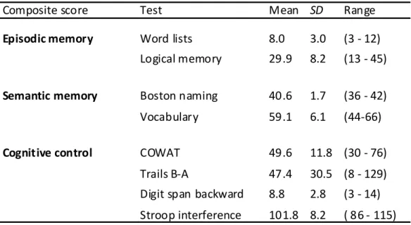
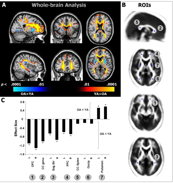
It is well established that healthy aging is accompanied by structural changes in many brain regions and functional decline in a number of cognitive domains. The goal of this study was to determine 1) whether the regional distribution of age-related brain changes is similar in gray matter (GM) and white matter (WM) regions, or whether these two tissue types are affected differently by aging, and 2) whether measures of cognitive performance are more closely linked to alterations in the cerebral cortex or in the underlying WM in older adults (OA). To address these questions, we collected high-resolution MRI data from a large sample of healthy young adults (YA; aged 18-28) and OA (aged 61-86 years). In addition, the OA completed a series of tasks selected to assess cognition in three domains: cognitive control, episodic memory, and semantic memory. Using advanced techniques for measuring cortical thickness and WM integrity, we found that healthy aging was accompanied by deterioration of both GM and WM, but with distinct patterns of change: Cortical thinning occurred primarily in primary sensory and motor cortices, whereas WM changes were localized to regions underlying association cortices. Further, in OA, we found a striking pattern of region-specific correlations between measures of cognitive performance and WM integrity, but not cortical thickness. Specifically, cognitive control correlated with integrity of frontal lobe WM, whereas episodic memory was related to integrity of temporal and parietal lobe WM. Thus, age-related impairments in specific cognitive capacities may arise from degenerative processes that affect the underlying connections of their respective neural networks.
Introduction
Healthy aging is characterized by myriad cognitive changes. Some of the most pronounced and consistently reported deficits are on tasks that require long-term memory or that challenge cognitive control processes and working memory (Hedden & Gabrieli, 2004; Piguet & Corkin, 2007). While the pattern of age-related cognitive changes is relatively well characterized, less is known about the neural bases of age-related changes in cognition.
Age-related structural changes in gray matter
Age-related cognitive decline is frequently attributed to deterioration of cortical gray matter (GM) structures. Magnetic resonance imaging (MRI)-based studies point to reduced global GM volumes in OA (Pfefferbaum et al., 1994; Raz et al., 1997; Salat et al., 1999; Bartzokis et al., 2001; Allen et al., 2005; Raz et al., 2005; Walhovd et al., 2005). Several studies that examined regional effects of age found that frontal areas showed greater volumetric reduction than posterior regions (Raz et al., 1997; Salat et al., 1999; Bartzokis et al., 2001; Allen et al., 2005). Particularly notable are volumetric losses in prefrontal cortex (PFC) (Raz et al., 1997; Salat et al., 1999; Raz et al., 2004a; Grieve et al., 2005). These findings have led to the hypothesis that age-related GM loss occurs along an anterior-to-posterior gradient (Jernigan et al., 1991; Raz et al., 1997; Sowell et al., 2003; Raz & Rodrigue, 2006). While this hypothesis tends to dominate the aging literature, degeneration has also been documented in all of the major lobes of the brain (Cowell et al., 1994; Bartzokis et al., 2001; Tisserand et al., 2002; Raz et al., 2004a; Van Petten, 2004; Van Petten et al., 2004; Allen et al., 2005).
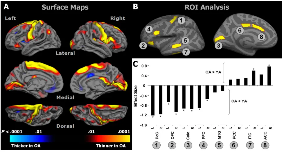
Advanced methods now allow assessment of changes in cortical thickness across the lifespan (Fischl & Dale, 2000). Similar to results from volumetric studies, cortical thickness of lateral PFC is reduced in OA. At the same time, cortical thinning is also found in the occipital lobe and precentral gyrus—areas that have not generally been associated with volumetric decline (Salat et al., 2004). The histopathological underpinnings of these macroscopic changes in cortical GM remain elusive: While early studies reported a loss of cortical neurons and decreased cell packing density (Pakkenberg & Gundersen, 1997), more advanced methods indicate that cell loss is relatively minimal in old age, overshadowed by a drastic loss of neuropil (Peters et al., 1998a).
Age-related structural changes in white matter
In addition to GM degeneration, WM changes likely play an important role in explaining age-related cognitive deficits (Peters, 2002; Hinman & Abraham, 2007). Many volumetric studies have documented reduced global and regional WM volumes in OA (Guttmann et al., 1998; Salat et al., 1999; Courchesne et al., 2000; Bartzokis et al., 2001; Jernigan et al., 2001; Allen et al., 2005; Piguet et al., 2009), but see (Pfefferbaum et al., 1994; Good et al., 2001; Sullivan et al., 2004). Evidence that WM volume loss is greatest in the frontal lobes is equivocal (Raz et al., 1997; Salat et al., 1999; Raz et al., 2004b; Allen et al., 2005; Piguet et al., 2009).
Other MRI-based markers of WM degeneration include an increase in hyperintensities on T2- and proton density-weighted images, with the greatest volume of hyperintensities typically found in the WM underlying the frontal lobes (DeCarli et al., 1995; de Groot et al., 2000;
Gunning-Dixon & Raz, 2000; Pfefferbaum et al., 2000; Tullberg et al., 2004; Nordahl et al., 2006; Yoshita et al., 2006). In addition, microstructural deterioration of WM has been assessed using diffusion tensor imaging (DTI), with numerous studies documenting widespread age-related decreases in fractional anisotropy (FA) (O'Sullivan et al., 2001; Sullivan & Pfefferbaum, 2003; Madden et al., 2004; Salat et al., 2005a; Benedetti et al., 2006; Charlton et al., 2006). FA measures the degree to which the diffusion of water molecules is restricted by microstructural elements, such as cell bodies, axons, or myelin and other glial cells (Beaulieu, 2002). Like WM hyperintensities, FA reductions tend to be most prominent anteriorly, such as in the genu and anterior portions of the corpus callosum and in the WM underlying PFC (O'Sullivan et al., 2001; Head et al., 2004; Pfefferbaum et al., 2005; Salat et al., 2005b; Sullivan & Pfefferbaum, 2006; Ardekani et al., 2007; Madden et al., 2007; Yoon et al., 2008). Other notable loci of decreased integrity include the internal capsule (Good et al., 2001; Salat et al., 2004), auditory pathways of the temporal lobes (Lutz et al., 2007), and cingulum bundle (Yoon et al., 2008). Postmortem studies reveal a number of pathologic factors that may cause changes in FA, including loss of small myelinated fibers (Tang et al., 1997; Marner et al., 2003) and myelin degradation (Peters, 2002), which likely contribute to volumetric change (Double et al., 1996; Guttmann et al., 1998; Ikram et al., 2008; Piguet et al., 2009).
In summary, evidence of morphological and microstructural changes in frontal areas appears consistently in the aging literature. In addition, regional alterations have been noted across wide regions of GM and WM, although the exact nature and magnitude of these changes remains a topic of debate. To explicitly test whether WM and GM exhibit similar or distinct
patterns of age-related change, measures of both structures must be examined in a single group of participants. The present study achieved that goal.
Linking GM changes to cognitive performance in OA
A prevalent view contends that age-related decline in episodic memory is related to deterioration of the hippocampus and other medial temporal lobe structures, and that cortical losses are more highly correlated with decrements in cognitive control processes (i.e., the frontal aging hypothesis) (West, 1996; Tisserand & Jolles, 2003). While a number of studies have reported correlations between hippocampal volume and episodic memory (Golomb et al., 1996; Kramer et al., 2007), some concerns have been raised about the robustness of the effect (Van Petten, 2004). Direct evidence in favor of the frontal aging hypothesis has also been difficult to demonstrate in humans (Greenwood, 2000; Van Petten et al., 2004; Raz & Rodrigue, 2006). Diminished attention and executive function in OA have been associated with decreased global cortical volumes and reduced volumes of lateral PFC and OFC (Zimmerman et al., 2006; Kramer et al., 2007), although an inverse correlation between working memory function and OFC volume has also been reported (Salat et al., 2002). In addition, PFC volume has been inversely correlated with perseverative errors in OA (Raz et al., 1998; Gunning-Dixon & Raz, 2003). In contrast, spatial and object working memory correlated with visual cortex volume (Raz et al., 1998), but neither spatial and object or verbal working memory (Gunning-Dixon & Raz, 2003) showed significant correlations with PFC volume.
