HAL Id: hal-00821095
https://hal.archives-ouvertes.fr/hal-00821095
Submitted on 7 May 2013
HAL is a multi-disciplinary open access
archive for the deposit and dissemination of
sci-entific research documents, whether they are
pub-lished or not. The documents may come from
teaching and research institutions in France or
abroad, or from public or private research centers.
L’archive ouverte pluridisciplinaire HAL, est
destinée au dépôt et à la diffusion de documents
scientifiques de niveau recherche, publiés ou non,
émanant des établissements d’enseignement et de
recherche français ou étrangers, des laboratoires
publics ou privés.
Assessment on experimental bacterial biofilms and in
clinical practice of the efficacy of sampling solutions for
microbiological testing of endoscopes
Claire Aumeran, E. Thibert, F. A. Chapelle, C. Hennequin, O. Lesens,
Ousmane Traore
To cite this version:
Claire Aumeran, E. Thibert, F. A. Chapelle, C. Hennequin, O. Lesens, et al.. Assessment on
experi-mental bacterial biofilms and in clinical practice of the efficacy of sampling solutions for microbiological
testing of endoscopes. Journal of Clinical Microbiology, American Society for Microbiology, 2012, 50
(3), pp.938-42. �10.1128/JCM.06221-11�. �hal-00821095�
Published Ahead of Print 14 December 2011.
10.1128/JCM.06221-11.
2012, 50(3):938. DOI:
J. Clin. Microbiol.
Lesens and O. Traoré
C. Aumeran, E. Thibert, F. A. Chapelle, C. Hennequin, O.
Microbiological Testing of Endoscopes
Efficacy of Sampling Solutions for
Biofilms and in Clinical Practice of the
Assessment on Experimental Bacterial
http://jcm.asm.org/content/50/3/938
Updated information and services can be found at:
These include:
REFERENCES
http://jcm.asm.org/content/50/3/938#ref-list-1
This article cites 21 articles, 2 of which can be accessed free at:
CONTENT ALERTS
more»
articles cite this article),
Receive: RSS Feeds, eTOCs, free email alerts (when new
http://journals.asm.org/site/misc/reprints.xhtml
Information about commercial reprint orders:
http://journals.asm.org/site/subscriptions/
To subscribe to to another ASM Journal go to:
on May 7, 2013 by INIST-CNRS BiblioVie
http://jcm.asm.org/
Assessment on Experimental Bacterial Biofilms and in Clinical Practice
of the Efficacy of Sampling Solutions for Microbiological Testing
of Endoscopes
C. Aumeran,aE. Thibert,aF. A. Chapelle,aC. Hennequin,cO. Lesens,band O. Traoréa
Service d’Hygiène Hospitalièreaand Maladies Infectieuses,bPôle REUNNHIR, CHU Clermont-Ferrand, Clermont-Ferrand, France, and Laboratoire de Bactériologie, UFR
Pharmacie, Clermont-Ferrand, Francec
Opinions differ on the value of microbiological testing of endoscopes, which varies according to the technique used. We com-pared the efficacy on bacterial biofilms of sampling solutions used for the surveillance of the contamination of endoscope chan-nels. To compare efficacy, we used an experimental model of a 48-h Pseudomonas biofilm grown on endoscope internal tubing. Sampling of this experimental biofilm was performed with a Tween 80-lecithin-based solution, saline, and sterile water. We also performed a randomized prospective study during routine clinical practice in our hospital sampling randomly with two differ-ent solutions the endoscopes after reprocessing. Biofilm recovery expressed as a logarithmic ratio of bacteria recovered on bacte-ria initially present in biofilm was significantly more effective with the Tween 80-lecithin-based solution than with saline solu-tion (P ⴝ 0.002) and sterile water (P ⴝ 0.002). There was no significant difference between saline and sterile water. In the randomized clinical study, the rates of endoscopes that were contaminated with the Tween 80-lecithin-based sampling solution and the saline were 8/25 and 1/25, respectively (P ⴝ 0.02), and the mean numbers of bacteria recovered were 281 and 19 CFU/100 ml (P ⴝ 0.001), respectively. In conclusion, the efficiency and therefore the value of the monitoring of endoscope reprocessing by microbiological cultures is dependent on the sampling solutions used. A sampling solution with a tensioactive action is more efficient than saline in detecting biofilm contamination of endoscopes.
E
ndoscopes have a high bioburden of microorganisms after use (8) and are difficult to clean and disinfect because of their complicated design, long narrow lumens and because of the ma-terials used in their manufacture (2). Endoscope reprocessing is a multistep procedure involving numerous factors that can inter-fere with its efficacy. To ensure the quality of the reprocessing, strict compliance with the disinfection procedure is mandatory, and a regular audit of all of the steps in reprocessing is crucial. Despite the publication of reprocessing guidelines, breaches in reprocessing practices continue to be reported, and failure to fol-low cleaning or disinfection guidelines can result in outbreaks involving a large number of patients (24). The microbiological safety of endoscopes can also be affected by occult endoscope damage and contaminated automated endoscope reprocessors, and thus quality control for endoscope reprocessing is extremely important. However, there is continuing debate about the role and value of surveillance cultures in the quality assurance pro-gram of endoscope reprocessing (24). Many authors recommend endoscope surveillance cultures, and several recent reports of endoscopy-related outbreaks have stressed the importance that these cultures have played or could have played in the prevention of these adverse events (4, 15, 19, 20). Even the recent guidelines of the American Society for Gastrointestinal Endoscopy (ASGE), which do not recommend routine microbiological testing of en-doscopes, state that this question warrants further studies (3). The value of surveillance cultures is likely dependent on how often endoscopes are sampled and by what technique. Samplings of in-ternal channels of endoscopes usually rely on flushing the chan-nels, generally with saline or sterile water, and sometimes in com-bination with brushing of the internal channels. Very few studies have attempted to evaluate the efficacy of the sampling methods of endoscope channels (17). In addition, and despite increasingevi-dence of the implication of biofilms in endoscope contamination, no published data are available on how efficient these methods are on bacterial biofilms (6, 22, 24).
In the present study, we compared the efficacy of several sam-pling solutions used for the microbial surveillance of the contam-ination of endoscope internal channels on bacterial biofilms. To compare efficacy, we used an experimental model of biofilm grown on endoscope internal tubing and performed an in-use evaluation sampling the endoscopes during routine clinical prac-tice with two different sampling solutions.
MATERIALS AND METHODS
Biofilm formation.A Pseudomonas aeruginosa (CIP 103.467; Collection Institut Pasteur, Paris, France) biofilm was produced over 48 h inside a flexible Teflon tube (Tygon, R3603; Cole-Parmer, Vernon Hills, IL) (Fig. 1). Our laboratory model of biofilm production was based on an experi-mental model described elsewhere (23). The sterile Teflon tube was con-nected to a sterile polyvinylchloride tube (Nalgene, Illkirch, France) to form a loop that was supplied with tryptone soy broth culture medium (TSB; CM129; Oxoid, Cambridge, England). The system was activated by two pumps (Watson Marlow 205S; La Queue, Lez Yvelines, France), one providing a continuous flow of TSB medium in the system and the second providing a homogenous diffusion of the TSB and of the P. aeruginosa
Received 26 October 2011 Returned for modification 22 November 2011 Accepted 6 December 2011
Published ahead of print 14 December 2011
Address correspondence to O. Traoré, [email protected]. Copyright © 2012, American Society for Microbiology. All Rights Reserved.
doi:10.1128/JCM.06221-11
938 jcm.asm.org 0095-1137/12/$12.00 Journal of Clinical Microbiology p. 938 –942
on May 7, 2013 by INIST-CNRS BiblioVie
http://jcm.asm.org/
suspension in the loop (Fig. 1). The circuit was inoculated with 20 ml ⫾ 1 ml of a suspension containing ca. 108P. aeruginosaorganisms per ml.
Recovery and numeration of viable bacteria from the biofilm.We used a mechanical technique based on scraping, vortexing, and ultrasoni-cation to recover the biofilm in a saline solution as previously described (23). The solution was then diluted and plated on Trypticase soy agar (TSA).
Endoscope sampling solutions.We tested a commercially available Letheen broth (VWR Prolabo, Fontenay Sous Bois, France) composed of Tween 80 (0.5% [vol/vol]), meat peptone (1% [wt/vol]), meat extract (0.5 [wt/vol]), sodium chloride (0.5% [wt/vol]) and lecithin (0.07% [wt/vol]), 0.9% sterile saline solution, and sterile water.
Portions (2 ml) of the sampling solutions tested were instilled with a syringe for 30 s in 3-cm portions of the Teflon tube, recovered, and diluted up to 10⫺6. Portions (500 l) of 10⫺4to 10⫺6dilutions were plated in duplicate on TSA plates that were incubated at 37°C for 24 h. The bacterial counts are expressed as CFU per cm2or log
10CFU/cm2.
Sampling of endoscopes after reprocessing in routine clinical prac-tice.The 61 endoscopes included in the prospective randomized clinical study came from the teaching hospital of Clermont-Ferrand, France. They were divided into six types: gastroscope, duodenoscope, colonoscope, echoendoscope, bronchoscope, and cystoscope. For each type, half of the endoscopes were randomized to a sterile saline solution group or to the Letheen broth group.
The sampling method is used routinely in our hospital and follows French guidelines (10). Sampling is performed aseptically by two people after alcohol-based hand-rubbing. The ends of the channels are disin-fected by 60°C alcohol with a sterile gauze. A total volume of 100 ml of the tested sampling solution is injected inside the operating, suction, and air/water channels. The pooled sample was then collected from the oper-ating channel. The identity of the endoscope, the duration of storage before sampling, the date of the last disinfection and/or the last cleaning, and the type of disinfection/cleaning (manual or automated) are re-corded.
Microbiological identification.The sample was filtered through a 0.45-m-pore-size membrane (EZ-PAK; Millipore, Molsheim, France) and then rinsed. The membrane was placed on Trypticase soy agar plates and incubated for 2 days at 30°C and then for 3 days at room temperature. The viable cell counts were made at 48 h and 5 days and were expressed as CFU per 100 ml.
The microorganisms were identified by standard procedures. Micro-bial identification was made by Gram staining for bacteria and scotch test for fungi.
Gram-positive cocci were identified by coagulase test (Becton Dickin-son, Le Pont-De-Claix, France) and Chapman plates (Oxoid, Dardilly,
France). Gram-negative bacilli were identified by oxidase testing (Bio-Rad, Marnes-la-Coquette, France) and the use of API 20 E and API 20 NE strips (bioMérieux, Lyon, France).
In accordance with our national guidelines (10), a sample was classi-fied as “unacceptable” if more than 5 CFU per 100 ml and/or the presence of pathogens (enterobacteriaceae, Pseudomonas aeruginosa,
Staphylococ-cus aureus, Aspergillus spp., and yeast) were detected.
Statistical analyses. (i) Experimental study.The quantity of biofilm initially present in the Teflon tube was determined in each trial as the mean bacterial counts (log10CFU/cm2) recovered from three tube por-tions taken as controls. The remaining tube porpor-tions (n ⫽ 7) were sam-pled by the solution tested: a logarithmic ratio for each tube portion was calculated with the bacterial count (log10CFU/cm2) recovered by the tested solution in each tube portion as the numerator and the mean bac-terial count (log10CFU/cm2) recovered from the three tube control por-tions as the denominator. Finally, the mean logarithmic ratios were cal-culated for the three sampling solutions tested and compared by the Mann-Whitney test. We also determined the percentage of biofilm recov-ery for each sampling solution using the mean bacterial count (CFU/cm2) obtained on the seven portions with the test solution as the numerator and the mean bacterial count (CFU/cm2) recovered from the three tube con-trol portions as the denominator.
(ii) Prospective clinical study.Wilcoxon test was used to compare the overall count of microorganisms (CFU/100 ml) recovered by the saline solution and the commercially available Letheen broth. The Fisher exact test was used to compare the proportion of unacceptable samples ob-tained with each sampling solution used.
Pvalues of ⬍0.05 were considered to indicate statistical significance. Analyses were performed using SAS software (SAS Institute, Inc.). RESULTS
Recovery of the 48 h P. aeruginosa biofilm according to solu-tions used.We first assessed the homogeneity of the biofilm pro-duced in the Teflon tubes analyzing the bacterial recovery from five to seven 3-cm samples of the Teflon tube that were cut from the straight portion and curved portions and at the air-water in-terface of the Teflon tube loop. The reproducibility of the biofilm formation in Teflon tubes in three different trials was also as-sessed. The recovery of viable bacteria from the biofilms produced in the three different trials were 7.98 ⫾ 0.21, 7.82 ⫾ 0.10, and 8.27 ⫾ 0.19, respectively. These results with low standard devia-tions show that biofilm formation was quantitatively uniform throughout the length of the Teflon tube and that it was reproduc-ible between each trial.
We then compared the bacterial recovery rates after the use of three test solutions (Letheen broth, 0.9% saline solution, and ster-ile water) in three independent 48-h Pseudomonas aeruginosa bio-film trials.
For each trial, the sampling solution was tested on seven por-tions of 3-cm Teflon tube. Three porpor-tions, collected at the ends and center of the Teflon tube, were used as controls. The viable cells in the controls were counted using the mechanical recovery technique. Biofilm formation in the controls was consistent with the results obtained during the development phase, as shown by low standard deviations (from 0.06 to 0.28 log10CFU/cm2). The
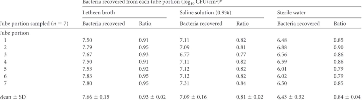
percentage of biofilm recovery was higher after instillation of the Letheen broth than with the other test solutions: 30.1% versus 2.2% for saline solution and 7.1% for sterile water. These results were confirmed by comparing, between the three tested solutions, the mean logarithmic ratios of bacterial counts obtained in each tube portion (Table 1). The ratios were significantly different be-tween Letheen broth and sterile water (0.93 versus 0.84; P ⫽
FIG 1 Model of biofilm formation in Teflon tube.
Sampling Biofilm in Endoscopes
March 2012 Volume 50 Number 3 jcm.asm.org 939
on May 7, 2013 by INIST-CNRS BiblioVie
http://jcm.asm.org/
0.002) and between Letheen broth and saline solution (0.93 versus 0.81; P ⫽ 0.002). There was no significant difference between sa-line solution and sterile water (0.81 versus 0.84; P ⫽ 0.12).
Randomized prospective study of endoscopes.We randomly sampled 50 (82%) of the 61 endoscopes from our hospital (Table 2). Most were digestive (n ⫽ 24) and bronchial (n ⫽ 22). Their disinfection procedure was performed for most cases in an auto-mated endoscope disinfector (41/50). The endoscopes came from different wards: gastroenterology (n ⫽ 20), pediatrics (n ⫽ 9), pneumology (n ⫽ 8), intensive care units (n ⫽ 8), urology (n ⫽ 4), and thoracic surgery (n ⫽ 1).
Most endoscopes (47/50) were sampled after 12 h of storage. A total of 38 microorganisms were found on 33 endoscopes. According to French guidelines, 34 of these microorganisms can be considered environmental contaminants (10) (coagulase-negative staphylococci [n ⫽ 15], Bacillus spp. [n ⫽ 8], mold [n ⫽ 6], non-Enterobacteriaceae Gram-negative bacilli [n ⫽ 2],
Micro-coccusspp. [n ⫽ 2], Corynebacterium sp. [n ⫽ 1]) and 4 can be considered potential pathogens (P. aeruginosa in a gastroscope [n ⫽ 1], Enterobacter cloacae in a duodenoscope [n ⫽ 1], and
Aspergillus versicolorin bronchoscopes [n ⫽ 2]). Most microor-ganisms (29/38), including all those considered pathogens, were found when the samplings were performed with Letheen broth.
The count of microorganisms by endoscope was usually low, and only 9 endoscopes of 50 (18%) were found to have an unac-ceptable result, with more than 5 CFU per 100 ml and/or the presence of pathogens. Eight of nine of the unacceptable samples were found with Letheen broth, which was significantly more ef-ficient than saline for identifying unacceptable contaminations (8/25 versus 1/25; P ⫽ 0.02). The overall CFU count obtained with the Letheen broth in 25 endoscopes was significantly higher than that obtained with the saline solution in the 25 other endoscopes (281 versus 19 CFU; P ⫽ 0.001).
DISCUSSION
In this study, both experimental evaluation on bacterial biofilms and in-use clinical results showed that, for the microbiological testing of internal channels of endoscopes, the use of a tensioactive sampling fluid was significantly more efficient than sterile water or saline.
The use of routine environmental microbiological testing of endoscopes for quality assurance of the cleaning and disinfection process of the endoscope has not been established and is a matter of wide debate. A consensus guideline from the European Society of Gastrointestinal Endoscopy (ESGE) and the European Society of Gastroenterology and Endoscopy Nurses and Associates (ESGENA) addresses the need for microbiological surveillance in endoscopy (5). Recommendations from several countries
TABLE 1Recovery of P. aeruginosa biofilm with each test solutiona
Tube portion sampled (n ⫽ 7)
Bacteria recovered from each tube portion (log10CFU/cm2)b
Letheen broth Saline solution (0.9%) Sterile water
Bacteria recovered Ratio Bacteria recovered Ratio Bacteria recovered Ratio
Tube portion 1 7.50 0.91 7.11 0.82 6.48 0.85 2 7.79 0.95 7.09 0.81 6.88 0.90 3 7.67 0.93 6.77 0.77 6.56 0.86 4 7.50 0.91 7.11 0.82 6.59 0.86 5 7.53 0.92 7.12 0.82 6.01 0.79 6 7.83 0.95 7.12 0.82 6.02 0.79 7 7.80 0.95 7.31 0.84 6.50 0.85 Mean ⫾ SD 7.66 ⫾ 0,15 0.93 ⫾ 0.02 7.09 ⫾ 0.16 0.81 ⫾ 0.02 6.43 ⫾ 0.32 0.84 ⫾ 0.04
aThe mean logarithmic counts in control samples (n ⫽ 3) for biofilm ⫾ the standard deviation (log
10CFU/cm2) for Letheen broth, saline solution, and sterile water were 8.20 ⫾
0.06, 8.71 ⫾ 0.28, and 7.64 ⫾ 0.21, respectively. The percentages of biofilm recovery (CFU/cm2) in these control samples for Letheen broth, saline solution, and sterile water were
30.1 (4.8 ⫻ 107/1.6 ⫻ 108), 2.2 (1.3 ⫻ 107/5.8 108), and 7.1 (3.3 ⫻ 106/4.7 ⫻ 107), respectively. The percent biofilm recovery for each sampling solution was calculated using the
mean bacterial count (CFU/cm2) obtained for the seven portions with the test solution as the numerator and the mean bacterial count (CFU/cm2) recovered from the three tube
control portions as the denominator.
bThe logarithmic ratio for each tube portion was calculated as the bacterial count (log
10CFU/cm2) recovered by the tested solution in each tube portion as the numerator and the
mean bacterial count (log10CFU/cm2) recovered from the three tube control portions as the denominator.
TABLE 2Results of prospective endoscope sampling using Letheen broth or 0.9% saline solution during routine clinical practice Test solution and
endoscope typea Determination
Storage duration (h) Letheen broth Bronchoscope (n ⫽ 11) 7 acceptable 36–480 4 unacceptable 12–60 Coloscope (n ⫽ 5) 5 acceptable 7–48 Gastroscope (n ⫽ 3) 1 acceptable 12 2 unacceptable 12 Duodenoscope (n ⫽ 3) 2 acceptable 12 1 unacceptable 12 Echoendoscope (n ⫽ 1) 1 unacceptable 12 Cystoscope (n ⫽ 2) 2 acceptable 12–72 Saline solution (0.9%) Bronchoscopes (n ⫽ 11) 11 acceptable 48–720 Coloscope (n ⫽ 3) 3 acceptable 12 Gastroscope (n ⫽ 6) 6 acceptable 7–48 Duodenoscope (n ⫽ 2) 1 acceptable 12 1 unacceptable 12 Echoendoscope (n ⫽ 1) 1 acceptable 2 Cystoscope (n ⫽ 2) 2 acceptable 12–72 an ⫽ number of samples. Aumeran et al.
940 jcm.asm.org Journal of Clinical Microbiology
on May 7, 2013 by INIST-CNRS BiblioVie
http://jcm.asm.org/
throughout the world advise microbiological testing of gastroin-testinal and respiratory endoscopes as a quality control (12, 14, 16). Conversely, microbiological surveillance testing of endo-scopes after reprocessing, during storage, or before use is not stip-ulated in current U.S. guidelines (3, 18, 21). However, the recent guideline of the American Society for Gastrointestinal Endoscopy (ASGE) stated that this question warrants further studies (3).
There are few documented reports on how to perform the rou-tine microbiological sampling of endoscopes and no recognized method for verifying the effectiveness of cleaning and disinfecting in clinical practice. However, selecting appropriate sampling and assay methods is essential for the results to be meaningful. The samplings of internal channels of endoscopes usually consist in flushing the channels with a fluid, usually saline or sterile water. Some guidelines favor the use of a neutralizing and more tensio-active solution based on polysorbate and lecithin (10). Recently, an antero-retrograde flushing technique with sterile water was developed to improve the effectiveness of sampling (6). In our in-use clinical study no retrograde flushing was performed, and therefore we were unable to compare the efficacy of retrograde versus anterograde flushing with our tensioactive solution. Most of the techniques proposed in current guidelines are empirical and, to our knowledge, very few published studies include a com-parative and comprehensive evaluation of the sampling tech-niques (17). Furthermore, no published studies have assessed the efficacy of the techniques on bacterial biofilms. Biofilm develops in all wet environments (11). It is now well established that if the routine cleaning procedure is not rigorous, particularly if an ac-curate drying procedure is not applied, the microbial contamina-tion of the endoscopes will be due to bacteria embedded in biofilm rather than to planktonic (in suspension) bacteria (22, 24). Bac-teria attached in biofilm are more difficult to kill by disinfectant than are unattached planktonic bacteria (11); such attached bac-teria also have very different physiological features (11), and the removal of biofilm from endoscope channels is much more diffi-cult than that of planktonic bacteria (1). It is therefore essential to assess the endoscope microbial sampling technique on biofilms because their presence requires highly sensitive methods of sam-pling.
Results from our laboratory contamination model showed that when the Letheen solution was used for sampling, it achieved a much higher recovery rate than saline or sterile water of biofilm contaminating endoscopes. These findings were strengthened by the results obtained from the random sampling of 50 endoscopes during daily clinical activity in our hospital. There were many positive cultures that yielded low counts of bacterial species such as coagulase-negative staphylococci or Bacillus. The clinical im-portance of these cultures may be low since it is likely that they do not represent a significant problem with the disinfection or clean-ing process. However, samplclean-ing with Letheen solution yielded sig-nificantly more positive results than with saline, whatever the pa-rameters: the total count of microorganisms recovered, the rate of overall positive samples, and the rate of samples with pathogens. Routine microbiological surveillance usually focuses on vegetative bacteria, fungi, and more rarely mycobacteria and usually ex-cludes fastidious bacteria, anaerobes, and viruses whose detection is complex and prohibitively expensive for routine surveillance purposes (6, 10, 14). Improving the sensitivity of detection of pathogens such as enteric organisms or Pseudomonas spp. may have a real direct impact on patient safety. We recently reported an
outbreak due to multiresistant Klebsiella pneumoniae contaminat-ing duodenoscopes durcontaminat-ing which routine surveillance cultures of duodenoscopes performed over several months by saline flushing failed to detect any contamination. Only when we modified the sampling procedure of the inner channels, replacing flushing with saline solution by a Tween 80-lecithin-based solution plus brush-ing, were we able to isolate the outbreak strains from a contami-nated endoscope (4).
Previous studies have shown that, in experimentally contami-nated endoscopes, a single flushing of internal channels with sa-line solution removes only a very small number of bacteria (9, 13). The main reason for the greater efficacy of Letheen broth is the tensioactive action against biofilm of polysorbate (Tween 80), which is frequently used for its detergent activity (25). In addition, the Letheen solution could also neutralize the antimicrobial activ-ity of residual traces of disinfectant present in endoscopes in rou-tine clinical practice (7).
In conclusion, our experimental data demonstrate that testing of endoscopes to detect biofilm contamination is much more ef-ficient with a tensioactive agent than with saline or water. The microbiological surveillance results obtained during routine clin-ical practice confirmed the greater efficacy of the tensioactive agent. If microbiological testing is implemented as a quality con-trol measure of endoscope reprocessing, the biofilm nature of bac-teria should be taken into account to assess safety.
ACKNOWLEDGMENTS
This study was supported by CHU Ferrand, Clermont-Ferrand, France.
We thank Jeffrey Watts for help in preparing the manuscript. REFERENCES
1. Alfa MJ, Degagne P, Olson N. 1999. Worst-case soiling levels for patient-used flexible endoscopes before and after cleaning. Am. J. Infect. Control
27:392– 401.
2. ASGE Standards of Practice Committee. 2008. Infection control during GI endoscopy. Gastrointest. Endosc. 67:781–790.
3. ASGE Quality Assurance in Endoscopy Committee. 2011. Multisociety guideline on reprocessing flexible gastrointestinal endoscopes: 2011. Gas-trointest. Endosc. 73:1075–1084.
4. Aumeran C, et al. 2010. Multidrug-resistant Klebsiella pneumoniae out-break after endoscopic retrograde cholangiopancreatography. Endoscopy
42:895– 899.
5. Beilenhoff U, et al. 2007. ESGE/ESGENA guideline for quality assurance in reprocessing: microbiological surveillance testing in endoscopy. Endos-copy 39:175–181.
6. Buss AJ, et al. 2008. Endoscope disinfection and its pitfalls: requirement for retrograde surveillance cultures. Endoscopy 40:327–332.
7. CEN. 2003. EN 13727 chemical disinfectants and antiseptics: quantitative suspension test for the evaluation of bactericidal activity of chemical dis-infectants for instruments used in the medical area: test method and re-quirements (phase 2/step 1), CEN, Brussels, Belgium.
8. Chu NS, McAlister D, Antonoplos PA. 1998. Natural bioburden levels detected on flexible gastrointestinal endoscopes after clinical use and manual cleaning. Gastrointest. Endosc. 48:137–142.
9. Corcoran GD, Holton J, Ridgway GL. 1994. Endoscope decontamina-tion: a comparison of the Wolf 35100 and DSD-91 systems. J. Hosp. Infect.
27:307–315.
10. CTINILS. 2007. Eléments d’assurance qualité en hygiène relatifs au con-trôle microbiologique des endoscopes et a` la traçabilité en endoscopie. Conseil Supérieur d’Hygiène Publique de France, Direction Générale de la Santé, Paris, France.
11. Donlan RM, Costerton JW. 2002. Biofilms: survival mechanisms of clin-ically relevant microorganisms. Clin. Microbiol. Rev. 15:167–193. 12. Endoscopy Working Group Infection Control Subcommittee. 2000.
Manitoba Advisory Committee on Infectious Disease guidelines for
infec-Sampling Biofilm in Endoscopes
March 2012 Volume 50 Number 3 jcm.asm.org 941
on May 7, 2013 by INIST-CNRS BiblioVie
http://jcm.asm.org/
tion prevention and control in endoscopy. Manitoba Public Health, Win-nipeg, Manitoba, Canada.
13. Felmingham D, Mowles J, Thomas K, Ridgway GL. 1985. Disinfection of gastrointestinal fibroscopes: an evaluation of the Pauldrach Endocleaner, and various chemical agents. J. Hosp. Infect. 6:379 –388.
14. Gastroenterological Society of Australia. 2008. Microbiological testing of endoscopes, p 67–72. In Cowen AE, et al. (ed), Guidelines: infection con-trol in endoscopy, 2nd ed. Gastroenterological Society of Australia, Syd-ney, Australia.
15. Kovaleva J, et al. 2009. Is bacteriologic surveillance in endoscope repro-cessing stringent enough? Endoscopy 41:913–916.
16. Leung J. 2000. Working Party Report: care of endoscopes. Reprocessing of flexible endoscopes. J. Gastroenterol. Hepatol. 15:73–77.
17. Luu Duc D, et al. 1998. Validation d’une méthode de prélèvement des canaux d’un endoscope souple contaminé expérimentalement. Pathol. Biol. 46:34 –38.
18. Mehta AC, et al. 2005. American College of Chest Physicians and Amer-ican Association for Bronchology consensus statement: prevention of flex-ible bronchoscopy-associated infection. Chest 128:1742–1755.
19. Merighi A, et al. 1996. Quality improvement in gastrointestinal
endos-copy: microbiologic surveillance of disinfection. Gastrointest. Endosc. 43: 457– 462.
20. Muscarella LF. 2006. Inconsistencies in endoscope-reprocessing and infection-control guidelines: the importance of endoscope drying. Am. J. Gastroenterol. 101:2147–2154.
21. Nelson DB, et al. 2003. Multi-society guideline for reprocessing flexible gastrointestinal endoscopes. Infect. Control. Hosp. Epidemiol. 24:532– 537.
22. Pajkos A, Vickery K, Cossart Y. 2004. Is biofilm accumulation on endo-scope tubing a contributor to the failure of cleaning and decontamination? J. Hosp. Infect. 58:224 –229.
23. Pineau L, Roques C, Luc J, Michel G. 1997. Automatic washer disinfec-tor for flexible endoscopes: a new evaluation process. Endoscopy 29:372– 379.
24. Seoane-Vazquez E, Rodriguez-Monguio R. 2008. Endoscopy-related in-fection: relic of the past? Curr. Opin. Infect. Dis. 21:362–366.
25. Toutain-Kidd CM, et al. 2009. Polysorbate 80 inhibition of Pseudomonas
aeruginosabiofilm formation and its cleavage by the secreted lipase LipA. Antimicrob. Agents Chemother. 53:136 –145.
Aumeran et al.
942 jcm.asm.org Journal of Clinical Microbiology